Search results
1000 results found for “HBsAg”
Name
Description
Product #
Price
Quantity
Shipping Method
- View Data Sheet
Name :
HIV-1 p24, HRPDescription:
HIV-1 p24 Recombinant, HRP Labeled
Product # :
HIV-109Price :
Quantity :
Shipping Method :
Shipped with Ice Packs
More Info
- description
- source
- formulation
- purity
- More Info
- Citations
Description
The E.coli derived HRP recombinant protein is a non-glycosylated polypeptide chain, containing the HIV-1 p24 immunodominant regions and fused to a GST tag at N-terminus.
Source
Escherichia Coli.
Formulation
1.5M urea, 25mM Tris-HCl pH-8.0 and 50% Glycerol.
Purity
Greater than 90.0% as determined by HPLC analysis and SDS-PAGE.
Citations
More Info
-
Introduction
Human immunodeficiency virus (HIV) is a retrovirusthat can lead to a condition in which the immune systembegins to fail, leading to opportunistic infections. HIV primarily infects vital cells in the humanimmune systemsuch as helper T cells(specifically CD4+ T cells), macrophagesand dendritic cells. HIV infection leads to low levels of CD4+ T cells through three main mechanisms: firstly, direct viral killing of infected cells; secondly, increased rates of apoptosisin infected cells; and thirdly, killing of infected CD4+ T cells by CD8 cytotoxic lymphocytesthat recognize infected cells. When CD4+ T cell numbers decline below a critical level, cell-mediated immunityis lost, and the body becomes progressively more susceptible to opportunistic infections. HIV was classified as a member of the genus Lentivirus, part of the family of Retroviridae. Lentiviruses have many common morphologies and biological properties. Many species are infected by lentiviruses, which are characteristically responsible for long-duration illnesses with a long incubation period. Lentiviruses are transmitted as single-stranded, positive-sense, enveloped RNA viruses. Upon entry of the target cell, the viral RNA genomeis converted to double-stranded DNAby a virally encoded reverse transcriptasethat is present in the virus particle. This viral DNA is then integrated into the cellular DNA by a virally encoded integraseso that the genome can be transcribed. Once the virus has infected the cell, two pathways are possible: either the virus becomes latentand the infected cell continues to function, or the virus becomes active and replicates, and a large number of virus particles are liberated that can then infect other cells.
-
Physical Appearance
Sterile filtered colorless clear solution.
-
Stability
HIV-1 p24 although stable at 4°C for 1 week, should be stored below -18°C. Please prevent freeze thaw cycles.
-
Applications
ELISA, Western blots, Flow-through.
ProSpec's products are furnished for LABORATORY RESEARCH USE ONLY. The product may not be used as drugs, agricultural or pesticidal products, food additives or household chemicals.
- View Data Sheet
Name :
HTATIP2 HumanDescription:
HIV-1 Tat Interactive Protein 2 Human Recombinant
TIP30, CC3, SDR44U1, HTATIP2, EC=1.1.1.-, HIV-1 TAT-interactive protein 2, FLJ26963.
Product # :
ENZ-546Price :
Quantity :
Shipping Method :
Shipped with Ice Packs
More Info
- description
- source
- formulation
- purity
- More Info
- Citations
Description
HTATIP2 Recombinant Human produced in E.Coli is a single, non-glycosylated polypeptide chain containing 262 amino acids (1-242 a.a.) and having a molecular mass of 29.3 kDa. The HTATIP2 is fused to a 20 amino acid His-Tag at N-terminus and purified by proprietary chromatographic techniques.
Source
Escherichia Coli.
Formulation
HTATIP2 protein solution (1mg/ml) containing 20mM Tris-HCl pH-8, 1mM DTT & 10% glycerol.
Purity
Greater than 95.0% as determined by SDS-PAGE.
Citations
More Info
-
Introduction
HTATIP2 is part of the short-chain dehydrogenases/reductases (SDR) family which acts as a tumor suppressor in metabolic suppression, inhibition of angiogenesis and induces the expression of apoptosis related genes Bad and Siva. HTATIP2 cooperates with the activation domain of HIV-1 TAT and enhances its transcription by phosphorylating RNA polymerase II (Pol II). Defects in HTATIP2 are related with hepatocellular carcinomas and apoptotic resistant tumor cells, implicating a probable use for HTATIP2 in antitumor therapy.
-
Synonyms
TIP30, CC3, SDR44U1, HTATIP2, EC=1.1.1.-, HIV-1 TAT-interactive protein 2, FLJ26963.
-
Physical Appearance
Sterile filtered colorless solution.
-
Stability
Store at 4°C if entire vial will be used within 2-4 weeks. Store, frozen at -20°C for longer periods of time. For long term storage it is recommended to add a carrier protein (0.1% HSA or BSA).Avoid multiple freeze-thaw cycles.
-
Amino Acid Sequence
MGSSHHHHHH SSGLVPRGSH MAETEALSKL REDFRMQNKS VFILGASGET GRVLLKEILE QGLFSKVTLI GRRKLTFDEE AYKNVNQEVV DFEKLDDYAS AFQGHDVGFC CLGTTRGKAG AEGFVRVDRD YVLKSAELAK AGGCKHFNLL SSKGADKSSN FLYLQVKGEV EAKVEELKFD RYSVFRPGVL LCDRQESRPG EWLVRKFFGS LPDSWARGHS VPVVTVVRAM LNNVVRPRDK QMELLENKAI HDLGKAHGSL KP.
ProSpec's products are furnished for LABORATORY RESEARCH USE ONLY. The product may not be used as drugs, agricultural or pesticidal products, food additives or household chemicals.
- View Data Sheet
Name :
GSTA1 HumanDescription:
Glutathione S-Transferase Alpha-1 Human Recombinant
GST2, GSTA1-1, GTH1, GSTA-1, GSTAI, GSTA-I, EC 2.5.1.18, Glutathione S-transferase A1, GST HA subunit 1, GST-epsilon, GST class-alpha member 1, GSTA1, MGC131939.
Product # :
ENZ-469Price :
Quantity :
Shipping Method :
Shipped with Ice Packs
More Info
- description
- source
- formulation
- purity
- biological activity
- More Info
- Citations
Description
GSTA1 Human Recombinant produced in E.Coli is a single, non-glycosylated, polypeptide chain containing 222 amino acids (1-222 a.a.) and having a molecular mass of 25.6 kDa. The GSTA1 is purified by proprietary chromatographic techniques.
Source
Escherichia Coli.
Formulation
The GSTA1 solution (1mg/ml) contains 20mM Tris-HCl buffer (pH 8.0), 1mM DTT and 10% glycerol.
Purity
Greater than 90.0% as determined by SDS-PAGE.
Biological Activity
Specific activity is 35,000 pmol/min/ug, and is defined as the amount of enzyme that conjugate 1.0 u mole of 1-chloro-2,4-dinitrobenzene (CDNB) with reduced glutathione per minute at pH 6.5 at 25°C.
Citations
More Info
-
Introduction
Membrane-bound & Cytosolic forms of GST are encoded by 2 separate supergene families. These enzymes function in the detoxification of electrophilic compounds, including carcinogens, therapeutic drugs, environmental toxins and products of oxidative stress, by conjugation with glutathione. There are 8 different classes of soluble cytoplasmic mammalian GST: alpha, kappa, mu, omega, pi, sigma, theta and zeta. The GSTA1 is found in a cluster mapped to chromosome 6, and is highly expressed in the liver. GSTA1 protects the cells from reactive oxygen species.
-
Synonyms
GST2, GSTA1-1, GTH1, GSTA-1, GSTAI, GSTA-I, EC 2.5.1.18, Glutathione S-transferase A1, GST HA subunit 1, GST-epsilon, GST class-alpha member 1, GSTA1, MGC131939.
-
Physical Appearance
Sterile Filtered colorless solution.
-
Stability
Store at 4°C if entire vial will be used within 2-4 weeks. Store, frozen at -20°C for longer periods of time. For long term storage it is recommended to add a carrier protein (0.1% HSA or BSA).Avoid multiple freeze-thaw cycles.
-
Amino Acid Sequence
MAEKPKLHYF NARGRMESTR WLLAAAGVEF EEKFIKSAED LDKLRNDGYL MFQQVPMVEI DGMKLVQTRA ILNYIASKYN LYGKDIKERA LIDMYIEGIA DLGEMILLLP VCPPEEKDAK LALIKEKIKN RYFPAFEKVL KSHGQDYLVG NKLSRADIHL VELLYYVEEL DSSLISSFPL LKALKTRISN LPTVKKFLQP GSPRKPPMDE KSLEEARKIF RF.
ProSpec's products are furnished for LABORATORY RESEARCH USE ONLY. The product may not be used as drugs, agricultural or pesticidal products, food additives or household chemicals.
- View Data Sheet
Name :
SARS MERS Spike AntibodyDescription:
Mouse Anti MERS-CoV Spike
Middle East respiratory syndrome coronavirus, Human betacoronavirus 2c EMC/2012, MERS-CoV, MERS, MERSCoV SP, Spike glycoprotein, S glycoprotein, S, Spike protein, E2, Peplomer protein.
Product # :
ANT-768Price :
Quantity :
Shipping Method :
Shipped with Ice Packs
More Info
- formulation
- More Info
- Citations
Formulation
1mg/ml containing Phosphate-Buffered Saline (pH 7.4), 0.02% Sodium Azide and 10% Glycerol.
Citations
More Info
-
Introduction
Since April 2012, cases of the Middle East Respiratory Syndrome Coronavirus (MERS-CoV) have been identified in various countries. Coronaviruses are the cause of the common cold, SARS (severe acute respiratory syndrome) and other severe illnesses with high mortality rates, all are classified into coronavirus family. MERS-CoV is a new type of SARS found in the coronavirus family causing severe pneumonia with sudden and serious respiratory illness with high mortality rates as well. Since January 27th 2015, the WHO has reported 956 human cases, including 351 deaths. More cases of the new coronavirus strain are expected. Like in other coronaviruses, large surface spike glycoprotein is a central structural protein of this virus; it is located above the virion surface to bind and enter into the target cell. Spike protein has 2 domains- S1 and S2. The S1 domain is responsible for cellular tropism and interaction with target cell, while the S2 domain is responsible for membrane fusion. The C-terminal of S1 domain contains a receptor binding domain, and is also a potential target for vaccine development and an antigen for diagnosis.
-
Synonyms
Middle East respiratory syndrome coronavirus, Human betacoronavirus 2c EMC/2012, MERS-CoV, MERS, MERSCoV SP, Spike glycoprotein, S glycoprotein, S, Spike protein, E2, Peplomer protein.
-
Physical Appearance
Sterile filtered colorless solution.
-
Immunogen
Recombinant MERS-CoV Spike (18-1296aa) purified from Baculovirus.
-
Ig Subclass
IgG1 kappa.
-
Clone
PAT40G7AT
-
Applications
SARS MERS Spike antibody has been tested by ELISA analysis to assure specificity and reactivity. Since application varies, however, each investigation should be titrated by the reagent to obtain optimal results.
-
Type
Mouse antibody Monoclonal.
-
Storage Procedures
For periods up to 1-month store at 4°C, for longer periods of time, store at -20°C. Prevent freeze thaw cycles.
-
Purification Method
SARS MERS Spike antibody was purified from mouse ascitic fluids by protein-A affinity chromatography.
ProSpec's products are furnished for LABORATORY RESEARCH USE ONLY. The product may not be used as drugs, agricultural or pesticidal products, food additives or household chemicals.
- View Data Sheet
Name :
Borrelia p41, Sf9Description:
Borrelia Burgdorferi p41 Recombinant, Sf9
Product # :
BOR-002Price :
Quantity :
Shipping Method :
Shipped with Ice Packs
More Info
- description
- source
- formulation
- purity
- More Info
- Citations
Description
Recombinant Borrelia Burgdorferi p41 produced in SF9 is a glycosylated, polypeptide chain having a calculated molecular mass of 36,578 Dalton. Borrelia p41 is expressed with a -6x His tag at N-terminus and purified by proprietary chromatographic techniques.
Source
Sf9 insect cells.
Formulation
Borrelia p41 (1.01mg/1ml) is supplied in 20mM HEPES buffer pH-7.5, 0.01mM EDTA and 0.02% SDS.
Purity
Greater than 80.0% as determined by SDS-PAGE.
Citations
More Info
-
Introduction
Borrelia belongs to a genus of bacteria of the spirochete phylum. Borrelia causes borreliosis, which is a zoonotic, vector-borne disease transmitted mainly by ticks and some by lice, depending on the species. Of the 36 known species of Borrelia, 12 are distinguished to cause Lyme disease or borreliosis and are transmitted by ticks. The main Borrelia species causing Lyme disease are Borrelia burgdorferi, Borrelia afzelii, and Borrelia garinii. The Borrelia genus members have a linear chromosome which is about 900 kbp in length as well as an excess of both linear and circular plasmids in the 5-220 kbp size range. The plasmids are atypical, as compared to most bacterial plasmids, since they contain many paralogous sequences, a large number of pseudogenes and, in some cases, essential genes. Moreover, a number of the plasmids have features suggesting that they are prophages.
-
Physical Appearance
Sterile Filtered clear solution.
-
Stability
Store at 4°C if entire vial will be used within 2-4 weeks. Store, frozen at -20°C for longer periods of time. Avoid multiple freeze-thaw cycles.
-
Applications
Western blot with Lyme positive plasma.
ProSpec's products are furnished for LABORATORY RESEARCH USE ONLY. The product may not be used as drugs, agricultural or pesticidal products, food additives or household chemicals.
- View Data Sheet
Name :
HDGF2 HumanDescription:
Hepatoma-Derived Growth Factor-2 Human Recombinant
Hepatoma-derived growth factor-related protein 3, HRP-3, Hepatoma-derived growth factor 2, HDGF-2, HDGFRP3, HDGF2, CGI-142.
Product # :
CYT-132Price :
Quantity :
Shipping Method :
Shipped with Ice Packs
More Info
- description
- source
- formulation
- purity
- More Info
- SDS-PAGE
- Citations
Description
HDGF2 Human Recombinant produced in E.Coli is a single, non-glycosylated polypeptide chain containing 227 amino acids (1-203 a.a.) and having a molecular mass of 25.1kDa (Molecular weight on SDS-PAGE will appear higher). HDGF2 is fused to a 24 amino acid His-tag at N-terminus & purified by proprietary chromatographic techniques.
Source
Escherichia Coli.
Formulation
HDGF2 protein solution (0.5mg/ml) containing 20mM Tris-HCl buffer (pH 8.0), 1mM DTT, 10% glycerol and 0.1M NaCl.
Purity
Greater than 90% as determined by SDS-PAGE.
SDS-PAGE
Citations
More Info
-
Introduction
Hepatoma-derived growth factor, related protein 3 (HDGFRP3/HDGF2) is the original member of a family of polypeptides labeled HDGF related proteins (HRPs). The HDGF2 protein augments DNA synthesis and may play a part in cell proliferation. HDGF2 is expressed primarily in the testis and the brain, to an intermediate extent in the heart, and to a slight extent in the ovaries, kidneys, spleen, and liver in humans.
-
Synonyms
Hepatoma-derived growth factor-related protein 3, HRP-3, Hepatoma-derived growth factor 2, HDGF-2, HDGFRP3, HDGF2, CGI-142.
-
Physical Appearance
Sterile Filtered clear solution.
-
Stability
Store at 4°C if entire vial will be used within 2-4 weeks. Store, frozen at -20°C for longer periods of time. For long term storage it is recommended to add a carrier protein (0.1% HSA or BSA).Avoid multiple freeze-thaw cycles.
-
Amino Acid Sequence
MGSSHHHHHH SSGLVPRGSH MGSHMARPRP REYKAGDLVF AKMKGYPHWP ARIDELPEGA VKPPANKYPI FFFGTHETAF LGPKDLFPYK EYKDKFGKSN KRKGFNEGLW EIENNPGVKF TGYQAIQQQS SSETEGEGGN TADASSEEEG DRVEEDGKGK RKNEKAGSKR KKSYTSKKSS
KQSRKSPGDE DDKDCKEEEN KSSSEGGDAG NDTRNTTSDL QKTSEGT. -
Background
What is the molecular weight/Mw of HDGF2 HUMAN Protein?
HDGF2 HUMAN Protein has a total Mw of 25.1kDa.
What is the source or expression system of HDGF2 HUMAN Protein?
Escherichia Coli.
What is the Purity of HDGF2 HUMAN Protein?
HDGF2 HUMAN Protein is >90% pure as determined by SDS-PAGE.
What is the Biological Activity of HDGF2 HUMAN Protein?
The biological functionality of HDGF2 HUMAN Protein will be determined in the future.
What is the amino acid sequence of HDGF2 HUMAN Protein?
MGSSHHHHHH SSGLVPRGSH MGSHMARPRP REYKAGDLVF AKMKGYPHWP ARIDELPEGA VKPPANKYPI FFFGTHETAF LGPKDLFPYK EYKDKFGKSN KRKGFNEGLW EIENNPGVKF TGYQAIQQQS SSETEGEGGN TADASSEEEG DRVEEDGKGK RKNEKAGSKR KKSYTSKKSS
KQSRKSPGDE DDKDCKEEEN KSSSEGGDAG NDTRNTTSDL QKTSEGT.
What applications can HDGF2 HUMAN Protein be used in?
HDGF2 HUMAN Protein can probably be used in western blot, ELISA and Lateral Flow.
What is the endotoxin level for HDGF2 HUMAN Protein?
The endotoxin level is minimal, HDGF2 HUMAN Protein was purified using conventional chromatography techniques.
ProSpec's products are furnished for LABORATORY RESEARCH USE ONLY. The product may not be used as drugs, agricultural or pesticidal products, food additives or household chemicals.
- View Data Sheet
Name :
SIV p55Description:
Simian Immunodeficiency Virus p55 Recombinant
Product # :
SIV-114Price :
Quantity :
Shipping Method :
Shipped with Ice Packs
More Info
- description
- source
- formulation
- purity
- More Info
- Citations
Description
Recombinant SIV p55- Strains: SIV mac 23g and SIV smH4 is glycosylated with N-linked sugars and produced using baculovirus vectors in insect cells.
Source
Baculovirus Insect Cells.
Formulation
SIV p55 Protein solution containing 10mM Tris, pH 8, 140mM NaCl & 400mM L-Arginine.
Purity
Greater than 90.0% as determined by:
(a) Analysis by RP-HPLC.
(b) Analysis by SDS-PAGE.Citations
More Info
-
Introduction
Simian immunodeficiency virus, SIV, is a retrovirusthat is found in strains, having the HIV-1and HIV-2specific strains that infect humans. SIV strains may cause an AIDS-like immune deficiency known as SAIDS (simian acquired immunodeficiency syndrome) if they cross species boundaries. HIV-2 is more similar to SIV strains than to HIV-1, suggesting for the first time the simian origin of HIV. HIV-2 is derived from the SIV strain found in sooty mangabeys whereas HIV-1, the predominant virus found in humans, is derived from SIV strains infecting chimpanzees.
-
Physical Appearance
Sterile Filtered colorless solution.
-
Stability
Recombinant SIV p55 although stable at 4°C for 3 weeks, should be stored below -18°C. For long term storage it is recommended to add a carrier protein (0.1% HSA). Please avoid freeze-thaw cycles.
ProSpec's products are furnished for LABORATORY RESEARCH USE ONLY. The product may not be used as drugs, agricultural or pesticidal products, food additives or household chemicals.
- View Data Sheet
Name :
H1N1 Puerto Rico RecombinantDescription:
H1N1 Influenza A- Virus Puerto Rico 08/1934 Recombinant
Product # :
IHA-036Price :
Quantity :
Shipping Method :
Shipped with Ice Packs
More Info
- description
- source
- formulation
- purity
- More Info
- Citations
Description
Recombinant Full-Length H1N1 Puerto Rico 08/1934 is glycosylated with N-linked sugars, produced using baculovirus vectors in insect cells and its Mw is approximately 62 kDa.
Source
Baculovirus Insect Cells.
Formulation
The Recombinant H1N1 A/Puerto Rico 08/1934 solution contains 10mM Sodium phosphate, pH 7.2, 150mM NaCl and 0.005% Tween-20.
Purity
Greater than 90.0% as determined by SDS-PAGE.
Citations
More Info
-
Introduction
H1N1 is a subtype specie of Influenza A virus. H1N1 Influenza Virus has mutated into various strains such as the Spanish Flustrain, mild humanflu strains, endemic pigstrains, and various strains found in birds. The Influenza A Virus is a globular particle about 100nm in diameter, sheathed in a lipid bilayer derived from the plasma membrane of its host. Studded in the lipid bilayer are two integral membrane proteins some 500 molecules of hemagglutinin ("H") and some 100 molecules of neuraminidase ("N"). Within the lipid bilayer are 3000 molecules of matrix protein and 8 pieces of RNA. Each of the 8 RNA molecules is associated with many copies of a nucleoprotein, several molecules of the three subunits of its RNA polymerase some "non-structural" protein molecules of uncertain function.
-
Physical Appearance
Sterile Filtered colorless solution.
-
Stability
H1N1 A/Puerto Rico 08/1934 recombinant should be stored at 4°C. Do not freeze!
ProSpec's products are furnished for LABORATORY RESEARCH USE ONLY. The product may not be used as drugs, agricultural or pesticidal products, food additives or household chemicals.
- View Data Sheet
Name :
H3N2 Wisconsin/67/05Description:
H3N2 Influenza-A Virus Wisconsin/67/05
Product # :
IHA-023Price :
Quantity :
Shipping Method :
Shipped with Ice Packs
More Info
- description
- formulation
- purity
- More Info
- Citations
Description
Allantoic fluid of 10 days old embryonated eggs, inoculated with influenza A virus, strain A/Wisconsin/67/05. The Influenza Virus was purified by Ultracentrifugation with 10-40 % sucrose gradient.
Formulation
The H3N2 A/Wisconsin/67/05 solution contains STE, 0.1% sodium azide (NaN3) and 0.005% thimerosal.
Purity
Greater than 90.0% as determined by SDS-PAGE.
Citations
More Info
-
Introduction
H3N2 is a subtype of the influenza A virus. Its name derives from the forms of the two kinds of proteinson the surface of its coat, hemagglutinin(H) and neuraminidase(N). H3N2 exchanges genes for internal proteins with other influenza subtypes. H3N2 has tended to dominate in prevalence over H1N1, H1N2, and influenza B. H3N2 strain descended from H2N2by antigenic shift, in which genes from multiple subtypes re-assorted to form a new virus. Both the H2N2and H3N2 strains contained genesfrom avian influenzaviruses.
-
Physical Appearance
Sterile Filtered colorless solution.
-
Stability
A/Wisconsin/67/05 although stable 4°C for 4 weeks, should be stored below -18°C. Please prevent freeze-thaw cycles.
-
Inactivation
Thimerosal and beta propiolactone treatmentThis product has been treated in a manner consistent with methods of inactivation. Generally accepted good laboratory practices appropriate to microbiological/viral safe handling practices and techniques are required at work.
-
Immunological Activity
Serological studies of influenza A virus, immunogen for antibody production.Tested with anti-influenza A monoclonal antibodies in ELISA.
ProSpec's products are furnished for LABORATORY RESEARCH USE ONLY. The product may not be used as drugs, agricultural or pesticidal products, food additives or household chemicals.
- View Data Sheet
Name :
HPSE ActiveDescription:
Recombinant Human Heparanase-1 Active
Product # :
ENZ-1032Price :
Quantity :
Shipping Method :
Shipped with Ice Packs
More Info
- description
- formulation
- purity
- biological activity
- More Info
- Citations
Description
Heparanase Active Enzyme is produced in CHO cells.The protein is purified by several orthogonal chromatography steps.
Formulation
Heparanase Active Enzyme is supplied in20mM Acetate buffer and 750mM NaCl pH 5.4.
Purity
Greater than 95.0% as determined by:
(a) Analysis by RP-HPLC.
(b) Analysis by SDS-PAGE.Biological Activity
The specific activity of Heparanase Active Enzyme in-house standard is about 0.7 Units (1 unit = 1 μmole of reducing ends of heparan sulfate substrate formed per minute per mg Heparanase Active Enzyme at 37°C). The enzymatic activity of each Heparanase Active Enzyme batch is comparable to the standard as determined by activity assay in which immobilized heparan, released due to heparanase activity, is quantified colorimetrically. Recommended reaction buffer: 20 mM Citrate Phosphate buffer, pH 5.4; 50mM NaCl; 1mM CaCl2.
Citations
More Info
-
Introduction
Heparanase is an endo β-D-glucuronidase, which degrades heparan sulfate side chains of heparan sulfate proteoglycans (HSPGs) in the extracellular matrix. Heparanase plays an important role in ECM degradation, facilitating the migration and extravasation of tumor cells and inflammatory leukocytes (1,2,3). Upon degradation, heparanase releases growth factors and cytokines that stimulate cell proliferation and chemotaxis (4,5). Heparanase is a heterodimer comprised of a 50 kDa subunit harboring the active site and a 8 kDa subunit. It is produced as a latent 65 kDa precursor and proteolytically processed to its active form (1,6). Heparanase is highly expressed in myeloid leukocytes (i.e. neutrophils) in platelets and in human placenta. Human heparanase was found to be upregulated in various types of primary tumors, correlating in some cases with increased tumor invasiveness and vascularity and with poor prospective survival (7,8).
-
Data Sheet
To view the FULL VERSION data sheet click Heparanase-1 Active Enzyme
-
Specificity
Heparanase Active Enzyme is identified by Western blot analysis with polyclonalrabbit anti-HPA1 antibodies as 2 subunits of 8-kDa and 50-kDa.
-
Shipping Conditions
Heparanase Active Enzyme is shipped frozen on dry ice unless stated otherwise by the customer. Shipping fees to N. America and W. Europe is $400 Shipping fees to Asia, Australia and E. Europe is $500
-
Storage Procedures
Store at –80ºC, avoid repeated freeze-thaw cycles.
ProSpec's products are furnished for LABORATORY RESEARCH USE ONLY. The product may not be used as drugs, agricultural or pesticidal products, food additives or household chemicals.
- View Data Sheet
Name :
Dengue Epitope 13Description:
Dengue Multiple Epitopes 13 Recombinant
Product # :
DEN-043Price :
Quantity :
Shipping Method :
Shipped with Ice Packs
More Info
- description
- source
- formulation
- purity
- More Info
- Citations
Description
Recombinant Dengue Multiple Epitopes 13 is genetically designed dengue multiple epitopes epi-13 designed especially for lateral flow product, they are selected from dengue genome. The rapid test prepared by this antigen has over 90% sensitivity and specificity over 90% to show quick and strong signal for both dengue IgM and IgG.
Source
Escherichia Coli.
Formulation
Phosphate buffered saline, pH 7.4 and 0.02% sodium nitrate.
Purity
Protein is >95% pure as determined by 12% PAGE (coomassie staining).
Citations
More Info
-
Introduction
Caused by one of four closely related virus serotypes of the genus Flavivirus, family Flaviviridae, each serotype is sufficiently different that there is no cross-protection and epidemics caused by multiple serotypes (hyperendemicity) can occur. In cell culture experiments and mice Morpholino antisense oligos have shown specific activity against Dengue virus.
-
Stability
Store at 4°C if entire vial will be used within 2-4 weeks. Store, frozen at -20°C for longer periods of time. For long term storage it is recommended to add a carrier protein (0.1% HSA or BSA).Avoid multiple freeze-thaw cycles.
ProSpec's products are furnished for LABORATORY RESEARCH USE ONLY. The product may not be used as drugs, agricultural or pesticidal products, food additives or household chemicals.
- View Data Sheet
Description:
Mycobacterium Tuberculosis RV1681, Polyclonal Rabbit Antibody
Product # :
ANT-493Price :
Quantity :
Shipping Method :
Shipped with Ice Packs
More Info
- description
- formulation
- purity
- More Info
- Citations
Description
The polyclonal antibody to Mycobacterium Tuberculosis RV1681 was collected from the rabbit igG and immunized with recombinant M. tuberculosis t RV-1681.
Formulation
Glycine-Tris buffer, pH7.5.
Purity
Pure Rabbit IgG, >95% pure as determined by 12% PAGE (coomassie staining).
Citations
More Info
-
Introduction
Mycobacterium tuberculosis, a member of the Mycobacteriaceae family, is an important infectious pathogenic bacteria and the cause for most cases of tuberculosis. It is the second common cause of death from an infectious disease in the world. Diagnosis using patient’s specimen culture of M. tuberculosis are still considered gold standard, but it is expensive and time-consuming. RV-1681 is a protein released from M. tuberculosis on infected organ, which filtrates through into the urine after circulating in the blood flow, to the kidney.RV-1681 is considered to be a reliable marker for M. tuberculosis diagnosis allowing easy low cost and time saving urine testing to screen large population for M. tuberculosis infection.
-
Physical Appearance
Sterile Filtered clear colorless solution.
-
Stability
Store at 4°C if entire vial will be used within 2-4 weeks. Store, frozen at -20°C for longer periods of time. For long term storage it is recommended to add a carrier protein (0.1% HSA or BSA).Avoid multiple freeze-thaw cycles.
-
Purification Method
Purified by affinity chromatographic technique.
ProSpec's products are furnished for LABORATORY RESEARCH USE ONLY. The product may not be used as drugs, agricultural or pesticidal products, food additives or household chemicals.
- View Data Sheet
Name :
CoV-2 Spike (318-542)Description:
Coronavirus 2019 Spike Receptor Binding Domain (318-542 a.a.) Recombinant
Product # :
SARS-057Price :
Quantity :
Shipping Method :
Shipped with Ice Packs
More Info
- description
- source
- formulation
- purity
- More Info
- Citations
Description
Recombinant Coronavirus 2019 Spike Receptor Binding Domain (318-542 aa) having a Mw of 25.7kDa was purified from E. coli.The CoV-2 Spike is fused to a 6xHis tag at its C terminal and purified by proprietary chromatographic technique.
Source
Escherichia Coli.
Formulation
CoV-2 Spike protein solution contains PBS and 25mM K2CO3.
Purity
Protein is >95% pure as determined by 10% PAGE (coomassie staining).
Citations
More Info
-
Introduction
A human infecting coronavirus (viral pneumonia) called 2019 novel coronavirus, 2019-nCoV was found in the fish market at the city of Wuhan, Hubei province of China on December 2019.The 2019-nCoV shares an 87% identity to the 2 bat-derived severe acute respiratory syndrome 2018 SARS-CoV-2 located in Zhoushan of eastern China. 2019-nCoV has an analogous receptor-BD-structure to that of 2018 SARS-CoV, even though there is a.a. diversity so thus the 2019-nCoV might bind to ACE2 receptor protein (angiotensin-converting enzyme 2) in humans.While bats are possibly the host of 2019-nCoV, researchers suspect that animal from the ocean sold at the seafood market was an intermediate host. RSCU analysis proposes that the 2019-nCoV is a recombinant within the viral spike glycoprotein between the bat coronavirus and an unknown coronavirus.
-
Physical Appearance
Sterile Filtered clear solution.
-
Stability
Store at 4°C if entire vial will be used within 2-4 weeks.Store, frozen at -20°C for longer periods of time.For long term storage it is recommended to add a carrier protein (0.1% HSA or BSA).Avoid multiple freeze-thaw cycles.
-
Amino Acid Sequence
HMRVQPTESI VRFPNITNLC PFGEVFNATR FASVYAWNRK RISNCVADYS VLYNSASFS TFKCYGVSPT KLNDLCFTNV YADSFVIRGD EVRQIAPGQT GKIADYNYKL PDDFTGCVI AWNSNNLDSKV GGNYNYLYRL FRKSNLKPFE RDISTEIYQA GSTPCNGVEG FNCYFPLQSY GFQPTNGVGY QPYRVVVLSF ELLHAPATVC GPKKSTNLVK NKCVNFNLE
-
Applications
ELISA, Western blot and lateral flow immunoassay.
ProSpec's products are furnished for LABORATORY RESEARCH USE ONLY. The product may not be used as drugs, agricultural or pesticidal products, food additives or household chemicals.
- View Data Sheet
Name :
CoV-NL63Description:
Coronavirus NL63 Recombinant
Product # :
SARS-003Price :
Quantity :
Shipping Method :
Shipped with Ice Packs
More Info
- description
- source
- formulation
- purity
- More Info
- Citations
Description
Recombinant Coronavirus NL63 c-terminal nucleoprotein produced in E. coli contains 221-340 amino acids and fused to a 6 a.a. His tag a c-terminal having a total of 130 a.a..
Source
Escherichia Coli.
Formulation
Coronavirus NL63 protein solution is supplied in PBS and 25mM K2CO3.
Purity
Protein is >95% pure as determined by 10% SDS-PAGE (coomassie staining).
Citations
More Info
-
Introduction
Human coronavirus NL63 also known as HCoV-NL63 is a type of coronavirus that was identified in 2004. Among the associated diseases are mild to moderate upper respiratory tract infections, bronchiolitis, severe lower respiratory tract infection and croup. Coronavirus NL63 appears mainly in young children, the elderly and immunocompromised patients with acute respiratory illness. Coronavirus NL63 has a seasonal association in temperate climates. Coronavirus NL63 is not an emerging virus, but rather one that continually circulates the human population. Transmission of HCoV-NL63 is likely through droplet expulsion from the respiratory tract, which may be cottages when close personal contact occurs. Coronavirus NL63 is able to endure for up to7 days in respiratory secretions and remains infective at room temperature. Once the virus has entered the host it binds to cellular receptors using spike proteins, similar to those found in HIV-1. Coronavirus NL63 is able to use Angiotensin-converting enzyme 2 (ACE2) as an entry receptor to target cells. Since coronavirus NL63 is a positive single-stranded RNA virus, the processes of replication via transcription and translation can be carried out in the cytoplasm of the infected cell.
-
Physical Appearance
Sterile Filtered clear solution.
-
Stability
Store at 4°C if entire vial will be used within 2-4 weeks. Store, frozen at -20°C for longer periods of time. For long term storage it is recommended to add a carrier protein (0.1% HSA or BSA). Avoid multiple freeze-thaw cycles.
-
Amino Acid Sequence
GSSQPRADKPSQLKKPRWKRVPTREENVIQCFGPRDFNHNMGDSDLVQNGVDA
KGFPQLAELIPNQAALFFDSEVSTDEVGDNVQITYTYKMLVAKDNKNLPKFIEQISAF
TKPSSIKEMQSLEHHHHHH
ProSpec's products are furnished for LABORATORY RESEARCH USE ONLY. The product may not be used as drugs, agricultural or pesticidal products, food additives or household chemicals.
- View Data Sheet
Name :
HIV Type-O EnvelopeDescription:
HIV Type-O Envelope
Product # :
HIV-132Price :
Quantity :
Shipping Method :
Shipped with Ice Packs
More Info
- description
- formulation
- purity
- More Info
- Citations
Description
HIV type-O Envelope is a chemically synthesized peptidehaving a Mw of 2.6kda containing the HIV type-O transmembrane envelope-derived MVP5180 and consensus sequence. Detects all clades of HIV-type O infected individuals responding to HIV-type O envelope proteins. Detects HIV-type O infected individuals responding to HIV envelope antibodies.
Formulation
1 mg/ml in water.
Purity
Greater than 95.0% as determined by HPLC analysis and SDS-PAGE.
Citations
More Info
-
Introduction
Human immunodeficiency virus (HIV) is a retrovirusthat can lead to a condition in which the immune systembegins to fail, leading to opportunistic infections. HIV primarily infects vital cells in the humanimmune systemsuch as helper T cells(specifically CD4+ T cells), macrophagesand dendritic cells. HIV infection leads to low levels of CD4+ T cells through three main mechanisms: firstly, direct viral killing of infected cells; secondly, increased rates of apoptosisin infected cells; and thirdly, killing of infected CD4+ T cells by CD8 cytotoxic lymphocytesthat recognize infected cells. When CD4+ T cell numbers decline below a critical level, cell-mediated immunityis lost, and the body becomes progressively more susceptible to opportunistic infections. HIV was classified as a member of the genus Lentivirus, part of the family of Retroviridae. Lentiviruses have many common morphologies and biological properties. Many species are infected by lentiviruses, which are characteristically responsible for long-duration illnesses with a long incubation period. Lentiviruses are transmitted as single-stranded, positive-sense, enveloped RNA viruses. Upon entry of the target cell, the viral RNA genomeis converted to double-stranded DNAby a virally encoded reverse transcriptasethat is present in the virus particle. This viral DNA is then integrated into the cellular DNA by a virally encoded integraseso that the genome can be transcribed. Once the virus has infected the cell, two pathways are possible: either the virus becomes latentand the infected cell continues to function, or the virus becomes active and replicates, and a large number of virus particles are liberated that can then infect other cells.
-
Physical Appearance
Sterile filtered colorless clear solution.
-
Stability
HIV type-O although stable at 20°C for 1 week, should be stored between 2-8°C. Please do NOT freeze.
-
Specificity
Immunoreactive with all sera of HIV type-O infected individuals.
ProSpec's products are furnished for LABORATORY RESEARCH USE ONLY. The product may not be used as drugs, agricultural or pesticidal products, food additives or household chemicals.
- View Data Sheet
Name :
SCGB2A2 Human, HEKDescription:
Mammaglobin-A Human Recombinant, HEK
Mammaglobin-A, Mammaglobin-1, Secretoglobin family 2A member 2, SCGB2A2, MGB1, UGB2, MGC71974.
Product # :
PRO-2038Price :
Quantity :
Shipping Method :
Shipped at Room temp
More Info
- description
- source
- formulation
- purity
- More Info
- Citations
Description
Mammaglobin-A Human Recombinant (Fc Chimera) produced in HEK cells is a single, glycosylated, polypeptide chain (Gly19-Phe93) containing a total of 321 amino acids, having a calculated molecular mass of 36.3kDa. The SCGB2A2 protein is fused to a 2 aa C-terminal linker, a 6 aa C-terminal His tag, a 7 aa TEV site and a 231 aa Human IgG1 fragment (Pro100-Lys330).
Source
HEK 293.
Formulation
SCGB2A2 was filtered (0,4µm) and lyophilized from 0.5mg/ml in PBS buffer and 5% (w/v) Threalose pH 7.4
Purity
Greater than 95.0% as determined by SDS-PAGE.
Citations
More Info
-
Introduction
The mammaglobin gene was first identified using a differential screening approach directed at the isolation of novel, human breast cancer-associated genes. Mammaglobin encodes a 10 kDa glycoprotein and is distantly relaetd to a family of epithelial secretory proteins that includes rat estramustine-binding protein/prostatein and human Clara cell 10 kDa protein (CC10)/uteroglobin. Mammaglobin, a mammary-specific member of the uterglobin family, is known to be overexpressed in human breast cancer. Studies suggest that mammaglobin is one of the first relatively mammary-specific and mammary-sensitive markers (85%). Mammaglobin may be valuable used in a panel with BRST-2 (GCDFP-15) and ER in evaluating tumors of unknown primary sites.
SCGB2A2 (the mammaglobin gene) is located on chromosome 11, at locus 11q13. SCGB2A2 is member of the secretoglobin superfamily of which is a group of small dimeric secreted and sometimes glycosylated proteins. Expressed mainly in mucosa, secretoglobins appear to be involved in cell signalling, immune response, chemotaxis, and might also serve as transporters for steroid hormones in humans. -
Synonyms
Mammaglobin-A, Mammaglobin-1, Secretoglobin family 2A member 2, SCGB2A2, MGB1, UGB2, MGC71974.
-
Physical Appearance
Sterile filtered lyophilized powder.
-
Stability
Store lyophilized protein at -20°C. Aliquot the product after reconstitution to avoid repeated freezing/thawing cycles. Reconstituted protein can be stored at 4°C for 5 days.
-
Solubility
It is recommended to add deionized water to prepare a working stock solution of approximately 0.5mg/ml and let the lyophilized pellet dissolve completely. Product is not sterile! Please filter the product by an appropriate sterile filter before using it on cell culture.
-
Amino Acid Sequence
GSGCPLLENV ISKTINPQVS KTEYKELLQE FIDDNATTNA IDELKECFLN QTDETLSNVE VFMQLIYDSS LCDLF KLENL YFQGPKSCDK THTCPPCPAP ELLGGPSVFL FPPKPKDTLM ISRTPEVTCV VVDVSHEDPE VKFNWYVDGV EVHNAKTKPR EEQYNSTYRV VSVLTVLHQD WLNGKEYKCK VSNKALPAPI EKTISKAKGQ PREPQVYTLP PSRDELTKNQ VSLTCLVKGF YPSDIAVEWE SNGQPENNYK TTPPVLDSDG SFFLYSKLTV DKSRWQQGNV FSCSVMHEAL HNHYTQKSLS LSPGKHHHHH H.
ProSpec's products are furnished for LABORATORY RESEARCH USE ONLY. The product may not be used as drugs, agricultural or pesticidal products, food additives or household chemicals.
- View Data Sheet
Name :
ARSA MouseDescription:
Arylsulfatase A Mouse Recombinant
Arylsulfatase A, ASA, Cerebroside-sulfatase.
Product # :
ENZ-814Price :
Quantity :
Shipping Method :
Shipped with Ice Packs
More Info
- description
- source
- formulation
- purity
- More Info
- Citations
Description
ARSA Mouse Recombinant produced in Sf9 Baculovirus cells is a single, glycosylated polypeptide chain containing 498 amino acids (18-506 a.a.) and having a molecular mass of 53.2kDa (Molecular size on SDS-PAGE will appear at approximately 50-70kDa). ARSA is expressed with a 6 amino acid His tag at C-Terminus and purified by proprietary chromatographic techniques.
Source
Sf9, Baculovirus cells.
Formulation
ARSA protein solution (0.5mg/ml) containing Phosphate Buffered Saline (pH 7.4) and 10% glycerol.
Purity
Greater than 95% as determined by SDS-PAGE.
Citations
More Info
-
Introduction
Arylsulfatase A (ARSA) hydrolyzes cerebrosidesulfate to cerebroside and sulfate. ARSA is inhibited by phosphate. The phosphate develops a covalent bond with the active site 3-oxoalanine. ARSA gene defects cause metachromatic leucodystrophy (MLD), a progressive demyelination disease which results in various neurological symptoms and ultimately death.
-
Synonyms
Arylsulfatase A, ASA, Cerebroside-sulfatase.
-
Physical Appearance
Sterile Filtered clear solution.
-
Stability
Store at 4°C if entire vial will be used within 2-4 weeks. Store, frozen at -20°C for longer periods of time. For long term storage it is recommended to add a carrier protein (0.1% HSA or BSA).Avoid multiple freeze-thaw cycles.
-
Amino Acid Sequence
ADPSPPNILL IFADDLGYGD LGSYGHPSST TPNLDQLAEG GLRFTDFYVP VSLCTPSRAA LLTGRLPVRS GMYPGVLGPS SQGGLPLEEV TLAEVLAARG YLTGMAGKWH LGVGPEGAFL PPHQGFHRFL GIPYSHDQGP CQNLTCFPPD IPCKGGCDQG LVPIPLLANL TVEAQPPWLP GLEARYVSFS RDLMADAQRQ GRPFFLYYAS HHTHYPQFSG QSFTKRSGRG PFGDSLMELD GAVGALMTTV GDLGLLEETL VIFTADNGPE LMRMSNGGCS GLLRCGKGTT FEGGVREPAL VYWPGHITPG VTHELASSLD LLPTLAALTG APLPNVTLDG VDISPLLLGT GKSPRKSVFF YPPYPDEIHG VFAVRNGKYK AHFFTQGSAH SDTTSDPACH AANRLTAHEP PLLYDLSQDP GENYNVLESI EGVSPEALQA LKHIQLLKAQ YDAAMTFGPS QIAKGEDPAL QICCQPSCTP HPVCCHCPGS QSHHHHHH.
ProSpec's products are furnished for LABORATORY RESEARCH USE ONLY. The product may not be used as drugs, agricultural or pesticidal products, food additives or household chemicals.
- View Data Sheet
Name :
TPA (311-562) HumanDescription:
Tissue Plasminogen Activator (311-562 a.a.) Human Recombinant
Tissue-type plasminogen activator, EC 3.4.21.68, tPA, t-PA, t-plasminogen activator, TPA, T-PA, DKFZp686I03148, PLAT and tPA.
Product # :
ENZ-1023Price :
Quantity :
Shipping Method :
Shipped with Ice Packs
More Info
- description
- source
- formulation
- purity
- More Info
- Citations
Description
TPA Human Recombinant produced in Sf9 Baculovirus cells is a single, glycosylated polypeptide chain containing 261 amino acids (311-562 a.a.) and having a molecular mass of 29.2kDa (Migrates at 28-40kDa on SDS-PAGE under reducing conditions).TPA is expressed with a 6 amino acid His tag at C-Terminus and purified by proprietary chromatographic techniques.
Source
Sf9, Baculovirus cells.
Formulation
TPA protein solution (0.5mg/ml) contains 50mM MES (pH 5.0), 5mM CaCl2, 1mM DTT, 0.5M NaCl and 30% glycerol.
Purity
Greater than 95.0% as determined by SDS-PAGE.
Citations
More Info
-
Introduction
Tissue plasminogen activator (abbreviated PLAT or tPA) is a secreted serine proteasewhich converts the proenzymeplasminogento plasmin, a fibrinolyticenzyme. Plasminogen is synthesized as a single chain which is cleaved by PLAT into the two chain disulfide linked plasmin.
This enzyme plays a role in cell migrationand tissue remodeling. Increased enzymatic activity causes hyperfibrinolysis, which manifests as excessive bleeding; decreased activity leads to hypofibrinolysiswhich can result in thrombosisor embolism. -
Synonyms
Tissue-type plasminogen activator, EC 3.4.21.68, tPA, t-PA, t-plasminogen activator, TPA, T-PA, DKFZp686I03148, PLAT and tPA.
-
Physical Appearance
Sterile Filtered colorless solution.
-
Stability
Store at 4°C if entire vial will be used within 2-4 weeks. Store, frozen at -20°C for longer periods of time. For long term storage it is recommended to add a carrier protein (0.1% HSA or BSA).Avoid multiple freeze-thaw cycles.
-
Amino Acid Sequence
ADPIKGGLFA DIASHPWQAA IFAKHRRSPG ERFLCGGILI SSCWILSAAH CFQERFPPHH LTVILGRTYR VVPGEEEQKF EVEKYIVHKE FDDDTYDNDI ALLQLKSDSS RCAQESSVVR TVCLPPADLQ LPDWTECELS GYGKHEALSP FYSERLKEAH VRLYPSSRCT SQHLLNRTVT DNMLCAGDTR SGGPQANLHD ACQGDSGGPL VCLNDGRMTL VGIISWGLGC GQKDVPGVYT KVTNYLDWIR DNMRPHHHHH H.
ProSpec's products are furnished for LABORATORY RESEARCH USE ONLY. The product may not be used as drugs, agricultural or pesticidal products, food additives or household chemicals.
- View Data Sheet
Name :
Dengue Envelope-3, InsectDescription:
Dengue Virus Subtype 3 Recombinant, Insect Cells
Product # :
DEN-035Price :
Quantity :
Shipping Method :
Shipped with Ice Packs
More Info
- description
- source
- formulation
- purity
- More Info
- Citations
Description
Recombinant Dengue Virus Subtype 3 produced in Insect Cells is a polypeptide chain containing amino acids 281-673 and having a molecular weight of approximately 50kDa. Dengue Envelope-3 is purified by proprietary chromatographic technique.
Source
Insect cells.
Formulation
Dengue Envelope-3 protein solution in 1xD-PBS, pH7.4, 0.1% Thimerosal, 5mM EDTA, 1µg/ml of Leupeptin, Aprotinin and Pepstatin A.
Purity
Protein is >90% pure as determined by 12.5% SDS-PAGE.
Citations
More Info
-
Introduction
Caused by one of four closely related virus serotypes of the genus Flavivirus, family Flaviviridae, each serotype is sufficiently different that there is no cross-protection and epidemics caused by multiple serotypes (hyperendemicity) can occur. In cell culture experiments and mice Morpholino antisense oligos have shown specific activity against Dengue virus.
-
Physical Appearance
Sterile filtered colorless solution.
-
Stability
Store at 4°C if entire vial will be used within 2-4 weeks. Store, frozen at -20°C for longer periods of time. For long term storage it is recommended to add a carrier protein (0.1% HSA or BSA).Avoid multiple freeze-thaw cycles.
ProSpec's products are furnished for LABORATORY RESEARCH USE ONLY. The product may not be used as drugs, agricultural or pesticidal products, food additives or household chemicals.
- View Data Sheet
Name :
ARSA HumanDescription:
Arylsulfatase A Human Recombinant
Arylsulfatase A, ASA, EC 3.1.6.8, Cerebroside-sulfatase, ARSA, MLD.
Product # :
ENZ-706Price :
Quantity :
Shipping Method :
Shipped with Ice Packs
More Info
- description
- source
- formulation
- purity
- More Info
- Citations
Description
ARSA Human Recombinant produced in E.coli is a single, non-glycosylated polypeptide chain containing 512 amino acids (21-509) and having a molecular mass of 54.3kDa.ARSA is fused to a 23 amino acid His-tag at N-terminus & purified by proprietary chromatographic techniques.
Source
Escherichia Coli.
Formulation
The ARSA solution (1mg/ml) contains 20mM Tris-HCl buffer (pH 8.0), 0.4M UREA and 10% glycerol.
Purity
Greater than 90% as determined by SDS-PAGE.
Citations
More Info
-
Introduction
Arylsulfatase A (ARSA) hydrolyzes cerebrosidesulfate to cerebroside and sulfate. ARSA is inhibited by phosphate. The phosphate develops a covalent bond with the active site 3-oxoalanine. ARSA gene defects cause metachromatic leucodystrophy (MLD), a progressive demyelination disease which results in various neurological symptoms and ultimately death.
-
Synonyms
Arylsulfatase A, ASA, EC 3.1.6.8, Cerebroside-sulfatase, ARSA, MLD.
-
Physical Appearance
Sterile Filtered clear solution.
-
Stability
Store at 4°C if entire vial will be used within 2-4 weeks. Store, frozen at -20°C for longer periods of time. For long term storage it is recommended to add a carrier protein (0.1% HSA or BSA).Avoid multiple freeze-thaw cycles.
-
Amino Acid Sequence
MGSSHHHHHH SSGLVPRGSH MGSRPPNIVL IFADDLGYGD LGCYGHPSST TPNLDQLAAG GLRFTDFYVP VSLCTPSRAA LLTGRLPVRM GMYPGVLVPS SRGGLPLEEV TVAEVLAARG YLTGMAGKWH LGVGPEGAFL PPHQGFHRFL GIPYSHDQGP CQNLTCFPPA TPCDGGCDQG LVPIPLLANL SVEAQPPWLP GLEARYMAFA HDLMADAQRQ DRPFFLYYAS HHTHYPQFSG QSFAERSGRG PFGDSLMELD AAVGTLMTAI GDLGLLEETL VIFTADNGPE TMRMSRGGCS GLLRCGKGTT YEGGVREPAL AFWPGHIAPG VTHELASSLD LLPTLAALAG APLPNVTLDG FDLSPLLLGT GKSPRQSLFF YPSYPDEVRG VFAVRTGKYK AHFFTQGSAH SDTTADPACH ASSSLTAHEP PLLYDLSKDP GENYNLLGGV AGATPEVLQA LKQLQLLKAQ LDAAVTFGPS QVARGEDPAL QICCHPGCTP RPACCHCPDP HA.
ProSpec's products are furnished for LABORATORY RESEARCH USE ONLY. The product may not be used as drugs, agricultural or pesticidal products, food additives or household chemicals.
- View Data Sheet
Name :
UBE2A HumanDescription:
Ubiquitin Conjugating Enzyme E2A Human Recombinant
HHR6A, HR6A, RAD6A, UBC2, UBE2A, EC=6.3.2.19, Ubiquitin-conjugating enzyme E2 A, Ubiquitin-protein ligase A, Ubiquitin carrier protein A, RAD6 homolog A.
Product # :
ENZ-514Price :
Quantity :
Shipping Method :
Shipped with Ice Packs
More Info
- description
- source
- formulation
- purity
- More Info
- Citations
Description
UBE2A Human Recombinant produced in E.Coli is a single, non-glycosylated, polypeptide chain containing 172 amino acids (1-152 a.a.) and having a molecular mass of 19.4 kDa. UBE2A protein is fused to a 20 amino acid His tag at N-terminus and is purified by standard chromatography.
Source
Escherichia Coli.
Formulation
UBE2A Human solution containing 20mM Tris HCl pH-8, 1mM DTT, 1mM EDTA and 20% glycerol.
Purity
Greater than 95% as determined by SDS-PAGE.
Citations
More Info
-
Introduction
The alteration of proteins with ubiquitin is a vital cellular mechanism for targeting atypical or short-lived proteins for degradation. Ubiquitination involves noT less than three classes of enzymes: ubiquitin-activating enzymes, or E1s, ubiquitin-conjugating enzymes, or E2s, and ubiquitin-protein ligases, or E3s. UBE2A is part of the E2 ubiquitin-conjugating enzyme family. UBE2A is necessary for post-replicative DNA damage repair. UBE2A catalyzes the covalent attachment of ubiquitin to other proteins. UBE2A is necessary for postreplication repair of UV-damaged DNA.
-
Synonyms
HHR6A, HR6A, RAD6A, UBC2, UBE2A, EC=6.3.2.19, Ubiquitin-conjugating enzyme E2 A, Ubiquitin-protein ligase A, Ubiquitin carrier protein A, RAD6 homolog A.
-
Physical Appearance
Sterile filtered colorless solution.
-
Stability
Store at 4°C if entire vial will be used within 2-4 weeks. Store, frozen at -20°C for longer periods of time. For long term storage it is recommended to add a carrier protein (0.1% HSA or BSA).Avoid multiple freeze-thaw cycles.
-
Amino Acid Sequence
MGSSHHHHHH SSGLVPRGSH MSTPARRRLM RDFKRLQEDP PAGVSGAPSE NNIMVWNAVI FGPEGTPFED GTFKLTIEFT EEYPNKPPTV RFVSKMFHPN VYADGSICLD ILQNRWSPTY DVSSILTSIQ SLLDEPNPNS PANSQAAQLY QENKREYEKR VSAIVEQSWR DC.
ProSpec's products are furnished for LABORATORY RESEARCH USE ONLY. The product may not be used as drugs, agricultural or pesticidal products, food additives or household chemicals.
- View Data Sheet
Name :
HRAS HumanDescription:
V-Ha-ras Harvey Rat Sarcoma Viral Oncogene Homolog Human Recombinant
C-BAS/HAS, C-H-RAS, C-HA-RAS1, CTLO, H-RASIDX, HAMSV, HRAS1, K-RAS, N-RAS, RASH1.
Product # :
PRO-822Price :
Quantity :
Shipping Method :
Shipped with Ice Packs
More Info
- description
- source
- formulation
- purity
- More Info
- Citations
Description
HRAS Human Recombinant produced in E.Coli is a single, non-glycosylated, polypeptide chain containing 194 amino acids (1-186 a.a.) and having a molecular mass of 22 kDa. HRAS protein is fused to an 8 amino acid His-Tag at C-terminus and purified by standard chromatography.
Source
Escherichia Coli.
Formulation
0.5mg/ml solution containing 20mM Tris-HCl pH-8, 0.1M NaCl & 20% glycerol.
Purity
Greater than 90% as determined by SDS-PAGE.
Citations
More Info
-
Introduction
HRAS is part of the Ras oncogene family, whose members are related to the transforming genes of mammalian sarcoma retroviruses. The products encoded by these genes participate in signal transduction pathways. These proteins can bind GTP and GDP, and they have intrinsic GTPase activity. HRAS go trough a continuous cycle of de- and re-palmitoylation, which mediates its rapid exchange between the plasma membrane and the Golgi apparatus. Mutations in HRAS result in Costello syndrome, a disease characterized by increased growth at the prenatal stage, growth deficiency at the postnatal stage, predisposition to tumor formation, mental retardation, skin and musculoskeletal abnormalities, distinctive facial appearance and cardiovascular abnormalities. Defects in this HRAS gene are implicated in a range of cancers, including bladder cancer, follicular thyroid cancer, and oral squamous cell carcinoma.
-
Synonyms
C-BAS/HAS, C-H-RAS, C-HA-RAS1, CTLO, H-RASIDX, HAMSV, HRAS1, K-RAS, N-RAS, RASH1.
-
Physical Appearance
Sterile filtered colorless solution.
-
Stability
Store at 4°C if entire vial will be used within 2-4 weeks. Store, frozen at -20°C for longer periods of time. For long term storage it is recommended to add a carrier protein (0.1% HSA or BSA).Avoid multiple freeze-thaw cycles.
-
Amino Acid Sequence
MTEYKLVVVG AGGVGKSALT IQLIQNHFVD EYDPTIEDSY RKQVVIDGET CLLDILDTAG QEEYSAMRDQ YMRTGEGFLC VFAINNTKSF EDIHQYREQI KRVKDSDDVP MVLVGNKCDL AARTVESRQA QDLARSYGIP YIETSAKTRQ GVEDAFYTLV REIRQHKLRK LNPPDESGPG CMSCKCLEHH HHHH.
ProSpec's products are furnished for LABORATORY RESEARCH USE ONLY. The product may not be used as drugs, agricultural or pesticidal products, food additives or household chemicals.
- View Data Sheet
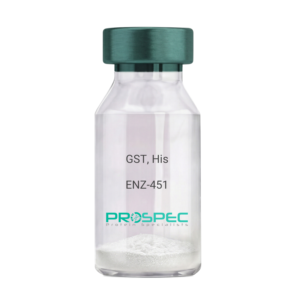
Name :
GST, HisDescription:
Glutathione S-Transferase Recombinant, His Tag
Glutathione S-Transferase, GST, Glutathione S-transferase class-mu 28 kDa isozyme, GST 28, EC 2.5.1.18, Sj28GST, Sj28 antigen.
Product # :
ENZ-451Price :
Quantity :
Shipping Method :
Shipped with Ice Packs
More Info
- description
- source
- formulation
- purity
- biological activity
- More Info
- Citations
Description
Recombinant Schistosoma japonicum GST full length protein contains a total of 244 amino acids (1-218 a.a.) expressed in E.coli, having a molecular mass of 28.3kDa. The GST protein is fused to a 20 amino acids His-Tag at N-terminus. The GST protein is purified by proprietary chromatographic techniques.
Source
Escherichia Coli.
Formulation
GST is supplied in PBS pH 7.4 & 10% glycerol.
Purity
Greater than 90% as determined by SDS-PAGE.
Biological Activity
>10 units/mg, & is defined as the amount of enzyme that conjugate 1.0 u mole of 1-chloro-2,4-dinitrobenzene (CDNB) with reduced glutathione per minute at pH-6.5 at 25C.
Citations
More Info
-
Introduction
Antioxidant enzyme Glutathione S- Transferase (GST) is thought to do the primary cellular defense mechanism against reactive oxygen species. GST reduces lipid hydroperoxides through its Se-independent glutathione peroxidase activity. The enzyme also detoxifies lipid peroxidation end products such as 4-hydroxynonenal (4-HNE).
The soluble GST is a 26 kDa protein which occurs as a dimer in all aerobic organisms. Each monomer has two domains, one that binds GSH and is an /-structure similar to thioredoxin and the other, all helical, that binds the hydrophobic substrate. The GST -fusion protein expression system is a widely used recombinant protein expression system that allows a peptide or a regulatory protein domain to be expressed as a fusion to the C-terminus of Schistosoma japonicum GST. Fusion proteins also possess GST -enzymatic activity and can undergo dimerization similar to in vivo. The fusion protein can be purified via GST -affinity column chromatography. In most cases, the desired peptides or domains are removed from GST by applying a specific protease that recognizes and cleaves the linker between the protein domain and GST. The technique has been widely used to generate different kinds of proteins for crystallization, molecular immunology studies, the production of vaccines and studies involving protein-protein and protein-DNA interactions. -
Synonyms
Glutathione S-Transferase, GST, Glutathione S-transferase class-mu 28 kDa isozyme, GST 28, EC 2.5.1.18, Sj28GST, Sj28 antigen.
-
Physical Appearance
Sterile Filtered clear solution.
-
Stability
Store at 4°C if entire vial will be used within 2-4 weeks. Store, frozen at -20°C for longer periods of time. For long term storage it is recommended to add a carrier protein (0.1% HSA or BSA).Avoid multiple freeze-thaw cycles.
-
Amino Acid Sequence
MGSSHHHHHH SSGLVPRGSH MSPILGYWKI KGLVQPTRLL LEYLEEKYEE HLYERDEGDK WRNKKFELGL EFPNLPYYID GDVKLTQSMAIIRYIADKHN MLGGCPKERA EISMLEGAVL DIRYGVSRIA YSKDFETLKV DFLSKLPEML KMFEDRLCHK TYLNGDHVTH PDFMLYDALDVVLYMDPMCL DAFPKLVCFK KRIEAIPQID KYLKSSKYIA WPLQGWQATF GGGDHPPKSD LVPR.
ProSpec's products are furnished for LABORATORY RESEARCH USE ONLY. The product may not be used as drugs, agricultural or pesticidal products, food additives or household chemicals.
- View Data Sheet
Name :
Gliadin Alpha WheatDescription:
Gliadin Alpha Wheat Recombinant
Product # :
PRO-2147Price :
Quantity :
Shipping Method :
Shipped with Ice Packs
More Info
- description
- source
- formulation
- purity
- More Info
- Citations
Description
Recombinant Wheat Gliadin Alpha protein produced in E.Coli and fused to a 6 His Tag at C-terminus, having a theoretical Mw of 34557.64 Dalton, pI 7.70. Purified by proprietary chromatographic technique.
Source
Escherichia Coli.
Formulation
Gliadin Alpha protein solution in 10mM Tris-HCl pH 7.2.
Purity
Protein is >90% pure.
Citations
More Info
-
Introduction
Wheat Gliadin and related gluten components from barley, rye and possibly oats can cause an abnormal immune response called Celiac disease which is a chronic gastrointestinal disorder. Celiac disease characteristics are flattening of the jejunal mucosa and intestinal lesions of variable severity in hereditarily inclined individuals. Even though Celiac disease is not a classic autoimmune disease it is related to anti-tissue transglutaminase antibodies and gliadin antibodies tests are most recommended in screening populations at risk for CD and other gluten-sensitive enteropathies. In the past, serologic tests for gliadin antibodies usually were not very precise and were not enough for accurate diagnosis due to missing deamidated epitopes within the authentic gliadin fraction traditionally used in diagnostic test kits. ProSpec's deamidated Gliadin isoform matches to the deamidated neo-epitopes, which in the natural antigen are formed by transglutaminase-mediated glutamine side chain deamidation.
-
Physical Appearance
Sterile Filtered clear solution.
-
Stability
Gliadin Alpha although stable at 4°C for 1 week, should be stored below -18°C. Please prevent freeze thaw cycles.
-
Amino Acid Sequence
MVRVPVPQLQPQNPSQQQPQEQVPLVQQQQFPGQQQPFPPQQPYPQPQPFPSQQPYLQ
LQPFPQPQLPYPQPQLPYPQPQLPYPQPQPFRPQQPYPQSQPQYSQPQQPISQQQQQQQQ
QQQQKQQQQQQQQILQQILQQQLIPCRDVVLQQHSIAYGSSQVLQQSTYQLVQQLCCQQL
WQIPEQSRCQAIHNVVHAIILHQQQQQQQQQQQQPLSQVSFQQPQQQYPSGQGSFQPSQ
QNPQAQGSVQPQQLPQFEEIRNLALETLPAMCNVYIPPYCTIAPVGIFGTNYRHHHHHH.
ProSpec's products are furnished for LABORATORY RESEARCH USE ONLY. The product may not be used as drugs, agricultural or pesticidal products, food additives or household chemicals.