Search results
1000 results found for “hemoglobin”
Name
Description
Product #
Price
Quantity
Shipping Method
- View Data Sheet
Name :
HLA-G HumanDescription:
Major Histocompatibility Complex Class I G Human Recombinant
Major Histocompatibility Complex, Class I, G, HLA-G Histocompatibility Antigen, Class I, G, MHC Class I Antigen G, B2 Microglobulin, HLA G Antigen, HLA Class I Histocompatibility Antigen, Alpha Chain G, Mutant MHC Class Ib Antigen, Mutant MHC Class I Antigen, MHC Class Ib Antigen, HLA-6.0, MHC-G, HLAG, HLA class I histocompatibility antigen, alpha chain G, HLA G antigen, MHC class I antigen G.
Product # :
PRO-2520Price :
Quantity :
Shipping Method :
Shipped with Ice Packs
More Info
- description
- source
- formulation
- purity
- More Info
Description
HLA-G Human Recombinant produced in E.Coli is a single, non-glycosylated polypeptide chain containing 309 amino acids (25-308 a.a) and having a molecular mass of 35.3kDa.HLA-G is fused to a 25 amino acid His-tag at N-terminus & purified by proprietary chromatographic techniques.
Source
Escherichia Coli.
Formulation
HLA-G protein solution (0.25mg/ml) contains 20% glycerol and PBS (pH 7.4).
Purity
Greater than 90% as determined by SDS-PAGE.
More Info
-
Introduction
HLA-G (Major Histocompatibility Complex Class I G) is a member of the HLA class I heavy chain paralogues. This class I molecule is a heterodimer which comprises a heavy chain as well as a light chain, beta-2 microglobulin, while the heavy chain is fixed in the membrane. HLA-G is expressed on fetal derived placental cells. HLA-G is a non-classical class-I HLA molecule linked with immuno-modulatory & anti-inflammatory properties which interacts with inhibitory receptors such as, ILT2/ILT4/KIR2DL4, that are present on different immune cells. HLA-G inhibits the proliferation of T cells, B cells & natural killer cells, moreover it also induces regulatory T cells.
-
Synonyms
Major Histocompatibility Complex, Class I, G, HLA-G Histocompatibility Antigen, Class I, G, MHC Class I Antigen G, B2 Microglobulin, HLA G Antigen, HLA Class I Histocompatibility Antigen, Alpha Chain G, Mutant MHC Class Ib Antigen, Mutant MHC Class I Antigen, MHC Class Ib Antigen, HLA-6.0, MHC-G, HLAG, HLA class I histocompatibility antigen, alpha chain G, HLA G antigen, MHC class I antigen G.
-
Physical Appearance
Sterile Filtered clear solution.
-
Stability
Store at 4°C if entire vial will be used within 2-4 weeks. Store, frozen at -20°C for longer periods of time. For long term storage it is recommended to add a carrier protein (0.1% HSA or BSA).Avoid multiple freeze-thaw cycles.
-
Amino Acid Sequence
MGSSHHHHHH SSGLVPRGSH MGSHMGSHSM RYFSAAVSRP GRGEPRFIAM GYVDDTQFVR FDSDSACPRM EPRAPWVEQE GPEYWEEETR NTKAHAQTDR MNLQTLRGYY NQSEASSHTL QWMIGCDLGS DGRLLRGYEQ YAYDGKDYLA LNEDLRSWTA ADTAAQISKR KCEAANVAEQ RRAYLEGTCV EWLHRYLENG KEMLQRADPP KTHVTHHPVF DYEATLRCWA LGFYPAEIIL TWQRDGEDQT QDVELVETRP AGDGTFQKWA AVVVPSGEEQ RYTCHVQHEG LPEPLMLRWK QSSLPTIPI.
ProSpec's products are furnished for LABORATORY RESEARCH USE ONLY. The product may not be used as drugs, agricultural or pesticidal products, food additives or household chemicals.
- View Data Sheet
Name :
Erythropoietin Human, ActiveDescription:
Erythropoietin Receptor Human Recombinant, Active
EPO-R, EPOR, Erythropoietin Receptor.
Product # :
CYT-997Price :
Quantity :
Shipping Method :
Shipped with Ice Packs
More Info
- description
- source
- formulation
- purity
- biological activity
- More Info
Description
EPOR produced in Sf9 Baculovirus cells is a single, glycosylated polypeptide chain containing 232 amino acids (25-250a.a.) and having a molecular mass of 25.6kDa (Molecular size on SDS-PAGE will appear at approximately 28-40kDa).EPOR is expressed with a 6 amino acid His-tag at C-Terminus and purified by proprietary chromatographic techniques.
Source
Sf9, Baculovirus cells.
Formulation
EPOR protein solution (0.5mg/ml) contains Phosphate Buffered Saline (pH 7.4) and 10% glycerol.
Purity
Greater than 95.0% as determined by analysis by SDS-PAGE.
Biological Activity
Measured by its ability to inhibit EPO dependent proliferation assay using TF-1 human erythroleukemic cells. The ED50 for this effect less or equal to 70ng/ml.More Info
-
Introduction
Erythropoietin receptor, also known as EPOR arbitrates erythropoietin-induced erythroblast proliferation as well as differentiation. During EPO binding, EPOR activates Jak2 tyrosine kinase which activates various intracellular pathways including: Ras/MAP kinase, phosphatidylinositol 3-kinase and STAT transcription factors. Furthermore, stimulated EPOR has a function in erythroid cell survival. Mutations in EPOR may possibly produce erythroleukemia and familial erythrocytosis. In addition, dysregulation of EPOR can affect on the growth of selected tumors.
-
Synonyms
EPO-R, EPOR, Erythropoietin Receptor.
-
Physical Appearance
Sterile Filtered clear solution.
-
Stability
Store at 4°C if entire vial will be used within 2-4 weeks. Store, frozen at -20°C for longer periods of time. For long term storage it is recommended to add a carrier protein (0.1% HSA or BSA).Avoid multiple freeze-thaw cycles.
-
Amino Acid Sequence
APPPNLPDPK FESKAALLAA RGPEELLCFT ERLEDLVCFW EEAASAGVGP GNYSFSYQLE DEPWKLCRLH QAPTARGAVR FWCSLPTADT SSFVPLELRV TAASGAPRYH RVIHINEVVL LDAPVGLVAR LADESGHVVL RWLPPPETPM TSHIRYEVDV SAGNGAGSVQ RVEILEGRTE CVLSNLRGRT RYTFAVRARM AEPSFGGFWS AWSEPVSLLT PSDLDPHHHH HH.
-
Background
What is the molecular weight/Mw of ERYTHROPOIETIN Protein?
ERYTHROPOIETIN Protein has a total Mw of 25.6kDa.
What is the source or expression system of ERYTHROPOIETIN Protein?
Sf9, Baculovirus cells.
What is the Purity of ERYTHROPOIETIN Protein?
ERYTHROPOIETIN Protein is >95% pure as determined by SDS-PAGE.
What is the Biological Activity of ERYTHROPOIETIN Protein?
Measured by its ability to inhibit EPO dependent proliferation assay using TF-1 human erythroleukemic cells. The ED50 for this effect less or equal to 70ng/ml.
What is the amino acid sequence of ERYTHROPOIETIN Protein?
APPPNLPDPK FESKAALLAA RGPEELLCFT ERLEDLVCFW EEAASAGVGP GNYSFSYQLE DEPWKLCRLH QAPTARGAVR FWCSLPTADT SSFVPLELRV TAASGAPRYH RVIHINEVVL LDAPVGLVAR LADESGHVVL RWLPPPETPM TSHIRYEVDV SAGNGAGSVQ RVEILEGRTE CVLSNLRGRT RYTFAVRARM AEPSFGGFWS AWSEPVSLLT PSDLDPHHHH HH.
What applications can ERYTHROPOIETIN Protein be used in?
ERYTHROPOIETIN Protein can probably be used in western blot, ELISA and Lateral Flow.
What is the endotoxin level for ERYTHROPOIETIN Protein?
The endotoxin level is minimal, ERYTHROPOIETIN Protein was purified using conventional chromatography techniques.
ProSpec's products are furnished for LABORATORY RESEARCH USE ONLY. The product may not be used as drugs, agricultural or pesticidal products, food additives or household chemicals.
- View Data Sheet
Name :
FLT1 Human, HEK ActiveDescription:
Vascular Endothelial Growth Factor receptor-1 Human Recombinant, HEK Active
FLT-1, FLT1, Tyrosine-protein kinase receptor FLT, Flt-1, Tyrosine-protein kinase FRT, Fms-like tyrosine kinase 1, VEGFR-1.
Product # :
PKA-134Price :
Quantity :
Shipping Method :
Shipped with Ice Packs
More Info
- description
- source
- formulation
- purity
- biological activity
- More Info
Description
FLT1 Human Recombinant is a single, glycosylated polypeptide chain containing 535 amino acids (27-328a.a) and having a molecular mass of 60.3kDa (calculated). FLT1 is fused to a 233 amino acid hIgG-Tag at C-terminus and is purified by proprietary chromatographic techniques.
Source
HEK293
Formulation
FLT1 protein solution (0.5mg/ml) contains 10% Glycerol and Phosphate-Buffered Saline (pH 7.4).
Purity
Greater than 90.0% as determined by SDS-PAGE.
Biological Activity
The ED50 range ≤ 60ng/ml and is measured by its ability to inhibit proliferation using HUVEC human umbilical vein endothelial cells in the
presence of Human VEGF165.More Info
-
Synonyms
FLT-1, FLT1, Tyrosine-protein kinase receptor FLT, Flt-1, Tyrosine-protein kinase FRT, Fms-like tyrosine kinase 1, VEGFR-1.
-
Physical Appearance
Filtered clear solution.
-
Stability
Store at 4°C if entire vial will be used within 2-4 weeks.Store, frozen at -20°C for longer periods of time.Avoid multiple freeze-thaw cycles.
-
Amino Acid Sequence
SKLKDPELSL KGTQHIMQAG QTLHLQCRGE AAHKWSLPEM VSKESERLSI TKSACGRNGK QFCSTLTLNT AQANHTGFYS CKYLAVPTSK KKETESAIYI FISDTGRPFV EMYSEIPEII HMTEGRELVI PCRVTSPNIT VTLKKFPLDT LIPDGKRIIW DSRKGFIISN ATYKEIGLLT CEATVNGHLY KTNYLTHRQT NTIIDVQIST PRPVKLLRGH TLVLNCTATT PLNTRVQMTW SYPDEKNKRA SVRRRIDQSN SHANIFYSVL TIDKMQNKDK GLYTCRVRSG PSFKSVNTSV HILEPKSCDK THTCPPCPAP ELLGGPSVFL FPPKPKDTLM ISRTPEVTCV VVDVSHEDPE VKFNWYVDGV EVHNAKTKPR EEQYNSTYRV VSVLTVLHQD WLNGKEYKCK VSNKALPAPI EKTISKAKGQ PREPQVYTLP PSRDELTKNQ VSLTCLVKGF YPSDIAVEWE SNGQPENNYK TTPPVLDSDG SFFLYSKLTV DKSRWQQGNV FSCSVMHEAL HNHYTQKSLS LSPGK.
-
Background
VEGFR-1 (Vascular Endothelial Growth Factor Receptor-1), also known as Flt-1 (Fms-like tyrosine kinase 1), is a critical receptor involved in angiogenesis and vascular development. This research paper delves into the structure, function, and therapeutic implications of VEGFR-1, shedding light on its multifaceted role in various physiological and pathological processes.
VEGFR-1 is a transmembrane receptor tyrosine kinase belonging to the VEGF receptor family. It is primarily expressed on endothelial cells and plays a pivotal role in mediating the cellular responses to VEGF ligands. Upon ligand binding, VEGFR-1 initiates intracellular signaling cascades that regulate endothelial cell proliferation, migration, and survival, ultimately contributing to the formation of new blood vessels.
The structure of VEGFR-1 comprises distinct domains, including an extracellular ligand-binding domain, a transmembrane domain, and an intracellular tyrosine kinase domain. The extracellular domain facilitates the interaction between VEGF ligands and the receptor, while the intracellular domain transduces downstream signals by phosphorylating specific tyrosine residues.
VEGFR-1 exhibits not only ligand-dependent but also ligand-independent functions. In addition to its role as a VEGF receptor, it can act as a decoy receptor, sequestering VEGF and modulating the bioavailability of VEGF ligands. This unique property allows VEGFR-1 to regulate VEGF signaling and influence angiogenic processes.
The signaling pathways activated by VEGFR-1 are diverse and intricate, involving multiple downstream effectors, such as PI3K/AKT, MAPK/ERK, and STAT proteins. These pathways regulate endothelial cell behaviors, including proliferation, migration, and differentiation, which are crucial for angiogenesis. Perturbations in VEGFR-1 signaling have been implicated in various pathological conditions, including cancer, retinopathy, and inflammatory disorders.
The therapeutic targeting of VEGFR-1 has gained considerable attention for its potential in managing angiogenesis-related diseases. Inhibitors specifically designed to block VEGFR-1 have been developed to suppress aberrant angiogenesis and impede tumor growth. Moreover, VEGFR-1-based therapies have been explored for ocular diseases like wet age-related macular degeneration (AMD) and diabetic retinopathy, aiming to alleviate pathological neovascularization.
The availability of VEGFR-1 human recombinant proteins has facilitated in-depth research and the development of potential therapeutic interventions. Recombinant VEGFR-1 proteins serve as valuable tools for investigating VEGF-VEGFR-1 interactions, screening drug candidates, and elucidating the underlying molecular mechanisms of VEGFR-1-mediated signaling pathways.
ProSpec's products are furnished for LABORATORY RESEARCH USE ONLY. The product may not be used as drugs, agricultural or pesticidal products, food additives or household chemicals.
- View Data Sheet
Name :
POLD4 HumanDescription:
Polymerase Delta 4 Human Recombinant
DNA polymerase delta subunit 4, p12, POLDS, DNA polymerase delta subunit p12, POLD4, Polymerase Delta 4.
Product # :
PRO-1667Price :
Quantity :
Shipping Method :
Shipped with Ice Packs
More Info
- description
- source
- formulation
- purity
- More Info
Description
POLD4 Human Recombinant produced in E.coli is a single, non-glycosylated polypeptide chain containing 130 amino acids (1-107) and having a molecular mass of 14.8kDa.POLD4 is fused to a 23 amino acid His-tag at N-terminus & purified by proprietary chromatographic techniques.
Source
Escherichia Coli.
Formulation
The POLD4 solution (0.25mg/ml) contains 20mM Tris-HCl buffer (pH 8.0), 0.2M NaCl, 40% glycerol and 2mM DTT.
Purity
Greater than 90.0% as determined by SDS-PAGE.
More Info
-
Introduction
Polymerase Delta 4 also known as POLD4 is the smallest subunit of DNA polymerase delta. DNA polymerase delta which takes a vital part in DNA replication and repair possesses both polymerase and 3' to 5' exonuclease activity. POLD4 increases the activity of DNA polymerase delta and takes a part in fork repair and stabilization during interactions with the DNA helicase Bloom syndrome protein. POLD4 contributes to PCNA-dependent activity of DNA polymerase delta.
-
Synonyms
DNA polymerase delta subunit 4, p12, POLDS, DNA polymerase delta subunit p12, POLD4, Polymerase Delta 4.
-
Physical Appearance
Sterile Filtered colorless solution.
-
Stability
Store at 4°C if entire vial will be used within 2-4 weeks. Store, frozen at -20°C for longer periods of time. For long term storage it is recommended to add a carrier protein (0.1% HSA or BSA).Avoid multiple freeze-thaw cycles.
-
Amino Acid Sequence
MGSSHHHHHH SSGLVPRGSH MGSMGRKRLI TDSYPVVKRR EGPAGHSKGE LAPELGEEPQ PRDEEEAELE LLRQFDLAWQ YGPCTGITRL QRWCRAKQMG LEPPPEVWQV LKTHPGDPRF QCSLWHLYPL.
ProSpec's products are furnished for LABORATORY RESEARCH USE ONLY. The product may not be used as drugs, agricultural or pesticidal products, food additives or household chemicals.
- View Data Sheet
Name :
Leptin Human, HisDescription:
Leptin Human Recombinant, His Tag
OB Protein, Obesity Protein, OBS, Obesity factor.
Product # :
CYT-287Price :
Quantity :
Shipping Method :
Shipped at Room temp
More Info
- description
- source
- formulation
- purity
- More Info
Description
Leptin Human Recombinant produced in E.Coli is a single, non-glycosylated, polypeptide chain containing amino acids 48-167 and having a total molecular mass of 19 kDa including the 4 kDa His tag.The Leptin is purified by proprietary chromatographic techniques.
Source
Escherichia Coli.
Formulation
The protein was lyophilized from a concentrated (1mg/ml) solution with 1X PBS, 0.1% SDS and 1mM DTT.
Purity
Greater than 90.0% as determined by SDS-PAGE.
More Info
-
Introduction
Leptin is a protein hormone with important effects in regulating body weight, metabolism and reproductive function. The protein is approximately~16 kDa in mass and encoded by the obese (ob)gene. leptin is expressed predominantly by adipocytes, which fits with the idea that body weight is sensed as the total mass of fat in the body. Smaller amounts of leptin are also secreted by cellsin the epithelium of the stomach and in the placenta. Leptin receptors are highly expressed in areas of the hypothalamus known to be important in regulating body weight, as well as in T lymphocytes and vascular endothelial cells.
-
Synonyms
OB Protein, Obesity Protein, OBS, Obesity factor.
-
Physical Appearance
Sterile Filtered White lyophilized (freeze-dried) powder.
-
Stability
Lyophilized Leptin although stable at room temperature, should be stored desiccated below 0°C. Reconstituted Leptin is best stored refrigerated at 4°C.Please avoid freeze-thaw cycles.
-
Solubility
The lyophilized Leptin is very soluble in water and most aqueous buffers below and above the isoelectric point.
ProSpec's products are furnished for LABORATORY RESEARCH USE ONLY. The product may not be used as drugs, agricultural or pesticidal products, food additives or household chemicals.
- View Data Sheet
Name :
HAO1 Human, ActiveDescription:
Hydroxyacid Oxidase 1 Human Recombinant, Active
Hydroxyacid oxidase 1, HAOX1, Glycolate oxidase, GOX, HAO1, GOX1.
Product # :
ENZ-1094Price :
Quantity :
Shipping Method :
Shipped with Ice Packs
More Info
- description
- source
- formulation
- purity
- biological activity
- More Info
Description
HAO1 Human Recombinant produced in E.Coli is a single, non-glycosylated polypeptide chain containing 406 amino acids (1-370 a.a) and having a molecular mass of 45.0kDa. HAO1 is fused to a 36 amino acid His-tag at N-terminus & purified by proprietary chromatographic techniques.
Source
Escherichia Coli.
Formulation
HAO1 protein solution (1mg/ml) contains 20% glycerol, 20mM Tris-Hcl (pH8.0) and 0.5M NaCl.
Purity
Greater than 95% as determined by SDS-PAGE.
Biological Activity
Specific activity is > 3000 pmol/min/ug, and defined as the amount of enzyme that oxidize glyoxylate at pH 8.0 at 25C.
More Info
-
Introduction
Glycolate oxidase (HAO1) is a part of the superfamily of the alpha hydroxy acid oxidases (HAO) enzymes. HAO1 catalyses the FMN mediated oxidation of glycolate to glyoxylate and glyoxylate to oxalate by reducing oxygen to hydrogen peroxide. HAO1 is expressed mainly in the liver and pancreas and is most active on twocarbon substrates such as glycolate. HAO1 isthe main cause of hyperoxaluria, a disorder in which large deposits of calcium oxalate form kidney stones.
-
Synonyms
Hydroxyacid oxidase 1, HAOX1, Glycolate oxidase, GOX, HAO1, GOX1.
-
Physical Appearance
Sterile filtered yellowish solution.
-
Stability
Store at 4°C if entire vial will be used within 2-4 weeks. Store, frozen at -20°C for longer periods of time. For long term storage it is recommended to add a carrier protein (0.1% HSA or BSA).Avoid multiple freeze-thaw cycles.
-
Amino Acid Sequence
MRGSHHHHHH GMASMTGGQQ MGRDLYDDDD KDRWGSMLPR LICINDYEQH AKSVLPKSIY DYYRSGANDE ETLADNIAAF SRWKLYPRMLRNVAETDLST SVLGQRVSMP ICVGATAMQR MAHVDGELAT VRACQSLGTG MMLSSWATSS IEEVAEAGPE ALRWLQLYIY KDREVTKKLVRQAEKMGYKA IFVTVDTPYL GNRLDDVRNR FKLPPQLRMK NFETSTLSFS PEENFGDDSG LAAYVAKAID PSISWEDIKW LRRLTSLPIVAKGILRGDDA REAVKHGLNG ILVSNHGARQ LDGVPATIDV LPEIVEAVEG KVEVFLDGGV RKGTDVLKAL ALGAKAVFVG RPIVWGLAFQGEKGVQDVLE ILKEEFRLAM ALSGCQNVKV IDKTLVRKNP LAVSKI.
ProSpec's products are furnished for LABORATORY RESEARCH USE ONLY. The product may not be used as drugs, agricultural or pesticidal products, food additives or household chemicals.
- View Data Sheet
Name :
COX5B HumanDescription:
Cytochrome C Oxidase Subunit Vb Human Recombinant
Cytochrome c oxidase subunit 5B, mitochondrial precursor, COXVB, COX5B, Mitochondrial, Cytochrome c oxidase polypeptide Vb.
Product # :
PRO-1513Price :
Quantity :
Shipping Method :
Shipped with Ice Packs
More Info
- description
- source
- formulation
- purity
- More Info
Description
COX5B Human Recombinant produced in E.Coli is a single, non-glycosylated polypeptide chain containing 121 amino acids (32-129a.a) and having a molecular mass of 13kDa. COX5B is fused to a 23 amino acid His-tag at N-terminus & purified by proprietary chromatographic techniques.
Source
Escherichia Coli.
Formulation
COX5B protein solution (0.5mg/ml) containing 20mM Tris-HCl buffer (pH8.0), 0.1M NaCl, 20% glycerol and 1mM DTT.
Purity
Greater than 95.0% as determined by SDS-PAGE.
More Info
-
Introduction
Cytochrome c oxidase subunit VB (COX5B) is the terminal enzyme of the mitochondrial respiratory chain. COX5B is a multi-subunit enzyme complex which couples the transfer of electrons from cytochrome c to molecular oxygen and contributes to a proton electrochemical gradient across the inner mitochondrial membrane. There are two isoforms of COX5:COX5a and COX5b. Transcription of COX5A (the aerobic isoform) is up-regulated as the rate of cellular respiration increases, when oxygen levels within the cell are high. However, when oxygen levels are low, COX5B (the hypoxic isoform) transcription increases and functions to maximize the turnover rate of the COX apoenzyme.
-
Synonyms
Cytochrome c oxidase subunit 5B, mitochondrial precursor, COXVB, COX5B, Mitochondrial, Cytochrome c oxidase polypeptide Vb.
-
Physical Appearance
Sterile filtered colorless solution.
-
Stability
Store at 4°C if entire vial will be used within 2-4 weeks. Store, frozen at -20°C for longer periods of time. For long term storage it is recommended to add a carrier protein (0.1% HSA or BSA).Avoid multiple freeze-thaw cycles.
-
Amino Acid Sequence
MGSSHHHHHH SSGLVPRGSH MGSASGGGVP TDEEQATGLE REIMLAAKKG LDPYNVLAPK GASGTREDPN LVPSISNKRI VGCICEEDNT SVVWFWLHKG EAQRCPRCGA HYKLVPQQLA H.
ProSpec's products are furnished for LABORATORY RESEARCH USE ONLY. The product may not be used as drugs, agricultural or pesticidal products, food additives or household chemicals.
- View Data Sheet
Name :
VSTM2L HumanDescription:
V-Set and Transmembrane Domain Containing 2 Like Human Recombinant
V-set and transmembrane domain-containing protein 2-like protein, VSTM2L, C20orf102, dJ1118M15.2.
Product # :
PRO-1018Price :
Quantity :
Shipping Method :
Shipped with Ice Packs
More Info
- description
- source
- formulation
- purity
- More Info
Description
VSTM2L Human Recombinant produced in E.Coli is a single, non-glycosylated polypeptide chain containing 205 amino acids (25-204 a.a.) and having a molecular mass of 22.6kDa. VSTM2L is fused to a 25 amino acid His-tag at N-terminus & purified by proprietary chromatographic techniques.
Source
E.coli.
Formulation
VSTM2L protein solution (0.25mg/ml) containing 20mM Tris-HCl buffer (pH8.0), 20% glycerol, 0.1M NaCl,1mM DTT and 0.1mM PMSF.
Purity
Greater than 90% as determined by SDS-PAGE.
More Info
-
Introduction
V-set and transmembrane domain containing 2 (VSTM2L) is a novel modulator of HN neuroprotective activity. VSTM2L colocalizes with HN in distinct brain areas as well as in primary cultured neurons, where it plays a part in the modulation of neuronal viability. When tested in HN neuroprotection bioassays, VSTM2L acts as a potent antagonist of HN neuroprotective activity. The VSTM2L protein is the first example of a secreted antagonist of HN and may have a role in the modulation of HN biological functions.
-
Synonyms
V-set and transmembrane domain-containing protein 2-like protein, VSTM2L, C20orf102, dJ1118M15.2.
-
Physical Appearance
Sterile Filtered colorless solution.
-
Stability
Store at 4°C if entire vial will be used within 2-4 weeks. Store, frozen at -20°C for longer periods of time. For long term storage it is recommended to add a carrier protein (0.1% HSA or BSA).Avoid multiple freeze-thaw cycles.
-
Amino Acid Sequence
MGSSHHHHHH SSGLVPRGSH MGSHMTRPAG HAPWDNHVSG HALFTETPHD MTARTGEDVE MACSFRGSGS PSYSLEIQWW YVRSHRDWTD KQAWASNQLK ASQQEDAGKE ATKISVVKVV GSNISHKLRL SRVKPTDEGS YECRVIDFSD GKARHHKVKA YLRVQPGENS VLHLPEAPPA APAPPPPKPG KELRKRSVDQ EACSL.
ProSpec's products are furnished for LABORATORY RESEARCH USE ONLY. The product may not be used as drugs, agricultural or pesticidal products, food additives or household chemicals.
- View Data Sheet
Name :
SPOP HumanDescription:
Speckle-Type POZ Protein Human Recombinant
Speckle-type POZ protein, HIB homolog 1, Roadkill homolog 1, SPOP, TEF2.
Product # :
PRO-195Price :
Quantity :
Shipping Method :
Shipped with Ice Packs
More Info
- description
- source
- formulation
- purity
- More Info
Description
SPOP Human Recombinant fused with a 20 amino acid His tag at N-terminus produced in E.Coli is a single, non-glycosylated, polypeptide chain containing 394 amino acids (1-374 a.a.) and having a molecular mass of 44.3kDa. The SPOP is purified by proprietary chromatographic techniques.
Source
Escherichia Coli.
Formulation
The SPOP solution (0.25mg/ml) contains 20mM Tris-HCl buffer (pH 8.0), 5mM DTT, 50% glycerol, 0.2M NaCl and 2mM EDTA.
Purity
Greater than 90.0% as determined by SDS-PAGE.
More Info
-
Introduction
Speckle-type POZ protein (SPOP) belongs to the Tdpoz family containing one N-terminal MATH (Meprin and TRAF homology) domain and one C-terminal BTB/POZ domain. SPOP inhibits IPF1/PDX1 transactivation of established target promoters, may be by recruiting a repressor complex. SPOP is involved in ubiquitinylation and protein degradation as a result of an interaction with CUL-3.
-
Synonyms
Speckle-type POZ protein, HIB homolog 1, Roadkill homolog 1, SPOP, TEF2.
-
Physical Appearance
SPOP is supplied as a sterile Filtered colorless solution.
-
Stability
Store at 4°C if entire vial will be used within 2-4 weeks. Store, frozen at -20°C for longer periods of time. For long term storage it is recommended to add a carrier protein (0.1% HSA or BSA).Avoid multiple freeze-thaw cycles.
-
Amino Acid Sequence
MGSSHHHHHH SSGLVPRGSH MSRVPSPPPP AEMSSGPVAE SWCYTQIKVV KFSYMWTINN FSFCREEMGE VIKSSTFSSG ANDKLKWCLR VNPKGLDEES KDYLSLYLLL VSCPKSEVRA KFKFSILNAK GEETKAMESQ RAYRFVQGKD WGFKKFIRRD FLLDEANGLL PDDKLTLFCE
VSVVQDSVNI SGQNTMNMVK VPECRLADEL GGLWENSRFT DCCLCVAGQE FQAHKAILAA RSPVFSAMFE HEMEESKKNR VEINDVEPEV FKEMMCFIYT GKAPNLDKMA DDLLAAADKY ALERLKVMCE DALCSNLSVE NAAEILILAD LHSADQLKTQ AVDFINYHAS DVLETSGWKS MVVSHPHLVA EAYRSLASAQ CPFLGPPRKR LKQS.
ProSpec's products are furnished for LABORATORY RESEARCH USE ONLY. The product may not be used as drugs, agricultural or pesticidal products, food additives or household chemicals.
- View Data Sheet
Name :
C3 HumanDescription:
Complement C3 Human
Complement C3, C3 and PZP-like alpha-2-macroglobulin domain-containing protein 1, C3, CPAMD1.
Product # :
PRO-2684Price :
Quantity :
Shipping Method :
Shipped with Ice Packs
More Info
- description
- source
- formulation
- purity
- More Info
Description
Human Complement C3 produced in Human plasma having a molecular mass of 185 kDa.
Source
Human Plasma.
Formulation
C3 solution contains phosphate buffer saline.
Purity
Greater than 94.0% as determined by SDS-PAGE.
More Info
-
Introduction
Native human C3 is a naturally glycosylated polypeptide containing two disulfide-linked chains. C3 is central to the activation of all 3 pathways of complement activation. Initiation of each pathway generates proteolytic enzyme complexes which binds the target surface. These enzymes cleave a peptide bond in C3 releasing the anaphylatoxin C3a and activating C3b. Most of the C3 activated during complement activation never attaches to the surface due to its thioester reaction with water forming fluid phase C3b which is rapidly inactivated by factors H and I forming iC3b. Surface-bound C3b is necessary in all 3 pathways for efficient activation of C5 and formation of C5b-9 complexes that lyse the target cell membrane.
-
Synonyms
Complement C3, C3 and PZP-like alpha-2-macroglobulin domain-containing protein 1, C3, CPAMD1.
-
Physical Appearance
Sterile Filtered solution.
-
Stability
C3 Human is stable at 4°C if entire vial will be used within 2-4 weeks.Store, frozen below -20°C for longer periods of time.For long term storage it is recommended to add a carrier protein (0.1% HSA or BSA).Avoid multiple freeze-thaw cycles.
-
Human Virus Test
Plasma from each donor has been tested and found negative for antibody to HIV-1,HIV-2, HCV, HTLV-I &II, STS and HBSAG.
ProSpec's products are furnished for LABORATORY RESEARCH USE ONLY. The product may not be used as drugs, agricultural or pesticidal products, food additives or household chemicals.
- View Data Sheet
Name :
GLO1 Human, ActiveDescription:
Glyoxalase-I Human Recombinant, Active
GLYI, GLOD1, GLO1, Glyoxalase-1, Lactoylglutathione lyase, Methylglyoxalase, Aldoketomutase, Ketone-aldehyde mutase, Glyoxalase I, S-D-lactoylglutathione methylglyoxal lyase, Glx I.
Product # :
ENZ-999Price :
Quantity :
Shipping Method :
Shipped with Ice Packs
More Info
- description
- source
- formulation
- purity
- biological activity
- More Info
Description
Glyoxalase-I Human Recombinant produced in E.Coli is a single, non-glycosylated, polypeptide chain containing 184 amino acids and having a molecular mass of 20.7 kDa. Glyoxalase-1 is purified by proprietary chromatographic techniques.
Source
Escherichia Coli.
Formulation
Glyoxalase-1 solution containing 20mM Tris-HCl pH-8, 1mM DTT and 10% glycerol.
Purity
Greater than 90.0% as determined by SDS-PAGE.
Biological Activity
Specific activity: > 400 units/mg. One unit will form 1.0umol of S-lactoylgutathione from methylglyoxal and reduced glutathione per minute at pH6.5 at 25CMore Info
-
Introduction
GLO1 is involved in the catalysis and formation of S-lactoyl-glutathione from methylglyoxal condensation and reduced glutatione. GLO1 is linked to HLA and is localized to 6p21.3-p21.1, between HLA and the centromere. GLO1 enzyme is ubundantly expressed and present in numerous tumor cell lines, in which its concentration is often upregulated ubiquitisly. GLO1 is a major susceptible gene for autism in an ethnic Chinese population from Taiwan. GLO1 might be involved in the pathophysiology of mood disorders. GLO1 plays a role in the pathophysiology of mood disorders. Overexpression of GLO1 is associated with kidney tumor.
-
Synonyms
GLYI, GLOD1, GLO1, Glyoxalase-1, Lactoylglutathione lyase, Methylglyoxalase, Aldoketomutase, Ketone-aldehyde mutase, Glyoxalase I, S-D-lactoylglutathione methylglyoxal lyase, Glx I.
-
Physical Appearance
Sterile filtered colorless solution.
-
Stability
Store at 4°C if entire vial will be used within 2-4 weeks. Store, frozen at -20°C for longer periods of time. For long term storage it is recommended to add a carrier protein (0.1% HSA or BSA).Avoid multiple freeze-thaw cycles.
-
Amino Acid Sequence
MAEPQPPSGG LTDEAALSCC SDADPSTKDF LLQQTMLRVK DPKKSLDFYT RVLGMTLIQK CDFPIMKFSL YFLAYEDKND IPKEKDEKIAWALSRKATLE LTHNWGTEDD ETQSYHNGNS DPRGFGHIGI AVPDVYSACK RFEELGVKFV KKPDDGKMKG LAFIQDPDGY WIEILNPNKM ATLM.
ProSpec's products are furnished for LABORATORY RESEARCH USE ONLY. The product may not be used as drugs, agricultural or pesticidal products, food additives or household chemicals.
- View Data Sheet
Name :
SPARC HumanDescription:
Secreted Protein Acidic & Rich in Cysteine Recombinant Human
Osteonectin, ON, Basement-membrane protein 40, BM-40, SPARC, Secreted Protein acidic and Rich in Cysteine.
Product # :
PRO-2602Price :
Quantity :
Shipping Method :
Shipped at Room temp
More Info
- description
- source
- formulation
- purity
- biological activity
- More Info
Description
Secreted Protein Acidic & Rich in Cysteine Human Recombinant produced in E.Coli is a single, non- glycosylated polypeptide chain containing 286 amino acids and having a molecular mass of 32.7kDa.SPARC is purified by proprietary chromatographic techniques.
Source
Escherichia Coli.
Formulation
Lyophilized from a 0.2μm filtered concentrated solution in PBS, pH 7.4.
Purity
Greater than 98.0% as determined by:
(a) Analysis by RP-HPLC.
(b) Analysis by SDS-PAGE.Biological Activity
The ED50 as determined by its ability to inhibit the cell growth of Mv1Lu mink lung epithelial cells is < 3.0 µg/mL, corresponding to a specific activity of > 333 IU/mg.
More Info
-
Introduction
SPARC, an acronym for “secreted protein, acidic and rich in cysteine”, is also known as osteonectin or BM-40. It is the founding member of a family of secreted matricellular proteins with similar domain structure. The 303 amino acid, 43 kDa protein contains a 17 aa signal sequence, an N-terminal acidic region that binds calcium, a follistatin domain containing Kazal-like sequences, and a C-terminal extracellular calcium (EC) binding domain with two EF-hand motifs. SPARC is produced by fibroblasts, capillary endothelial cells, platelets and macrophages, especially in areas of tissue morphogenesis and remodeling. SPARC shows context-specific effects, but generally inhibits adhesion, spreading and proliferation, and promotes collagen matrix formation. For endothelial cells, SPARC disrupts focal adhesions and binds and sequesters PDGF and VEGF. SPARC is abundantly expressed in bone, where it promotes osteoblast differentiation and inhibits adipogenesis.
-
Synonyms
Osteonectin, ON, Basement-membrane protein 40, BM-40, SPARC, Secreted Protein acidic and Rich in Cysteine.
-
Physical Appearance
Sterile Filtered White lyophilized (freeze-dried) powder.
-
Stability
Lyophilized SPARC although stable at room temperature for 3 weeks, should be stored desiccated below -18°C. Upon reconstitution Secreted Protein Acidic & Rich in Cysteine should be stored at 4°C between 2-7 days and for future use below -18°C.Please prevent freeze-thaw cycles.
-
Solubility
It is recommended to reconstitute the lyophilized SPARC in sterile 18MΩ-cm H2O not less than 100µg/ml, which can then be further diluted to other aqueous solutions.
-
Amino Acid Sequence
APQQEALPDE TEVVEETVAE VTEVSVGANP VQVEVGEFDD GAEETEEEVV AENPCQNHHC KHGKVCELDE NNTPMCVCQD PTSCPAPIGE FEKVCSNDNK TFDSSCHFFA TKCTLEGTKK GHKLHLDYIG PCKYIPPCLD SELTEFPLRM RDWLKNVLVT LYERDEDNNL LTEKQKLRVK KIHENEKRLE AGDHPVELLA RDFEKNYNMY IFPVHWQFGQ LDQHPIDGYL SHTELAPLRA PLIPMEHCTT RFFETCDLDN DKYIALDEWA GCFGIKQKDI DKDLVI.
ProSpec's products are furnished for LABORATORY RESEARCH USE ONLY. The product may not be used as drugs, agricultural or pesticidal products, food additives or household chemicals.
- View Data Sheet
Name :
EPHB2 HumanDescription:
EPH Receptor B2 Human Recombinant
EPHB2, CAPB, DRT, EK5, EPHT3, ERK, Hek5, PCBC, Tyro5, Developmentally-regulated Eph-related tyrosine kinase, ELK-related tyrosine kinase, EPH tyrosine kinase 3, EPH-like kinase 5, hEK5, Renal carcinoma antigen NY-REN-47, Tyrosine-protein kinase TYRO5, Tyrosine-protein kinase receptor EPH-3.
Product # :
PRO-2379Price :
Quantity :
Shipping Method :
Shipped with Ice Packs
More Info
- description
- source
- formulation
- purity
- More Info
Description
EPHB2 Human Recombinant produced in Sf9 Baculovirus cells is a single, glycosylated polypeptide chain containing 533 amino acids (19-543a.a) and having a molecular mass of 59.1kDa (Molecular size on SDS-PAGE will appear at approximately 50-70kDa). EPHB2 is fused to a 8 amino acid His-tag at C-terminus & purified by proprietary chromatographic techniques.
Source
Sf9, Baculovirus cells.
Formulation
EPHB2 protein solution (0.5mg/ml) containing Phosphate Buffered Saline (pH 7.4), 1mM DTT and 20% glycerol.
Purity
Greater than 90% as determined by SDS-PAGE.
More Info
-
Introduction
EPH Receptor B2 (EPHB2) is a part of the transmembrane Eph receptor tyrosine kinase family (RTKs) which binds proteins of the Ephrin family on adjacent cells. The interaction leads to contact-dependent bidirectional signaling into neighboring cells. Hippocampal neurons can release vesicles containing full length EPHB2, and these are taken up by neighboring glial cells. EPHB2 takes part in the guidance of commissural axons through the embryonic midline and regulates dendritic spines development and maturation and stimulates the formation of excitatory synapses.
-
Synonyms
EPHB2, CAPB, DRT, EK5, EPHT3, ERK, Hek5, PCBC, Tyro5, Developmentally-regulated Eph-related tyrosine kinase, ELK-related tyrosine kinase, EPH tyrosine kinase 3, EPH-like kinase 5, hEK5, Renal carcinoma antigen NY-REN-47, Tyrosine-protein kinase TYRO5, Tyrosine-protein kinase receptor EPH-3.
-
Physical Appearance
Sterile Filtered colorless solution.
-
Stability
Store at 4°C if entire vial will be used within 2-4 weeks. Store, frozen at -20°C for longer periods of time. For long term storage it is recommended to add a carrier protein (0.1% HSA or BSA).Avoid multiple freeze-thaw cycles.
-
Amino Acid Sequence
VEETLMDSTT ATAELGWMVH PPSGWEEVSG YDENMNTIRT YQVCNVFESS QNNWLRTKFI RRRGAHRIHV EMKFSVRDCS SIPSVPGSCK ETFNLYYYEA DFDSATKTFP NWMENPWVKV DTIAADESFS QVDLGGRVMK INTEVRSFGP VSRSGFYLAF QDYGGCMSLI AVRVFYRKCP RIIQNGAIFQ ETLSGAESTS LVAARGSCIA NAEEVDVPIK LYCNGDGEWL VPIGRCMCKA GFEAVENGTV CRGCPSGTFK ANQGDEACTH CPINSRTTSE GATNCVCRNG YYRADLDPLD MPCTTIPSAP QAVISSVNET SLMLEWTPPR DSGGREDLVY NIICKSCGSG RGACTRCGDN VQYAPRQLGL TEPRIYISDL LAHTQYTFEI QAVNGVTDQS PFSPQFASVN ITTNQAAPSA VSIMHQVSRT VDSITLSWSQ PDQPNGVILD YELQYYEKEL SEYNATAIKS PTNTVTVQGL KAGAIYVFQV RARTVAGYGR YSGKMYFQTM TEAEYQTSIQ EKLPLLEHHH HHH.
ProSpec's products are furnished for LABORATORY RESEARCH USE ONLY. The product may not be used as drugs, agricultural or pesticidal products, food additives or household chemicals.
- View Data Sheet
Name :
ELOB MouseDescription:
Elongin B Mouse Recombinant
Transcription elongation factor B polypeptide 2, TCEB2, RNA polymerase IItranscription factor SIII subunit B, SIII p18, EloB, Elongin 18 kDa subunit.
Product # :
PRO-2550Price :
Quantity :
Shipping Method :
Shipped with Ice Packs
More Info
- description
- source
- formulation
- purity
- More Info
Description
ELOB Mouse Recombinant produced in E.Coli is a single, non-glycosylated polypeptide chain of 141 amino acids ( 1-118 a.a.) having a molecular mass of 15.6 kDa. The Recombinant Mouse ELOB is fused to a 23 amino acid His-tag at N-terminus & purified by proprietary chromatographic techniques.
Source
Escherichia Coli.
Formulation
The protein (1mg/ml) contains PBS pH-7.4 containing 1mM DTT and 10% Glycerol.
Purity
Greater than 95.0% as determined by SDS-PAGE.
More Info
-
Introduction
Elongin B (Elob) is a subunit of the transcription factor B (SIII) complex. SIII complex is a general transcription elongation factor that increases the RNA polymerase II transcription elongation past template-encoded arresting sites. The SIII complex comprised of a transcriptionally active subunit (A) and 2 regulatory subunits (B and C). Subunit A is transcriptionally active and its transcription activity is enhanced by binding to the dimeric complex of the SIII regulatory subunits B and C. The von Hippel-Lindau tumor suppressor protein binds to elongin B and C and inhibits transcription elongation.
-
Synonyms
Transcription elongation factor B polypeptide 2, TCEB2, RNA polymerase IItranscription factor SIII subunit B, SIII p18, EloB, Elongin 18 kDa subunit.
-
Physical Appearance
Sterile filtered colorless solution.
-
Stability
Store at 4°C if entire vial will be used within 2-4 weeks. Store, frozen at -20°C for longer periods of time. For long term storage it is recommended to add a carrier protein (0.1% HSA or BSA). Avoid multiple freeze-thaw cycles.
-
Amino Acid Sequence
MGSSHHHHHH SSGLVPRGSH MGSMDVFLMI RRHKTTIFTD AKESSTVFEL KRIVEGILKR PPEEQRLYKD DQLLDDGKTL GECGFTSQTA RPQAPATVGL AFRADDTFEA LRIEPFSSPP ELPDVMKPQD SGGSANEQAV Q
ProSpec's products are furnished for LABORATORY RESEARCH USE ONLY. The product may not be used as drugs, agricultural or pesticidal products, food additives or household chemicals.
- View Data Sheet
Name :
Gly m 4.0101Description:
Stress-Induced Protein SAM22 Recombinant
Stress-induced protein SAM22, Starvation-associated message 22, Gly m 4.
Product # :
PRO-2278Price :
Quantity :
Shipping Method :
Shipped with Ice Packs
More Info
- description
- source
- formulation
- purity
- More Info
Description
Recombinant Stress-Induced Protein SAM22 produced in SF9 is a glycosylated, polypeptide chain having a calculated molecular mass of 19,484 Dalton. Gly m 4.0101 is expressed with a 10xHis tag at N-terminus and purified by proprietary chromatographic techniques.
Source
Sf9 insect cells.
Formulation
Gly m 4.0101 is supplied in in 20mM HEPES buffer pH-7.9 and 6M Urea.
Purity
Greater than 80.0% as determined by SDS-PAGE.
More Info
-
Introduction
Stress-Induced Protein SAM22 (Gly m 4.0101) causes an allergic reaction in humans. Gly m 4 is the main soy allergen for patients allergic to birch pollen with soy allergy.
-
Synonyms
Stress-induced protein SAM22, Starvation-associated message 22, Gly m 4.
-
Physical Appearance
Sterile Filtered clear solution.
-
Stability
Store at 4°C if entire vial will be used within 2-4 weeks. Store, frozen at -20°C for longer periods of time. Avoid multiple freeze-thaw cycles.
-
Immunological Functions
1. Binds IgE type human antibodies. 2. Immunodot test with positive/negative sera panels.
ProSpec's products are furnished for LABORATORY RESEARCH USE ONLY. The product may not be used as drugs, agricultural or pesticidal products, food additives or household chemicals.
- View Data Sheet
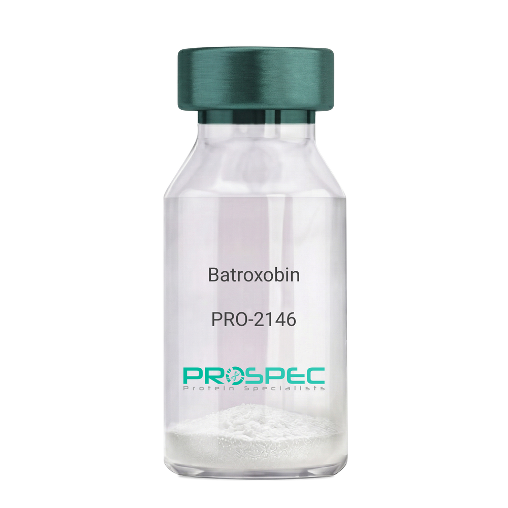
Name :
BatroxobinDescription:
Batroxobin
Thrombin-like enzyme batroxobin, EC 3.4.21.74, BX, Bothrops atrox serine proteinase, Venombin-A, Batroxobin.
Product # :
PRO-2146Price :
Quantity :
Shipping Method :
Shipped at Room temp
More Info
- description
- formulation
- More Info
Description
Batroxobin, isolated from Bothrops atrox snake venom, has an Mw of approximately 43kDa.
Formulation
The Batroxobin protein was lyophilized from a concentrated (1mg/ml) solution with no additives.
More Info
-
Introduction
Batroxobin is a serin protease that reduces fibronogen levels and is originally extracted from snake venom of Bothrops Atrox. Batroxobin is used in defibrinogenation and thrombolysis and also has an effect on c-fos gene and growth factor.
Batroxobin can efficiently restrain proliferation of VSMCs, by blocking the release and uptake of Ca2+, thus influencing [Ca2+]i.
Batroxobin converts fibrinogen to fibrin through the restricted release of fibrinopeptide-A from fibrinogen to promote blood to clot. Unlike thrombin, it is not affected by heparin and hirudin. -
Synonyms
Thrombin-like enzyme batroxobin, EC 3.4.21.74, BX, Bothrops atrox serine proteinase, Venombin-A, Batroxobin.
-
Physical Appearance
Sterile Filtered white lyophilized powder.
-
Stability
Store the lyophilized Batroxobin between 2-8°C. Do not freeze!
-
Solubility
It is recommended to reconstitute the lyophilized Batroxobin in sterile 18MΩ-cm H2O not less than 100µg/ml or more than 10mg/ml solutions.
-
Unit Definition
100BU [Batroxobin Units]=1mg.
ProSpec's products are furnished for LABORATORY RESEARCH USE ONLY. The product may not be used as drugs, agricultural or pesticidal products, food additives or household chemicals.
- View Data Sheet
Name :
C9 HumanDescription:
Complement C9 Human
Product # :
PRO-2696Price :
Quantity :
Shipping Method :
Shipped with Ice Packs
More Info
- description
- source
- formulation
- purity
- More Info
Description
Human Complement C9 produced in Human plasma is glycosylated polypeptide chain having a total molecular mass of 71kDa.
Source
Human Plasma.
Formulation
C9 protein 1mg/ml solution contains PBS pH 7.2.
Purity
Greater than 95.0% as determined by SDS-PAGE.
More Info
-
Introduction
C9 binds to the C5b-8 complex and formson cell membranes the mature membrane attack complex. Each pathway of complement activation generates proteolytic enzyme complexes which binds the target surface. These enzymes cleave a peptide bond in the larger alpha chain of C5 releasing the anaphylatoxin C5a and activating C5b. This is the only proteolytic step in the assembly of the C5b-9 complex. Although C5b is unstable it remains bound to the activating complex for a few minutes during which it binds a single C6 from the surrounding fluid or it decays and is no longer capable of forming MAC.The C5b,6 complex may also remain connected to the C3/C5 convertase where the binding of a single C7 exposes a membrane-binding region and C5b,6,7 can enter into the bilipid layer of the target cell. Each C5b-7 complex can bind 1 molecule of C8 causing the complex to enter more firmly into the membrane. The C5b-8 complex can cause lysis without C9,though it will take longer andwill require many more complexes per cell than with C9. The main role of C8 is to catalyze the binding of C9 and each can bind another C9 initiating formation of a ring structure containing up to 18 C9molecules.C5b-9 complexes with 1 or more C9 are called the Membrane Attack Complex of complement.
-
Physical Appearance
Sterile filtered solution.
-
Stability
C9 Human is stable at 4°C if entire vial will be used within 2-4 weeks.Store, frozen below -20°C for longer periods of time.For long term storage it is recommended to add a carrier protein (0.1% HSA or BSA).Avoid multiple freeze-thaw cycles.
-
Human Virus Test
Plasma from each donor has been tested and found negative for antibody to HIV-1, HIV-2, HCV and HBSAG.
ProSpec's products are furnished for LABORATORY RESEARCH USE ONLY. The product may not be used as drugs, agricultural or pesticidal products, food additives or household chemicals.
- View Data Sheet
Name :
C4c HumanDescription:
Complement Component C4c Human
Complement C4c, Complement Component C4c, C4c.
Product # :
PRO-556Price :
Quantity :
Shipping Method :
Shipped with Ice Packs
More Info
- description
- source
- formulation
- purity
- More Info
Description
Human C4c produced in Human plasma having a molecular mass of 210 kDa.
Source
Human Plasma.
Formulation
The Human Complement C4c solution is in Sodium Phosphate buffer pH 7.0 containing 0.15M NaCl and 0.09% NaN3.
Purity
Greater than 99.0%.
More Info
-
Introduction
Complement C4c is a degradation product of C4b which is cleaved by C4b/C3b inactivator to yield C4d and C4c.
Complement C4c interacts with C1 and C2 to form C3 convertase of the classic activation pathway. Systemic lupus erythematosus (SLE) is often associated with congenital C4 deficiency. Reduced levels of components of the classic pathway (C1, C2, C4, C3) are common after such activation, e.g. in SLE, acute serum sickness and conditions associated with circulating immune complexes. -
Synonyms
Complement C4c, Complement Component C4c, C4c.
-
Physical Appearance
Sterile Filtered clear solution.
-
Stability
Human C4c although stable at 4°C for 1 week, should be stored at -15°C.Please avoid freeze-thaw cycles.
-
Human Virus Test
Plasma from each donor has been tested and found negative for HIV-1 & 2 antibodies, Hepatatis B surface antigen, and Hepatatis C antibodies.
ProSpec's products are furnished for LABORATORY RESEARCH USE ONLY. The product may not be used as drugs, agricultural or pesticidal products, food additives or household chemicals.
- View Data Sheet
Name :
HIV-2 gp32, HRPDescription:
HIV-2 gp32 Recombinant, HRP Labeled
Product # :
HIV-137Price :
Quantity :
Shipping Method :
Shipped with Ice Packs
More Info
- description
- source
- formulation
- purity
- More Info
Description
HIV-2 gp32 HRP Labeled recombinant- contains the full-length sequence of HIV-2 envelope immunodominant regions gp32 having a Mw of 32kDa and fused to a beta-galactosidase at N-terminus.
Source
Escherichia Coli.
Formulation
0.01M Na2CO3, 10mM EDTA, 14mM beta-ME and 0.02% Sarcosyl.
Purity
Greater than 95.0% as determined by SDS-PAGE.
More Info
-
Introduction
HIV-1 and HIV-2 appear to package their RNA differently. HIV-1 binds to any appropriate RNA whereas HIV-2 preferentially binds to mRNA which creates the Gag protein itself. This means that HIV-1 is better able to mutate. HIV-2 is transmitted in the same ways as HIV-1: Through exposure to bodily fluids such as blood, semen, tears and vaginal fluids.
Immunodeficiency develops more slowly with HIV-2.
HIV-2 is less infectious in the early stages of the virus than with HIV-1.
The infectiousness of HIV-2 increases as the virus progresses.
Major differences include reduced pathogenicity of HIV-2 relative to HIV-1, enhanced immune control of HIV-2 infection and often some degree of CD4-independence. Despite considerable sequence and phenotypic differences between HIV-1 and 2 envelopes, structurally they are quite similar. Both membrane-anchored proteins eventually form the 6-helix bundles from the N-terminal and C-terminal regions of the ectodomain, which is common to many viral and cellular fusion proteins and which seems to drive fusion.
HIV-1 gp41 helical regions can form more stable 6-helix bundles than
HIV-2 gp41 helical regions however HIV-2 fusion occurs at a lower threshold temperature (25°C), does not require Ca2+ in the medium, is insensitive to treatment of target cells with cytochalasin B, and is not affected by target membrane glycosphingolipid composition. -
Physical Appearance
Sterile filtered colorless clear solution.
-
Stability
HIV-2 gp-32 although stable at room temperature for 3 weeks, should be stored at 4°C.
ProSpec's products are furnished for LABORATORY RESEARCH USE ONLY. The product may not be used as drugs, agricultural or pesticidal products, food additives or household chemicals.
- View Data Sheet
Name :
CFB-a HumanDescription:
Complement Factor B Fragment a Human
Complement factor B (EC:3.4.21.47), C3/C5 convertase, Glycine-rich beta glycoprotein, GBG, PBF2, Properdin factor B, Complement factor B Ba fragment, Complement factor B Bb fragment, CFB, Complement Factor B, BFD, AHUS4, BF, BFD, CFAB, FB, FBI12, H2-Bf.
Product # :
PRO-2735Price :
Quantity :
Shipping Method :
Shipped with Ice Packs
More Info
- description
- source
- formulation
- purity
- More Info
Description
CFB-a Human produced in Human Plasma having a molecular mass of 33 kDa.
Source
Human Plasma.
Formulation
CFB-a solution (1mg/ml) contains Phosphate-buffered saline, pH 7.2.
Purity
Greater than 95.0% as determined by SDS-PAGE.
More Info
-
Introduction
Complement Factor B, also known as CFB, encodes complement factor B which is a component of the alternative pathway of complement activation. Factor B circulates in the blood as a single chain polypeptide. Once the alternative pathway is activated it is cleaved by complement factor D yielding the noncatalytic chain Ba and the catalytic subunit Bb. The active subunit Bb is a serine protease which connects with C3b to form the alternative pathway C3 convertase. Also, Bb is involved in the proliferation of preactivated B lymphocytes, while Ba inhibits their proliferation.
-
Synonyms
Complement factor B (EC:3.4.21.47), C3/C5 convertase, Glycine-rich beta glycoprotein, GBG, PBF2, Properdin factor B, Complement factor B Ba fragment, Complement factor B Bb fragment, CFB, Complement Factor B, BFD, AHUS4, BF, BFD, CFAB, FB, FBI12, H2-Bf.
-
Physical Appearance
Sterile filtered solution.
-
Stability
CFB-a Human is stable at 4°C if entire vial will be used within 2-4 weeks. Store, frozen below -20°C for longer periods of time.For long term storage it is recommended to add a carrier protein (0.1% HSA or BSA).Avoid multiple freeze-thaw cycles.
-
Human Virus Test
Plasma from each donor has been tested and found negative for antibody to HIV-1, HIV-2, HCV and HBSAG.
ProSpec's products are furnished for LABORATORY RESEARCH USE ONLY. The product may not be used as drugs, agricultural or pesticidal products, food additives or household chemicals.
- View Data Sheet
Name :
ErbB2 (146-192) HumanDescription:
Tyrosine Kinase ErbB-2 (146-192) Human Recombinant
erbB-2, EC 2.7.10.1, p185erbB2, C-erbB-2.
Product # :
PKA-136Price :
Quantity :
Shipping Method :
Shipped at Room temp
More Info
- description
- source
- formulation
- purity
- More Info
Description
The ErbB2 Human is created as a recombinant protein with a 4kda N-terminal fusion of His Tag. The ErbB2 His-Tagged Fusion Protein, produced in E. coli, is a 14kDa protein containing 47 amino acid residues of the ErbB2 Human, 146-192amino acids.
Source
Escherichia Coli.
Formulation
Each mg was lyophilized with 1xPBS, 0.4% SDS and 4mM DTT.
Purity
Greater than 90% as determined by SDS-PAGE.
More Info
-
Synonyms
erbB-2, EC 2.7.10.1, p185erbB2, C-erbB-2.
-
Physical Appearance
Filtered White lyophilized (freeze-dried) powder.
-
Stability
Store lyophilized ErbB2 at -20°C. Aliquot the product after reconstitution to avoid repeated freezing/thawing cycles. Reconstituted protein can be stored at 4°C for a limited period of time; it does not show any change after two weeks at 4°C.
-
Solubility
It is recommended to add deionized water to prepare a working stock solution of approximately 0.5mg/ml and let the lyophilized pellet dissolve completely. Product is not sterile! Please filter the product by an appropriate sterile filter before using it on cell culture.
-
Background
HER2 / ErbB2 (human epidermal growth factor receptor 2) is a member of the ErbB family of receptor tyrosine kinases. It plays a role in cell signalling related to growth, differentiation and proliferation.
erbB-2 encodes a 185-kDa orphan receptor tyrosine kinase that is constitutively active as a dimer and displays potent oncogenic activity when overexpressed.
ErbB2 works as a receptor that, upon ligand binding activates downstream signalling pathways such as the MAPK and PI3K/AKT pathways, promoting cell proliferation and survival.
The activation of ErbB2 leads to enhanced cell growth and survival, making it critical in normal cellular functions and in the pathogenesis of cancer.
Herstatin, as the product of alternative HER-2 transcript, retains intron 8.
The herstatin mRNA is expressed in normal human fetal kidney and liver, but is at reduced levels relative to p185HER-2 mRNA in carcinoma cells that contain an amplified HER-2 gene. Herstatin appears to be an inhibitor of p185HER-2, because it disrupts dimers, reduces tyrosine phosphorylation of p185, and inhibits the anchorage-independent growth of transformed cells that overexpress HER-2.
ProSpec's products are furnished for LABORATORY RESEARCH USE ONLY. The product may not be used as drugs, agricultural or pesticidal products, food additives or household chemicals.
- View Data Sheet
Name :
CMV gBDescription:
Cytomegalo Virus gB Recombinant
Product # :
CMV-211Price :
Quantity :
Shipping Method :
Shipped with Ice Packs
More Info
- description
- source
- formulation
- purity
- More Info
Description
The E.Coli derived recombinant artificial mosaic protein contains the CMV gB immunodominant regions 11-67 amino acids, fused with a 26 kDa GST Tag, the protein weight is 6.5kDa, having a total weight of 32.5 kDa.
Source
Escherichia Coli.
Formulation
50mM Tris pH 7.2, 1mM EDTA and 50% glycerol.
Purity
CMV gB protein is >95% pure as determined by 10% PAGE (coomassie staining).
More Info
-
Introduction
MV belongs to the Beta herpesvirinae subfamily of Herpesviridae which includes herpes simplex virustypes 1 and 2, varicella-zoster virus, and Epstein-Barrvirus. The herpesviruses share a characteristic ability to remain latentover long periods.CMV is a double-stranded linear DNA virus with 162 hexagonal protein capsomeres surrounded by a lipid membrane. CMV has the largest genome of the herpes viruses, ranging from 230-240 kilobase pairs. Human CMV is composed of unique and inverted repeats that include the existence of 4 genome isomers caused by inversion of L-S genome components (class E). Replication may be divided into immediate early, delayed early, and late gene expression based on time of synthesis after infection. The DNA is replicated by rolling circles. In vitro, CMV replicates in human fibroblasts.
-
Stability
CMV gB protein although stable at 4°C for 1 week, should be stored below -18°C. Please prevent freeze thaw cycles.
-
Specificity
Immunoreactive with sera of CMV-infected individuals.
-
Purification Method
CMV gB was purified by proprietary chromatographic technique.
ProSpec's products are furnished for LABORATORY RESEARCH USE ONLY. The product may not be used as drugs, agricultural or pesticidal products, food additives or household chemicals.
- View Data Sheet
Name :
SPP1 Human, HEKDescription:
Osteopontin Human Recombinant, HEK
Secreted Phosphoprotein-1, OPN, BNSP, BSPI, ETA-1, MGC110940, SPP-1, Osteopontin, Bone sialoprotein 1, Urinary stone protein, Nephropontin, Uropontin, SPP1.
Product # :
CYT-047Price :
Quantity :
Shipping Method :
Shipped at Room temp
More Info
- description
- source
- formulation
- purity
- More Info
Description
Osteopontin Human Recombinant is a single, glycosylated, polypeptide chain produced in HEK293 cells, is a full length protein (amino acids 17-314) fused with a polyhistidine tag at the C-terminus, having a total calculated molecular mass of 34.5kDa (The actual molecular mass may be approximately 60-65kDa in SDS-PAGE under reducing conditions due to glycosylation).Osteopontin is purified by proprietary chromatographic techniques.
Source
HEK293 cells.
Formulation
Osteopontin was lyophilized from a 0.2µM filtered solution of 20mM PBS and 150mM NaCl, pH 7.2.
Purity
Greater than 95.0% as determined by SDS-PAGE.
More Info
-
Introduction
Osteopontin is a glycoprotein that was first identified in osteoblasts and is involved in bone remodeling, immune functions in fibroblasts, macrophages, and lymphocytes during inflammation and wound healing. SPP1 binds tightly to hydroxyapatite. SPP1 forms an integral part of the mineralized matrix. SPP1 is vital to cell-matrix interaction.
Secreted Phosphoprotein-1 protects against cardiac ischemia-reperfusion injury via late preconditioning. Expression of both Ostepontin and CD44 in hepatocellular carcinoma is linked with advanced tumor stage and contributes to prognosis information. SPP1 is the most over-expressed gene in intrahepatic cholangiocarcinoma. Secreted Phosphoprotein-1 overexpression is related with interstitial lung diseases. -
Synonyms
Secreted Phosphoprotein-1, OPN, BNSP, BSPI, ETA-1, MGC110940, SPP-1, Osteopontin, Bone sialoprotein 1, Urinary stone protein, Nephropontin, Uropontin, SPP1.
-
Physical Appearance
Sterile Filtered White lyophilized (freeze-dried) powder.
-
Stability
Store at 4°C if entire vial will be used within 2-4 weeks. Store, frozen at -20°C for longer periods of time.Please avoid freeze thaw cycles.
-
Solubility
It is recommended to reconstitute the lyophilized SPP1 in 1xPBS to a concentration no less than 100 µg/ml, which can then be further diluted to other aqueous solutions.
-
Amino Acid Sequence
IPVKQADSGSSEEKQLYNKYPDAVATWLNPDPSQKQNLLAPQNAVSSEETNDFKQETL
PSKSNESHDHMDDMDDEDDDDHVDSQDSIDSNDSDDVDDTDDSHQSDESHHSDESDEL
VTDFPTDLPATEVFTPVVPTVDTYDGRGDSVVYGLRSKSKKFRRPDIQYPDATDEDIT
SHMESEELNGAYKAIPVAQDLNAPSDWDSRGKDSYETSQLDDQSAETHSHKQSRLYKRK
ANDESNEHSDVIDSQELSKVSREFHSHEFHSHEDMLVVDPKSKEEDKHLKFRISHELDS
ASSEVNVDHHHHHH.
ProSpec's products are furnished for LABORATORY RESEARCH USE ONLY. The product may not be used as drugs, agricultural or pesticidal products, food additives or household chemicals.
- View Data Sheet
Name :
HBV core (1-183)Description:
Hepatitis B Virus Core (1-183 a.a.) Recombinant
Product # :
HBV-269Price :
Quantity :
Shipping Method :
Shipped with Ice Packs
More Info
- description
- formulation
- purity
- More Info
Description
The E.Coli derived 18 kDa recombinant protein contains the HBV core ayw immunodominant region, amino acids 1-183.
Formulation
1x PBS (pH 7.2), 75 mM NaCl, 0.05% Proclin 300 and 50% glycerol.
Purity
HBV Core protein is >95% pure as determined by SDS-PAGE.
More Info
-
Introduction
Hepatitis B is one of a few known non-retroviralviruses which employ reverse transcriptionas a part of its replication process. (HIV, a completely unrelated virus, also uses reverse transcription, but it is a retrovirus.) HBV invades the cell by binding to surface receptor and become internalized. The viral core particles then migrate to the hepatocyte nucleus and the partially double-stranded, relaxed circular genomes (RC-DNA) are repaired to form a covalently closed circular DNA (cccDNA), which is the template for viral genomic and sub-genomic RNAs by cellular RNA polymerase II. Of these, the pregenomic RNA (pgRNA is selectively packaged into progeny capsids and is then reverse-transcribed into new RC-DNA. The core can either bud into the endoplasmic reticulum to be enveloped or exported from the cell or recycled back into the genome for conversion to cccDNA.
-
Stability
HBV Core protein although stable at 4°C for 1 week, should be stored below -18°C. Please prevent freeze thaw cycles.
-
Amino Acid Sequence
MDIDPYKEFG ASVELLSFLP SDFFPSIRDL LDTASALYRE ALESPEHCSPHHTALRQAIL CWGELMNLAT WVGSNLEDPA SRELVVSYVN VNMGLKFRQL LWFHVSCLTF GRETVLEYLV SFGVWIRTPP AYRPPNAPIL STLPETTVVR RRGRSPRRRT PSPRRRRSQS PRRRRSQSRE SQC.
-
Specificity
Immunoreactive with sera HBV-infected individuals.
-
Purification Method
HBV Core protein was purified by proprietary chromatographic technique.
ProSpec's products are furnished for LABORATORY RESEARCH USE ONLY. The product may not be used as drugs, agricultural or pesticidal products, food additives or household chemicals.