Skip to content

High-quality research proteins.

  • 1-800-805-6531

  • Mon - Sat 8.00-18.00, Sun - Closed

  • info@prospecbio.com

  • PO Box 6591 East Brunswick NJ 08816

  • Products
    • Cytokines
    • Growth factors
    • Chemokines
    • Cd antigens
    • Neurotrophins
    • Hormones
    • Enzymes
    • Viral antigens
    • Recombinant proteins
    • Natural proteins
    • Monoclonal antibodies
    • Polyclonal antibodies
    • Thermal Packaging
  • About
    • Company
    • Founder
    • Contribution
  • Customers
    • Eastern Asia
    • Eastern Europe
    • North America
    • South America
    • South East Asia
    • Southern Asia
    • Western Europe
  • Ordering
    • Ordering Method
    • Ordering - Credit Card
    • Ordering - PO
    • Shipping Fees
    • FAQs
  • Services
    • DNA Cloning
    • Protein Expression
    • Monoclonal Antibody
    • Fermentation
    • Technology License
  • Contact us
Log in

Country/region

  • USD $ | Albania
  • USD $ | Andorra
  • USD $ | Argentina
  • USD $ | Armenia
  • USD $ | Australia
  • USD $ | Austria
  • USD $ | Azerbaijan
  • USD $ | Belarus
  • USD $ | Belgium
  • USD $ | Bolivia
  • USD $ | Brazil
  • USD $ | Bulgaria
  • USD $ | Cambodia
  • USD $ | Canada
  • USD $ | Chile
  • USD $ | China
  • USD $ | Colombia
  • USD $ | Costa Rica
  • USD $ | Croatia
  • USD $ | Cyprus
  • USD $ | Czechia
  • USD $ | Denmark
  • USD $ | Estonia
  • USD $ | Finland
  • USD $ | France
  • USD $ | Georgia
  • USD $ | Germany
  • USD $ | Greece
  • USD $ | Hong Kong SAR
  • USD $ | Hungary
  • USD $ | Iceland
  • USD $ | India
  • USD $ | Ireland
  • USD $ | Israel
  • USD $ | Italy
  • USD $ | Japan
  • USD $ | Kazakhstan
  • USD $ | Latvia
  • USD $ | Liechtenstein
  • USD $ | Lithuania
  • USD $ | Madagascar
  • USD $ | Malta
  • USD $ | Mexico
  • USD $ | Moldova
  • USD $ | Monaco
  • USD $ | Netherlands
  • USD $ | New Zealand
  • USD $ | North Macedonia
  • USD $ | Norway
  • USD $ | Panama
  • USD $ | Paraguay
  • USD $ | Peru
  • USD $ | Philippines
  • USD $ | Poland
  • USD $ | Portugal
  • USD $ | Romania
  • USD $ | Singapore
  • USD $ | Slovakia
  • USD $ | Slovenia
  • USD $ | South Africa
  • USD $ | South Korea
  • USD $ | Spain
  • USD $ | Sri Lanka
  • USD $ | Sweden
  • USD $ | Switzerland
  • USD $ | Taiwan
  • USD $ | Thailand
  • USD $ | Türkiye
  • USD $ | Ukraine
  • USD $ | United Kingdom
  • USD $ | United States
  • USD $ | Uruguay
  • USD $ | Vatican City
  • USD $ | Venezuela
  • USD $ | Vietnam
  • Facebook
  • Instagram
logoProSpec - Recombinant Protein Manufacturer for academic and R&D industry
Become a distributor
Account Log in
Wishlist
Cart Cart(0)
  • Products
    +
    • Cytokines
    • Growth factors
    • Chemokines
    • Cd antigens
    • Neurotrophins
    • Hormones
    • Enzymes
    • Viral antigens
    • Recombinant proteins
    • Natural proteins
    • Monoclonal antibodies
    • Polyclonal antibodies
    • Thermal Packaging
  • About
    +
    • Company
    • Founder
    • Contribution
  • Customers
    +
    • Eastern Asia
    • Eastern Europe
    • North America
    • South America
    • South East Asia
    • Southern Asia
    • Western Europe
  • Ordering
    +
    • Ordering Method
    • Ordering - Credit Card
    • Ordering - PO
    • Shipping Fees
    • FAQs
  • Services
    +
    • DNA Cloning
    • Protein Expression
    • Monoclonal Antibody
    • Fermentation
    • Technology License
  • Contact us

Item added to your cart

View cart
View All
  • Cytokines
  • Growth factors
  • Chemokines
  • Cd antigens
  • Neurotrophins
  • Hormones
  • Enzymes
  • Viral antigens
  • Recombinant proteins
  • Natural proteins
  • Monoclonal antibodies
  • Polyclonal antibodies
  • Thermal Packaging
  • Tumor Necrosis Factor

    Tumor Necrosis Factor

    View All
  • 4-1BB

    4-1BB

    View All
  • Adiponectin

    Adiponectin

    View All
  • AIF1

    AIF1

    View All
  • Other Cytokines

    Other Cytokines

    View All
  • AITRL

    AITRL

    View All
  • Angiopoietin

    Angiopoietin

    View All
  • Apolipoprotein

    Apolipoprotein

    View All
  • B-Cell Activating Factor

    B-Cell Activating Factor

    View All
  • Beta Defensin

    Beta Defensin

    View All
  • Bone Morphogenetic Protein

    Bone Morphogenetic Protein

    View All
  • B type Natriuretic Peptide

    B type Natriuretic Peptide

    View All
  • BST

    BST

    View All
  • Betacellulin

    Betacellulin

    View All
  • Cardiotrophin

    Cardiotrophin

    View All
  • Activin

    Activin

    View All
  • Fibroblast Growth Factor

    Fibroblast Growth Factor

    View All
  • Other Growth Factors

    Other Growth Factors

    View All
  • CSF

    CSF

    View All
  • CTGF

    CTGF

    View All
  • IGFBP

    IGFBP

    View All
  • VEGF Protein

    VEGF Protein

    View All
  • EGF

    EGF

    View All
  • Epigen

    Epigen

    View All
  • Erythropoietin

    Erythropoietin

    View All
  • Insulin-Like Growth Factor

    Insulin-Like Growth Factor

    View All
  • Growth Hormone

    Growth Hormone

    View All
  • HDGF

    HDGF

    View All
  • Hepatocyte Growth Factor

    Hepatocyte Growth Factor

    View All
  • Insulin

    Insulin

    View All
  • BCA-1/ BLC (CXCL13)

    BCA-1/ BLC (CXCL13)

    View All
  • BRAK (CXCL14)

    BRAK (CXCL14)

    View All
  • C-10 (CCL6)

    C-10 (CCL6)

    View All
  • Other Chemokines

    Other Chemokines

    View All
  • MEC (CCL28)

    MEC (CCL28)

    View All
  • LD78-beta (CCL3L1)

    LD78-beta (CCL3L1)

    View All
  • CTACK (CCL27)

    CTACK (CCL27)

    View All
  • CXCL16

    CXCL16

    View All
  • CXCL17

    CXCL17

    View All
  • Platelet Factor-4 (CXCL4)

    Platelet Factor-4 (CXCL4)

    View All
  • ENA-78 (CXCL5)

    ENA-78 (CXCL5)

    View All
  • Eotaxin (CCL11,24,26)

    Eotaxin (CCL11,24,26)

    View All
  • Exodus-2 (CCL21)

    Exodus-2 (CCL21)

    View All
  • Fractalkine (CX3CL1)

    Fractalkine (CX3CL1)

    View All
  • CXCL6

    CXCL6

    View All
  • Other CD Antigens

    Other CD Antigens

    View All
  • CD14

    CD14

    View All
  • CD164

    CD164

    View All
  • CD1

    CD1

    View All
  • CD2

    CD2

    View All
  • CD200

    CD200

    View All
  • CD207

    CD207

    View All
  • CD226

    CD226

    View All
  • CD23

    CD23

    View All
  • CD244

    CD244

    View All
  • CD247

    CD247

    View All
  • CD27

    CD27

    View All
  • CD274

    CD274

    View All
  • CD300

    CD300

    View All
  • CD33

    CD33

    View All
  • Other Neurotrophins

    Other Neurotrophins

    View All
  • Beta-NGF

    Beta-NGF

    View All
  • BDNF

    BDNF

    View All
  • CDNF

    CDNF

    View All
  • CNTF

    CNTF

    View All
  • GDNF

    GDNF

    View All
  • Glia Maturation Factor

    Glia Maturation Factor

    View All
  • MANF

    MANF

    View All
  • Midkine

    Midkine

    View All
  • Neuroglobin

    Neuroglobin

    View All
  • Neurotrophic factors

    Neurotrophic factors

    View All
  • Neuregulin

    Neuregulin

    View All
  • Neuropilin

    Neuropilin

    View All
  • Pigment Epithelium-Derived Factor

    Pigment Epithelium-Derived Factor

    View All
  • Pleiotrophin

    Pleiotrophin

    View All
  • Peptide Hormones

    Peptide Hormones

    View All
  • Other Hormones

    Other Hormones

    View All
  • HCG

    HCG

    View All
  • Endothelin

    Endothelin

    View All
  • Exendin

    Exendin

    View All
  • FSH

    FSH

    View All
  • Glucagon

    Glucagon

    View All
  • GHRP

    GHRP

    View All
  • GLP

    GLP

    View All
  • Thyrostimulin

    Thyrostimulin

    View All
  • Inhibin A

    Inhibin A

    View All
  • LHRH

    LHRH

    View All
  • Melanotan

    Melanotan

    View All
  • Procalcitonin

    Procalcitonin

    View All
  • PTH

    PTH

    View All
  • Peroxiredoxin

    Peroxiredoxin

    View All
  • Peptidase

    Peptidase

    View All
  • Synthetase

    Synthetase

    View All
  • Transferase

    Transferase

    View All
  • Hydrolase

    Hydrolase

    View All
  • Dehydrogenase

    Dehydrogenase

    View All
  • ACE2

    ACE2

    View All
  • Esterase

    Esterase

    View All
  • Protein Kinases

    Protein Kinases

    View All
  • Other Enzymes

    Other Enzymes

    View All
  • Phosphatase

    Phosphatase

    View All
  • Deaminase

    Deaminase

    View All
  • Oxygenase

    Oxygenase

    View All
  • Adenylate Kinase

    Adenylate Kinase

    View All
  • Lyase

    Lyase

    View All
  • Chagas

    Chagas

    View All
  • SARS

    SARS

    View All
  • Influenza

    Influenza

    View All
  • ASFV

    ASFV

    View All
  • Astrovirus

    Astrovirus

    View All
  • Borrelia

    Borrelia

    View All
  • Helicobacter Pylori

    Helicobacter Pylori

    View All
  • Chikungunya

    Chikungunya

    View All
  • Chlamydia

    Chlamydia

    View All
  • Cytomegalo

    Cytomegalo

    View All
  • Dengue

    Dengue

    View All
  • EBV

    EBV

    View All
  • Ebola

    Ebola

    View All
  • Feline Leukemia Virus

    Feline Leukemia Virus

    View All
  • Hepatitis A

    Hepatitis A

    View All
  • Other

    Other

    View All
  • Actin

    Actin

    View All
  • ADAM

    ADAM

    View All
  • Complement Component

    Complement Component

    View All
  • Ag85

    Ag85

    View All
  • Eukaryotic Translation Initiation Factor

    Eukaryotic Translation Initiation Factor

    View All
  • Anterior Gradient Protein

    Anterior Gradient Protein

    View All
  • Heat Shock Protein

    Heat Shock Protein

    View All
  • Alpha-2-HS-Glycoprotein

    Alpha-2-HS-Glycoprotein

    View All
  • Hemoglobin

    Hemoglobin

    View All
  • Allergy

    Allergy

    View All
  • Anaplasma

    Anaplasma

    View All
  • Angiogenin

    Angiogenin

    View All
  • Ankyrin Repeat Domain

    Ankyrin Repeat Domain

    View All
  • Annexin

    Annexin

    View All
  • Other Natural Proteins

    Other Natural Proteins

    View All
  • Aprotinin

    Aprotinin

    View All
  • Transferrin

    Transferrin

    View All
  • Avidin

    Avidin

    View All
  • Anti Coagulation Factors

    Anti Coagulation Factors

    View All
  • Natural Albumin

    Natural Albumin

    View All
  • Natural Coagulation Factors

    Natural Coagulation Factors

    View All
  • Fibronectin

    Fibronectin

    View All
  • Thrombin

    Thrombin

    View All
  • Anti Human Enzyme

    Anti Human Enzyme

    View All
  • Other Monoclonal Antibodies

    Other Monoclonal Antibodies

    View All
  • Anti Human Cytokine

    Anti Human Cytokine

    View All
  • Anti Human Heat Shock Protein

    Anti Human Heat Shock Protein

    View All
  • Anti Mouse Lymphocyte

    Anti Mouse Lymphocyte

    View All
  • Anti Human Chemokine

    Anti Human Chemokine

    View All
  • Anti Human Lymphocyte

    Anti Human Lymphocyte

    View All
  • Anti Viral Monoclonal

    Anti Viral Monoclonal

    View All
  • Anti Mouse Cytokine

    Anti Mouse Cytokine

    View All
  • Anti-GST Monoclonal

    Anti-GST Monoclonal

    View All
  • Other Polyclonal Antibodies

    Other Polyclonal Antibodies

    View All
  • Anti Viral

    Anti Viral

    View All
  • Anti-GST Monoclonal

    Anti-GST Monoclonal

    View All
Back

Search results

1000 results found for “Other Chemokines”

Name

Description

Product #

Price

Quantity

Shipping Method

  • View Data Sheet

    Name :

    CoV-2-Spike (1-260)

    Description:

    Coronavirus 2019 Spike (1-260 a.a.), Recombinant

    Product # :

    SARS-022

    Price :

    Quantity :

    Shipping Method :

    Ice Icon

    Shipped with Ice Packs

    Add To Cart

    More Info

    • description
    • source
    • formulation
    • purity
    • More Info

    Description

    The HEK293 derived recombinant protein contains the Coronavirus 2019-Spike N-Terminal Domain, Wuhan-Hu-1 strain, amino acids 1-260 fused to Fc tag at C-terminal.

    Source

    HEK293

    Formulation

    CoV-2 spike protein solution is supplied in DPBS.

    Purity

    Protein is >95% pure as determined SDS-PAGE.

    More Info

    • Introduction

      A human infecting coronavirus (viral pneumonia) called 2019 novel coronavirus, 2019-nCoV was found in the fish market at the city of Wuhan, Hubei province of China on December 2019.

      The 2019-nCoV shares an 87% identity to the 2 bat-derived severe acute respiratory syndrome 2018 SARS-CoV-2 located in Zhoushan of eastern China. 2019-nCoV has an analogous receptor-BD-structure to that of 2018 SARS-CoV, even though there is a.a. diversity so thus the 2019-nCoV might bind to ACE2 receptor protein (angiotensin-converting enzyme 2) in humans.

      While bats are possibly the host of 2019-nCoV, researchers suspect that animal from the ocean sold at the seafood market was an intermediate host. RSCU analysis proposes that the 2019-nCoV is a recombinant within the viral spike glycoprotein between the bat coronavirus and an unknown coronavirus.

    • Physical Appearance

      Sterile Filtered clear solution.

    • Stability

      CoV-2 Spike Protein is shipped on ice packs. Upon arrival, Store at -20°C.

    • Purification Method

      Purified by Protein-G chromatographic technique.

    ProSpec's products are furnished for LABORATORY RESEARCH USE ONLY. The product may not be used as drugs, agricultural or pesticidal products, food additives or household chemicals.

    product_image.jpg
  • View Data Sheet

    Name :

    CoV-2 N (196 a.a.)

    Description:

    Coronavirus 2019 Nucleocapsid (196 a.a.), Recombinant

    Product # :

    SARS-042

    Price :

    Quantity :

    Shipping Method :

    Ice Icon

    Shipped with Ice Packs

    Add To Cart

    More Info

    • description
    • source
    • formulation
    • purity
    • More Info

    Description

    The E.Coli derived recombinant protein contains the Coronavirus 2019 middle region a.a. from the Nucleocapsid protein and fused to GST-6xHis tag at N-terminal and having a Mw. Of 48.4 kDa.

    Source

    E.Coli.

    Formulation

    CoV-2 Nucleocapsid protein solution is supplied in 50mM Tris-HCl pH 8, 1M Urea, and 50% Glycerol.

    Purity

    CoV-2 Nucleocapsid protein is >95% pure as determined SDS-PAGE.

    More Info

    • Introduction

      A human infecting coronavirus (viral pneumonia) called 2019 novel coronavirus, 2019-nCoV was found in the fish market at the city of Wuhan, Hubei province of China on December 2019.

      The 2019-nCoV shares an 87% identity to the 2 bat-derived severe acute respiratory syndrome 2018 SARS-CoV-2 located in Zhoushan of eastern China. 2019-nCoV has an analogous receptor-BD-structure to that of 2018 SARS-CoV, even though there is a.a. diversity so thus the 2019-nCoV might bind to ACE2 receptor protein (angiotensin-converting enzyme 2) in humans.

      While bats are possibly the host of 2019-nCoV, researchers suspect that animal from the ocean sold at the seafood market was an intermediate host. RSCU analysis proposes that the 2019-nCoV is a recombinant within the viral spike glycoprotein between the bat coronavirus and an unknown coronavirus.

    • Physical Appearance

      Sterile Filtered clear solution.

    • Stability

      CoV-2 Spike Protein is shipped on ice packs. Upon arrival, Store at -20°C.

    • Purification Method

      NTA Sepharose-Affinity Purification.

    ProSpec's products are furnished for LABORATORY RESEARCH USE ONLY. The product may not be used as drugs, agricultural or pesticidal products, food additives or household chemicals.

    product_image.jpg
  • View Data Sheet

    Name :

    IL 28A Mouse

    Description:

    Interleukin-28A Mouse Recombinant

    IFN-lambda-2, Interleukin-28A, IL-28A, Ifnl2, Il28a, EG330496.

    Product # :

    CYT-741

    Price :

    Quantity :

    Shipping Method :

    Room Temp Icon

    Shipped at Room temp

    Add To Cart

    More Info

    • description
    • source
    • formulation
    • purity
    • biological activity
    • More Info

    Description

    Recombinant Mouse Interleukin-28A produced in E.Coli is a single, non-glycosylated polypeptide chain containing 174 amino acids and having a total molecular mass of 19.7kDa.The IL-28A is purified by proprietary chromatographic techniques.

    Source

    Escherichia Coli.

    Formulation

    The Mouse IL-28A is lyophilized from a 0.2µm filtered concentrated solution in PBS, pH 7.4 and 5% trehalose.

    Purity

    Greater than 95% as determined by:
    (a) Analysis by RP-HPLC.
    (b) Analysis by SDS-PAGE.

    Biological Activity

    The ED50 as determined by an anti-viral assay using human HepG2 cells infected with encephalomyocarditis(EMC) virus is less than 3 ng/ml, corresponding to a specific activity of > 3.3 × 10⁵ IU/mg.

    More Info

    • Introduction

      IL-28A is distantly related to the IL-10 family. Expression of IL-28A is induced by viral infection which interacts with a heterodimeric class II cytokine receptor that consists of interleukin 10 receptor, beta (IL10RB) and interleukin 28 receptor, alpha. IL-28A exhibits common features with type I IFNs such as antiviral activity, antiproliferative activity and in vivo antitumour activity.
      IL-28A acts similarly to IFNs, but is less effective generally and has activity in a more limited range of cell lines. IFN-ambda 1, IFN-lambda 2 and IFN-lambda 3 are closely positioned genes on human chromosome 19.
      IL-28A induces ELR(-) CXC chemokine mRNA in human peripheral blood mononuclear cells, in an IFN-gamma-independent manner.
      IL-28A is able to generate tolerogenic DCs, an activity that could thwart IFN-beta functions. IL-28A produced in response to viral infection, activates both monocytes and macrophages producing a restricted panel of cytokines and therefore is an important factor in activating innate immune responses at the site of viral infection.

    • Synonyms

      IFN-lambda-2, Interleukin-28A, IL-28A, Ifnl2, Il28a, EG330496.

    • Physical Appearance

      Sterile Filtered White lyophilized (freeze-dried) powder.

    • Stability

      Lyophilized IFN-Lambda 2 although stable at room temperature for 3 weeks, should be stored desiccated below -18°C. Upon reconstitution IFN-Lambda 2 Recombinant should be stored at 4°C between 2-7 days and for future use below -18°C.For long term storage it is recommended to add a carrier protein (0.1% HSA or BSA).Please prevent freeze-thaw cycles.

    • Solubility

      It is recommended to reconstitute the lyophilized IL-28A in sterile 18MΩ-cm H2O not less than 100µg/ml, which can then be further diluted to other aqueous solutions.

    • Amino Acid Sequence

      DPVPRATRLP VEAKDCHIAQ FKSLSPKELQ AFKKAKDAIE KRLLEKDMRC SSHLISRAWD LKQLQVQERP KALQAEVALT LKVWENMTDS ALATILGQPL HTLSHIHSQL QTCTQLQATA EPKPPSRRLS RWLHRLQEAQ SKETPGCLED SVTSNLFRLL TRDLKCVASG DQCV.

    ProSpec's products are furnished for LABORATORY RESEARCH USE ONLY. The product may not be used as drugs, agricultural or pesticidal products, food additives or household chemicals.

    Il 28A Mouse
  • View Data Sheet

    Name :

    CoV-2 Spike (318-542)

    Description:

    Coronavirus 2019 Spike Receptor Binding Domain (318-542 a.a.) Recombinant

    Product # :

    SARS-057

    Price :

    Quantity :

    Shipping Method :

    Ice Icon

    Shipped with Ice Packs

    Add To Cart

    More Info

    • description
    • source
    • formulation
    • purity
    • More Info

    Description

    Recombinant Coronavirus 2019 Spike Receptor Binding Domain (318-542 aa) having a Mw of 25.7kDa was purified from E. coli.The CoV-2 Spike is fused to a 6xHis tag at its C terminal and purified by proprietary chromatographic technique.

    Source

    Escherichia Coli.

    Formulation

    CoV-2 Spike protein solution contains PBS and 25mM K2CO3.

    Purity

    Protein is >95% pure as determined by 10% PAGE (coomassie staining).

    More Info

    • Introduction

      A human infecting coronavirus (viral pneumonia) called 2019 novel coronavirus, 2019-nCoV was found in the fish market at the city of Wuhan, Hubei province of China on December 2019.The 2019-nCoV shares an 87% identity to the 2 bat-derived severe acute respiratory syndrome 2018 SARS-CoV-2 located in Zhoushan of eastern China. 2019-nCoV has an analogous receptor-BD-structure to that of 2018 SARS-CoV, even though there is a.a. diversity so thus the 2019-nCoV might bind to ACE2 receptor protein (angiotensin-converting enzyme 2) in humans.While bats are possibly the host of 2019-nCoV, researchers suspect that animal from the ocean sold at the seafood market was an intermediate host. RSCU analysis proposes that the 2019-nCoV is a recombinant within the viral spike glycoprotein between the bat coronavirus and an unknown coronavirus.

    • Physical Appearance

      Sterile Filtered clear solution.

    • Stability

      Store at 4°C if entire vial will be used within 2-4 weeks.Store, frozen at -20°C for longer periods of time.For long term storage it is recommended to add a carrier protein (0.1% HSA or BSA).Avoid multiple freeze-thaw cycles.

    • Amino Acid Sequence

      HMRVQPTESI VRFPNITNLC PFGEVFNATR FASVYAWNRK RISNCVADYS VLYNSASFS TFKCYGVSPT KLNDLCFTNV YADSFVIRGD EVRQIAPGQT GKIADYNYKL PDDFTGCVI AWNSNNLDSKV GGNYNYLYRL FRKSNLKPFE RDISTEIYQA GSTPCNGVEG FNCYFPLQSY GFQPTNGVGY QPYRVVVLSF ELLHAPATVC GPKKSTNLVK NKCVNFNLE

    • Applications

      ELISA, Western blot and lateral flow immunoassay.

    ProSpec's products are furnished for LABORATORY RESEARCH USE ONLY. The product may not be used as drugs, agricultural or pesticidal products, food additives or household chemicals.

    product_image.jpg
  • View Data Sheet

    Name :

    CD100 Human HEK

    Description:

    CD100 Human Recombinant HEK

    Thesemaphorin family containing 1 Ig-like C2-type domain, 1 PSI domain and 1 Sema domai,Semaphorin-4D, A8, BB18, GR3, SEMA4D, C9orf164, CD100, SEMAJ.

    Product # :

    PRO-1640

    Price :

    Quantity :

    Shipping Method :

    Room Temp Icon

    Shipped at Room temp

    Add To Cart

    More Info

    • description
    • source
    • formulation
    • purity
    • More Info

    Description

    CD100 Human Recombinant produced by mammalian expression system in human cells is a single polypeptide chain containing 721 amino acids (22-734). CD100 is fused to an 8 amino acid His-tag at C-terminus & purified by proprietary chromatographic techniques.

    Source

    HEK293 cells.

    Formulation

    CD100 was lyophilized from a 0.2 µM filtered solution of 20mM PB and 150mM NaCl, PH 7.4.

    Purity

    Greater than 95% as determined by SDS-PAGE.

    More Info

    • Introduction

      Semaphorin-4D (CD100) is a member of the semaphorin family containing 1 Ig-like C2-type domain, 1 PSI domain and 1 Sema domain. CD100 is the cell surface receptor forPLXN1B and PLXNB2 which plays a central role in cell-cell signaling. CD100 stimulates the migration of cerebellar granule cells and of endothelial cells, regulates dendrite and axon branching and morphogenesis. In addition, CD100 has a role in the immune system; it promotes signaling via SRC and PTK2B/PYK2, which subsequently mediates activation of phosphatidylinositol 3-kinase and of the AKT1 signaling cascade.

    • Synonyms

      Thesemaphorin family containing 1 Ig-like C2-type domain, 1 PSI domain and 1 Sema domai,Semaphorin-4D, A8, BB18, GR3, SEMA4D, C9orf164, CD100, SEMAJ.

    • Physical Appearance

      Sterile Filtered White lyophilized (freeze-dried) powder.

    • Stability

      Lyophilized CD100 although stable at room temperature for 3 weeks, should be stored desiccated below -18°C. Upon reconstitution CD100 should be stored at 4°C between 2-7 days and for future use below -18°C. Please prevent freeze-thaw cycles.

    • Solubility

      It is recommended to reconstitute the lyophilized CCD100 in 1xPBS to a concentration no less than 100 µg/ml, which can then be further diluted to other aqueous solutions.

    • Amino Acid Sequence

      MAFAPIPRITWEHREVHLVQFHEPDIYNYSALLLSEDKDTLYIGAREAVFAVNALNISEKQHE
      VYWKVSEDKKAKCAEKGKSKQTECLNYIRVLQPLSATSLYVCGTNAFQPACDHLNLTSFKFLG
      KNEDGKGRCPFDPAHSYTSVMVDGELYSGTSYNFLGSEPIISRNSSHSPLRTEYAIPWLNEPSF
      VFADVIRKSPDSPDGEDDRVYFFFTEVSVEYEFVFRVLIPRIARVCKGDQGGLRTLQKKWTSFL
      KARLICSRPDSGLVFNVLRDVFVLRSPGLKVPVFYALFTPQLNNVGLSAVCAYNLSTAEEVFSH
      GKYMQSTTVEQSHTKWVRYNGPVPKPRPGACIDSEARAANYTSSLNLPDKTLQFVKDHPLMDDSV
      TPIDNRPRLIKKDVNYTQIVVDRTQALDGTVYDVMFVSTDRGALHKAISLEHAVHIIEETQLFQD
      FEPVQTLLLSSKKGNRFVYAGSNSGVVQAPLAFCGKHGTCEDCVLARDPYCAWSPPTATCVALHQ
      TESPSRGLIQEMSGDASVCPDKSKGSYRQHFFKHGGTAELKCSQKSNSEKTMYLKSSDNRVDHHHHHH.

    ProSpec's products are furnished for LABORATORY RESEARCH USE ONLY. The product may not be used as drugs, agricultural or pesticidal products, food additives or household chemicals.

    Cd100 Human Hek
  • View Data Sheet

    Name :

    Adiponectin Human, HEK

    Description:

    Adiponectin Human Recombinant, HEK

    Acrp30, AdipoQ, GBP-28, APM-1, ACDC.

    Product # :

    CYT-434

    Price :

    Quantity :

    Shipping Method :

    Room Temp Icon

    Shipped at Room temp

    Add To Cart

    More Info

    • description
    • source
    • formulation
    • purity
    • biological activity
    • More Info
    • sds-page

    Description

    Acrp30 Human Recombinant produced in HEK cells is a glycosylated polypeptide chain containing 234 amino acids (19-244a.a.) and having a molecular mass of 25.5kDa. Acrp30 is expressed with a FLAG tag at C-Terminus and purified by proprietary chromatographic techniques.

    Source

    HEK293 (Human Embryonic Kidney cell line).

    Formulation

    Filtered (0.4µm) and lyophilized from 0.5mg/ml in 50mM phosphate Buffer, 75mM NaCl, pH 7.4.

    Purity

    Greater than 98% as determined by HPLC and SDS PAGE.

    Biological Activity

    In vitro gluconeogenesis assay in primary hepatocytes was performed, showing the Adiponectin human derived from mammalian cells can inhibit glucose production. The ED50 was ~6 µg/ml.

    sds-page

    adiponectin human HEK sds-page - Product image 1

    More Info

    • Introduction

      Adiponectin is a recently discovered 244 amino acid protein, the product of the apM1 gene, which is physiologically active and specifically and highly expressed in adipose cells (Adipokine). The protein belongs to the soluble defense collagen super family; it has a collagen-like domain structurally homologous with collagen VIII and X and complement factor C1q-like globular domain. APM-1 forms homotrimers, which are the building blocks for higher order complexes found circulating in serum.

    • Synonyms

      Acrp30, AdipoQ, GBP-28, APM-1, ACDC.

    • Physical Appearance

      White lyophilized (freeze-dried) powder.

    • Stability

      For long term, store lyophilized AdipoQ at -20°C. Aliquot the product after reconstitution to avoid repeated freezing/thawing cycles. Reconstituted protein can be stored at 4°C for a limited period of time; it does not show any change after two weeks at 4°C. The lyophilized protein remains stable for 24 months when stored at -20°C.

    • Solubility

      Add deionized water to prepare a working stock solution of approximately 0.5mg/ml and let the lyophilized pellet dissolve completely. Product not sterile! Please filter the product by an appropriate sterile filter before using it in cell culture.

    • Amino Acid Sequence

      ETTTQGPGVL LPLPKGACTG WMAGIPGHPG HNGAPGRDGR DGTPGEKGEK GDPGLIGPKG DIGETGVPGA EGPRGFPGIQ GRKGEPGEGA YVYRSAFSVG LETYVTIPNM PIRFTKIFYN QQNHYDGSTG KFHCNIPGLY YFAYHIVYMK DVKVSLFKKD KAMLFTYDQY QENNVDQASG SVLLHLEVGD QVWLQVYGEG ERNGLYADND NDSTFTGFLL YHDTNDYKDDDDK.

    • Background

      What is the molecular weight/Mw of adiponectin Protein?
      Adiponectin Protein has a total Mw of 25.5kDa.

      What is the source or expression system of adiponectin Protein?
      HEK293.

      What is the Purity of adiponectin Protein?
      adiponectin Protein is >98% pure as determined by SDS-PAGE.

      What is the Biological Activity of adiponectin Protein?
      In vitro gluconeogenesis assay in primary hepatocytes was performed, showing the Adiponectin human derived from mammalian cells can inhibit glucose production. The ED50 was ~6 µg/ml.

      What is the amino acid sequence of adiponectin Protein?
      ETTTQGPGVL LPLPKGACTG WMAGIPGHPG HNGAPGRDGR DGTPGEKGEK GDPGLIGPKG DIGETGVPGA EGPRGFPGIQ GRKGEPGEGA YVYRSAFSVG LETYVTIPNM PIRFTKIFYN QQNHYDGSTG KFHCNIPGLY YFAYHIVYMK DVKVSLFKKD KAMLFTYDQY QENNVDQASG SVLLHLEVGD QVWLQVYGEG ERNGLYADND NDSTFTGFLL YHDTNDYKDDDDK.

      What applications can adiponectin Protein be used in?
      adiponectin Protein can probably be used in western blot, ELISA and Lateral Flow.

      What is the endotoxin level for adiponectin Protein?
      The endotoxin level is minimal, adiponectin Protein was purified using conventional chromatography techniques.

    ProSpec's products are furnished for LABORATORY RESEARCH USE ONLY. The product may not be used as drugs, agricultural or pesticidal products, food additives or household chemicals.

    Adiponectin Human Hek
  • View Data Sheet

    Name :

    IFNAR1 Mouse

    Description:

    Interferon Alpha And Beta Receptor Mouse Recombinant

    IFN-R-1, IFN-alpha/beta receptor 1, Type I interferon receptor 1, Ifar, Ifnar, Infar CD118 and Ifnar1.

    Product # :

    CYT-994

    Price :

    Quantity :

    Shipping Method :

    Ice Icon

    Shipped with Ice Packs

    Add To Cart

    More Info

    • description
    • source
    • formulation
    • purity
    • More Info
    • SDS-PAGE

    Description

    IFNAR1 Mouse Recombinant produced in Sf9 Baculovirus cells is a single, glycosylated polypeptide chain containing 411 amino acids (27-429a.a) and having a molecular mass of 46.8kDa (Molecular size on SDS-PAGE will appear at approximately 40-57kDa).IFNAR1 is fused to an 6 amino acid His-tag at C-terminus & purified by proprietary chromatographic techniques.

    Source

    Sf9, Baculovirus cells.

    Formulation

    IFNAR1 protein solution (0.5mg/ml) contains Phosphate buffered saline (pH7.4) and 10% glycerol.

    Purity

    Greater than 90.0% as determined by SDS-PAGE.

    SDS-PAGE

    IFNAR1 Mouse - Product image 1

    More Info

    • Introduction

      Interferon Alpha And Beta Receptor, also known IFNAR1, belongs to the class II cytokine receptor family. IFNAR1 protein forms one of the two chains of a receptor for interferons alpha as well as beta. IFNAR1 binds and activates the receptor which stimulates Janus protein kinases, which in turn phosphorylate some proteins, including STAT1 & STAT2. Furthermore, IFNAR1 also functions as an antiviral factor.

    • Synonyms

      IFN-R-1, IFN-alpha/beta receptor 1, Type I interferon receptor 1, Ifar, Ifnar, Infar CD118 and Ifnar1.

    • Physical Appearance

      Sterile filtered colorless solution.

    • Stability

      Store at 4°C if entire vial will be used within 2-4 weeks. Store, frozen at -20°C for longer periods of time. For long term storage it is recommended to add a carrier protein (0.1% HSA or BSA).Avoid multiple freeze-thaw cycles.

    • Amino Acid Sequence

      ENLKPPENID VYIIDDNYTL KWSSHGESMG SVTFSAEYRT KDEAKWLKVP ECQHTTTTKC EFSLLDTNVY IKTQFRVRAE EGNSTSSWNE VDPFIPFYTA HMSPPEVRLE AEDKAILVHI SPPGQDGNMW ALEKPSFSYT IRIWQKSSSD KKTINSTYYV EKIPELLPET TYCLEVKAIH PSLKKHSNYS TVQCISTTVA NKMPVPGNLQ VDAQGKSYVL KWDYIASADV LFRAQWLPGY SKSSSGSRSD KWKPIPTCAN VQTTHCVFSQ DTVYTGTFFL HVQASEGNHT SFWSEEKFID SQKHILPPPP VITVTAMSDT LLVYVNCQDS TCDGLNYEII FWENTSNTKI SMEKDGPEFT LKNLQPLTVY CVQARVLFRA LLNKTSNFSE KLCEKTRPGS FSTLEHHHHH H

    • Background

      What is the molecular weight/Mw of IFNAR1 MOUSE Protein?
      IFNAR1 MOUSE Protein has a total Mw of 46.8kDa.

      What is the source or expression system of IFNAR1 MOUSE Protein?
      Sf9, Baculovirus cells.
      What is the Purity of IFNAR1 MOUSE Protein?
      IFNAR1 MOUSE Protein is >90% pure as determined by SDS-PAGE.

      What is the Biological Activity of IFNAR1 MOUSE Protein?
      The biological functionality of IFNAR1 MOUSE Protein will be determined in the future.

      What is the amino acid sequence of IFNAR1 MOUSE Protein?
      ENLKPPENID VYIIDDNYTL KWSSHGESMG SVTFSAEYRT KDEAKWLKVP ECQHTTTTKC EFSLLDTNVY IKTQFRVRAE EGNSTSSWNE VDPFIPFYTA HMSPPEVRLE AEDKAILVHI SPPGQDGNMW ALEKPSFSYT IRIWQKSSSD KKTINSTYYV EKIPELLPET TYCLEVKAIH PSLKKHSNYS TVQCISTTVA NKMPVPGNLQ VDAQGKSYVL KWDYIASADV LFRAQWLPGY SKSSSGSRSD KWKPIPTCAN VQTTHCVFSQ DTVYTGTFFL HVQASEGNHT SFWSEEKFID SQKHILPPPP VITVTAMSDT LLVYVNCQDS TCDGLNYEII FWENTSNTKI SMEKDGPEFT LKNLQPLTVY CVQARVLFRA LLNKTSNFSE KLCEKTRPGS FSTLEHHHHH H

      What applications can IFNAR1 MOUSE Protein be used in?
      IFNAR1 MOUSE Protein can probably be used in western blot, ELISA and Lateral Flow.

      What is the endotoxin level for IFNAR1 MOUSE Protein?
      The endotoxin level is minimal, IFNAR1 MOUSE Protein was purified using conventional chromatography techniques.


    ProSpec's products are furnished for LABORATORY RESEARCH USE ONLY. The product may not be used as drugs, agricultural or pesticidal products, food additives or household chemicals.

    Ifnar1 Mouse
  • View Data Sheet

    Name :

    STMN2 Human

    Description:

    Stathmin Like-2 Human Recombinant

    SCG10, STMB2, SCGN10, AI159727, STMN-2, Stathmin-2, Superior cervical ganglion-10 protein, Protein SCG10, STMN2, SGC10, stathmin-like 2.

    Product # :

    PRO-752

    Price :

    Quantity :

    Shipping Method :

    Ice Icon

    Shipped with Ice Packs

    Add To Cart

    More Info

    • description
    • source
    • formulation
    • purity
    • More Info

    Description

    Recombinant Human Stathmin Like-2 produced in E.Coli is a single, non-glycosylated polypeptide chain containing 142 amino acids (39-179 a.a) and having a molecular mass of 16.4 kDa. STMN2 is purified by conventional chromatography techniques.

    Source

    Escherichia Coli.

    Formulation

    The STMN2 protein solution contains 50mM MES, pH-6.0, 0.1mM PMSF, 1 mM EDTA and 10% Glycerol.

    Purity

    Greater than 85.0% as determined by SDS-PAGE.

    More Info

    • Introduction

      STMN2 is a neuronal growth-associated protein which shares a majority of its amino acid sequence with the phosphoprotein stathmin. STMN2 is involved in neuronal differentiation and in modulating membrane interaction with the cytoskeleton through neurite outgrowth. STMN2 is necessary for maintaining the anchorage-independent growth state of beta-catenin/TCF-activated hepatoma cells. SCG10 is a novel indicator of osteogenesis and osteoblast that takes part in the regulation of the adipocyte/osteoblast balance. STMN2 plays a role as an effector downstream of Rnd1 to regulate axon extensions by modulating microtubule organization.

    • Synonyms

      SCG10, STMB2, SCGN10, AI159727, STMN-2, Stathmin-2, Superior cervical ganglion-10 protein, Protein SCG10, STMN2, SGC10, stathmin-like 2.

    • Physical Appearance

      Sterile filtered colorless solution.

    • Stability

      Store at 4°C if entire vial will be used within 2-4 weeks. Store, frozen at -20°C for longer periods of time. For long term storage it is recommended to add a carrier protein (0.1% HSA or BSA).Avoid multiple freeze-thaw cycles.

    • Amino Acid Sequence

      MDMEVKQINK RASGQAFELI LKPPSPISEA PRTLASPKKK DLSLEEIQKK LEAAEERRKS QEAQVLKQLA EKREHEREVL QKALEENNNF SKMAEEKLIL KMEQIKENRE ANLAAIIERL QEKERHAAEV RRNKELQVEL SG.

    ProSpec's products are furnished for LABORATORY RESEARCH USE ONLY. The product may not be used as drugs, agricultural or pesticidal products, food additives or household chemicals.

    Stmn2 Human
  • View Data Sheet

    Name :

    AIMP1 Human

    Description:

    Aminoacyl tRNA Synthetase Complex-Interacting Multifunctional Protein 1 Human Recombinant

    Aminoacyl tRNA synthase complex-interacting multifunctional protein 1, Multisynthase complex auxiliary component p43, AIMP1, EMAP2, SCYE1, p43, EMAPII.

    Product # :

    CYT-021

    Price :

    Quantity :

    Shipping Method :

    Ice Icon

    Shipped with Ice Packs

    Add To Cart

    More Info

    • description
    • source
    • formulation
    • purity
    • More Info
    • sds-page

    Description

    AIMP1 Human Recombinant fused with a 20 amino acid His tag at N-terminus produced in E.Coli is a single, non-glycosylated, polypeptide chain containing 356 amino acids (1-336 a.a.) and having a molecular mass of 39.2kDa (Molecular size on SDS-PAGE will appear higher). The AIMP1 is purified by proprietary chromatographic techniques.

    Source

    Escherichia Coli.

    Formulation

    The AIMP1 solution (0.25mg/ml) 20mM Tris-HCl buffer (pH8.0), 0.2M NaCl, 2mM DTT and 10% glycerol.

    Purity

    Greater than 85.0% as determined by SDS-PAGE.

    sds-page

    AIMP1 Human-sds-page - Product image 1

    More Info

    • Introduction

      AIMP1 (EMPA2 or p43) is a cytokine that is specifically induced by apoptosis, and it is involved in the control of angiogenesis, inflammation, and wound healing. The release of the AIMP1 cytokine renders the tumor-associated vasculature sensitive to tumor necrosis factor. Furthermore, AIMP1 is involved in the stimulation of inflammatory responses after proteolytic cleavage in tumor cells.

    • Synonyms

      Aminoacyl tRNA synthase complex-interacting multifunctional protein 1, Multisynthase complex auxiliary component p43, AIMP1, EMAP2, SCYE1, p43, EMAPII.

    • Physical Appearance

      Sterile Filtered colorless solution.

    • Stability

      Store at 4°C if entire vial will be used within 2-4 weeks. Store, frozen at -20°C for longer periods of time.For long term storage it is recommended to add a carrier protein (0.1% HSA or BSA).Please avoid freeze thaw cycles.

    • Amino Acid Sequence

      MGSSHHHHHH SSGLVPRGSH MLPAVAVSEP VVLRFMIFCR LLAKMANNDA VLKRLEQKGA EADQIIEYLK QQVSLLKEKA ILQATLREEK KLRVENAKLK KEIEELKQEL IQAEIQNGVK QIPFPSGTPL HANSMVSENV IQSTAVTTVS SGTKEQIKGG TGDEKKAKEK IEKKGEKKEK KQQSIAGSAD SKPIDVSRLD LRIGCIITAR KHPDADSLYV EEVDVGEIAP RTVVSGLVNH VPLEQMQNRM VILLCNLKPA KMRGVLSQAM VMCASSPEKI EILAPPNGSV PGDRITFDAF PGEPDKELNP KKKIWEQIQP DLHTNDECVA TYKGVPFEVK GKGVCRAQTM SNSGIK.

    • Background

      What is the molecular weight/Mw of AIMP1 HUMAN Protein?
      AIMP1 HUMAN Protein has a total Mw of 39.2kDa.

      What is the source or expression system of AIMP1 HUMAN Protein?
      Escherichia Coli.

      What is the Purity of AIMP1 HUMAN Protein?
      AIMP1 HUMAN Protein is >85% pure as determined by SDS-PAGE.

      What is the Biological Activity of AIMP1 HUMAN Protein?
      The biological functionality of AIMP1 HUMAN Protein will be determined in the future.

      What is the amino acid sequence of AIMP1 HUMAN Protein?
      MGSSHHHHHH SSGLVPRGSH MLPAVAVSEP VVLRFMIFCR LLAKMANNDA VLKRLEQKGA EADQIIEYLK QQVSLLKEKA ILQATLREEK KLRVENAKLK KEIEELKQEL IQAEIQNGVK QIPFPSGTPL HANSMVSENV IQSTAVTTVS SGTKEQIKGG TGDEKKAKEK IEKKGEKKEK KQQSIAGSAD SKPIDVSRLD LRIGCIITAR KHPDADSLYV EEVDVGEIAP RTVVSGLVNH VPLEQMQNRM VILLCNLKPA KMRGVLSQAM VMCASSPEKI EILAPPNGSV PGDRITFDAF PGEPDKELNP KKKIWEQIQP DLHTNDECVA TYKGVPFEVK GKGVCRAQTM SNSGIK.

      What applications can AIMP1 HUMAN Protein be used in?
      AIMP1 HUMAN Protein can probably be used in western blot, ELISA and Lateral Flow.

      What is the endotoxin level for AIMP1 HUMAN Protein?
      The endotoxin level is minimal, AIMP1 HUMAN Protein was purified using conventional chromatography techniques..

    ProSpec's products are furnished for LABORATORY RESEARCH USE ONLY. The product may not be used as drugs, agricultural or pesticidal products, food additives or household chemicals.

    Aimp1 Human
  • View Data Sheet

    Name :

    CoV-2 Spike (300-600)

    Description:

    Coronavirus 2019 Spike Receptor Binding Domain (300-600 a.a.) Recombinant

    Product # :

    SARS-018

    Price :

    Quantity :

    Shipping Method :

    Ice Icon

    Shipped with Ice Packs

    Add To Cart

    More Info

    • description
    • source
    • formulation
    • purity
    • More Info

    Description

    The E.Coli derived recombinant protein contains the Coronavirus 2019 Spike Receptor Binding Domain (300-600 a.a.) immunodominant region, fused to 6xHis tag at C-terminal

    Source

    Escherichia Coli.

    Formulation

    CoV 2019 Spike Receptor Binding Domain Protein 1mg/ml solution is supplied in 1x PBS.

    Purity

    CoV 2019 Spike Receptor Binding Domain Protein is >90% pure as determined SDS-PAGE.

    More Info

    • Introduction

      A human infecting coronavirus (viral pneumonia) called 2019 novel coronavirus, 2019-nCoV was found in the fish market at the city of Wuhan, Hubei province of China on December 2019.

      The 2019-nCoV shares an 87% identity to the 2 bat-derived severe acute respiratory syndrome 2018 SARS-CoV-2 located in Zhoushan of eastern China. 2019-nCoV has an analogous receptor-BD-structure to that of 2018 SARS-CoV, even though there is a.a. diversity so thus the 2019-nCoV might bind to ACE2 receptor protein (angiotensin-converting enzyme 2) in humans.

      While bats are possibly the host of 2019-nCoV, researchers suspect that animal from the ocean sold at the seafood market was an intermediate host. RSCU analysis proposes that the 2019-nCoV is a recombinant within the viral spike glycoprotein between the bat coronavirus and an unknown coronavirus.

    • Physical Appearance

      Sterile Filtered clear solution.

    • Stability

      CoV 2019 Spike Receptor Binding Domain Protein is shipped on ice packs. Upon arrival, Store at -20°C.

    ProSpec's products are furnished for LABORATORY RESEARCH USE ONLY. The product may not be used as drugs, agricultural or pesticidal products, food additives or household chemicals.

    product_image.jpg
  • View Data Sheet

    Name :

    NCEH1 Human

    Description:

    Neutral Cholesterol Ester Hydrolase 1 Human Recombinant

    AADACL1, NCEH, Neutral cholesterol ester hydrolase 1, Arylacetamide deacetylase-like, KIAA1363, NCEH1.

    Product # :

    PRO-1393

    Price :

    Quantity :

    Shipping Method :

    Ice Icon

    Shipped with Ice Packs

    Add To Cart

    More Info

    • description
    • source
    • formulation
    • purity
    • More Info

    Description

    NCEH1 Human Recombinant produced in E.Coli is a single, non-glycosylated polypeptide chain containing 298 amino acids (1-275a.a) and having a molecular mass of 33.6kDa. NCEH1 is fused to a 23 amino acid His-tag at N-terminus & purified by proprietary chromatographic techniques.

    Source

    E.coli.

    Formulation

    NCEH1 protein solution (1mg/ml) containing 20mM Tris-HCl buffer (pH 8.0), 0.4M urea and 10% glycerol.

    Purity

    Greater than 90% as determined by SDS-PAGE.

    More Info

    • Introduction

      Neutral cholesterol ester hydrolase 1 (NCEH1), hydrolyzes 2-acetyl monoalkylglycerol ether, the penultimate precursor of the pathway for de novo synthesis of platelet-activating factor. NCEH1 is responsible for cholesterol ester hydrolysis in macrophages, by this means contributing to the development of atherosclerosis. NCEH1 contributes also to cancer pathogenesis by promoting tumor cell migration. NCEH1 is involved in organ detoxification by hydrolyzing exogenous organophosphorus compounds.

    • Synonyms

      AADACL1, NCEH, Neutral cholesterol ester hydrolase 1, Arylacetamide deacetylase-like, KIAA1363, NCEH1.

    • Physical Appearance

      Sterile Filtered colorless solution.

    • Stability

      Store at 4°C if entire vial will be used within 2-4 weeks. Store, frozen at -20°C for longer periods of time. For long term storage it is recommended to add a carrier protein (0.1% HSA or BSA).Avoid multiple freeze-thaw cycles.

    • Amino Acid Sequence

      MGSSHHHHHH SSGLVPRGSH MGSMAEELNA VIVSIEYRLV PKVYFPEQIH DVVRATKYFL KPEVLQKYMV DPGRICISGD SAGGNLAAAL GQQFTQDASL KNKLKLQALI YPVLQALDFN TPSYQQNVNT PILPRYVMVK YWVDYFKGNY DFVQAMIVNN HTSLDVEEAA AVRARLNWTS LLPASFTKNY KPVVQTTGNA RIVQELPQLL DARSAPLIAD QAVLQLLPKT YILTCEHDVL RDDGIMYAKR LESAGVEVTL DHFEDGFHGC MIFTSWPTNF SVGIRTRNSY IKWLDQNL.

    ProSpec's products are furnished for LABORATORY RESEARCH USE ONLY. The product may not be used as drugs, agricultural or pesticidal products, food additives or household chemicals.

    Nceh1 Human
  • View Data Sheet

    Name :

    CoV-2 N-Mosaic

    Description:

    Coronavirus 2019 Nucleocapsid Mosaic Recombinant

    Product # :

    SARS-015

    Price :

    Quantity :

    Shipping Method :

    Ice Icon

    Shipped with Ice Packs

    Add To Cart

    More Info

    • description
    • source
    • formulation
    • purity
    • More Info

    Description

    The E.Coli derived recombinant protein contains the Coronavirus 2019 full length nuclepocapsid Mosaic immunodominant regions [ full length N-antigen ], fused to 6xHis tag at C-terminal.

    Source

    Escherichia Coli.

    Formulation

    CoV 2019 Nucleocapsid-Mosaic Protein solution is supplied in 1x PBS.

    Purity

    CoV 2019 Nucleocapsid-Mosaic Protein ein is >90% pure as determined SDS-PAGE.

    More Info

    • Introduction

      A human infecting coronavirus (viral pneumonia) called 2019 novel coronavirus, 2019-nCoV was found in the fish market at the city of Wuhan, Hubei province of China on December 2019.

      The 2019-nCoV shares an 87% identity to the 2 bat-derived severe acute respiratory syndrome 2018 SARS-CoV-2 located in Zhoushan of eastern China. 2019-nCoV has an analogous receptor-BD-structure to that of 2018 SARS-CoV, even though there is a.a. diversity so thus the 2019-nCoV might bind to ACE2 receptor protein (angiotensin-converting enzyme 2) in humans.

      While bats are possibly the host of 2019-nCoV, researchers suspect that animal from the ocean sold at the seafood market was an intermediate host. RSCU analysis proposes that the 2019-nCoV is a recombinant within the viral spike glycoprotein between the bat coronavirus and an unknown coronavirus.

    • Physical Appearance

      Sterile Filtered clear solution.

    • Stability

      CoV 2019 Nucleocapsid-Mosaic Protein is shipped on ice packs. Upon arrival, Store at -20°C.

    • Specificity

      Reactivity with SARS infected individuals not tested.

    ProSpec's products are furnished for LABORATORY RESEARCH USE ONLY. The product may not be used as drugs, agricultural or pesticidal products, food additives or household chemicals.

    product_image.jpg
  • View Data Sheet

    Name :

    IL 17 Rat

    Description:

    Interleukin-17 Rat Recombinant

    CTLA-8, IL-17, IL-17A, Cytotoxic T-lymphocyte-associated antigen 8, Interleukin-17A.

    Product # :

    CYT-542

    Price :

    Quantity :

    Shipping Method :

    Room Temp Icon

    Shipped at Room temp

    Add To Cart

    More Info

    • description
    • source
    • formulation
    • purity
    • biological activity
    • More Info

    Description

    Interleukin-17A Rat Recombinant produced in E.Coli is a homodimeric, non-glycosylated polypeptide chain having a molecular mass of 30 kDa. The IL-17A Rat is purified by proprietary chromatographic techniques.

    Source

    Escherichia Coli.

    Formulation

    Lyophilized from a concentrated (1mg/ml) solution containing 10mM NaCitrate, pH-3.0.

    Purity

    Greater than 97.0% as determined by:
    (a) Analysis by RP-HPLC.
    (b) Analysis by SDS-PAGE.

    Biological Activity

    The ED50 as determined by dose-dependent induction of IL-6 by mouse NIH 3T3 fibroblasts is 0.44-0.66ng/ml.

    More Info

    • Introduction

      IL17 is a proinflammatory cytokine produced by activated T cells. IL-17 regulates the activities of NF-kappaB and mitogen-activated protein kinases. Interleukin-17 can stimulate the expression of IL6 and cyclooxygenase-2 (PTGS2/COX-2), as well as enhance the production of nitric oxide (NO). High levels of IL-17 are associated with several chronic inflammatory diseases including rheumatoid arthritis, psoriasis and multiple sclerosis.

    • Synonyms

      CTLA-8, IL-17, IL-17A, Cytotoxic T-lymphocyte-associated antigen 8, Interleukin-17A.

    • Physical Appearance

      Sterile Filtered White lyophilized (freeze-dried) powder.

    • Stability

      Lyophilized Interleukin Rat 17A although stable at room temperature for 3 weeks, should be stored desiccated below -18°C. Upon reconstitution Rat IL17 should be stored at 4°C between 2-7 days and for future use below -18°C. For long term storage it is recommended to add a carrier protein (0.1% HSA or BSA).Please prevent freeze-thaw cycles.

    • Solubility

      It is recommended to reconstitute the lyophilized Interleukin Rat 17A in sterile 18MΩ-cm H2O not less than 100µg/ml, which can then be further diluted to other aqueous solutions.

    • Amino Acid Sequence

      MAVLIPQSSV CPNAEANNFL QNVKVNLKVL NSLSSKASSR RPSDYLNRST SPWTLSRNED PDRYPSVIWE AQCRHQRCVN AEGKLDHHMN SVLIQQEILV LKREPEKCPF TFRVEKMLVG VGCTCVSSIV RHAS.

    ProSpec's products are furnished for LABORATORY RESEARCH USE ONLY. The product may not be used as drugs, agricultural or pesticidal products, food additives or household chemicals.

    Il 17 Rat
  • View Data Sheet

    Name :

    IL36G Human His

    Description:

    Interleukin-36 Gamma Human Recombinant, His Tag

    Interleukin 36 gamma, IL1F9, interleukin 1 family member 9, Interleukin-1 epsilon, IL-1RP2, IL-1H1, IL1E, interleukin 1-related protein 2, Interleukin-1 homolog 1.

    Product # :

    CYT-782

    Price :

    Quantity :

    Shipping Method :

    Ice Icon

    Shipped with Ice Packs

    Add To Cart

    More Info

    • description
    • source
    • formulation
    • purity
    • More Info

    Description

    IL36G Human Recombinant produced in E.coli is a single, non-glycosylated polypeptide chain containing 192 amino acids (1-169) and having a molecular mass of 21.1kDa.IL36G is fused to a 23 amino acid His-tag at N-terminus & purified by proprietary chromatographic techniques.

    Source

    Escherichia Coli.

    Formulation

    The IL36G solution contains 20mM Tris-HCl buffer (pH 8.0), 0.15M NaCl and 10% glycerol.

    Purity

    Greater than 95% as determined by SDS-PAGE.

    More Info

    • Introduction

      IL-36gamma belongs to the IL-1 family which includes IL-1b, IL-1a, IL-1ra, IL-18, IL-36 Ra (IL-1F5), IL-36a (IL-1F6), IL-36b (IL-1F8), IL-37 (IL-1F7) and IL-1F10. ). The IL-1 family members display a 12 b-strand, b-trefoil configuration, and are thought to have ascended from a mutual ancestral gene. IL-36g is an 18-22 kDa, 169aa intracellular and secreted protein which holds no signal sequence, no prosegment and no potential N-linked glycosylation sites. Human IL-36g shares 58%- 69% aa sequence homology with mouse, rat, bovine and equine IL-36g, and 23 - 57% aa sequence homology with other family members. The IL-36g receptor is a mixture of IL-1 Rrp2, mostly located in epithelia and keratinocytes, and the extensively expressed IL-1 RAcP. All IL-36 (a, b and g) activate N F-?B and MAPK pathways in an IL-1 Rrp2 dependent reaction. Additionally, IL-36g induces production of inflammatory cytokines and chemokines like CXCL8/IL-8.

    • Synonyms

      Interleukin 36 gamma, IL1F9, interleukin 1 family member 9, Interleukin-1 epsilon, IL-1RP2, IL-1H1, IL1E, interleukin 1-related protein 2, Interleukin-1 homolog 1.

    • Physical Appearance

      Sterile filtered colorless solution.

    • Stability

      Store at 4°C if entire vial will be used within 2-4 weeks. Store, frozen at -20°C for longer periods of time. For long term storage it is recommended to add a carrier protein (0.1% HSA or BSA).Avoid multiple freeze-thaw cycles.

    • Amino Acid Sequence

      MGSSHHHHHH SSGLVPRGSH MGSMRGTPGD ADGGGRAVYQ SMCKPITGTI NDLNQQVWTL QGQNLVAVPR SDSVTPVTVA VITCKYPEAL EQGRGDPIYL GIQNPEMCLY CEKVGEQPTL QLKEQKIMDL YGQPEPVKPF LFYRAKTGRT STLESVAFPD WFIASSKRDQ PIILTSELGK SYNTAFELNI ND

    ProSpec's products are furnished for LABORATORY RESEARCH USE ONLY. The product may not be used as drugs, agricultural or pesticidal products, food additives or household chemicals.

    Il36G Human His
  • View Data Sheet

    Name :

    CHMP6 Human

    Description:

    Charged Multivesicular Body Protein 6 Human Recombinant

    Charged multivesicular body protein 6, Chromatin-modifying protein 6, Vacuolar protein sorting-associated protein 20, Vps20, hVps20, CHMP6.

    Product # :

    PRO-1085

    Price :

    Quantity :

    Shipping Method :

    Ice Icon

    Shipped with Ice Packs

    Add To Cart

    More Info

    • description
    • source
    • formulation
    • purity
    • More Info

    Description

    CHMP6 Human Recombinant produced in E.Coli is a single, non-glycosylated polypeptide chain containing 225 amino acids (1-201 a.a) and having a molecular mass of 26.1kDa (Molecular size on SDS-PAGE will appear higher).CHMP6 is fused to a 24 amino acid His-tag at N-terminus & purified by proprietary chromatographic techniques.

    Source

    Escherichia Coli.

    Formulation

    CHMP6 protein solution (1mg/ml) containing 20mM Tris-HCl buffer (pH 8.0), 2mM DTT, 10% glycerol and 100mM NaCl.

    Purity

    Greater than 95.0% as determined by SDS-PAGE.

    More Info

    • Introduction

      Charged multivesicular body protein 6 (CHMP6) is a member of the SNF7 family. The CHMP6 protein is a core component of the endosomal sorting necessary for transport complex III (ESCRT-III) that is involved in multivesicular bodies (MVBs) formation and sorting of endosomal cargo proteins into MVBs. MVBs contain intraluminal vesicles (ILVs) which are produced by invagination and scission from the limiting membrane of the endosome and generally are transported to lysosomes facilitating degradation of membrane proteins, such as stimulated growth factor receptors, lysosomal enzymes and lipids.

    • Synonyms

      Charged multivesicular body protein 6, Chromatin-modifying protein 6, Vacuolar protein sorting-associated protein 20, Vps20, hVps20, CHMP6.

    • Physical Appearance

      Sterile Filtered colorless solution.

    • Stability

      Store at 4°C if entire vial will be used within 2-4 weeks. Store, frozen at -20°C for longer periods of time. For long term storage it is recommended to add a carrier protein (0.1% HSA or BSA).Avoid multiple freeze-thaw cycles.

    • Amino Acid Sequence

      MGSSHHHHHH SSGLVPRGSH MGSHMGNLFG RKKQSRVTEQ DKAILQLKQQ RDKLRQYQKR IAQQLERERA LARQLLRDGR KERAKLLLKK KRYQEQLLDR TENQISSLEA MVQSIEFTQI EMKVMEGLQF GNECLNKMHQ VMSIEEVERI LDETQEAVEY QRQIDELLAG SFTQEDEDAI LEELSAITQE QIELPEVPSE PLPEKIPENV PVKARPRQAE LVAAS.

    ProSpec's products are furnished for LABORATORY RESEARCH USE ONLY. The product may not be used as drugs, agricultural or pesticidal products, food additives or household chemicals.

    Chmp6 Human
  • View Data Sheet

    Name :

    CTGF Human, HEK

    Description:

    Connective Tissue Growth Factor Human Recombinant , HEK

    CCN2, NOV2, HCS24, IGFBP8, MGC102839, CTGF.

    Product # :

    CYT-687

    Price :

    Quantity :

    Shipping Method :

    Ice Icon

    Shipped with Ice Packs

    Add To Cart

    More Info

    • description
    • source
    • formulation
    • purity
    • More Info

    Description

    The CTGF Human Recombinant produced in HEK293 cells, is 36kDa protein containing a total of 329 amino acid residues (aa 27-349) including a C-terminal 6×His tag.

    Source

    HEK293 cells.

    Formulation

    CTGF filtered (0.2µm) solution in 0.1M Citrate buffer pH 4.7 and 20% (w/v) glycerol.

    Purity

    Greater than 90.0% as determined by SDS-PAGE.

    More Info

    • Introduction

      Connective Tissue Growth Factor belongs to the CCN family of proteins. The CCN family presently consists of six members in human also known as: Cyr61 (Cystein rich 61), CTGF (Connective Tissue Growth Factor), Nov (Nephroblastoma Overexpressed gene), WISP-1, 2 and 3 (Wnt-1 Induced Secreted Proteins). The CCN genes encode secreted proteins associated with the Extracellular Matrix (ECM) and cell membrane.
      CCN proteins are matricellular proteins which are involved in the regulation of various cellular functions including: proliferation, differentiation, survival, adhesion and migration. They are expressed in derivatives of the three embryonic sheets and are implicated in the development of kidney, nervous system, muscle, bone marrow, cartilage and bone. During adulthood, they are implicated in wound healing, bone fracture repair, and pathologies such as: fibrosis, vascular ailments and tumorigenesis.
      Full length secreted CCN proteins can show an antiproliferative activity, whereas truncated isoforms are likely to stimulate proliferation and behave as oncogenes.
      The full length protein consists of four modules: Module I shares partial identity with the N-terminal part of the Insulin-like Growth Factor Binding Proteins (IGFBPs).
      Module II includes a stretch of 70amino acid residues – which shares sequence identity with the Von Willebrand Factor Type C repeat (VWC).
      Module III contains sequences sharing identity with the Thrombospondin type 1 repeat (TSP1) (WSXCSXXCG), which is thought to be implicated in the binding of sulfated glycoconjugates and to be important for cell adhesion.
      Module IV, also designated CT, is encoded by exon5. It is the leasts conserved one of the four domains at the level of nucleotide sequence, but it appears to be critical for several of the biological functions attributed to the CCN proteins. Module IV resembles the CT domain of several extracellular protein including, Von Willebrand's factor and mucins. Sequence similarities to heparin-binding motifs are also found within this domain.
      Proteolysis of the secreted full-length CCN proteins that has been reported in the case of CCN2 and CCN3 might result in the production of CCN-derived peptides with high affinity for ligands that full-length CNN proteins bind only poorly. Amino-truncated CCN2 isoforms were biologically active whereas no specific biological activity has been attributed to the truncated CCN3. Although the molecular processes underlying the production of these secreted isoforms is presently unknown, it is important to note that proteolysis occur at the same amino acid residues in both CCN2 and CCN3. An elevated expression of CCN2 has also been detected by Northern blotting in human invasive mammary ductal carcinomas, dermatofibromas, pyogenic granuloma, endothelial cells of angiolipomas and angioleiomyomas, and in pancreatic tumors. A study performed with chondrosarcomas representative of various histological grades established that CCN2 expression was closely correlated with increasing levels of malignancy.
      In agreement with CCN2 playing a role in brain tumor angiogenesis, immunocytochemistry studies indicated that both glioblastoma tumor cells and proliferating endothelial cells stained positive for CCN2. In astrocytomas, CCN2 expression was particularly elevated in high grade tumors, with a marked effect of CCN2 on cell proliferation. Downregulation of CCN2 expression in these cells was associated with a growth arrest at the G1/S transition while over-expression of CCN2 induced a two-fold increase of the number of cells in the G1 phase. Gene profiling analysis allowed to identify a set of about 50 genes whose expression might account for the proliferative activity of CCN2 in these cells.
      CCN2 was seen in a higher proportion of mononuclear cells of patients with acute lymphoblastic leukemia.

    • Synonyms

      CCN2, NOV2, HCS24, IGFBP8, MGC102839, CTGF.

    • Physical Appearance

      Filtered colorless solution.

    • Stability

      Store at 4°C if entire vial will be used within 2-4 weeks. Store, frozen at -20°C for longer periods of time. For long term storage it is recommended to add a carrier protein (0.1% HSA or BSA).Avoid multiple freeze-thaw cycles.

    • Amino Acid Sequence

      QNCSGPCRCP DEPAPRCPAG VSLVLDGCGC CRVCAKQLGE LCTERDPCDP HKGLFCHFGS PANRKIGVCT AKDGAPCIFG GTVYRSGESF QSSCKYQCTC LDGAVGCMPL CSMDVRLPSP DCPFPRRVKL PGKCCEEWVC DEPKDQTVVG PALAAYRLED TFGPDPTMIR ANCLVQTTEW SACSKTCGMG ISTRVTNDNA SCRLEKQSRL CMVRPCEADL EENIKKGKKC IRTPKISKPI KFELSGCTSM KTYRAKFCGV CTDGRCCTPH RTTTLPVEFK CPDGEVMKKN MMFIKTCACH YNCPGDNDIF ESLYYRKMYG DMA HHHHHH.

    • Background

      What is the molecular weight/Mw of CTGF Protein?
      CTGF Protein has a total Mw of 36kDa.

      What is the source or expression system of CTGF Protein?
      HEK293 cells.

      What is the Purity of CTGF Protein?
      CTGF Protein is >90% pure as determined by SDS-PAGE.

      What is the Biological Activity of CTGF Protein?
      The biological functionality of CTGF Protein will be determined in the future.

      What is the amino acid sequence of CTGF Protein?
      QNCSGPCRCP DEPAPRCPAG VSLVLDGCGC CRVCAKQLGE LCTERDPCDP HKGLFCHFGS PANRKIGVCT AKDGAPCIFG GTVYRSGESF QSSCKYQCTC LDGAVGCMPL CSMDVRLPSP DCPFPRRVKL PGKCCEEWVC DEPKDQTVVG PALAAYRLED TFGPDPTMIR ANCLVQTTEW SACSKTCGMG ISTRVTNDNA SCRLEKQSRL CMVRPCEADL EENIKKGKKC IRTPKISKPI KFELSGCTSM KTYRAKFCGV CTDGRCCTPH RTTTLPVEFK CPDGEVMKKN MMFIKTCACH YNCPGDNDIF ESLYYRKMYG DMA HHHHHH.

      What applications can CTGF Protein be used in?
      CTGF Protein can probably be used in western blot, ELISA and Lateral Flow.

      What is the endotoxin level for CTGF Protein?
      The endotoxin level is minimal, CTGF Protein was purified using conventional chromatography techniques.

    ProSpec's products are furnished for LABORATORY RESEARCH USE ONLY. The product may not be used as drugs, agricultural or pesticidal products, food additives or household chemicals.

    Ctgf Humam Hek
  • View Data Sheet

    Name :

    IL11RA Rat

    Description:

    Interleukin 11 Receptor Subunit Alpha Rat Recombinant

    Interleukin-11 receptor subunit alpha, IL-11 receptor subunit alpha, IL-11R subunit alpha, IL-11R-alpha, IL-11RA, Il11ra1.

    Product # :

    CYT-990

    Price :

    Quantity :

    Shipping Method :

    Ice Icon

    Shipped with Ice Packs

    Add To Cart

    More Info

    • description
    • source
    • formulation
    • purity
    • More Info

    Description

    Interleukin 11 Receptor Subunit Alpha Rat Recombinant produced in Sf9 Baculovirus cells is a single, glycosylated polypeptide chain containing 356 amino acids (24-371a.a.) and having a molecular mass of 39.2kDa (Molecular size on SDS-PAGE will appear at approximately 40-57kDa).IL11RA is fused with an 8 amino acids His tag at C-Terminus and purified by proprietary chromatographic techniques.

    Source

    Sf9, Baculovirus cells.

    Formulation

    IL11RA protein solution (0.25mg/ml) contains Phosphate Buffered Saline (pH7.4) and 10% glycerol.

    Purity

    Greater than 85.0% as determined by SDS-PAGE.

    More Info

    • Introduction

      Interleukin 11 Receptor Subunit Alpha (IL11RA) which belongs to the hematopoietic cytokine receptor family is a receptor for interleukin-11. Interleukin 11 is a stromal cell-derived cytokine which is a member of a family of pleiotropic and redundant cytokines that employ the gp130 transducing subunit in their high affinity receptors. This specific receptor is quite similar to ciliary neurotrophic factor, since both contain an extracellular region with a two-domain structure composed of an immunoglobulin-like domain and a cytokine receptor-like domain. The receptor systems for IL6, LIF, OSM, CNTF, IL11 and CT1 are able to utilize IL6ST for initiating signal transmission. The IL11/IL11RA/IL6ST complex is involved in the control of proliferation and/or differentiation of skeletogenic progenitor or additional mesenchymal cells.

    • Synonyms

      Interleukin-11 receptor subunit alpha, IL-11 receptor subunit alpha, IL-11R subunit alpha, IL-11R-alpha, IL-11RA, Il11ra1.

    • Physical Appearance

      Sterile filtered colorless solution.

    • Stability

      Store at 4°C if entire vial will be used within 2-4 weeks. Store, frozen at -20°C for longer periods of time. For long term storage it is recommended to add a carrier protein (0.1% HSA or BSA).Avoid multiple freeze-thaw cycles.

    • Amino Acid Sequence

      TPCPQAWGPP GVQYGQPGRP VMLCCPGVNA GTPVSWFRDG DSRLLQGPDS GLGHRLVLAQ VDSRDEGTYV CRTLDGVFGG MVTLKLGSPP ARPEVSCQAV DYENFSCTWS PGRVSGLPTR YLTSYRKKTL PGAESQRESP STGPWPCPQD PLEASRCVVH GAEFWSEYRI NVTEVNPLGA STCLLDVRLQ RILRPDPPQG LRVESVPGYP RRLHASWTYP ASWRRQPHFL LKFRLQYRPA QHPAWSTVEP IGLEELITDA VAGLPHAVRV SARDFLDAGT WSAWSPEAWG TPSTGPLRDE VPDGSRGHEQ KLEAAAQEDS PAPPSPSLQP DPRPLDHRDP LEQVAVLAVE HHHHHH.

    ProSpec's products are furnished for LABORATORY RESEARCH USE ONLY. The product may not be used as drugs, agricultural or pesticidal products, food additives or household chemicals.

    Il11Ra Rat
  • View Data Sheet

    Name :

    IL3RA Human

    Description:

    Interleukin-3 Receptor Subunit Alpha Human Recombinant

    Interleukin 3 Receptor Subunit Alpha, Interleukin 3 Receptor, Alpha (Low Affinity), IL-3 Receptor Subunit Alpha, IL-3R Subunit Alpha, CD123 Antigen, IL-3R-Alpha, IL-3RA, IL3R, Interleukin-3 Receptor Subunit Alpha, IL-3 Receptor Alpha SP2 Isoform, HIL-3Ra, IL3RAY, CD123, IL3RX, IL3RY, IL3RA.

    Product # :

    CYT-1049

    Price :

    Quantity :

    Shipping Method :

    Ice Icon

    Shipped with Ice Packs

    Add To Cart

    More Info

    • description
    • source
    • formulation
    • purity
    • More Info

    Description

    IL3RA produced in Sf9 Baculovirus cells is a single, glycosylated polypeptide chain (20-305 a.a.) and fused to a 6 aa His Tag at C-terminus containing a total of 295 amino acids and having a molecular mass of 34.1kDa.IL3RA is purified by proprietary chromatographic techniques.

    Source

    Sf9, Baculovirus cells.

    Formulation

    IL3RA protein solution (0.25mg/ml) contains 10% glycerol & Phosphate buffered saline (pH7.4).

    Purity

    Greater than 90.0% as determined by SDS-PAGE.

    More Info

    • Introduction

      IL3RA, also known as Interleukin 3 Receptor Subunit Alpha, is a single-pass type 1 membrane protein which is a member of the type 1 cytokine receptor family as well as type 5 subfamily. IL3RA is a pleiotropic cytokine which is produced mainly by activated T cells or mast cells. Moreover, the specific alpha subunit of the interleukin 3 receptor is strongly expressed in a variety of leukemic blasts as well as leukemic stem cells and appears to be a great target for the therapy of leukemias.

    • Synonyms

      Interleukin 3 Receptor Subunit Alpha, Interleukin 3 Receptor, Alpha (Low Affinity), IL-3 Receptor Subunit Alpha, IL-3R Subunit Alpha, CD123 Antigen, IL-3R-Alpha, IL-3RA, IL3R, Interleukin-3 Receptor Subunit Alpha, IL-3 Receptor Alpha SP2 Isoform, HIL-3Ra, IL3RAY, CD123, IL3RX, IL3RY, IL3RA.

    • Physical Appearance

      Sterile filtered colorless solution.

    • Stability

      Store at 4°C if entire vial will be used within 2-4 weeks. Store, frozen at -20°C for longer periods of time. For long term storage it is recommended to add a carrier protein (0.1% HSA or BSA).Avoid multiple freeze-thaw cycles.

    • Amino Acid Sequence

      ADLKEDPNPP ITNLRMKAKA QQLTWDLNRN VTDIECVKDA DYSMPAVNNS YCQFGAISLC EVTNYTVRVA NPPFSTWILF PENSGKPWAG AENLTCWIHD VDFLSCSWAV GPGAPADVQY DLYLNVANRR QQYECLHYKT DAQGTRIGCR FDDISRLSSG SQSSHILVRG RSAAFGIPCT
      DKFVVFSQIE ILTPPNMTAK CNKTHSFMHW KMRSHFNRKF RYELQIQKRM QPVITEQVRD RTSFQLLNPG TYTVQIRARE RVYEFLSAWS TPQRFECDQE EGANTRAWRH HHHHH.

    ProSpec's products are furnished for LABORATORY RESEARCH USE ONLY. The product may not be used as drugs, agricultural or pesticidal products, food additives or household chemicals.

    Il3Ra Human
  • View Data Sheet

    Name :

    FSTL1 Human

    Description:

    Follistatin Like 1 Human Recombinant

    Follistatin-related protein 1, Follistatin-like protein 1, FSTL1, FRP, Follistatin Like 1, FSL1.

    Product # :

    CYT-792

    Price :

    Quantity :

    Shipping Method :

    Ice Icon

    Shipped with Ice Packs

    Add To Cart

    More Info

    • description
    • source
    • formulation
    • purity
    • More Info
    • sds-page

    Description

    FSTL1 Human Recombinant produced in E. coli is a single polypeptide chain containing 309 amino acids (21-308) and having a molecular mass of 34.9 kDa.FSTL1 is fused to a 21 amino acid His-tag at N-terminus & purified by proprietary chromatographic techniques.

    Source

    E.coli.

    Formulation

    The FSTL1 solution (0.5mg/1ml) contains 20mM Tris-HCl buffer (pH 8.0), 0.15M NaCl, 20% glycerol and 1mM DTT.

    Purity

    Greater than 90% as determined by SDS-PAGE.

    sds-page

    FSTL1  Human-sds-page - Product image 1

    More Info

    • Introduction

      FSTL1 protein resembles follistatin, an ACTV-binding protein. FSTL1 is an autoantigen associated with rheumatoid arthritis and it holds an FS section, a follistatin-like sequence having 10 conserved cysteine residues.

    • Synonyms

      Follistatin-related protein 1, Follistatin-like protein 1, FSTL1, FRP, Follistatin Like 1, FSL1.

    • Physical Appearance

      Sterile Filtered colorless solution.

    • Stability

      Store at 4°C if entire vial will be used within 2-4 weeks. Store, frozen at -20°C for longer periods of time. For long term storage it is recommended to add a carrier protein (0.1% HSA or BSA).Avoid multiple freeze-thaw cycles.

    • Amino Acid Sequence

      MGSSHHHHHH SSGLVPRGSH MEEELRSKSK ICANVFCGAG RECAVTEKGE PTCLCIEQCK PHKRPVCGSN GKTYLNHCEL HRDACLTGSK IQVDYDGHCK EKKSVSPSAS PVVCYQSNRD ELRRRIIQWL EAEIIPDGWF SKGSNYSEIL DKYFKNFDNG DSRLDSSEFL KFVEQNETAI NITTYPDQEN NKLLRGLCVD ALIELSDENA DWKLSFQEFL KCLNPSFNPP EKKCALEDET YADGAETEVD CNRCVCACGN WVCTAMTCDG KNQKGAQTQT EEEMTRYVQE LQKHQETAEK TKRVSTKEI.

    • Background

      What is the molecular weight/Mw of FSTL1 HUMAN Protein?
      FSTL1 HUMAN Protein has a total Mw of 34.9kDa.

      What is the source or expression system of FSTL1 HUMAN Protein?
      Escherichia Coli.

      What is the Purity of FSTL1 HUMAN Protein?
      FSTL1 HUMAN Protein is >90% pure as determined by SDS-PAGE.

      What is the Biological Activity of FSTL1 HUMAN Protein?
      The biological functionality of FSTL1 HUMAN Protein will be determined in the future.

      What is the amino acid sequence of FSTL1 HUMAN Protein?
      MGSSHHHHHH SSGLVPRGSH MEEELRSKSK ICANVFCGAG RECAVTEKGE PTCLCIEQCK PHKRPVCGSN GKTYLNHCEL HRDACLTGSK IQVDYDGHCK EKKSVSPSAS PVVCYQSNRD ELRRRIIQWL EAEIIPDGWF SKGSNYSEIL DKYFKNFDNG DSRLDSSEFL KFVEQNETAI NITTYPDQEN NKLLRGLCVD ALIELSDENA DWKLSFQEFL KCLNPSFNPP EKKCALEDET YADGAETEVD CNRCVCACGN WVCTAMTCDG KNQKGAQTQT EEEMTRYVQE LQKHQETAEK TKRVSTKEI.

      What applications can FSTL1 HUMAN Protein be used in?
      FSTL1 HUMAN Protein can probably be used in western blot, ELISA and Lateral Flow.

      What is the endotoxin level for FSTL1 HUMAN Protein?
      The endotoxin level is minimal, FSTL1 HUMAN Protein was purified using conventional chromatography techniques.

    ProSpec's products are furnished for LABORATORY RESEARCH USE ONLY. The product may not be used as drugs, agricultural or pesticidal products, food additives or household chemicals.

    Fstl1 Human
  • View Data Sheet

    Name :

    IL-10 Human, Sf9

    Description:

    Interleukin 10 Human Recombinant, Sf9, Active

    Interleukin-10, IL-10, Cytokine synthesis inhibitory factor, CSIF, IL10, GVHDS, IL10A, TGIF, T-Cell Growth Inhibitory Factor.

    Product # :

    CYT-1147

    Price :

    Quantity :

    Shipping Method :

    Ice Icon

    Shipped with Ice Packs

    Add To Cart

    More Info

    • description
    • source
    • formulation
    • purity
    • biological activity
    • More Info

    Description

    IL-10 Human produced in Sf9 Baculovirus cells is a single, glycosylated polypeptide chain containing 166 amino acids (19-178 aa) and having a molecular mass of 19.4kDa. IL-10 is fused to a 6 amino acid His tag at C-terminus and purified by proprietary chromatographic techniques.

    Source

    Sf9, Baculovirus cells.

    Formulation

    The IL-10 solution (0.5mg/ml) contains 10% Glycerol and Phosphate-Buffered Saline (pH 7.4).

    Purity

    Greater than 95.0% as determined by SDS-PAGE.

    Biological Activity

    Measured in a cell proliferation assay using MC/9 mouse mast cells. The ED50 range < 5 ng/ml.

    More Info

    • Introduction

      Interleukin 10 or IL-10 or human cytokine synthesis inhibitory factor, is a cytokine (anti-inflammatory). IL10 gene is coding for il-10 In humans. Interleukin 10 has a receptor complex that is built from a couple of IL-10 receptor-1 and a couple of IL-10 receptor-2 proteins. therfore, the whole receptor unit consists of four IL-10 receptor molecules. IL-10 binds the receptor and causes STAT3 signalling through the phosphorylation of the cytoplasmic ends of IL-10 receptor 1 and IL-10 receptor 2 through JAK1 and Tyk2.

    • Synonyms

      Interleukin-10, IL-10, Cytokine synthesis inhibitory factor, CSIF, IL10, GVHDS, IL10A, TGIF, T-Cell Growth Inhibitory Factor.

    • Physical Appearance

      Sterile Filtered colorless solution.

    • Stability

      Store at 4°C if entire vial will be used within 2-4 weeks. Store, frozen at -20°C for longer periods of time. For long term storage it is recommended to add a carrier protein (0.1% HSA or BSA).Avoid multiple freeze-thaw cycles.

    • Amino Acid Sequence

      SPGQGTQSEN SCTHFPGNLP NMLRDLRDAF SRVKTFFQMK DQLDNLLLKE SLLEDFKGYL GCQALSEMIQ FYLEEVMPQA ENQDPDIKAH VNSLGENLKT LRLRLRRCHR FLPCENKSKA VEQVKNAFNK LQEKGIYKAM SEFDIFINYI EAYMTMKIRN HHHHHH.

    ProSpec's products are furnished for LABORATORY RESEARCH USE ONLY. The product may not be used as drugs, agricultural or pesticidal products, food additives or household chemicals.

    Il10 Protein
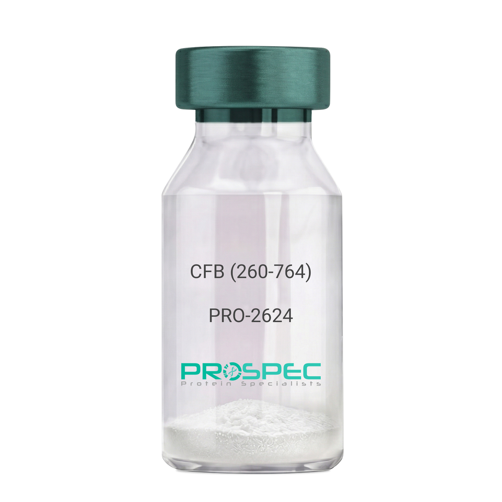
  • View Data Sheet

    Name :

    CFB (260-764) Human, Sf9

    Description:

    Complement Factor B (260-764 a.a.) Human Recombinant, Sf9

    CFB, C3/C5 convertase, Glycine-rich beta glycoprotein, GBG, PBF2, Properdin factor B, BF, BFD, AHUS4, ARMD14, CFAB, CFBD, FB, FBI12, GBG, H2-Bf.

    Product # :

    PRO-2624

    Price :

    Quantity :

    Shipping Method :

    Ice Icon

    Shipped with Ice Packs

    Add To Cart

    More Info

    • description
    • source
    • formulation
    • purity
    • More Info

    Description

    CFB Human Recombinant produced in Sf9 Baculovirus cells is a single, glycosylated polypeptide chain containing 514 amino acids (260-764 a.a) and having a molecular mass of 58.1kDa.CFB is fused to an 9 amino acid His-tag at C-terminus & purified by proprietary chromatographic techniques.

    Source

    Sf9, Baculovirus cells.

    Formulation

    CFB protein solution (0.5mg/ml) containing Phosphate Buffered Saline (pH 7.4) and 20% glycerol.

    Purity

    Greater than 90.0% as determined by SDS-PAGE.

    More Info

    • Introduction

      Factor B is a protein, build from a single chain that circulates in the blood. Complement factor D cleaves and activates factor B, thus creating a catalytic subunit Bb and noncatalytic chain Ba. Subunit Bb is a serine protease that can bind to C3b and forms alternative pathway C3 convertase. Subunit Ba acts as an inhibitor for proliferation of lymphocytes, when Bb is part of the lymphocytes’ proliferation. The gene that codes for this protein is located on chromosome six.

    • Synonyms

      CFB, C3/C5 convertase, Glycine-rich beta glycoprotein, GBG, PBF2, Properdin factor B, BF, BFD, AHUS4, ARMD14, CFAB, CFBD, FB, FBI12, GBG, H2-Bf.

    • Physical Appearance

      Sterile Filtered colorless solution.

    • Stability

      Store at 4°C if entire vial will be used within 2-4 weeks. Store, frozen at -20°C for longer periods of time. For long term storage it is recommended to add a carrier protein (0.1% HSA or BSA).Avoid multiple freeze-thaw cycles.

    • Amino Acid Sequence

      ADPKIVLDPS GSMNIYLVLD GSDSIGASNF TGAKKCLVNL IEKVASYGVK PRYGLVTYAT
      YPKIWVKVSE ADSSNADWVT KQLNEINYED HKLKSGTNTK KALQAVYSMM SWPDDVPPEG
      WNRTRHVIIL MTDGLHNMGG DPITVIDEIR DLLYIGKDRK NPREDYLDVY VFGVGPLVNQ
      VNINALASKK DNEQHVFKVK DMENLEDVFY QMIDESQSLS LCGMVWEHRK GTDYHKQPWQ
      AKISVIRPSK GHESCMGAVV SEYFVLTAAH CFTVDDKEHS IKVSVGGEKR DLEIEVVLFH
      PNYNINGKKE AGIPEFYDYD VALIKLKNKL KYGQTIRPIC LPCTEGTTRA LRLPPTTTCQ
      QQKEELLPAQ DIKALFVSEE EKKLTRKEVY IKNGDKKGSC ERDAQYAPGY DKVKDISEVV
      TPRFLCTGGV SPYADPNTCR GDSGGPLIVH KRSRFIQVGV ISWGVVDVCK NQKRQKQVPA HARDFHINLF QVLPWLKEKL QDEDLGFLHH HHHH

    ProSpec's products are furnished for LABORATORY RESEARCH USE ONLY. The product may not be used as drugs, agricultural or pesticidal products, food additives or household chemicals.

    Complement Factor B
  • View Data Sheet

    Name :

    Mouse OX40L

    Description:

    OX40 Ligand Mouse Recombinant

    Tumor necrosis factor ligand superfamily member 4, OX40 ligand, OX40L, OX-40L, CD252, Tnfsf4, Ox40l, Txgp1l, gp3, OX4, Ath-1, Ath1, CD134L, gp34, Tnlg2b, Txgp1l

    Product # :

    CYT-1191

    Price :

    Quantity :

    Shipping Method :

    Ice Icon

    Shipped with Ice Packs

    Add To Cart

    More Info

    • description
    • source
    • formulation
    • purity
    • biological activity
    • More Info

    Description

    OX40L Mouse produced in Sf9 Baculovirus cells is a single, glycosylated polypeptide chain containing 157 amino acids (51-198 aa) and having a molecular mass of 17.7kDa.OX40L is fused to a 6 amino acid His tag at C-terminus and purified by proprietary chromatographic techniques.

    Source

    Sf9, Baculovirus cells.

    Formulation

    The OX40L solution (1mg/ml) contains 10% Glycerol and Phosphate-Buffered Saline (pH 7.4).

    Purity

    Greater than 90.0% as determined by SDS-PAGE.

    Biological Activity

    Determined by its binding ability in a functional ELISA with Human TNFRSF4 (CAT# cyt-1039).

    More Info

    • Introduction

      OX40 Ligandis a part of the tumor necrosis factor (TNF)receptor family. OX40L receptor is OX40 which is expressed predominantly on activated CD4+ T cells.OX40 Ligandis expressed on various antigen-presenting cells such asmacrophages, DC2sand activated Blymphocytes. Expression of OX40 is momentary following engagement of T cell receptors. The ligation of OX40-OX40L stimulates proliferation anddifferentiation of activated B cells,increases immunoglobulin secretion and is also a sourceof survival signal for T cells and enables the development of memory T cells.

    • Synonyms

      Tumor necrosis factor ligand superfamily member 4, OX40 ligand, OX40L, OX-40L, CD252, Tnfsf4, Ox40l, Txgp1l, gp3, OX4, Ath-1, Ath1, CD134L, gp34, Tnlg2b, Txgp1l

    • Physical Appearance

      Sterile filtered colorless solution.

    • Stability

      Store at 4°C if entire vial will be used within 2-4 weeks.Store, frozen at -20°C for longer periods of time.For long term storage it is recommended to add a carrier protein (0.1% HSA or BSA).Avoid multiple freeze-thaw cycles.

    • Amino Acid Sequence

      ADPSSSPAKD PPIQRLRGAV TRCEDGQLFI SSYKNEYQTM EVQNNSVVIK CDGLYIIYLK GSFFQEVKID LHFREDHNPI SIPMLNDGRR IVFTVVASLA FKDKVYLTVN APDTLCEHLQ INDGELIVVQ LTPGYCAPEG SYHSTVNQVP LHHHHHH

    ProSpec's products are furnished for LABORATORY RESEARCH USE ONLY. The product may not be used as drugs, agricultural or pesticidal products, food additives or household chemicals.

    Ox40L Mouse
  • View Data Sheet

    Name :

    CTF1 Rat

    Description:

    Cardiotrophin-1 Rat Recombinant

    Cardiotrophin-1, CT-1, Ctf1.

    Product # :

    CYT-199

    Price :

    Quantity :

    Shipping Method :

    Room Temp Icon

    Shipped at Room temp

    Add To Cart

    More Info

    • description
    • source
    • formulation
    • purity
    • biological activity
    • More Info

    Description

    Cardiotrophin-1 Rat Recombinant produced in E.Coli is a single, non-glycosylated, polypeptide chain containing 203 amino acids and having a molecular mass of 21.4kDa.The CTF1 Rat is purified by proprietary chromatographic techniques.

    Source

    Escherichia Coli.

    Formulation

    Lyophilized from a 0.2µm filtered concentrated solution in PBS, pH 7.4.

    Purity

    Greater than 95.0% as determined by SDS-PAGE and HPLC analyses.

    Biological Activity

    The ED50 as determined by the dose-dependent proliferation of TF-1 cells was found to be < 0.5ng/ml, corresponding to a specific activity of > 2.0×10⁶ units/mg.

    More Info

    • Introduction

      Cardiotrophin 1 (CT-1) is a 201 amino acid member of the interleukin-6 superfamily. It was identified by its ability to induce hypertrophic response in cardiac myocytes. CT-1 mRNA levels were found both in cardiac myocytes and in cardiac nonmyocytes. CT 1 was also detected in abundance in normal adult human lung and was expressed in both fetal and adult airway smooth muscle cells. CT 1 activates gp130 dependent signaling and stimulates the Janus kinase/signal transducers and activators of transcription (JAK/STAT) pathway to transduce hypertrophic and cytoprotective signals in cardiac myocytes.
      CT 1 has also a neurotrophic function. CTF1 deficiency causes increased motoneuron cell death in spinal cord and brainstem nuclei of mice during a period between embryonic day 14 and the first postnatal week. Moreover, CT-1 is a hepatocyte survival factor that efficiently reduces hepatocellular damage in animal models of acute liver injury. Cardiotrophin 1 expression is augmented after hypoxic stimulation and it can protect cardiac cells when added either prior to simulated ischaemia or at the time of reoxygenation following simulated ischaemia. Cardiotrophin 1 can induce expression of the protective heat shock proteins (hsps) in cardiac cells.
      Cardiotrophin-1 increased ventricular expression of ANP, brain natriuretic peptide (BNP) and angiotensinogen mRNA.
      Cardiophin 1 levels were significantly elevated in patients with heart failure, patients with dilatative cardiomyopathy, moderate/severe mitral regurgitation, stable and unstable angina and after acute myocardial infarction.

    • Synonyms

      Cardiotrophin-1, CT-1, Ctf1.

    • Physical Appearance

      Sterile Filtered White lyophilized (freeze-dried) powder.

    • Stability

      Lyophilized Cardiotrophin-1 Rat although stable at room temperature for 3 weeks, should be stored desiccated below -18°C. Upon reconstitution CTF1 Rat should be stored at 4°C between 2-7 days and for future use below -18°C.Please prevent freeze-thaw cycles.

    • Solubility

      It is recommended to reconstitute the lyophilized CTF1 in sterile 4mM HCl not less than 100µg/ml, which can then be further diluted to other aqueous solutions.

    • Amino Acid Sequence

      MSQREGSLED HQTDSSFSFL PHLEAKIRQT HNLARLLTKY ADQLLEEYVQ QQGEPFGLPG FSPPRLPLAG LSGPAPSHAG LPVSERLRQD AAALSALPAL LDAVRRRQAE LNPRAPRLLR SLEDAARQVR ALGAAVETVL AALGAAARGP VPEPVATSAL FTSNSAAGVF SAKVLGLHVC GLYGEWVSRT EGDLGQLVPG GVA.

    • Background

      What is the molecular weight/Mw of CTF1 Protein?
      CTF1 Protein has a total Mw of 21.4kDa.

      What is the source or expression system of CTF1 Protein?
      Escherichia Coli.

      What is the Purity of CTF1 Protein?
      CTF1 Protein is >95% pure as determined by SDS-PAGE.

      What is the Biological Activity of CTF1 Protein?
      The ED50 as determined by the dose-dependent proliferation of TF-1 cells was found to be < 0.5ng/ml, corresponding to a specific activity of > 2.0×10⁶ units/mg.

      What is the amino acid sequence of CTF1 Protein?
      MSQREGSLED HQTDSSFSFL PHLEAKIRQT HNLARLLTKY ADQLLEEYVQ QQGEPFGLPG FSPPRLPLAG LSGPAPSHAG LPVSERLRQD AAALSALPAL LDAVRRRQAE LNPRAPRLLR SLEDAARQVR ALGAAVETVL AALGAAARGP VPEPVATSAL FTSNSAAGVF SAKVLGLHVC GLYGEWVSRT EGDLGQLVPG GVA.

      What applications can CTF1 Protein be used in?
      CTF1 Protein can probably be used in western blot, ELISA and Lateral Flow.

      What is the endotoxin level for CTF1 Protein?
      The endotoxin level is minimal, CTF1 Protein was purified using conventional chromatography techniques.

    ProSpec's products are furnished for LABORATORY RESEARCH USE ONLY. The product may not be used as drugs, agricultural or pesticidal products, food additives or household chemicals.

    Ct 1 Rat
  • View Data Sheet

    Name :

    EBI3 Human

    Description:

    Epstein Barr Virus Induced 3 Human Recombinant

    Interleukin-27 subunit beta, IL-27 subunit beta, IL-27B, Epstein-Barr virus-induced gene 3 protein, EBV-induced gene 3 protein, EBI3, IL27B.

    Product # :

    CYT-367

    Price :

    Quantity :

    Shipping Method :

    Room Temp Icon

    Shipped at Room temp

    Add To Cart

    More Info

    • description
    • source
    • formulation
    • purity
    • biological activity
    • More Info

    Description

    EBI3 Human Recombinant produced in E.Coli is a single, non-glycosylated, Polypeptide chain containing 209 amino acids fragment (21-229) having a molecular weight of 23.3kDa. The EBI3 is purified by proprietary chromatographic techniques.

    Source

    Escherichia Coli.

    Formulation

    EBI3 Human Recombinant was lyophilized from a solution containing 10mM Acetic Acid and 0.5% Mannitol.

    Purity

    Greater than 90% as determined by
    (a) Analysis by RP-HPLC.
    (b) Analysis by SDS-PAGE.

    Biological Activity

    Assay data for Human recombinant EBI3 is based upon qualitative binding to anti-EBI3 antibody.

    More Info

    • Introduction

      EBI3 has an induced expression in B lymphocytes in reaction to Epstein-Barr virus infection. EBI3 encodes a secreted glycoprotein belonging to the hematopoietin receptor family, and heterodimerizes with a 28 kDa protein to form iIL-27. EBI3 drives rapid clonal expansion of naive cd4(+) t-cells. EBI3 strongly synergizes with IL-12 to activate IFN-gamma production of naive cd4(+) t-cells. EBI3 mediates its biologic effects through the cytokine receptor wsx-1/tccr.

    • Synonyms

      Interleukin-27 subunit beta, IL-27 subunit beta, IL-27B, Epstein-Barr virus-induced gene 3 protein, EBV-induced gene 3 protein, EBI3, IL27B.

    • Physical Appearance

      Sterile Filtered White lyophilized (freeze-dried) powder.

    • Stability

      Lyophilized EBI3 although stable at room temperature for 3 weeks, should be stored desiccated below -18°C. Upon reconstitution EBI3 should be stored at 4°C between 2-7 days and for future use below -18°C. Please prevent freeze-thaw cycles.

    • Solubility

      It is recommended to reconstitute the lyophilized EBI3 in sterile 10mM Acetic acid not less than 100µg/ml, which can then be further diluted to other aqueous solutions.

    • Amino Acid Sequence

      RKGPPAALTLPRVQCRASRYPIAVDCSWTLPPAPNSTSPVSF
      IATYRLGMAARGHSWPCLQQTPTSTSCTITDVQLFSMAPYVL
      NVTAVHPWGSSSSFVPFITEHIIKPDPPEGVRLSPLAERQLQ
      VQWEPPGSWPFPEIFSLKYWIRYKRQGAARFHRVGPIEATSF
      ILRAVRPRARYYVQVAAQDLTDYGELSDWSLPATATMSLGK.

    • Background

      Title: Epstein-Barr Virus Induced 3 Human Recombinant: Unveiling its Role in Epstein-Barr Virus-Associated Diseases

      Abstract:


      Epstein-Barr Virus Induced 3 (EBI3) is a crucial cytokine involved in the immune response against Epstein-Barr virus (EBV) and various other pathogens. This research paper provides an extensive analysis of human recombinant EBI3, focusing on its production, characterization, and potential applications in understanding EBV-associated diseases. The paper highlights the significance of EBI3 in modulating immune responses and explores its role in the pathogenesis of EBV-related malignancies. Furthermore, it discusses ongoing research and clinical trials investigating the therapeutic potential of recombinant EBI3 in immune disorders and cancer. The information presented in this paper aims to enhance our understanding of human recombinant EBI3 and its utility as a research tool and a potential immunotherapeutic agent.

      Introduction:


      Epstein-Barr Virus Induced 3 (EBI3) is a cytokine that plays a critical role in the immune response against EBV. Human recombinant EBI3, produced through genetic engineering techniques, provides a valuable tool for studying its immunomodulatory properties and exploring its potential therapeutic applications.

      Production and Characterization:


      Recombinant EBI3 is typically generated using expression systems such as mammalian cells or bacteria. The protein is then purified and characterized to ensure its structural integrity and functional activity. Rigorous quality control measures are implemented to confirm the specificity and potency of the recombinant EBI3.

      Role in EBV-Associated Diseases:


      EBI3 is involved in the regulation of immune responses during EBV infection. It acts as a subunit of the heterodimeric cytokine interleukin-27 (IL-27), which plays a crucial role in anti-viral immunity. Recombinant EBI3 serves as a valuable tool for investigating the mechanisms underlying EBI3-mediated immune regulation and its potential implications in EBV-associated diseases, including infectious mononucleosis, nasopharyngeal carcinoma, and EBV-related lymphomas.

      Therapeutic Implications:


      Dysregulation of the immune response is implicated in various immune disorders and cancers. Recombinant EBI3 holds promise as a potential immunotherapeutic agent due to its immunomodulatory properties. Ongoing research and clinical trials are investigating the therapeutic applications of recombinant EBI3 in conditions such as autoimmune diseases, viral infections, and cancer.

      Conclusion:


      Human recombinant EBI3 represents a valuable research tool and a potential immunotherapeutic agent. Its production, characterization, and applications in understanding EBV-associated diseases contribute to our understanding of immune regulation and the development of targeted therapeutic interventions. Continued research and clinical trials exploring the therapeutic potential of recombinant EBI3 offer promising avenues for improving outcomes in immune disorders and EBV-related malignancies.

      What is the molecular weight/Mw of EBI3 Protein?
      EBI3 Protein has a total Mw of 23.3kDa.

      What is the source or expression system of EBI3 Protein?
      Escherichia Coli.

      What is the Purity of EBI3 Protein?
      EBI3 Protein is >90% pure as determined by SDS-PAGE.

      What is the Biological Activity of EBI3 Protein?
      Assay data for Human recombinant EBI3 is based upon qualitative binding to anti-EBI3 antibody.

      What is the amino acid sequence of EBI3 Protein?
      RKGPPAALTLPRVQCRASRYPIAVDCSWTLPPAPNSTSPVSF
      IATYRLGMAARGHSWPCLQQTPTSTSCTITDVQLFSMAPYVL
      NVTAVHPWGSSSSFVPFITEHIIKPDPPEGVRLSPLAERQLQ
      VQWEPPGSWPFPEIFSLKYWIRYKRQGAARFHRVGPIEATSF
      ILRAVRPRARYYVQVAAQDLTDYGELSDWSLPATATMSLGK.

      What applications can EBI3 Protein be used in?
      EBI3 Protein can probably be used in western blot, ELISA and Lateral Flow.

      What is the endotoxin level for EBI3 Protein?
      The endotoxin level is minimal, EBI3 Protein was purified using conventional chromatography techniques.

    ProSpec's products are furnished for LABORATORY RESEARCH USE ONLY. The product may not be used as drugs, agricultural or pesticidal products, food additives or household chemicals.

    Ebi3 Human
  • 1
  • …
  • 16
  • 17
  • 18
  • 19
  • 20
  • …
  • 42
Your browser does not support the video tag.

Products

  • Cytokines
  • Growth Factors
  • Chemokines
  • CD Antigens
  • Neurotrophins
  • Hormones
  • Enzymes
  • Viral Antigens
  • Recombinant Proteins
  • Natural Proteins
  • Monoclonal Antibodies
  • Polyclonal Antibodies
  • Thermal Packaging

About

  • Company
  • Founder
  • Contribution
  • Contact Us
  • My Account

Ordering

  • Ordering Method
  • Ordering - Credit Card
  • Ordering PO
  • Shipping Fees
  • FAQ's

Legal

  • Terms & Conditions
  • Privacy
  • Site Map
  • info@prospecbio.com
  • Facebook
  • Instagram
  • Linkedin Profile

© 2026 ProSpec-Tany TechnoGene Ltd. All Rights Reserved.

  • Choosing a selection results in a full page refresh.
  • Opens in a new window.