Search results
1000 results found for “Ectodysplasin”
Name
Description
Product #
Price
Quantity
Shipping Method
- View Data Sheet
Name :
TGFB3 MouseDescription:
Transforming Growth Factor-Beta 3 Mouse Recombinant
Transforming Growth Factor-beta3, TGFB3, ARVD, FLJ16571, TGF-beta3.
Product # :
CYT-143Price :
Quantity :
Shipping Method :
Shipped with Ice Packs
More Info
- description
- source
- formulation
- purity
- biological activity
- More Info
Description
TGF-β 3 Mouse Recombinant produced in E.Coli is a disulfide-linked homodimeric, non-glycosylated, polypeptide chain containing two 113 amino acid chains and having a total molecular mass of 25.7kDa. The TGF-β 3 is purified by standard chromatographic techniques.
Source
Escherichia Coli.
Formulation
The Mouse TGFB3 protein solution contains 20% Ethanol and 10mM Acetic acid.
Purity
Greater than 95.0% as determined by SDS-PAGE.
Biological Activity
The activity is determined by the ability to induce chondrogenic differentiation.More Info
-
Introduction
Transforming growth factor betas (TGF Betas) mediate many cell-cell interactions that occur during embryonic development. Three TGF Betas have been identified in mammals. TGF Beta 1, TGF Beta 2 and TGF Beta 3 are each synthesized as precursor proteins that are very similar in that each is cleaved to yield a 112 amino acid polypeptide that remains associated with the latent portion of the molecule.
-
Synonyms
Transforming Growth Factor-beta3, TGFB3, ARVD, FLJ16571, TGF-beta3.
-
Physical Appearance
Sterile Filtered clear solution.
-
Stability
Mouse TGF-beta 3 although stable at room temperature for 3 weeks, should be stored at 4°C.For long term storage it is recommended to add a carrier protein (0.1% HSA or BSA).
-
Amino Acid Sequence
MALDTNYCFRN LEENCCVRPL YIDFRQDLGW KWVHEPKGYY ANFCSGPCPY LRSADTTHST VLGLYNTLNP EASASPCCVP QDLEPLTILY YVGRTPKVEQ LSNMVVKSCK CS.
-
Protein content
Protein quantitation was carried out by two independent methods:1. UV spectroscopy at 280 nm using the absorbency value of 1.718 as the extinction coefficient for a 0.1% (1mg/ml) solution. This value is calculated by the PC GENE computer analysis program of protein sequences (IntelliGenetics). 2. Analysis by RP-HPLC, using a calibrated solution of TGF-b 3 as a Reference Standard.
ProSpec's products are furnished for LABORATORY RESEARCH USE ONLY. The product may not be used as drugs, agricultural or pesticidal products, food additives or household chemicals.
- View Data Sheet
Name :
Treponema p17 16.4kDaDescription:
Treponema pallidum p17, 16.4kDa Recombinant
Product # :
TRP-252Price :
Quantity :
Shipping Method :
Shipped at Room temp
More Info
- description
- source
- formulation
- purity
- More Info
Description
Recombinant Treponema p17 produced in E.coli is a non-glycosylated polypeptide chain having a molecular mass of 16.4kDa and fused to a His tag at N-terminus.
Source
Escherichia Coli.
Formulation
Lyophilized from 1mg/ml in 20mM sodium carbonate pH-10.
Purity
Greater than 95.0% as determined by SDS-PAGE.
More Info
-
Introduction
Treponema pallidum is a gram-negative spirochaete bacterium and is considered to be metabolically crippled. There are at least four known subspecies: T. pallidum pallidum, T. pallidum pertenue, T. pallidum carateum and T. pallidum endemicum. The helical structure of T. pallidum pallidum allows it to move in a corkscrew motion through viscous mediums such as mucus. Treponema pallidum sub sp. pallidum has one of the smallest bacterial genomes at 1.14 million base pairs (Mb) and has limited metabolic capabilities, reflecting its adaptation through genome reduction to the rich environment of mammalian tissue.
-
Physical Appearance
Sterile Filtered White lyophilized (freeze-dried) powder.
-
Stability
Treponema p17 although stable at room temperature for 4 weeks, should be stored below -18°C. Please prevent freeze thaw cycles.
-
Solubility
It is recommended to reconstitute the lyophilized Treponema p17 in sterile 18M-cm H2O not less than 100µg/ml, which can then be further diluted to other aqueous solutions.
ProSpec's products are furnished for LABORATORY RESEARCH USE ONLY. The product may not be used as drugs, agricultural or pesticidal products, food additives or household chemicals.
- View Data Sheet
Name :
MINA HumanDescription:
MYC Induced Nuclear Antigen Human Recombinant
MYC Induced Nuclear Antigen, MINA53, MDIG, 60S Ribosomal Protein L27a Histidine Hydroxylase, Mineral Dust-Induced Gene Protein, Histone Lysine Demethylase MINA Ribosomal Oxygenase MINA, Nucleolar Protein 52, NO52, ROX, Bifunctional Lysine-Specific Demethylase And Histidyl-Hydroxylase MINA, Myc-Induced Nuclear Antigen, 53 KDa, Mineral Dust Induced Gene Protein, MYC-Induced Nuclear Antigen, EC 1.14.11.-, Bifunctional lysine-specific demethylase and histidyl-hydroxylase MINA.
Product # :
PRO-2078Price :
Quantity :
Shipping Method :
Shipped with Ice Packs
More Info
- description
- source
- formulation
- purity
- More Info
Description
MINA Human Recombinant produced in E.Coli is a single, non-glycosylated polypeptide chain containing 485 amino acids (1-465 a.a) and having a molecular mass of 54.9kDa. MINA is fused to a 20 amino acid His-tag at N-terminus & purified by proprietary chromatographic techniques.
Source
Escherichia Coli.
Formulation
MINA protein solution (0.25mg/ml) containing 20mM Tris-HCl buffer (pH 8.0), 0.15M NaCl, 10% glycerol and 1mM DTT.
Purity
Greater than 90.0% as determined by SDS-PAGE.
More Info
-
Introduction
MYC Induced Nuclear Antigen, also known as MINA is an oxygenase which can function both as a histone lysine demethylase and a ribosomal histidine hydroxylase. MINA is involved in the demethylation of trimethylated Lys-9 on histone H3 (H3K9me3), leading to an increase in ribosomal RNA expression. MINA also catalyzes the hydroxylation of 60S ribosomal protein L27a on His-39. In addition, MINA plays a significant role in cell growth and survival. MINA is implicated in ribosome biogenesis, probably in the duration of the assembly process of pre-ribosomal particles.
-
Synonyms
MYC Induced Nuclear Antigen, MINA53, MDIG, 60S Ribosomal Protein L27a Histidine Hydroxylase, Mineral Dust-Induced Gene Protein, Histone Lysine Demethylase MINA Ribosomal Oxygenase MINA, Nucleolar Protein 52, NO52, ROX, Bifunctional Lysine-Specific Demethylase And Histidyl-Hydroxylase MINA, Myc-Induced Nuclear Antigen, 53 KDa, Mineral Dust Induced Gene Protein, MYC-Induced Nuclear Antigen, EC 1.14.11.-, Bifunctional lysine-specific demethylase and histidyl-hydroxylase MINA.
-
Physical Appearance
Sterile Filtered clear solution.
-
Stability
Store at 4°C if entire vial will be used within 2-4 weeks. Store, frozen at -20°C for longer periods of time. For long term storage it is recommended to add a carrier protein (0.1% HSA or BSA).Avoid multiple freeze-thaw cycles.
-
Amino Acid Sequence
MGSSHHHHHH SSGLVPRGSH MPKKAKPTGS GKEEGPAPCK QMKLEAAGGP SALNFDSPSS LFESLISPIK TETFFKEFWE QKPLLIQRDD PALATYYGSL FKLTDLKSLC SRGMYYGRDV NVCRCVNGKK KVLNKDGKAH FLQLRKDFDQ KRATIQFHQP QRFKDELWRI QEKLECYFGS LVGSNVYITP AGSQGLPPHY DDVEVFILQL EGEKHWRLYH PTVPLAREYS VEAEERIGRP VHEFMLKPGD LLYFPRGTIH QADTPAGLAH STHVTISTYQ NNSWGDFLLD TISGLVFDTA KEDVELRTGI PRQLLLQVES TTVATRRLSG FLRTLADRLE GTKELLSSDM KKDFIMHRLP PYSAGDGAEL STPGGKLPRL DSVVRLQFKD HIVLTVLPDQ DQSDETQEKM VYIYHSLKNS RETHMMGNEE ETEFHGLRFP LSHLDALKQI WNSPAISVKD LKLTTDEEKE SLVLSLWTEC LIQVV.
ProSpec's products are furnished for LABORATORY RESEARCH USE ONLY. The product may not be used as drugs, agricultural or pesticidal products, food additives or household chemicals.
- View Data Sheet
Name :
DGCR6L HumanDescription:
DiGeorge Syndrome Critical Region Gene 6-Like Human Recombinant
Protein DGCR6L, DiGeorge syndrome critical region 6-like protein, DGCR6L.
Product # :
PRO-1454Price :
Quantity :
Shipping Method :
Shipped with Ice Packs
More Info
- description
- source
- formulation
- purity
- More Info
Description
DGCR6L Human Recombinant produced in E.Coli is a single, non-glycosylated polypeptide chain containing 243 amino acids (1-220 a.a) and having a molecular mass of 27.3kDa.DGCR6L is fused to a 23 amino acid His-tag at N-terminus & purified by proprietary chromatographic techniques.
Source
Escherichia Coli.
Formulation
DGCR6L protein solution (0.5mg/ml) containing 20mM Tris-HCl buffer (pH 8.0), 0.2M NaCl, 50% glycerol, 1mM DTT and 250mM Imidazole.
Purity
Greater than 85.0% as determined by SDS-PAGE.
More Info
-
Introduction
DiGeorge syndrome critical region 6-like protein (DGCR6L) shares homology with the Drosophila gonadal protein, it is expressed in gonadal tissues and germ cells, and with the human laminin gamma-1 chain which functions in cell attachment and migration. DGCR6L is located in a region of chromosome 22 involved in the DiGeorge syndrome, one aspect of a wider collection of anomalies referred to as the CATCH 22 syndrome. Moreover, it is one of two functional genes encoding nearly identical proteins which have analogous expression patterns.
-
Synonyms
Protein DGCR6L, DiGeorge syndrome critical region 6-like protein, DGCR6L.
-
Physical Appearance
Sterile Filtered colorless solution.
-
Stability
Store at 4°C if entire vial will be used within 2-4 weeks. Store, frozen at -20°C for longer periods of time. For long term storage it is recommended to add a carrier protein (0.1% HSA or BSA).Avoid multiple freeze-thaw cycles.
-
Amino Acid Sequence
MGSSHHHHHH SSGLVPRGSH MGSMERYAAA LEEVADGARQ QERHYQLLSA LQSLVKELPS SFQQRLSYTT LSDLALALLD GTVFEIVQGL LEIQHLTEKS LYNQRLRLQN EHRVLRQALR QKHQEAQQAC RPHNLPVVQA AQQRELEAVE HRIREEQRAM DQKIILELDR KVADQQSTLE KAGVAGFYVT TNPQELMLQM NLLELIRKLQ QRGCRAGNAA LGLGGPWQSP AAQCDQKGSP VPP.
ProSpec's products are furnished for LABORATORY RESEARCH USE ONLY. The product may not be used as drugs, agricultural or pesticidal products, food additives or household chemicals.
- View Data Sheet
Name :
SIL1 HumanDescription:
SIL1 Human Recombinant
SIL1 homolog endoplasmic reticulum chaperone (S. cerevisiae), nucleotide exchange factor SIL1, Marinesco-Sjogren syndrome, BiP-associated protein, ULG5, MSS, BAP.
Product # :
PRO-1187Price :
Quantity :
Shipping Method :
Shipped with Ice Packs
More Info
- description
- source
- formulation
- purity
- More Info
Description
SIL1 Human Recombinant produced in E. coli is a single polypeptide chain containing 439 amino acids (32-461) and having a molecular mass of 50.0 kDa.SIL1 is fused to a 8 amino acid His-tag at C-terminus & purified by proprietary chromatographic techniques.
Source
E.coli.
Formulation
The SIL1 solution (0.5mg/ml) contains 20mM Tris-HCl buffer (pH 8.0), 100mM Nacl and 10% glycerol.
Purity
Greater than 90% as determined by SDS-PAGE.
More Info
-
Introduction
SIL1 is localized to endoplasmic reticulum (ER). SIL1 is an N-linked glycoprotein with an N-terminal ER targeting sequence, 2 putative N-glycosylation sites, and a C-terminal ER retention signal. SIL1 acts as a nucleotide exchange factor for another unfolded protein response protein. Mutations in SIL1 are linked to Marinesco-Sjogren syndrome.
-
Synonyms
SIL1 homolog endoplasmic reticulum chaperone (S. cerevisiae), nucleotide exchange factor SIL1, Marinesco-Sjogren syndrome, BiP-associated protein, ULG5, MSS, BAP.
-
Physical Appearance
Sterile Filtered colorless solution.
-
Stability
Store at 4°C if entire vial will be used within 2-4 weeks. Store, frozen at -20°C for longer periods of time. For long term storage it is recommended to add a carrier protein (0.1% HSA or BSA).Avoid multiple freeze-thaw cycles.
-
Amino Acid Sequence
MHQNLKEFAL TNPEKSSTKE TERKETKAEE ELDAEVLEVF HPTHEWQALQ PGQAVPAGSH VRLNLQTGER EAKLQYEDKF RNNLKGKRLD INTNTYTSQD LKSALAKFKE GAEMESSKED KARQAEVKRL FRPIEELKKD FDELNVVIET DMQIMVRLIN KFNSSSSSLE EKIAALFDLE YYVHQMDNAQ DLLSFGGLQV VINGLNSTEP LVKEYAAFVL GAAFSSNPKV QVEAIEGGAL QKLLVILATE QPLTAKKKVL FALCSLLRHF PYAQRQFLKL GGLQVLRTLV QEKGTEVLAV RVVTLLYDLV TEKMFAEEEA ELTQEMSPEK LQQYRQVHLL PGLWEQGWCE ITAHLLALPE HDAREKVLQT LGVLLTTCRD RYRQDPQLGR TLASLQAEYQ VLASLELQDG EDEGYFQELL GSVNSLLKEL RLEHHHHHH.
ProSpec's products are furnished for LABORATORY RESEARCH USE ONLY. The product may not be used as drugs, agricultural or pesticidal products, food additives or household chemicals.
- View Data Sheet
Name :
STX1A (1-265) HumanDescription:
Syntaxin-1A (1-265 a.a) Human Recombinant
Syntaxin 1A, Neuron-Specific Antigen HPC-1, Syntaxin 1A (Brain), STX1, Syntaxin-1A, HPC-1, P35-1, SYN1A.
Product # :
PRO-2335Price :
Quantity :
Shipping Method :
Shipped with Ice Packs
More Info
- description
- source
- formulation
- purity
- More Info
Description
STX1A Human Recombinant produced in E.Coli is a single, non-glycosylated polypeptide chain containing 265 amino acids (1-265 a.a) and having a molecular mass of 30.7kDa. STX1A is purified by proprietary chromatographic techniques.
Source
Escherichia Coli.
Formulation
STX1A protein solution (1mg/ml) containing phosphate buffered saline (pH 7.4), 10% glycerol and 1mM DTT.
Purity
Greater than 95.0% as determined by SDS-PAGE.
More Info
-
Introduction
Syntaxin is membrane integrated Q-SNARE protein participating in exocytosis. Syntaxin is composed of an N-terminal regulatory domain (Habc), a SNARE domain (known as H3), and a single C-terminal transmembrane domain. The SNARE (H3) domain binds to both synaptobrevin and SNAP-25 forming the core SNARE complex. Synaptic vesicles store neurotransmitters that are released during calcium-regulated exocytosis. The specificity of neurotransmitter release requires the localization of both synaptic vesicles and calcium channels to the presynaptic active zone. Syntaxins function in this vesicle fusion process. Syntaxins also serve as a substrate for botulinum neurotoxin type C, a metalloprotease that blocks exocytosis and has high affinity for a molecular complex that includes the alpha-latrotoxin receptor which produces exocytosis.
-
Synonyms
Syntaxin 1A, Neuron-Specific Antigen HPC-1, Syntaxin 1A (Brain), STX1, Syntaxin-1A, HPC-1, P35-1, SYN1A.
-
Physical Appearance
Sterile Filtered colorless solution.
-
Stability
Store at 4°C if entire vial will be used within 2-4 weeks. Store, frozen at -20°C for longer periods of time. For long term storage it is recommended to add a carrier protein (0.1% HSA or BSA). Avoid multiple freeze-thaw cycles.
-
Amino Acid Sequence
MKDRTQELRT AKDSDDDDDV AVTVDRDRFM DEFFEQVEEI RGFIDKIAEN VEEVKRKHSA ILASPNPDEK TKEELEELMS DIKKTANKVR SKLKSIEQSI EQEEGLNRSS ADLRIRKTQH STLSRKFVEV MSEYNATQSD YRERCKGRIQ RQLEITGRTT TSEELEDMLE SGNPAIFASG IIMDSSISKQ ALSEIETRHS EIIKLENSIR ELHDMFMDMA MLVESQGEMI DRIEYNVEHA VDYVERAVSD TKKAVKYQSK ARRKK.
ProSpec's products are furnished for LABORATORY RESEARCH USE ONLY. The product may not be used as drugs, agricultural or pesticidal products, food additives or household chemicals.
- View Data Sheet
Name :
PEDF Human, HEKDescription:
Pigment Epithelium-Derived Factor Human Recombinant, HEK
Pigment epithelium-derived factor, PEDF, Serpin-F1, SerpinF1, EPC-1, EPC1, PIG35.
Product # :
CYT-553Price :
Quantity :
Shipping Method :
Shipped at Room temp
More Info
- description
- source
- formulation
- purity
- More Info
Description
PEDF Human Recombinant produced in HEK cells is a single, glycosylated, polypeptide chain containing a total of 410 amino acids, having a molecular mass of 45.6 kDa and fused to an 11 aa FLAG tag at C-Terminus.The Human PEDF is purified by proprietary chromatographic techniques.
Source
HEK 293.
Formulation
The filtered (0.4µm) concentrated (0.5mg/ml) protein solution was lyophilized with 20mM Tris & 20mM NaCl pH-7.5.
Purity
Greater than 95% as determined by SDS-PAGE.
More Info
-
Introduction
PEDF is a neurotrophic protein that induces extensive neuronal differentiation in retinoblastoma cells. SerpinF1 is a potent inhibitor of angiogenesis. EPC1 doesn’t undergo the stressed to relaxed conformation transition characteristic as of the active serpins since it exhibits no serine protease inhibitory activity.
Aqueous humour level of asymmetric dimethylarginine is correlated with PEDF in humans. ADMA and PEDF levels are increased in response to inflammation in uveitis.
Lack of PEDF expression is a potent factor for the enhancement of tumor growth and angiogenesis in breast cancer.
PEDF & VEGF genes contribute to the development of diabetic retinopathy.
PEDF and VEGF structural changes in blood vessel wall play an important role in the pathophysiology of PD patients.
PEDF-overexpressing tumors exhibited reduced intratumoral angiogenesis.
SerpinF1 is a new promising approach for the treatment of osteosarcoma.
Levels of the natural ocular anti-angiogenic factor SentrinF1 (PEDF) is associated with proliferative retinopathy.
VEGF secreted by retinal pigment epithelial cells upregulates PEDF expression via VEGFR-1 in an autocrine manner.
Sentrin-F1 concentration in the aqueous humor of diabetic patients predicts who will develop progression of retinopathy.
PEDF blocks angiogenic effects of leptin through its anti-oxidative properties. -
Synonyms
Pigment epithelium-derived factor, PEDF, Serpin-F1, SerpinF1, EPC-1, EPC1, PIG35.
-
Physical Appearance
Filtered White lyophilized (freeze-dried) powder.
-
Stability
Store lyophilized protein at -20°C. Aliquot the product after reconstitution to avoid repeated freezing/thawing cycles. Reconstituted protein can be stored at 4°C for a limited period of time.
-
Solubility
It is recomnded to add deionized water to a working concentration of 0.5mg/ml and let the lyophilized pellet dissolve completely. Product is not sterile! Please filter the product by an appropriate sterile filter before using it in the cell culture.
-
Amino Acid Sequence
QNPASPPEEG SPDPDSTGAL VEEEDPFFKV PVNKLAAAVS NFGYDLYRVR SSTSPTTNVL LSPLSVATAL SALSLGAEQR TESIIHRALY YDLISSPDIH GTYKELLDTV TAPQKNLKSA SRIVFEKKLR IKSSFVAPLE KSYGTRPRVL TGNPRLDLQE INNWVQAQMK GKLARSTKEI PDEISILLLG VAHFKGQWVT KFDSRKTSLE DFYLDEERTV RVPMMSDPKA VLRYGLDSDL SCKIAQLPLT GSMSIIFFLP LKVTQNLTLI EESLTSEFIH DIDRELKTVQ AVLTVPKLKL SYEGEVTKSL QEMKLQSLFD SPDFSKITGK PIKLTQVEHR AGFEWNEDGA GTTPSPGLQP AHLTFPLDYH LNQPFIFVLR DTDTGALLFI GKILDPRGPAAADYKDDDDK.
ProSpec's products are furnished for LABORATORY RESEARCH USE ONLY. The product may not be used as drugs, agricultural or pesticidal products, food additives or household chemicals.
- View Data Sheet
Name :
VEGF Mouse (121 a.a.), YeastDescription:
Vascular Endothelial Growth Factor (121 a.a.) Mouse Recombinant, Yeast
Vascular endothelial growth factor A, VEGF-A, Vascular permeability factor, VPF, VEGF, MGC70609.
Product # :
CYT-1125Price :
Quantity :
Shipping Method :
Shipped at Room temp
More Info
- description
- source
- formulation
- purity
- biological activity
- More Info
Description
Vascular Endothelial Growth Factor (121 a.a.) Mouse Recombinant produced in yeast is a disulfide-linked homodimer consisting of 2x121 amino acid polypeptide chains, having a molecular mass of approximately 20.7kDa each.VEGF (121 a.a.) is purified by proprietary chromatographic techniques.
Source
Saccharomyces cerevisiae
Formulation
Lyophilized from a 0.2μm filtered concentrated solution in PBS, pH 7.4.
Purity
Greater than 95.0% as determined by:
(a) Analysis by RP-HPLC.
(b) Analysis by SDS-PAGE.Biological Activity
The activity was measured in a cell proliferation assay using HUVEC human umbilical vein endothelial cells and was found to be 1-4 ng/ml.
More Info
-
Introduction
Vascular endothelial growth factor is an important signaling protein involved in both angiogenesis and vasculogenesis.VEGF activity has been mostly studied on cells of the vascular endothelium, although it does have effects on a number of other cell types (e.g. stimulation monocyte/macrophage migration, neurons, cancer cells, kidney epithelial cells ).VEGF mediates increased vascular permeability, induces angiogenesis, vasculogenesis and endothelial cell growth, promotes cell migration, and inhibits apoptosis. In vitro, VEGF has been shown to stimulate endothelial cell mitogenesis and cell migration. VEGF is also a vasodilator and increases microvascular permeability and was originally referred to as vascular permeability factor.
Elevated levels of VEGF is linked to POEMS syndrome, also known as Crow-Fukase syndrome. Mutations in VEGF have been associated with proliferative and nonproliferative diabetic retinopathy. -
Synonyms
Vascular endothelial growth factor A, VEGF-A, Vascular permeability factor, VPF, VEGF, MGC70609.
-
Physical Appearance
Sterile Filtered White lyophilized (freeze-dried) powder.
-
Stability
Lyophilized VEGF (121 a.a.) although stable at room temperature for 3 weeks, should be stored desiccated below -18°C. Upon reconstitution Vascular Endothelial Growth Factor (121 a.a.) should be stored at 4°C between 2-7 days and for future use below -18°C.Please prevent freeze-thaw cycles.
-
Solubility
It is recommended to reconstitute the lyophilized Vascular Endothelial Growth Factor (121 a.a.) in sterile 18MΩ-cm H2O not less than 100µg/ml, which can then be further diluted to other aqueous solutions.
-
Amino Acid Sequence
MAPTTEGEQK SHEVIKFMDV YQRSYCRPIE TLVDIFQEYP DEIEYIFKPS CVPLMRCAGC CNDEALECVP TSESNITMQI MRIKPHQSQH IGEMSFLQHS RCECRPKKDR TKPEKCDKPR R.
ProSpec's products are furnished for LABORATORY RESEARCH USE ONLY. The product may not be used as drugs, agricultural or pesticidal products, food additives or household chemicals.
- View Data Sheet
Name :
TROVE2 Bovine, BiotinDescription:
TROVE Domain Family Member 2 Human Recombinant, Biotinlyated
60 kDa SS-A/Ro ribonucleoprotein, 60 kDa ribonucleoprotein Ro, RoRNP, 60 kDa Ro protein, Ro 60 kDa autoantigen, TROVE domain family member 2, Sjoegren syndrome type A antigen, SS-A, Sjoegren syndrome antigen A2, TROVE2, RO60, SSA2, RO-60.
Product # :
PRO-2561Price :
Quantity :
Shipping Method :
Shipped with Ice Packs
More Info
- description
- source
- formulation
- purity
- More Info
Description
TROVE2 is a biotinlyated natural protein having a molecular mass of 58kDa. TROVE2 is purified from bovine tissues by protein-chemical methods.
Source
Bovine tissues.
Formulation
TROVE2 is supplied in 20mM HEPES buffer pH-8.0, 150mM NaCl and 20% glycerol.
Purity
Greater than 83.0% as determined by SDS-PAGE.
More Info
-
Introduction
Ro 60 kDa autoantigen is an RNA-binding protein that binds to several small cytoplasmic RNA molecules known as Y RNAs. SSA2 may stabilize these RNAs from degradation. Sera taken from patients with Systemic Lupus Erythematosus (SLE) often contains antibodies that react with the normal cellular SSA2 protein as if this antigen was foreign.
-
Synonyms
60 kDa SS-A/Ro ribonucleoprotein, 60 kDa ribonucleoprotein Ro, RoRNP, 60 kDa Ro protein, Ro 60 kDa autoantigen, TROVE domain family member 2, Sjoegren syndrome type A antigen, SS-A, Sjoegren syndrome antigen A2, TROVE2, RO60, SSA2, RO-60.
-
Physical Appearance
Sterile Filtered clear solution.
-
Stability
Store at 4°C if entire vial will be used within 2-4 weeks. Store, frozen at -20°C for longer periods of time. Avoid multiple freeze-thaw cycles.
ProSpec's products are furnished for LABORATORY RESEARCH USE ONLY. The product may not be used as drugs, agricultural or pesticidal products, food additives or household chemicals.
- View Data Sheet
Name :
HSP90B1 HumanDescription:
Heat Shock Protein 90kDa Beta (GRP94) Member 1 Human Recombinant
ECGP, GP96, TRA1, GRP94, HSP90B1, Endoplasmin, Heat shock protein 90 kDa beta member 1, 94 kDa glucose-regulated protein, gp96 homolog, Tumor rejection antigen 1.
Product # :
HSP-091Price :
Quantity :
Shipping Method :
Shipped with Ice Packs
More Info
- description
- source
- formulation
- purity
- More Info
Description
HSP90B1 produced in E.Coli is a single, non-glycosylated polypeptide chain containing 819 amino acids (22-803 a.a.) and having a molecular mass of 94.4 kDa.HSP90B1 is expressed with a 36 amino acid His tag at N-Terminus and purified by proprietary chromatographic techniques.
Source
Escherichia Coli.
Formulation
The GRP94 protein solution (1mg/ml) contains 20mM Tris-HCl, pH-8, 1mM EDTA, 0.1M NaCl, 1mM DTT and 10% Glycerol.
Purity
Greater than 90.0% as determined by SDS-PAGE.
More Info
-
Introduction
HSP90B1 is an abundant molecular chaperone resident endoplasmic reticulum (ER) lumenal stress protein which is part of the Hsp90 family. HSP90B1 is involved in maintaining protein homeostasis in the secretory pathway as well as functioning in the intracellular trafficking of peptides from the extracellular space to the MHC class I antigen processing pathway of antigen presentation cells. HSP90B1 has key roles in signal transduction, protein folding, protein degradation, and morphologic evolution. HSP90B1 protein associates with numerous cochaperones and involved in folding of newly synthesized proteins or stabilizing and refolding denatured proteins after stress. HSP90B1 is highly expressed in human gastric carcinoma BGC-823 cells during the whole cell cycle.
-
Synonyms
ECGP, GP96, TRA1, GRP94, HSP90B1, Endoplasmin, Heat shock protein 90 kDa beta member 1, 94 kDa glucose-regulated protein, gp96 homolog, Tumor rejection antigen 1.
-
Physical Appearance
Sterile filtered colorless solution.
-
Stability
Store at 4°C if entire vial will be used within 2-4 weeks. Store, frozen at -20°C for longer periods of time. For long term storage it is recommended to add a carrier protein (0.1% HSA or BSA).Avoid multiple freeze-thaw cycles.
-
Amino Acid Sequence
MRGSHHHHHH GMASMTGGQQ MGRDLYDDDD KDRWGSMDDE VDVDGTVEED LGKSREGSRT DDEVVQREEE AIQLDGLNAS QIRELREKSE KFAFQAEVNR MMKLIINSLY KNKEIFLREL ISNASDALDK IRLISLTDEN ALSGNEELTV KIKCDKEKNL LHVTDTGVGM TREELVKNLG TIAKSGTSEF LNKMTEAQED GQSTSELIGQ FGVGFYSAFL VADKVIVTSK HNNDTQHIWE SDSNEFSVIA DPRGNTLGRG TTITLVLKEE ASDYLELDTI KNLVKKYSQF INFPIYVWSS KTETVEEPME EEEAAKEEKE ESDDEAAVEE EEEEKKPKTK KVEKTVWDWE LMNDIKPIWQ RPSKEVEEDE YKAFYKSFSK ESDDPMAYIH FTAEGEVTFK SILFVPTSAP RGLFDEYGSK KSDYIKLYVR RVFITDDFHD MMPKYLNFVK GVVDSDDLPL NVSRETLQQH KLLKVIRKKL VRKTLDMIKK IADDKYNDTF WKEFGTNIKL GVIEDHSNRT RLAKLLRFQS SHHPTDITSL DQYVERMKEK QDKIYFMAGS SRKEAESSPF VERLLKKGYE VIYLTEPVDE YCIQALPEFD GKRFQNVAKE GVKFDESEKT KESREAVEKE FEPLLNWMKD KALKDKIEKA VVSQRLTESP CALVASQYGW SGNMERIMKA QAYQTGKDIS TNYYASQKKT FEINPRHPLI RDMLRRIKED EDDKTVLDLA VVLFETATLR SGYLLPDTKA YGDRIERMLR LSLNIDPDAK VEEEPEEEPE ETAEDTTEDT EQDEDEEMDV GTDEEEETAK ESTAEKDEL.
ProSpec's products are furnished for LABORATORY RESEARCH USE ONLY. The product may not be used as drugs, agricultural or pesticidal products, food additives or household chemicals.
- View Data Sheet
Name :
Leptin-A TilapiaDescription:
Leptin-A Tilapia Recombinant
Product # :
CYT-1109Price :
Quantity :
Shipping Method :
Shipped at Room temp
More Info
- description
- source
- formulation
- purity
- biological activity
- More Info
Description
Leptin-A Tilapia Recombinant produced in E.Coli is a single, non-glycosylated polypeptide chain containing 161 amino acids and having a molecular mass of 16,491 Dalton. The Leptin-A Tilapia is purified by proprietary chromatographic techniques.
Source
Escherichia Coli.
Formulation
Lyophilized from a concentrated (1mg/ml) solution containing NaHCO3 at 1:2 salt: protein ratio.
Purity
Greater than 95.0% as determined by:
(a) Gel filtration analysis.
(b) Analysis by SDS-PAGE.Biological Activity
Tilapia leptins were found to be biologically active in promoting proliferation of BAF/3 cells stably transfected with the long form of human leptin receptor, but their activity was lower than that of mammalian leptin. Furthermore, the Tilapia leptins were biologically active in promoting STAT‐LUC activation in COS7 cells transfected with Tilapia leptin receptor but not in cells transfected with human leptin receptor. Tilapia Leptin A was more active than Tilapia Leptin B.
More Info
-
Introduction
Leptin is a protein hormone. It is mainly produced in adipose cells that regulate energy homeostasis by restraining hunger. Leptin ties to nuclear receptors in the hypothalamus (arcuate nucleus). Similar to insulin resistance in type II diabetes, in obesity there is a decrease in the sensitivity towards leptin, ending in a failure to identify satiety, even in high levels of energy stores or leptin itself. Full-length cDNA encoding 2 leptin sequences (tLepA and tLepB) and 1 leptin receptor sequence (tLepR) exists in tilapia (Oreochromis niloticus). The full-length cDNA of tLepR is 3423 bp, encoding a protein of 1140 amino acid which contained all functionally important domains conserved among vertebrate leptin receptors. The cDNAs of tLepA and tLepB are 486 bp and 459 bp in length, encoding proteins of 161 aa and 152 aa, respectively. The three-dimensional structures of tLepA and tLepB demonstrates strong conservation of tertiary structure with that of human leptin comprised of 4 helixes.
-
Physical Appearance
Sterile Filtered White lyophilized (freeze-dried) powder.
-
Stability
Lyophilized Leptin-A Tilapia although stable at room temperature for 3 weeks, should be stored desiccated below -18°C. Upon reconstitution Leptin-A Tilapia should be stored at 4°C between 2-7 days and for future use below -18°C. For long term storage it is recommended to add a carrier protein (0.1% HSA or BSA). Please prevent freeze-thaw cycles.
-
Solubility
It is recommended to reconstitute the lyophilized Leptin-A Tilapia in sterile water or 0.4% NaHCO3 adjusted to pH 8-9, not less than 100µg/ml, which can then be further diluted to other aqueous solutions.
-
Amino Acid Sequence
The first six N-terminal amino acids of recombinant Tilapia leptin A are Ala-Pro-Leu-Pro-Val-Glu.
-
Protein content
Protein quantitation was carried out by UV spectroscopy at 280 nm using the absorbency value of 1.33 for 1 mg/ml Leptin-A Tilapia as the extinction coefficient for a 0.1% (1mg/ml) solution at pH 8.0. This value is calculated by the DNAman computer analysis program of protein sequences.
ProSpec's products are furnished for LABORATORY RESEARCH USE ONLY. The product may not be used as drugs, agricultural or pesticidal products, food additives or household chemicals.
- View Data Sheet
Name :
SPARCL1 MouseDescription:
SPARC Like 1 Mouse Recombinant
Sparcl1, Ecm2, hevin, mast9, Sc1, SPARC-like protein 1, Extracellular matrix protein 2, Matrix glycoprotein Sc1.
Product # :
PRO-2617Price :
Quantity :
Shipping Method :
Shipped with Ice Packs
More Info
- description
- source
- formulation
- purity
- More Info
Description
SPARCL1 Mouse Recombinant produced in Sf9 Baculovirus cells is a single, glycosylated polypeptide chain containing 642 amino acids (17-650 a.a) and having a molecular mass of 71.7kDa.SPARCL1 is fused to an 8 amino acid His-tag at C-terminus & purified by proprietary chromatographic techniques.
Source
Sf9, Baculovirus cells.
Formulation
SPARCL1 protein solution (0.25mg/ml) containing Phosphate Buffered Saline (pH 7.4) and 10% glycerol.
Purity
Greater than 90.0% as determined by SDS-PAGE.
More Info
-
Introduction
SPARC-like protein 1 or SPARC1 is an anti-adhesive protein with a similarity to the SPARC protein’s structure. SPARC1 is highly expressed in several tissues such as brain, lungs, kidney, heart, although it is not found in the liver tissue. The protein inhibits spreading or adhesion of many substrates and is considered to lead to antiadhesive signaling that stops neuronal migration, that occurs during development or as a trauma response, consistent with production by glial and neuronal cells.
-
Synonyms
Sparcl1, Ecm2, hevin, mast9, Sc1, SPARC-like protein 1, Extracellular matrix protein 2, Matrix glycoprotein Sc1.
-
Physical Appearance
Sterile Filtered colorless solution.
-
Stability
Store at 4°C if entire vial will be used within 2-4 weeks. Store, frozen at -20°C for longer periods of time. For long term storage it is recommended to add a carrier protein (0.1% HSA or BSA).Avoid multiple freeze-thaw cycles.
-
Amino Acid Sequence
IPTSTRFLSD HSNPTTATLV TPEDATVPIA GVEATADIEN HPSDKAEKPS ALNSEEETHE
QSTEQDKTYS FEVDLKDEED GDGDLSVDPT EGTLTLDLQE GTSEPQQKSL PENGDFPATV
STSYVDPNQR ANITKGKESQ EQPVSDSHQQ PNESSKQTQD LKAEESQTQD PDIPNEEEEE
EEDEEEEEEE EPEDIGAPSD NQEEGKEPLE EQPTSKWEGN REQSDDTLEE SSQPTQISKT
EKHQSEQGNQ GQESDSEAEG EDKAAGSKEH IPHTEQQDQE GKAGLEAIGN QKDTDEKAVS
TEPTDAAVVP RSHGGAGDNG GGDDSKHGAG DDYFIPSQEF LEAERMHSLS YYLKYGGGEE
TTTGESENRR EAADNQEAKK AESSPNAEPS DEGNSREHSA GSCTNFQCKR GHICKTDPQG
KPHCVCQDPE TCPPAKILDQ ACGTDNQTYA SSCHLFATKC RLEGTKKGHQ LQLDYFGACK
SIPACTDFEV AQFPLRMRDW LKNILMQLYE PNPKHGGYLN EKQRSKVKKI YLDEKRLLAG
HPIELLLRD FKKNYHMYVY PVHWQFNELD QHPADRILTH SELAPLRASL VPMEHCITRF
FEECDPNKDK HITLKEWGHC FGIKEEDIDE NLLFLEHHHH HH
ProSpec's products are furnished for LABORATORY RESEARCH USE ONLY. The product may not be used as drugs, agricultural or pesticidal products, food additives or household chemicals.
- View Data Sheet
Name :
DTL HumanDescription:
Denticleless E3 Ubiquitin Protein Ligase Human Recombinant
L2DTL, DCAF2, RAMP, CDT2.
Product # :
PRO-2826Price :
Quantity :
Shipping Method :
Shipped at Room temp
More Info
- description
- source
- formulation
- purity
- More Info
Description
The DTL Human is created as a recombinant protein with a 4kda N-terminal fusion of His Tag. The DTL His-Tagged Fusion Protein, produced in E. coli, is a 31kDa protein containing 126 amino acid residues of the DTL Human, 1-231 amino acids.
Source
Escherichia Coli.
Formulation
Each mg was lyophilized with 1xPBS, 0.4% SDS and 4mM DTT.
Purity
Greater than 90% as determined by SDS-PAGE.
More Info
-
Synonyms
L2DTL, DCAF2, RAMP, CDT2.
-
Physical Appearance
Filtered White lyophilized (freeze-dried) powder.
-
Stability
Store lyophilized DTL at -20°C. Aliquot the product after reconstitution to avoid repeated freezing/thawing cycles. Reconstituted protein can be stored at 4°C for a limited period of time; it does not show any change after two weeks at 4°C.
-
Solubility
It is recommended to add deionized water to prepare a working stock solution of approximately 0.5mg/ml and let the lyophilized pellet dissolve completely. Product is not sterile! Please filter the product by an appropriate sterile filter before using it on cell culture.
-
Background
Denticleless E3 ubiquitin protein ligase (DTL) is part of the E3 ubiquitin ligase family, fundamental to the ubiquitin-proteasome system, which adjusts protein degradation and modification in cells. DTL also participates in numerous critical biological processes, which comprises cell cycle progression, DNA repair, as well as embryonic development. More than a few diseases have been linked with the dysregulation of DTL in particular Cancer, this highlights the importance of DTL as a potential therapeutic goal. DTL takes an important part in regulating the cell cycle, mainly the transition from the G1 phase to the S phase. The degradation of cell cycle regulators, such as cyclins, is also mediated by DTL in that way assuring appropriate cell division in addition to preventing uncontrolled proliferation. DTL is also part of the DNA damage response mechanisms. It ubiquitinates vital proteins which are involved in DNA repair pathways, thus influencing the cellular response to genotoxic stress and preserving genomic integrity.
ProSpec's products are furnished for LABORATORY RESEARCH USE ONLY. The product may not be used as drugs, agricultural or pesticidal products, food additives or household chemicals.
- View Data Sheet
Name :
Lymphotactin RatDescription:
Lymphotactin (XCL1) Rat Recombinant
XCL1, Cytokine SCM-1, ATAC, Lymphotaxin, SCM-1-alpha, Small inducible cytokine C1, XC chemokine ligand 1, LTN, LPTN, SCM1, SCM-1, SCYC1, SCM-1a.
Product # :
CHM-038Price :
Quantity :
Shipping Method :
Shipped at Room temp
More Info
- description
- source
- formulation
- purity
- biological activity
- More Info
Description
Lymphotactin (XCL1) Rat Recombinant produced in E.Coli is a non-glycosylated polypeptide chain containing 93 amino acids and having a molecular mass of approximately 10.0kDa.Lymphotactin is purified by proprietary chromatographic techniques.
Source
Escherichia Coli.
Formulation
Lyophilized from a 0.2μm filtered concentrated solution in PBS, pH 7.4.
Purity
Greater than 97.0% as determined by:
(a) Analysis by RP-HPLC.
(b) Analysis by SDS-PAGE.Biological Activity
The ED50 as determined by a chemotaxis bioassay using human XCR1 transfected murine BaF3 cells < 100 ng/ml, corresponding to a specific activity of > 1.0 × 10⁴ IU/mg.
More Info
-
Introduction
XCL1 is a small cytokine belongs to the XC chemokine family that is also known as lymphotactin. XCL1 is found in high levels in spleen, thymus, intestine and peripheral blood leukocytes, and at lower levels in lung, prostate gland and ovary. Cellular sources for XCL1 include activated thymic and peripheral blood CD8+ T cells. This chemokine attracts T cells. In humans, XCL1 is closely related to XCL2, whose gene is found at the same locus on chromosome 1. XCL1 induces it chemotactic function by binding to a chemokine receptor called XCR1.
-
Synonyms
XCL1, Cytokine SCM-1, ATAC, Lymphotaxin, SCM-1-alpha, Small inducible cytokine C1, XC chemokine ligand 1, LTN, LPTN, SCM1, SCM-1, SCYC1, SCM-1a.
-
Physical Appearance
Sterile Filtered White lyophilized (freeze-dried) powder.
-
Stability
Lyophilized XCL1 although stable at room temperature for 3 weeks, should be stored desiccated below -18°C. Upon reconstitution Lymphotactin should be stored at 4°C between 2-7 days and for future use below -18°C.Please prevent freeze-thaw cycles.
-
Solubility
It is recommended to reconstitute the lyophilized Lymphotactin in sterile 18MΩ-cm H2O not less than 100µg/ml, which can then be further diluted to other aqueous solutions.
-
Amino Acid Sequence
VGTEVLQESI CVSLRTQRLP VQKIKTYTIK EGAMRAVIFV TKRGLRICAD PQAKWVKTAI KTVDGRASAS KSKAETIPTQ AQRSASTAVT LTG.
ProSpec's products are furnished for LABORATORY RESEARCH USE ONLY. The product may not be used as drugs, agricultural or pesticidal products, food additives or household chemicals.
- View Data Sheet
Name :
T.gondii MIC-3Description:
Toxoplasma Gondii MIC 3 Recombinant
Product # :
TOX-261Price :
Quantity :
Shipping Method :
Shipped with Ice Packs
More Info
- description
- source
- formulation
- purity
- More Info
Description
The E.Coli derived recombinant protein contains the MIC3 immunodominant regions, amino acids 234-306. MIC3 protein is fused to a 26Kda Glutathione transferase tag.
Source
Escherichia Coli.
Formulation
50mM Tris-HCl pH 8.0, 1mM EDTA and 8M urea.
Purity
Toxoplasma protein is >95% pure as determined by 10% PAGE (coomassie staining).
More Info
-
Introduction
The life cycle of Toxoplasma gondii has two phases. The coccidia like takes place only in members of the Felidae family which makes these animals the parasite's primary host. The asexual part of the life cycle can take place in any warm-blooded animal, like other mammals(including felines) and birds. T. gondii constructing daughter scaffolds within the mother cell. In the intermediate hosts (including felines), the parasite invades cells, forming intracellular so-called parasitophorous vacuoles containing bradyzoites, the slowly replicating form of the parasit. Vacuoles form tissue cysts mainly within the muscles and brain. Since they are within cells, the host's immune system does not detect these cysts. Resistance to antibiotics varies, but the cysts are very difficult to eradicate entirely. Within these vacuoles T. gondii propagates by a series of binary fissions until the infected cell eventually bursts and tachyzoites are released. Tachyzoites are the motile, asexually reproducing form of the parasite. Unlike the bradyzoites, the free tachyzoites are usually efficiently cleared by the host's immune response, although some manage to infect cells and form bradyzoites, thus maintaining the infection.
-
Stability
Toxoplasma protein although stable at 4°C for 1 week, should be stored below -18°C. Please prevent freeze thaw cycles.
-
Applications
Toxoplasma antigen is suitable in ELISA and Western blots, excellent antigen for detection of Toxoplasma gondii- with minimal specificity problems.
-
Specificity
Immunoreactive with sera of Toxoplasma gondii-infected individuals.
-
Purification Method
Toxoplasama protein was purified by GS-4B Sepharose-Affinity Purification.
ProSpec's products are furnished for LABORATORY RESEARCH USE ONLY. The product may not be used as drugs, agricultural or pesticidal products, food additives or household chemicals.
- View Data Sheet
Name :
LGALS3 Mouse, ActiveDescription:
Galectin-3 Mouse Recombinant, BioActive
Lectin galactose binding soluble 3, Lectin, galactose binding, soluble 3, CBP35, GAL3, GALBP, GALIG, LGALS2, MAC2.
Product # :
CYT-1151Price :
Quantity :
Shipping Method :
Shipped with Ice Packs
More Info
- description
- source
- formulation
- purity
- biological activity
- More Info
- sds-page
Description
LGALS3 Mouse Recombinant produced in E.Coli is a single, non-glycosylated polypeptide chain containing 287 amino acids ( 1-264 a.a) and having a molecular mass of 29.8kDa.LGALS3 is fused to a 23 amino acid His-tag at N-terminus & purified by proprietary chromatographic techniques.
Source
E.coli.
Formulation
LGALS3 protein (0.5mg/ml) contains 20mM Tris-HCl buffer (pH 8.0), 0.15M NaCl, 50% glycerol,1mM DTT and 2mM EDTA.
Purity
Greater than 95% as determined by SDS-PAGE.
Biological Activity
Measured by its ability to agglutinate human red blood cells. The ED50 for this effect is ≥ 25ug/ml.
sds-page
More Info
-
Introduction
Galectin 3, or LGALS3, is a protein which belongs to the animal lectins family, that binds betagalactoside residues selectively. LGALS3 is originated and leaves cells through ectocytosis. The protein is capable of inhibition apoptosis and the development of cancer. Galectin 3 is found in epithelial tissues in organisms, it can be located in dendritic cells, Kupffer cells, macrophages etc. LGALS3 levels elevated when inflammation is generating, cell proliferation, trans-activation by viral proteins and cell differentiation.
-
Synonyms
Lectin galactose binding soluble 3, Lectin, galactose binding, soluble 3, CBP35, GAL3, GALBP, GALIG, LGALS2, MAC2.
-
Physical Appearance
Sterile Filtered colorless solution.
-
Stability
Store at 4°C if entire vial will be used within 2-4 weeks. Store, frozen at -20°C for longer periods of time. For long term storage it is recommended to add a carrier protein (0.1% HSA or BSA).Avoid multiple freeze-thaw cycles.
-
Amino Acid Sequence
MGSSHHHHHH SSGLVPRGSH MGSMADSFSL NDALAGSGNP NPQGYPGAWG NQPGAGGYPG AAYPGAYPGQ APPGAYPGQA PPGAYPGQAP PSAYPGPTAP GAYPGPTAPG AYPGSTAPGA FPGQPGAPGA YPSAPGGYPA AGPYGVPAGP LTVPYDLPLP GGVMPRMLIT IMGTVKPNAN RIVLDFRRGN DVAFHFNPRF NENNRRVIVC NTKQDNNWGK EERQSAFPFE SGKPFKIQVL VEADHFKVAV NDAHLLQYNH RMKNLREISQ LGISGDITLT SANHAMI.
-
Background
What is the molecular weight/Mw of LGALS3 MOUSE Protein?
LGALS3 MOUSE Protein has a total Mw of 29.8kDa.
What is the source or expression system of LGALS3 MOUSE Protein?
Escherichia Coli.
What is the Purity of LGALS3 MOUSE Protein?
LGALS3 MOUSE Protein is >95% pure as determined by SDS-PAGE.
What is the Biological Activity of LGALS3 MOUSE Protein?
Measured by its ability to agglutinate human red blood cells. The ED50 for this effect is ≥ 25ug/ml.
What is the amino acid sequence of LGALS3 MOUSE Protein?
MGSSHHHHHH SSGLVPRGSH MGSMADSFSL NDALAGSGNP NPQGYPGAWG NQPGAGGYPG AAYPGAYPGQ APPGAYPGQA PPGAYPGQAP PSAYPGPTAP GAYPGPTAPG AYPGSTAPGA FPGQPGAPGA YPSAPGGYPA AGPYGVPAGP LTVPYDLPLP GGVMPRMLIT IMGTVKPNAN RIVLDFRRGN DVAFHFNPRF NENNRRVIVC NTKQDNNWGK EERQSAFPFE SGKPFKIQVL VEADHFKVAV NDAHLLQYNH RMKNLREISQ LGISGDITLT SANHAMI.
What applications can LGALS3 MOUSE Protein be used in?
LGALS3 MOUSE Protein can probably be used in western blot, ELISA and Lateral Flow.
ProSpec's products are furnished for LABORATORY RESEARCH USE ONLY. The product may not be used as drugs, agricultural or pesticidal products, food additives or household chemicals.
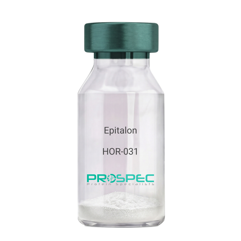
- View Data Sheet
Name :
EpitalonDescription:
Epitalon
Product # :
HOR-031Price :
Quantity :
Shipping Method :
Shipped at Room temp
More Info
- description
- formulation
- purity
- More Info
Description
Epithalon Synthetic is a single, non-glycosylated polypeptide chain containing 4 amino acids, having a molecular mass of 390.35 Dalton and a Molecular formula of C14H22N4O9.
Formulation
The protein was lyophilized with no additives.
Purity
Greater than 97.0% as determined by analysis by RP-HPLC.
More Info
-
Physical Appearance
Sterile Filtered White lyophilized (freeze-dried) powder.
-
Stability
Lyophilized Epithalon although stable at room temperature for 3 weeks, should be stored desiccated below -18°C. Upon reconstitution Epithalon should be stored at 4°C between 2-7 days and for future use below -18°C.For long term storage it is recommended to add a carrier protein (0.1% HSA or BSA). Please prevent freeze-thaw cycles.
-
Solubility
It is recommended to reconstitute the lyophilized Epithalon in sterile 18MΩ-cm H2O not less than 100 µg/ml, which can then be further diluted to other aqueous solutions.
-
Amino Acid Sequence
H-Ala-Glu-Asp-Gly-OH .
-
Background
Epitalon, also known as Epithalon, is a synthetic tetrapeptide that has been a focal point in the field of aging and longevity research. This peptide, consisting of four amino acids, is derived from the pineal gland and has been associated with a variety of biological effects, most notably its potential role in extending the lifespan of cells.
Epitalon's primary function is its interaction with telomeres, the protective caps at the ends of chromosomes that shorten as cells divide. By stimulating the production of telomerase, an enzyme that can repair and lengthen telomeres, Epitalon may slow down the aging process at a cellular level.
Numerous studies have explored the potential of Epitalon in extending the lifespan of organisms. For instance, Khavinson and colleagues (2003) found that Epitalon increased the lifespan of fruit flies, while Anisimov et al. (2003) reported similar results in mice. These findings suggest that Epitalon could potentially have similar effects in humans.
Epitalon has also been studied for its role in regulating circadian rhythms. Korkushko et al. (2011) found that Epitalon could restore disrupted circadian rhythms in elderly people, suggesting potential applications in sleep disorders and other conditions related to circadian rhythm disruption.
Beyond its potential role in aging and longevity, Epitalon has been explored for its potential therapeutic applications. Anisimov et al. (2011) found that Epitalon could reduce the incidence of spontaneous tumors in mice, suggesting potential applications in cancer prevention.
While research on Epitalon is promising, it is important to note that most studies have been conducted in animals or in vitro. More research is needed to fully understand the potential effects and applications of Epitalon in humans. However, the existing body of research suggests that Epitalon could be a promising tool in the fight against aging and age-related diseases.
ProSpec's products are furnished for LABORATORY RESEARCH USE ONLY. The product may not be used as drugs, agricultural or pesticidal products, food additives or household chemicals.
- View Data Sheet
Name :
T.pallidum p47 (Partial)Description:
Treponema pallidum p47 (Partial) Recombinant
Product # :
TRP-249Price :
Quantity :
Shipping Method :
Shipped with Ice Packs
More Info
- description
- source
- formulation
- purity
- More Info
Description
The E.Coli derived recombinant protein is fused at the C- terminus with a 6xHis Tag and contains the T.Pallidum p47 immunodominant regions.
Source
Escherichia Coli.
Formulation
70mM Tris-HCl pH8.0, 50mM NaCl, 50% Glycerol and 1.5M urea.
Purity
Treponema Pallidum protein is >95% pure as determined by 10% PAGE (coomassie staining).
More Info
-
Introduction
Treponema pallidum is a gram-negative spirochaete bacterium and is considered to be metabolically crippled. There are at least four known subspecies: T. pallidum pallidum, T. pallidum pertenue, T. pallidum carateum and T. pallidum endemicum. The helical structure of T. pallidum pallidum allows it to move in a corkscrew motion through viscous mediums such as mucus. Treponema pallidum subsp. pallidum has one of the smallest bacterial genomes at 1.14 million base pairs (Mb) and has limited metabolic capabilities, reflecting its adaptation through genome reduction to the rich environment of mammalian tissue.
-
Stability
Treponema Pallidum although stable at 4°C for 1 week, should be stored below -18°C. Please prevent freeze thaw cycles.
-
Applications
Treponema Pallidum protein is suitable for ELISA and Western blots, excellent antigen for detection of T.Pallidum with minimal specificity problems.
-
Specificity
Immunoreactive with sera of T.Pallidum infected individuals.
-
Purification Method
Treponema Pallidum protein was purified by proprietary chromatographic technique.
ProSpec's products are furnished for LABORATORY RESEARCH USE ONLY. The product may not be used as drugs, agricultural or pesticidal products, food additives or household chemicals.
- View Data Sheet
Name :
IL 12 p40 HumanDescription:
Interleukin-12 p40 Human Recombinant
NKSF2, CTL maturation factor (TCMF), Cytotoxic lymphocyte maturation factor 40 kDa subunit (CLMF p40), TSF, Edodekin-alpha, IL-12 p40, IL-12B, IL-12 subunit p40, NK cell stimulatory factor chain 2.
Product # :
CYT-488Price :
Quantity :
Shipping Method :
Shipped with Ice Packs
More Info
- description
- source
- formulation
- purity
- More Info
Description
Interleukin-12 p40 His Human Recombinant produced in E.Coli is single, a non-glycosylated, polypeptide chain containing 306 amino acids fragment (23-328) with an amino-terminal hexahistidine tag. The IL-12 p40 His is purified by proprietary chromatographic techniques.
Source
Escherichia Coli.
Formulation
Interleukin-12 p40 His is supplied in 1xPBS and 50% glycerol.
Purity
Greater than 95.0% as determined by SDS-PAGE.
More Info
-
Introduction
Active IL-12 is a p70 disulphide-linked dimer composed of p35 and p40 subunits. The protein is a pleiotropic cytokine produced primarily by antigen presenting cells and has multiple effects on T lymphocytes and natural killer cells in terms of stimulating cytotoxicity, proliferation, production of other cytokines and Th1 subset differentiation.
-
Synonyms
NKSF2, CTL maturation factor (TCMF), Cytotoxic lymphocyte maturation factor 40 kDa subunit (CLMF p40), TSF, Edodekin-alpha, IL-12 p40, IL-12B, IL-12 subunit p40, NK cell stimulatory factor chain 2.
-
Physical Appearance
Sterile Filtered clear solution.
-
Stability
Store at 4°C if entire vial will be used within 2-4 weeks. Store, frozen at -20°C for longer periods of time. Please avoid freeze thaw cycles.
ProSpec's products are furnished for LABORATORY RESEARCH USE ONLY. The product may not be used as drugs, agricultural or pesticidal products, food additives or household chemicals.
- View Data Sheet
Name :
ITGB1 AntibodyDescription:
Mouse Anti Human Integrin Beta 1
Integrin beta-1, Fibronectin receptor subunit beta, Glycoprotein IIa, GPIIA, VLA-4 subunit beta, CD29, ITGB1, FNRB, MDF2, MSK12, Integrin beta 1, CD29, VLAB.
Product # :
ANT-715Price :
Quantity :
Shipping Method :
Shipped with Ice Packs
More Info
- formulation
- More Info
Formulation
1mg/ml containing PBS, pH-7.4, 10% Glycerol and 0.02% Sodium Azide.
More Info
-
Introduction
Integrins are heterodimeric proteins consist of alpha and beta subunits. There are more than 18 alpha and 8 beta subunits discovered in mammals. Integrin family members are membrane receptors that participates in cell adhesion and recognition in a variety of processes including embryogenesis, hemostasis, tissue repair, immune response and metastatic diffusion of tumor cells. ITGB1 encodes a beta subunit.
-
Synonyms
Integrin beta-1, Fibronectin receptor subunit beta, Glycoprotein IIa, GPIIA, VLA-4 subunit beta, CD29, ITGB1, FNRB, MDF2, MSK12, Integrin beta 1, CD29, VLAB.
-
Physical Appearance
Sterile filtered colorless solution.
-
Immunogen
Anti-human ITGB1 mAb, is derived from hybridization of mouse F0 myeloma cells with spleen cells from BALB/c mice immunized with a recombinant human ITGB1 protein 21-461 amino acids purified from E. coli.
-
Ig Subclass
Mouse IgG1 heavy chain and k light chain.
-
Clone
PAT47E2AT.
-
Applications
ITGB1 antibody has been tested by ELISA and Western blot analysis to assure specificity and reactivity. Since application varies, however, each investigation should be titrated by the reagent to obtain optimal results.
-
Type
Mouse Anti Human Monoclonal.
-
Storage Procedures
For periods up to 1 month store at 4°C, for longer periods of time, store at -20°C. Prevent freeze thaw cycles.
-
Purification Method
ITGB1 antibody was purified from mouse ascitic fluids by protein-A affinity chromatography.
ProSpec's products are furnished for LABORATORY RESEARCH USE ONLY. The product may not be used as drugs, agricultural or pesticidal products, food additives or household chemicals.
- View Data Sheet
Name :
PSMD10 AntibodyDescription:
Gankyrin, Mouse Anti Human
26S proteasome non-ATPase regulatory subunit 10, 26S proteasome regulatory subunit p28, Gankyrin, PSMD10, p28, dJ889N15.2.
Product # :
ANT-609Price :
Quantity :
Shipping Method :
Shipped with Ice Packs
More Info
- formulation
- More Info
Formulation
1mg/ml containing PBS, pH-7.4, 10% Glycerol and 0.02% Sodium Azide.
More Info
-
Introduction
Gankyrin (proteasome 26S subunit) is a multicatalytic proteinase oncoprotein commonly overexpressed in most hepatocellular carcinomas. Proteasomes are found throughout eukaryotic cells at a high concentrations and cleave peptides in an ATP/ubiquitin-dependent process in a non-lysosomal pathway. Gankyrin interacts with S6 ATPase of the 19S regulatory particle of the 26S proteasome. Gankyrin is involved in theregulation of the phosphorylation of the retinoblastoma protein by CDK4, and to enhance the ubiquitinylation of p53 by MDM2. Gankyrin consists of 7 ankyrin repeats and is structurally similar to I kappa Bs. Gankyrin acts as a regulatory subunit of the 26s proteasome which is involved in the atp-dependent degradation of ubiquitinated proteins. Gankyrin is involved in progression of esophageal squamous cell carcinoma. gankyrin plays an oncogenic role especially in early stages of human epatocarcinogenesis. Gankyrin binds to NF-kappaB and suppresses its activity at the transcription level by modulating acetylation through SIRT1. Structural comparison between Gankyrin & p16(INK4A) identified numerous residues of gankyrin that are potentially important for CDK4 binding.
-
Synonyms
26S proteasome non-ATPase regulatory subunit 10, 26S proteasome regulatory subunit p28, Gankyrin, PSMD10, p28, dJ889N15.2.
-
Physical Appearance
Sterile filtered colorless solution.
-
Immunogen
Anti-human PSMD10 mAb, is derived from hybridization of mouse F0 myeloma cells with spleen cells from BALB/c mice immunized with a recombinant human PSMD10 protein 1-226 amino acids purified from E.coli.
-
Ig Subclass
Mouse IgG1 heavy chain and k light chain.
-
Clone
PAT1F4AT.
-
Applications
PSMD10 antibody has been tested by ELISA, Western blot analysis, Flow cytometry and ICC/IF to assure specificity and reactivity. Since application varies, however, each investigation should be titrated by the reagent to obtain optimal results.
-
Type
Mouse Anti Human Monoclonal.
-
Storage Procedures
For periods up to 1 month store at 4°C, for longer periods of time, store at -20°C. Prevent freeze thaw cycles.
-
Purification Method
PSMD10 antibody was purified from mouse ascitic fluids by protein-A affinity chromatography.
ProSpec's products are furnished for LABORATORY RESEARCH USE ONLY. The product may not be used as drugs, agricultural or pesticidal products, food additives or household chemicals.
- View Data Sheet
Name :
RUNX3 HumanDescription:
Runt-Related Transcription Factor 3 Human Recombinant
AML2, CBFA3, PEBP2aC, PEBP2A3, FLJ34510, MGC16070, RUNX3, Oncogene AML-2, Runt-related transcription factor 3, Core-binding factor subunit alpha-3, Acute myeloid leukemia 2 protein, Polyomavirus enhancer-binding protein 2 alpha C subunit, SL3-3 enhancer factor 1 alpha C subunit, SL3/AKV core-binding factor alpha C subunit.
Product # :
PRO-836Price :
Quantity :
Shipping Method :
Shipped with Ice Packs
More Info
- description
- source
- formulation
- purity
- More Info
Description
RUNX3 Recombinant Human produced in E.Coli is a single, non-glycosylated polypeptide chain containing 155 amino acids (53-186 a.a.) and having a molecular mass of 17.1 kDa. The RUNX3 is fused to 21 amino acid His-Tag at N-terminus and purified by proprietary chromatographic techniques.
Source
Escherichia Coli.
Formulation
RUNX3 Human solution (0.5mg/ml) containing 20mM Tris-HCl buffer (pH8.0), 20% glycerol, 0.1M NaCl and 1mM DTT.
Purity
Greater than 95.0% as determined by SDS-PAGE.
More Info
-
Introduction
RUNX3 is part of the RUNX family that mediates the expression of genes which participate in cellular differentiation and cell cycle progression. RUNX3 heterodimer and the beta subunit form a complex that binds to the core DNA sequence 5''-PYGPYGGT-3'' found in several enhancers and promoters, and can either activate or suppress transcription. RUNX3 interacts with additional transcription factors. RUNX3 is a good candidate for gastric cancer tumor suppressor diagnosis.
-
Synonyms
AML2, CBFA3, PEBP2aC, PEBP2A3, FLJ34510, MGC16070, RUNX3, Oncogene AML-2, Runt-related transcription factor 3, Core-binding factor subunit alpha-3, Acute myeloid leukemia 2 protein, Polyomavirus enhancer-binding protein 2 alpha C subunit, SL3-3 enhancer factor 1 alpha C subunit, SL3/AKV core-binding factor alpha C subunit.
-
Physical Appearance
Sterile filtered colorless solution.
-
Stability
Store at 4°C if entire vial will be used within 2-4 weeks. Store, frozen at -20°C for longer periods of time. For long term storage it is recommended to add a carrier protein (0.1% HSA or BSA).Avoid multiple freeze-thaw cycles.
-
Amino Acid Sequence
MGSSHHHHHH SSGLVPRGSH MRSMVDVLAD HAGELVRTDS PNFLCSVLPS HWRCNKTLPV AFKVVALGDV PDGTVVTVMA GNDENYSAEL RNASAVMKNQ VARFNDLRFV GRSGRGKSFT LTITVFTNPT QVATYHRAIK VTVDGPREPR RHRQK.
ProSpec's products are furnished for LABORATORY RESEARCH USE ONLY. The product may not be used as drugs, agricultural or pesticidal products, food additives or household chemicals.
- View Data Sheet
Name :
AITRL Human, T7Description:
AITRL Human Recombinant, T7 Tag
Osteostat, TNFSF18, Activation-induced TNFR member Ligand, GITRL,TL6, AITRL, Glucocorticoid-induced TNF-related ligand, hGITRL, Tumor necrosis factor ligand superfamily member 18, MGC138237.
Product # :
CYT-807Price :
Quantity :
Shipping Method :
Shipped with Ice Packs
More Info
- description
- source
- formulation
- purity
- More Info
Description
AITRL Human Recombinant produced in E.Coli is a single, non-glycosylated polypeptide chain containing 134 amino acids (81-199 a.a) and having a molecular mass of 15kDa.AITRL is fused to a 15 amino acid T7-tag at N-terminus & purified by proprietary chromatographic techniques.
Source
Escherichia Coli.
Formulation
AITRL protein solution (0.25mg/ml) contains 20mM Tris-HCl buffer (pH 8.0), 10% glycerol and 0.4M Urea.
Purity
Greater than 90% as determined by SDS-PAGE.
More Info
-
Introduction
Osteostat is the cytokine that binds to TNFRSF18/AITR/GITR and is important for interactions between activated T-lymphocytes and endothelial cells and may modulate T-lymphocyte survival in peripheral tissues. Osteostat is expressed at high levels in the small intestine, ovary, testis, kidney and endothelial cells after stimulation by lipopolysaccharides.
Osteostat protein is detectable in human microvascular EC and is highly up-regulated by IFN-alpha and IFN-beta. Osteostat inhibit differentiation of osteoclasts from monocytic precursor cells. Osteostat suppresses the early stage of osteoclastogenesis via inhibition of macrophage colony-stimulating factorinduced receptor activator of NF-kappaB (RANK) expression in the osteoclast precursor cells. Osteostat does not inhibit lipopolysaccharide-induced RANK expression in monocytes and dendritic cells, or activation-induced RANK expression in T cells. Osteostat is a novel regulator of osteoclast generation and substantiate the major role played by the endothelium in bone physiology. -
Synonyms
Osteostat, TNFSF18, Activation-induced TNFR member Ligand, GITRL,TL6, AITRL, Glucocorticoid-induced TNF-related ligand, hGITRL, Tumor necrosis factor ligand superfamily member 18, MGC138237.
-
Physical Appearance
Sterile Filtered colorless solution.
-
Stability
Store at 4°C if entire vial will be used within 2-4 weeks. Store, frozen at -20°C for longer periods of time. For long term storage it is recommended to add a carrier protein (0.1% HSA or BSA).Avoid multiple freeze-thaw cycles.
-
Amino Acid Sequence
MASMTGGQQM GRGSHMAKFG PLPSKWQMAS SEPPCVNKVS DWKLEILQNG LYLIYGQVAP NANYNDVAPF EVRLYKNKDM IQTLTNKSKI QNVGGTYELH VGDTIDLIFN SEHQVLKNNT YWGIILLANP QFIS.
-
Background
What is the molecular weight/Mw of AITRL Protein?
AITRL Protein has a total Mw of 15kDa.
What is the source or expression system of AITRL Protein?
Escherichia Coli.
What is the Purity of AITRL Protein?
AITRL Protein is >90% pure as determined by SDS-PAGE.
What is the Biological Activity of AITRL Protein?
The biological functionality of AITRL Protein will be determined in the future.
What is the amino acid sequence of AITRL Protein?
MASMTGGQQM GRGSHMAKFG PLPSKWQMAS SEPPCVNKVS DWKLEILQNG LYLIYGQVAP NANYNDVAPF EVRLYKNKDM IQTLTNKSKI QNVGGTYELH VGDTIDLIFN SEHQVLKNNT YWGIILLANP QFIS.
What applications can AITRL Protein be used in?
AITRL Protein can probably be used in western blot, ELISA and Lateral Flow.
What is the endotoxin level for AITRL Protein?
The endotoxin level is minimal, AITRL Protein was purified using conventional chromatography techniques.
ProSpec's products are furnished for LABORATORY RESEARCH USE ONLY. The product may not be used as drugs, agricultural or pesticidal products, food additives or household chemicals.
- View Data Sheet
Name :
DLK1 HumanDescription:
Delta-Like 1 Human Recombinant
DLK1, Delta-Like 1 Homolog (Drosophila), PREF1, DLK-1, FA1, DLK, PG2, Delta-Like Homolog (Drosophila), Protein Delta Homolog 1, Preadipocyte Factor 1, Fetal Antigen 1, Secredeltin, DELTA1, Pref-1, ZOG.
Product # :
PRO-2256Price :
Quantity :
Shipping Method :
Shipped with Ice Packs
More Info
- description
- source
- formulation
- purity
- More Info
Description
DLK1 Human Recombinant produced in Sf9 Baculovirus cells is a single, glycosylated polypeptide chain containing 288 amino acids (24-303 a.a) and having a molecular mass of 30.7kDa.DLK1 is fused to an 8 amino acid His-tag at C-terminus & purified by proprietary chromatographic techniques.
Source
Sf9, Baculovirus cells.
Formulation
DLK1 protein solution (0.25mg/ml) containing Phosphate Buffered Saline (pH7.4) and 10% glycerol.
Purity
Greater than 95.0% as determined by SDS-PAGE.
More Info
-
Introduction
Delta-Like 1, also known as DLK1 is a member of the Notch/Delta/Serrate family of epidermal growth factor (EGF)-like repeat-containing proteins which act as ligands in the Notch signaling pathway. Furthermore, DLK1 has six highly conserved epidermal-growth-factor motifs and it also contains up to ten O-glycosylation and N-glycosylation locations, six of them are differentially glycosylated.
-
Synonyms
DLK1, Delta-Like 1 Homolog (Drosophila), PREF1, DLK-1, FA1, DLK, PG2, Delta-Like Homolog (Drosophila), Protein Delta Homolog 1, Preadipocyte Factor 1, Fetal Antigen 1, Secredeltin, DELTA1, Pref-1, ZOG.
-
Physical Appearance
Sterile Filtered clear solution.
-
Stability
Store at 4°C if entire vial will be used within 2-4 weeks. Store, frozen at -20°C for longer periods of time. For long term storage it is recommended to add a carrier protein (0.1% HSA or BSA).Avoid multiple freeze-thaw cycles.
-
Amino Acid Sequence
AECFPACNPQ NGFCEDDNVC RCQPGWQGPL CDQCVTSPGC LHGLCGEPGQ CICTDGWDGE LCDRDVRACS SAPCANNGTC VSLDDGLYEC SCAPGYSGKD CQKKDGPCVI NGSPCQHGGT CVDDEGRASH ASCLCPPGFS GNFCEIVANS CTPNPCENDG VCTDIGGDFR CRCPAGFIDK TCSRPVTNCA SSPCQNGGTC LQHTQVSYEC LCKPEFTGLT CVKKRALSPQ QVTRLPSGYG LAYRLTPGVH ELPVQQPEHR ILKVSMKELN KKTPLLTEGQ LEHHHHHH.
ProSpec's products are furnished for LABORATORY RESEARCH USE ONLY. The product may not be used as drugs, agricultural or pesticidal products, food additives or household chemicals.