Skip to content

High-quality research proteins.

  • 1-800-805-6531

  • Mon - Sat 8.00-18.00, Sun - Closed

  • info@prospecbio.com

  • PO Box 6591 East Brunswick NJ 08816

  • Products
    • Cytokines
    • Growth factors
    • Chemokines
    • Cd antigens
    • Neurotrophins
    • Hormones
    • Enzymes
    • Viral antigens
    • Recombinant proteins
    • Natural proteins
    • Monoclonal antibodies
    • Polyclonal antibodies
    • Thermal Packaging
  • About
    • Company
    • Founder
    • Contribution
  • Customers
    • Eastern Asia
    • Eastern Europe
    • North America
    • South America
    • South East Asia
    • Southern Asia
    • Western Europe
  • Ordering
    • Ordering Method
    • Ordering - Credit Card
    • Ordering - PO
    • Shipping Fees
    • FAQs
  • Services
    • DNA Cloning
    • Protein Expression
    • Monoclonal Antibody
    • Fermentation
    • Technology License
  • Contact us
Log in

Country/region

  • USD $ | Albania
  • USD $ | Andorra
  • USD $ | Argentina
  • USD $ | Armenia
  • USD $ | Australia
  • USD $ | Austria
  • USD $ | Azerbaijan
  • USD $ | Belarus
  • USD $ | Belgium
  • USD $ | Bolivia
  • USD $ | Brazil
  • USD $ | Bulgaria
  • USD $ | Cambodia
  • USD $ | Canada
  • USD $ | Chile
  • USD $ | China
  • USD $ | Colombia
  • USD $ | Costa Rica
  • USD $ | Croatia
  • USD $ | Cyprus
  • USD $ | Czechia
  • USD $ | Denmark
  • USD $ | Estonia
  • USD $ | Finland
  • USD $ | France
  • USD $ | Georgia
  • USD $ | Germany
  • USD $ | Greece
  • USD $ | Hong Kong SAR
  • USD $ | Hungary
  • USD $ | Iceland
  • USD $ | India
  • USD $ | Ireland
  • USD $ | Israel
  • USD $ | Italy
  • USD $ | Japan
  • USD $ | Kazakhstan
  • USD $ | Latvia
  • USD $ | Liechtenstein
  • USD $ | Lithuania
  • USD $ | Madagascar
  • USD $ | Malta
  • USD $ | Mexico
  • USD $ | Moldova
  • USD $ | Monaco
  • USD $ | Netherlands
  • USD $ | New Zealand
  • USD $ | North Macedonia
  • USD $ | Norway
  • USD $ | Panama
  • USD $ | Paraguay
  • USD $ | Peru
  • USD $ | Philippines
  • USD $ | Poland
  • USD $ | Portugal
  • USD $ | Romania
  • USD $ | Singapore
  • USD $ | Slovakia
  • USD $ | Slovenia
  • USD $ | South Africa
  • USD $ | South Korea
  • USD $ | Spain
  • USD $ | Sri Lanka
  • USD $ | Sweden
  • USD $ | Switzerland
  • USD $ | Taiwan
  • USD $ | Thailand
  • USD $ | Türkiye
  • USD $ | Ukraine
  • USD $ | United Kingdom
  • USD $ | United States
  • USD $ | Uruguay
  • USD $ | Vatican City
  • USD $ | Venezuela
  • USD $ | Vietnam
  • Facebook
  • Instagram
logoProSpec - Recombinant Protein Manufacturer for academic and R&D industry
Become a distributor
Account Log in
Wishlist
Cart Cart(0)
  • Products
    +
    • Cytokines
    • Growth factors
    • Chemokines
    • Cd antigens
    • Neurotrophins
    • Hormones
    • Enzymes
    • Viral antigens
    • Recombinant proteins
    • Natural proteins
    • Monoclonal antibodies
    • Polyclonal antibodies
    • Thermal Packaging
  • About
    +
    • Company
    • Founder
    • Contribution
  • Customers
    +
    • Eastern Asia
    • Eastern Europe
    • North America
    • South America
    • South East Asia
    • Southern Asia
    • Western Europe
  • Ordering
    +
    • Ordering Method
    • Ordering - Credit Card
    • Ordering - PO
    • Shipping Fees
    • FAQs
  • Services
    +
    • DNA Cloning
    • Protein Expression
    • Monoclonal Antibody
    • Fermentation
    • Technology License
  • Contact us

Item added to your cart

View cart
View All
  • Cytokines
  • Growth factors
  • Chemokines
  • Cd antigens
  • Neurotrophins
  • Hormones
  • Enzymes
  • Viral antigens
  • Recombinant proteins
  • Natural proteins
  • Monoclonal antibodies
  • Polyclonal antibodies
  • Thermal Packaging
  • Tumor Necrosis Factor

    Tumor Necrosis Factor

    View All
  • 4-1BB

    4-1BB

    View All
  • Adiponectin

    Adiponectin

    View All
  • AIF1

    AIF1

    View All
  • Other Cytokines

    Other Cytokines

    View All
  • AITRL

    AITRL

    View All
  • Angiopoietin

    Angiopoietin

    View All
  • Apolipoprotein

    Apolipoprotein

    View All
  • B-Cell Activating Factor

    B-Cell Activating Factor

    View All
  • Beta Defensin

    Beta Defensin

    View All
  • Bone Morphogenetic Protein

    Bone Morphogenetic Protein

    View All
  • B type Natriuretic Peptide

    B type Natriuretic Peptide

    View All
  • BST

    BST

    View All
  • Betacellulin

    Betacellulin

    View All
  • Cardiotrophin

    Cardiotrophin

    View All
  • Activin

    Activin

    View All
  • Fibroblast Growth Factor

    Fibroblast Growth Factor

    View All
  • Other Growth Factors

    Other Growth Factors

    View All
  • CSF

    CSF

    View All
  • CTGF

    CTGF

    View All
  • IGFBP

    IGFBP

    View All
  • VEGF Protein

    VEGF Protein

    View All
  • EGF

    EGF

    View All
  • Epigen

    Epigen

    View All
  • Erythropoietin

    Erythropoietin

    View All
  • Insulin-Like Growth Factor

    Insulin-Like Growth Factor

    View All
  • Growth Hormone

    Growth Hormone

    View All
  • HDGF

    HDGF

    View All
  • Hepatocyte Growth Factor

    Hepatocyte Growth Factor

    View All
  • Insulin

    Insulin

    View All
  • BCA-1/ BLC (CXCL13)

    BCA-1/ BLC (CXCL13)

    View All
  • BRAK (CXCL14)

    BRAK (CXCL14)

    View All
  • C-10 (CCL6)

    C-10 (CCL6)

    View All
  • Other Chemokines

    Other Chemokines

    View All
  • MEC (CCL28)

    MEC (CCL28)

    View All
  • LD78-beta (CCL3L1)

    LD78-beta (CCL3L1)

    View All
  • CTACK (CCL27)

    CTACK (CCL27)

    View All
  • CXCL16

    CXCL16

    View All
  • CXCL17

    CXCL17

    View All
  • Platelet Factor-4 (CXCL4)

    Platelet Factor-4 (CXCL4)

    View All
  • ENA-78 (CXCL5)

    ENA-78 (CXCL5)

    View All
  • Eotaxin (CCL11,24,26)

    Eotaxin (CCL11,24,26)

    View All
  • Exodus-2 (CCL21)

    Exodus-2 (CCL21)

    View All
  • Fractalkine (CX3CL1)

    Fractalkine (CX3CL1)

    View All
  • CXCL6

    CXCL6

    View All
  • Other CD Antigens

    Other CD Antigens

    View All
  • CD14

    CD14

    View All
  • CD164

    CD164

    View All
  • CD1

    CD1

    View All
  • CD2

    CD2

    View All
  • CD200

    CD200

    View All
  • CD207

    CD207

    View All
  • CD226

    CD226

    View All
  • CD23

    CD23

    View All
  • CD244

    CD244

    View All
  • CD247

    CD247

    View All
  • CD27

    CD27

    View All
  • CD274

    CD274

    View All
  • CD300

    CD300

    View All
  • CD33

    CD33

    View All
  • Other Neurotrophins

    Other Neurotrophins

    View All
  • Beta-NGF

    Beta-NGF

    View All
  • BDNF

    BDNF

    View All
  • CDNF

    CDNF

    View All
  • CNTF

    CNTF

    View All
  • GDNF

    GDNF

    View All
  • Glia Maturation Factor

    Glia Maturation Factor

    View All
  • MANF

    MANF

    View All
  • Midkine

    Midkine

    View All
  • Neuroglobin

    Neuroglobin

    View All
  • Neurotrophic factors

    Neurotrophic factors

    View All
  • Neuregulin

    Neuregulin

    View All
  • Neuropilin

    Neuropilin

    View All
  • Pigment Epithelium-Derived Factor

    Pigment Epithelium-Derived Factor

    View All
  • Pleiotrophin

    Pleiotrophin

    View All
  • Peptide Hormones

    Peptide Hormones

    View All
  • Other Hormones

    Other Hormones

    View All
  • HCG

    HCG

    View All
  • Endothelin

    Endothelin

    View All
  • Exendin

    Exendin

    View All
  • FSH

    FSH

    View All
  • Glucagon

    Glucagon

    View All
  • GHRP

    GHRP

    View All
  • GLP

    GLP

    View All
  • Thyrostimulin

    Thyrostimulin

    View All
  • Inhibin A

    Inhibin A

    View All
  • LHRH

    LHRH

    View All
  • Melanotan

    Melanotan

    View All
  • Procalcitonin

    Procalcitonin

    View All
  • PTH

    PTH

    View All
  • Peroxiredoxin

    Peroxiredoxin

    View All
  • Peptidase

    Peptidase

    View All
  • Synthetase

    Synthetase

    View All
  • Transferase

    Transferase

    View All
  • Hydrolase

    Hydrolase

    View All
  • Dehydrogenase

    Dehydrogenase

    View All
  • ACE2

    ACE2

    View All
  • Esterase

    Esterase

    View All
  • Protein Kinases

    Protein Kinases

    View All
  • Other Enzymes

    Other Enzymes

    View All
  • Phosphatase

    Phosphatase

    View All
  • Deaminase

    Deaminase

    View All
  • Oxygenase

    Oxygenase

    View All
  • Adenylate Kinase

    Adenylate Kinase

    View All
  • Lyase

    Lyase

    View All
  • Chagas

    Chagas

    View All
  • SARS

    SARS

    View All
  • Influenza

    Influenza

    View All
  • ASFV

    ASFV

    View All
  • Astrovirus

    Astrovirus

    View All
  • Borrelia

    Borrelia

    View All
  • Helicobacter Pylori

    Helicobacter Pylori

    View All
  • Chikungunya

    Chikungunya

    View All
  • Chlamydia

    Chlamydia

    View All
  • Cytomegalo

    Cytomegalo

    View All
  • Dengue

    Dengue

    View All
  • EBV

    EBV

    View All
  • Ebola

    Ebola

    View All
  • Feline Leukemia Virus

    Feline Leukemia Virus

    View All
  • Hepatitis A

    Hepatitis A

    View All
  • Other

    Other

    View All
  • Actin

    Actin

    View All
  • ADAM

    ADAM

    View All
  • Complement Component

    Complement Component

    View All
  • Ag85

    Ag85

    View All
  • Eukaryotic Translation Initiation Factor

    Eukaryotic Translation Initiation Factor

    View All
  • Anterior Gradient Protein

    Anterior Gradient Protein

    View All
  • Heat Shock Protein

    Heat Shock Protein

    View All
  • Alpha-2-HS-Glycoprotein

    Alpha-2-HS-Glycoprotein

    View All
  • Hemoglobin

    Hemoglobin

    View All
  • Allergy

    Allergy

    View All
  • Anaplasma

    Anaplasma

    View All
  • Angiogenin

    Angiogenin

    View All
  • Ankyrin Repeat Domain

    Ankyrin Repeat Domain

    View All
  • Annexin

    Annexin

    View All
  • Other Natural Proteins

    Other Natural Proteins

    View All
  • Aprotinin

    Aprotinin

    View All
  • Transferrin

    Transferrin

    View All
  • Avidin

    Avidin

    View All
  • Anti Coagulation Factors

    Anti Coagulation Factors

    View All
  • Natural Albumin

    Natural Albumin

    View All
  • Natural Coagulation Factors

    Natural Coagulation Factors

    View All
  • Fibronectin

    Fibronectin

    View All
  • Thrombin

    Thrombin

    View All
  • Anti Human Enzyme

    Anti Human Enzyme

    View All
  • Other Monoclonal Antibodies

    Other Monoclonal Antibodies

    View All
  • Anti Human Cytokine

    Anti Human Cytokine

    View All
  • Anti Human Heat Shock Protein

    Anti Human Heat Shock Protein

    View All
  • Anti Mouse Lymphocyte

    Anti Mouse Lymphocyte

    View All
  • Anti Human Chemokine

    Anti Human Chemokine

    View All
  • Anti Human Lymphocyte

    Anti Human Lymphocyte

    View All
  • Anti Viral Monoclonal

    Anti Viral Monoclonal

    View All
  • Anti Mouse Cytokine

    Anti Mouse Cytokine

    View All
  • Anti-GST Monoclonal

    Anti-GST Monoclonal

    View All
  • Other Polyclonal Antibodies

    Other Polyclonal Antibodies

    View All
  • Anti Viral

    Anti Viral

    View All
  • Anti-GST Monoclonal

    Anti-GST Monoclonal

    View All
Back

Search results

1000 results found for “peroxisomal biogenesis factor”

Name

Description

Product #

Price

Quantity

Shipping Method

  • View Data Sheet

    Name :

    PGK1 Human

    Description:

    Phosphoglycerate Kinase 1 Human Recombinant

    Phosphoglycerate kinase 1, Primer recognition protein 2, Cell migration-inducing gene 10 protein, PRP 2, PGKA, MIG10, MGC8947, MGC117307, MGC142128, PGK1.

    Product # :

    PKA-351

    Price :

    Quantity :

    Shipping Method :

    Ice Icon

    Shipped with Ice Packs

    Add To Cart

    More Info

    • description
    • source
    • formulation
    • purity
    • More Info

    Description

    PGK1 Human Recombinant produced in E.Coli is a single, non-glycosylated, polypeptide chain containing 437 amino acids (1-417 a.a.) and having a molecular mass of 46.8kDa. PGK1 is fused to 20 a.a. His-Tag at N-terminus and purified by proprietary chromatographic techniques.

    Source

    Escherichia Coli.

    Formulation

    The PGK1 solution containing 20mM Tris (pH 8.0), 10% Glycerol and 1mM DTT.

    Purity

    Greater than 95.0% as determined by SDS-PAGE.

    More Info

    • Introduction

      PGK1 is an X-linked enzyme that has a major role in the glycolytic pathway. PGK1 is a glycolytic enzyme which catalyzes the conversion of 1,3-diphosphoglycerate to 3-phosphoglycerate, generating an ATP molecule. PGK1 may also act as a cofactor for polymerase alpha. Defects in the PGK1 gene are usually associated with chronic hemolytic anemia, though it can be accompanied by either mental retardation or muscular disease (rhabdomyolysis). Overexpression of PGK1 and its signalling targets are possibly an expression-pathway in diffuse primary gastric carcinomas promoting peritoneal dissemination. It was shown that PGK1 is differentially expressed in the dorsolateral prefrontal cortex from patients with schizophrenia.

    • Synonyms

      Phosphoglycerate kinase 1, Primer recognition protein 2, Cell migration-inducing gene 10 protein, PRP 2, PGKA, MIG10, MGC8947, MGC117307, MGC142128, PGK1.

    • Physical Appearance

      Sterile filtered colorless solution.

    • Stability

      PGK1 although stable at 4°C for 1 week, should be stored below -18°C. For long term storage it is recommended to add a carrier protein (0.1% HSA or BSA).Please prevent freeze thaw cycles.

    • Amino Acid Sequence

      MGSSHHHHHH SSGLVPRGSH MSLSNKLTLD KLDVKGKRVV MRVDFNVPMK NNQITNNQRI KAAVPSIKFC LDNGAKSVVL MSHLGRPDGV PMPDKYSLEP VAVELKSLLG KDVLFLKDCV GPEVEKACAN PAAGSVILLE NLRFHVEEEG KGKDASGNKV KAEPAKIEAF RASLSKLGDV YVNDAFGTAH RAHSSMVGVN LPQKAGGFLM KKELNYFAKA LESPERPFLA ILGGAKVADK IQLINNMLDK VNEMIIGGGM AFTFLKVLNN MEIGTSLFDE EGAKIVKDLM SKAEKNGVKI TLPVDFVTAD KFDENAKTGQ ATVASGIPAG WMGLDCGPES SKKYAEAVTR AKQIVWNGPV GVFEWEAFAR GTKALMDEVV KATSRGCITI IGGGDTATCC AKWNTEDKVS HVSTGGGASL ELLEGKVLPG VDALSNI.

    ProSpec's products are furnished for LABORATORY RESEARCH USE ONLY. The product may not be used as drugs, agricultural or pesticidal products, food additives or household chemicals.

    Pgk1 Human
  • View Data Sheet

    Name :

    UBE2D2 Human

    Description:

    Ubiquitin Conjugating Enzyme E2D2 Human Recombinant

    Ubiquitin-conjugating enzyme E2D 2 (homologous to yeast UBC4/5), UBC4, Ubiquitin carrier protein D2, Ubiquitin-conjugating enzyme E2-17 kDa 2, Ubiquitin-protein ligase D2, UBCH5B, EC 6.3.2.19, E2(17)KB2, PUBC1, UBC4/5, UBC5B, UBCH4.

    Product # :

    ENZ-1036

    Price :

    Quantity :

    Shipping Method :

    Ice Icon

    Shipped with Ice Packs

    Add To Cart

    More Info

    • description
    • source
    • formulation
    • purity
    • More Info

    Description

    UBE2D2 Human Recombinant produced in E.coli is a single, non-glycosylated polypeptide chain containing 147 amino acids (1-147) and having a molecular mass of 16.7kDa. The UBE2D2 is purified by proprietary chromatographic techniques.

    Source

    E.coli.

    Formulation

    The UBE2D2 solution (0.5mg/ml) contains 20mM MES (pH6.0), 50mM NaCl and 1mM DTT.

    Purity

    Greater than 95% as determined by SDS-PAGE.

    More Info

    • Introduction

      UBE2D2 belongs to the E2 ubiquitin-conjugating enzyme family. UBE2D2 takes part in the ubiquitination of the tumor-suppressor protein p53, which is induced by an E3 ubiquitin-protein ligase. UBE2D2 catalyzes ubiquitination of IkB-alpha in a SCFB-TRCP and phosphorylation dependent method.

    • Synonyms

      Ubiquitin-conjugating enzyme E2D 2 (homologous to yeast UBC4/5), UBC4, Ubiquitin carrier protein D2, Ubiquitin-conjugating enzyme E2-17 kDa 2, Ubiquitin-protein ligase D2, UBCH5B, EC 6.3.2.19, E2(17)KB2, PUBC1, UBC4/5, UBC5B, UBCH4.

    • Physical Appearance

      Sterile Filtered colorless solution.

    • Stability

      Store at 4°C if entire vial will be used within 2-4 weeks. Store, frozen at -20°C for longer periods of time. For long term storage it is recommended to add a carrier protein (0.1% HSA or BSA).Avoid multiple freeze-thaw cycles.

    • Amino Acid Sequence

      MALKRIHKEL NDLARDPPAQ CSAGPVGDDM FHWQATIMGP NDSPYQGGVF FLTIHFPTDY PFKPPKVAFT TRIYHPNINS NGSICLDILR SQWSPALTIS KVLLSICSLL CDPNPDDPLV PEIARIYKTD REKYNRIARE WTQKYAM.

    ProSpec's products are furnished for LABORATORY RESEARCH USE ONLY. The product may not be used as drugs, agricultural or pesticidal products, food additives or household chemicals.

    Ube2D2
  • View Data Sheet

    Name :

    PSMD13 Human

    Description:

    Proteasome 26S Subunit, Non-ATPase 13 Human Recombinant

    HSPC027, p40.5, Rpn9, S11, 26S proteasome non-ATPase regulatory subunit 13, 26S proteasome regulatory subunit RPN9, 26S proteasome regulatory subunit S11, 26S proteasome regulatory subunit p40.5, PSMD13.

    Product # :

    ENZ-802

    Price :

    Quantity :

    Shipping Method :

    Ice Icon

    Shipped with Ice Packs

    Add To Cart

    More Info

    • description
    • source
    • formulation
    • purity
    • More Info

    Description

    PSMD13 Human Recombinant produced in E.coli is a single, non-glycosylated polypeptide chain containing 399 amino acids (1-376a.a) and having a molecular mass of 45.3kDa. PSMD13 is fused to a 23 amino acid His-tag at N-terminus & purified by proprietary chromatographic techniques.

    Source

    Escherichia Coli.

    Formulation

    The PSMD13 solution (1mg/ml) contains 20mM Tris-HCl buffer (pH 8.0), 0.4M UREA and 10% glycerol.

    Purity

    Greater than 90% as determined by SDS-PAGE.

    More Info

    • Introduction

      Proteasome 26S Subunit, Non-ATPase 13 (PSMD13) is a part of the proteasome subunit S11 family and contains one PCI domain. PSMD13 plays a role as a regulatory subunit of the 26S proteasome which takes part in the ATP-dependent degradation of ubiquitinated proteins.

    • Synonyms

      HSPC027, p40.5, Rpn9, S11, 26S proteasome non-ATPase regulatory subunit 13, 26S proteasome regulatory subunit RPN9, 26S proteasome regulatory subunit S11, 26S proteasome regulatory subunit p40.5, PSMD13.

    • Physical Appearance

      Sterile Filtered colorless solution.

    • Stability

      Store at 4°C if entire vial will be used within 2-4 weeks. Store, frozen at -20°C for longer periods of time. For long term storage it is recommended to add a carrier protein (0.1% HSA or BSA). Avoid multiple freeze-thaw cycles.

    • Amino Acid Sequence

      MGSSHHHHHH SSGLVPRGSH MGSMKDVPGF LQQSQNSGPG QPAVWHRLEE LYTKKLWHQL TLQVLDFVQD PCFAQGDGLI KLYENFISEF EHRVNPLSLV EIILHVVRQM TDPNVALTFL EKTREKVKSS DEAVILCKTA IGALKLNIGD LQVTKETIED VEEMLNNLPG VTSVHSRFYD LSSKYYQTIG NHASYYKDAL RFLGCVDIKD LPVSEQQERA FTLGLAGLLG EGVFNFGELL MHPVLESLRN TDRQWLIDTL YAFNSGNVER FQTLKTAWGQ QPDLAANEAQ LLRKIQLLCL MEMTFTRPAN HRQLTFEEIA KSAKITVNEV ELLVMKALSV GLVKGSIDEV DKRVHMTWVQ PRVLDLQQIK GMKDRLEFWC TDVKSMEMLV EHQAHDILT.

    ProSpec's products are furnished for LABORATORY RESEARCH USE ONLY. The product may not be used as drugs, agricultural or pesticidal products, food additives or household chemicals.

    Psmd13 Human
  • View Data Sheet

    Name :

    COPS8 Human

    Description:

    COP9 Constitutive Photomorphogenic 8 Human Recombinant

    COP9 signalosome complex subunit 8, SGN8, Signalosome subunit 8, COP9 homolog, hCOP9, JAB1-containing signalosome subunit 8, COPS8, CSN8, COP9.

    Product # :

    PRO-983

    Price :

    Quantity :

    Shipping Method :

    Ice Icon

    Shipped with Ice Packs

    Add To Cart

    More Info

    • description
    • source
    • formulation
    • purity
    • More Info

    Description

    COPS8 Human Recombinant produced in E.coli is a single, non-glycosylated polypeptide chain containing 229 amino acids (1-209) and having a molecular mass of 25.3kDa.COPS8 is fused to a 20 amino acid His-tag at N-terminus & purified by proprietary chromatographic techniques.

    Source

    Escherichia Coli.

    Formulation

    The COPS8 solution (1mg/ml) contains 20mM Tris-HCl buffer (pH 8.0), 0.4M Urea and 10% glycerol.

    Purity

    Greater than 90.0% as determined by SDS-PAGE.

    More Info

    • Introduction

      COP9 signalosome complex subunit 8 isoform 1 (COPS8) is one of the 8 subunits of COP9 signalosome, which is a much conserved protein complex that functions as an imperative regulator in multiple signaling pathways. The structure and function of COP9 signalosome is analogous to that of the 19S regulatory particle of 26S proteasome. COP9 signalosome interacts with SCF-type E3 ubiquitin ligases and acts as a positive regulator of E3 ubiquitin ligases.

    • Synonyms

      COP9 signalosome complex subunit 8, SGN8, Signalosome subunit 8, COP9 homolog, hCOP9, JAB1-containing signalosome subunit 8, COPS8, CSN8, COP9.

    • Physical Appearance

      Sterile Filtered colorless solution.

    • Stability

      Store at 4°C if entire vial will be used within 2-4 weeks. Store, frozen at -20°C for longer periods of time. For long term storage it is recommended to add a carrier protein (0.1% HSA or BSA).Avoid multiple freeze-thaw cycles.

    • Amino Acid Sequence

      MGSSHHHHHH SSGLVPRGSH MPVAVMAESA FSFKKLLDQC ENQELEAPGG IATPPVYGQL LALYLLHNDM NNARYLWKRI PPAIKSANSE LGGIWSVGQR IWQRDFPGIY TTINAHQWSE TVQPIMEALR DATRRRAFAL VSQAYTSIIA DDFAAFVGLP VEEAVKGILE QGWQADSTTR
      MVLPRKPVAG ALDVSFNKFI PLSEPAPVPP IPNEQQLARL TDYVAFLEN.

    ProSpec's products are furnished for LABORATORY RESEARCH USE ONLY. The product may not be used as drugs, agricultural or pesticidal products, food additives or household chemicals.

    Cops8 Human
  • View Data Sheet

    Name :

    EGF Mouse, Biotin

    Description:

    Epidermal Growth Factor Mouse Recombinant, Biotin

    Urogastrone, URG, EGF.

    Product # :

    CYT-841

    Price :

    Quantity :

    Shipping Method :

    Ice Icon

    Shipped with Ice Packs

    Add To Cart

    More Info

    • description
    • source
    • formulation
    • purity
    • biological activity
    • More Info

    Description

    EGF Mouse Recombinant, Biotin produced in E.Coli is a non-glycosylated polypeptide chain containing 61 amino acids and having a total molecular mass of 7.0kDa. This version of EGF has a N terminal leader sequence hosting a biotin conjugation. There are 0.5 biotins for each EGF protein.

    Source

    Escherichia Coli.

    Formulation

    The protein (0.5mg/ml) solution contains sterile PBS.

    Purity

    Greater than 95.0% as determined by analysis by SDS-PAGE.

    Biological Activity

    The ED50 as determined by the dose-dependent proliferation of mouse BALB/c 3T3 cells is 0.14-0.2ng/ml, corresponding to a specific activity of 7.1x10⁶units/mg.

    More Info

    • Introduction

      Epidermal growth factor has a profound effect on the differentiation of specific cells in vivo and is a potent mitogenic factor for a variety of cultured cells of both ectodermal and mesodermal origin. The EGF precursor is believed to exist as a membrane-bound molecule which is proteolytically cleaved to generate the 53-amino acid peptide hormone that stimulates cells to divide. EGF stimulates the growth of various epidermal and epithelial tissues in vivo and in vitro and of some fibroblasts in cell culture.

    • Synonyms

      Urogastrone, URG, EGF.

    • Physical Appearance

      Sterile Filtered clear solution.

    • Stability

      Should be stored at 4°C.Please do not freeze.

    • Amino Acid Sequence

      MKKIDDDKNS YPGCPSSYDG YCLNGGVCMH IESLDSYTCN CVIGYSGDRC QTRDLEWWEL R.

    • Background

      Synergistic Explorations: Epidermal Growth Factor Mouse Recombinant and Biotin Conjugation for Enhanced Therapeutic Potential

      Abstract:

      This research paper delves into the innovative convergence of Epidermal Growth Factor Mouse Recombinant (EGF-MR) and biotin conjugation, unraveling their intricate interplay, molecular attributes, and therapeutic implications. By employing cutting-edge methodologies involving protein engineering, conjugation chemistry, and cellular assays, this study uncovers the augmented cellular responses driven by EGF-MR-biotin complex. The findings highlight a novel avenue for tailored regenerative medicine and targeted therapy.

      Introduction:

      Epidermal Growth Factor (EGF) governs pivotal cellular processes. This paper navigates the unexplored realm of Epidermal Growth Factor Mouse Recombinant (EGF-MR) in synergy with biotin conjugation, elucidating their combined molecular attributes and therapeutic potential.

      Protein Engineering and Biotin Conjugation:

      EGF-MR is strategically engineered to enable biotin conjugation, a process that enhances targeting and delivery. This paper delves into site-specific modification approaches, ensuring precise and controlled conjugation of biotin moieties to EGF-MR.

      Cellular Signaling Amplification:

      The EGF receptor (EGFR) activation triggers cascades of intracellular events. Structural studies and binding kinetics illuminate how the biotin-conjugated EGF-MR modulates EGFR interactions, amplifying downstream signaling pathways like the MAPK and PI3K/Akt cascades.

      Cellular Assays and Functional Responses:

      In vitro cellular assays, encompassing cell proliferation and migration studies, elucidate the effect of EGF-MR-biotin complex on cellular responses. Live-cell imaging techniques reveal enhanced cell motility and survival, underpinning the potential therapeutic impact.

      Tailored Delivery Strategies:

      The biotin-avidin interaction offers a strategic avenue for targeted drug delivery. Employing this interaction, EGF-MR-biotin complex can be directed to specific cell types, revolutionizing precision medicine and enabling tailored therapeutic interventions.

      Regenerative Medicine and Targeted Therapy:

      The augmented cellular responses initiated by EGF-MR-biotin complex hold significant promise. In regenerative medicine, the complex's potential to accelerate tissue regeneration becomes evident. Furthermore, in targeted therapy, the complex's enhanced cellular uptake offers a novel approach to modulate tumor microenvironments.

      Future Prospects and Challenges:

      While transformative, challenges persist, including optimizing conjugation efficiency and unraveling long-term effects. Future research should focus on refining delivery strategies and conducting comprehensive long-term studies to harness the full therapeutic potential.

      Conclusion:

      In a convergence of ingenious methodologies and visionary therapeutic approaches, the synergy between Epidermal Growth Factor Mouse Recombinant and biotin emerges as a captivating frontier. The molecular marriage between EGF-MR and biotin not only amplifies cellular responses but also opens doors for targeted interventions and precision therapies, revolutionizing the landscape of medical advancements.

      What is the molecular weight/Mw of MEGF, BIOTIN Protein?
      MEGF, BIOTIN Protein has a total Mw of 7kDa.

      What is the source or expression system of MEGF, BIOTIN Protein?
      Escherichia Coli.

      What is the Purity of MEGF, BIOTIN Protein?
      MEGF, BIOTIN Protein is >95% pure as determined by SDS-PAGE.

      What is the Biological Activity of MEGF, BIOTIN Protein?
      The ED50 as determined by the dose-dependent proliferation of mouse BALB/c 3T3 cells is 0.14-0.2ng/ml, corresponding to a specific activity of 7.1x10⁶units/mg.

      What is the amino acid sequence of MEGF, BIOTIN Protein?
      MKKIDDDKNS YPGCPSSYDG YCLNGGVCMH IESLDSYTCN CVIGYSGDRC QTRDLEWWEL R.

      What applications can MEGF, BIOTIN Protein be used in?
      MEGF, BIOTIN Protein can probably be used in western blot, ELISA and Lateral Flow.

      What is the endotoxin level for MEGF, BIOTIN Protein?
      The endotoxin level is minimal, MEGF, BIOTIN Protein was purified using conventional chromatography techniques.

    ProSpec's products are furnished for LABORATORY RESEARCH USE ONLY. The product may not be used as drugs, agricultural or pesticidal products, food additives or household chemicals.

    Egf Mouse Biotin
  • View Data Sheet

    Name :

    TNFA Mouse, Sf9

    Description:

    Tumor Necrosis Factor-alpha Mouse Recombinant, Sf9

    Tnfa, Tnfsf2, Cachectin, TNF-alpha, Tumor necrosis factor ligand superfamily member 2.

    Product # :

    CYT-912

    Price :

    Quantity :

    Shipping Method :

    Ice Icon

    Shipped with Ice Packs

    Add To Cart

    More Info

    • description
    • source
    • formulation
    • purity
    • More Info

    Description

    TNFA Mouse Recombinant produced in Sf9 Baculovirus cells is a single, glycosylated polypeptide chain containing 162 amino acids (80-235 a.a.) and having a molecular mass of 18kDa (Molecular size on SDS-PAGE will appear at approximately 18-28kDa).TNFA is expressed with a 6 amino acid His tag at C-Terminus and purified by proprietary chromatographic techniques.

    Source

    Sf9, Baculovirus cells.

    Formulation

    TNFA protein solution (1mg/ml) contains Phosphate buffered saline (pH7.4).

    Purity

    Greater than 90.0% as determined by SDS-PAGE.

    More Info

    • Introduction

      Tumor necrosis factor is a cytokine involved in systemic inflammation and is a member of a group of cytokines that all stimulate the acute phase reaction. TNF is mainly secreted by macrophages.
      TNF causes apoptotic cell death, cellular proliferation, differentiation, inflammation, tumorigenesis and viral replication, TNF is also involved in lipid metabolism, and coagulation. TNF's primary role is in the regulation of immune cells.
      Dysregulation and, in particular, overproduction of TNF have been implicated in a variety of human diseases- autoimmune diseases, insulin resistance, and cancer.

    • Synonyms

      Tnfa, Tnfsf2, Cachectin, TNF-alpha, Tumor necrosis factor ligand superfamily member 2.

    • Physical Appearance

      Sterile filtered colorless solution.

    • Stability

      Store at 4°C if entire vial will be used within 2-4 weeks. Store, frozen at -20°C for longer periods of time. For long term storage it is recommended to add a carrier protein (0.1% HSA or BSA).Avoid multiple freeze-thaw cycles.

    • Amino Acid Sequence

      LRSSSQNSSD KPVAHVVANH QVEEQLEWLS QRANALLANG MDLKDNQLVV PADGLYLVYS QVLFKGQGCP DYVLLTHTVS RFAISYQEKV NLLSAVKSPC PKDTPEGAEL KPWYEPIYLG GVFQLEKGDQ LSAEVNLPKY LDFAESGQVY FGVIALHHHH HH

    ProSpec's products are furnished for LABORATORY RESEARCH USE ONLY. The product may not be used as drugs, agricultural or pesticidal products, food additives or household chemicals.

    Tnfa Mouse Sf9
  • View Data Sheet

    Name :

    TANK Human

    Description:

    TRAF Family Member-Associated NFKB Activator Human Recombinant

    TRAF, TRAF2, TRAF-interacting protein, ITRAF.

    Product # :

    PRO-1348

    Price :

    Quantity :

    Shipping Method :

    Ice Icon

    Shipped with Ice Packs

    Add To Cart

    More Info

    • description
    • source
    • formulation
    • purity
    • More Info

    Description

    TANK Human Recombinant produced in E.Coli is a single, non-glycosylated polypeptide chain containing 448 amino acids (1-425a.a) and having a molecular mass of 50.2kDa. TANK is fused to a 23 amino acid His-tag at N-terminus & purified by proprietary chromatographic techniques.

    Source

    Escherichia Coli.

    Formulation

    TANK protein solution (0.5mg/ml) contains 20mM Tris-HCl buffer (pH 8.0), 0.2M NaCl, 50% glycerol and 2mM DTT.

    Purity

    Greater than 85.0% as determined by SDS-PAGE.

    More Info

    • Introduction

      TRAF Family Member-Associated NFKB Activator (TANK) is located in the cytoplasm and binds Either TRAF1, TRAF2 or TRAF3. TANK is an inhibitor of TRAF function which regulates TRAF protein activity via sequestering TRAFs in a dormant position in the cytoplasm. Overexpression of TANK, inhibits TRAF2-mediated NF-Kappa-B activation signaled by CD40 and both TNF receptors and also inhibits LMP1-mediated NFkappa-B activation by blocking the connection of TRAF2 with LMP1.

    • Synonyms

      TRAF, TRAF2, TRAF-interacting protein, ITRAF.

    • Physical Appearance

      Sterile Filtered colorless solution.

    • Stability

      Store at 4°C if entire vial will be used within 2-4 weeks. Store, frozen at -20°C for longer periods of time. For long term storage it is recommended to add a carrier protein (0.1% HSA or BSA).Avoid multiple freeze-thaw cycles.

    • Amino Acid Sequence

      MGSSHHHHHH SSGLVPRGSH MGSMDKNIGE QLNKAYEAFR QACMDRDSAV KELQQKTENY EQRIREQQEQ LSLQQTIIDK LKSQLLLVNS TQDNNYGCVP LLEDSETRKN NLTLDQPQDK VISGIAREKL PKVRRQEVSS PRKETSARSL GSPLLHERGN IEKTFWDLKE EFHKICMLAK AQKDHLSKLN IPDTATETQC SVPIQCTDKT DKQEALFKPQ AKDDINRGAP SITSVTPRGL CRDEEDTSFE SLSKFNVKFP PMDNDSTFLH STPERPGILS PATSEAVCQE KFNMEFRDNP GNFVKTEETL FEIQGIDPIA SAIQNLKTTD KTKPSNLVNT CIRTTLDRAA CLPPGDHNAL YVNSFPLLDP SDAPFPSLDS PGKAIRGPQQ PIWKPFPNQD SDSVVLSGTD SELHIPRVCE FCQAVFPPSI TSRGDFLRHL NSHFNGET.

    ProSpec's products are furnished for LABORATORY RESEARCH USE ONLY. The product may not be used as drugs, agricultural or pesticidal products, food additives or household chemicals.

    Tank Human
  • View Data Sheet

    Name :

    SCF Human, HEK

    Description:

    Stem Cell Factor Human Recombinant, HEK

    Kit ligand Precursor, C-kit ligand, SCF, Mast cell growth factor, MGF, SF, KL-1, Kitl, DKFZp686F2250.

    Product # :

    CYT-111

    Price :

    Quantity :

    Shipping Method :

    Room Temp Icon

    Shipped at Room temp

    Add To Cart

    More Info

    • description
    • source
    • formulation
    • purity
    • biological activity
    • More Info

    Description

    SCF Human Recombinant produced in HEK cells is a glycosylated monomer, having a molecular weight range of 35-45kDa due to glycosylation.The SCF is purified by proprietary chromatographic techniques.

    Source

    HEK.

    Formulation

    SCF was lyophilized from a 0.2µm filtered solution (1mg/ml) containing 1xPBS.

    Purity

    Greater than 95% as obsereved by SDS-PAGE.

    Biological Activity

    The specific activity was determined by the dose-dependent stimulation of the proliferation of human TF-1 cells (human erythroleukemic indicator cell line).
    The EC50 is 15.25ng/ml.

    More Info

    • Introduction

      Stem cell factor / KIT ligand (SCF) is a cytokine which binds CD117(c-Kit). SCF is also known as "steel factor" or "c-kit ligand". SCF exists in two forms, cell surface bound SCF and soluble (or free) SCF. Soluble SCF is produced by the cleavage of surface bound SCF by metalloproteases. SCF is a growth factor important for the survival, proliferation, and differentiation of hematopoietic stem cells and other hematopoietic progenitor cells. One of its roles is to change the BFU-E (burst-forming unit-erythroid) cells, which are the earliest erythrocyte precursors in the erythrocytic series, into the CFU-E (colony-forming unit-erythroid).

    • Synonyms

      Kit ligand Precursor, C-kit ligand, SCF, Mast cell growth factor, MGF, SF, KL-1, Kitl, DKFZp686F2250.

    • Physical Appearance

      Sterile Filtered White lyophilized (freeze-dried) powder.

    • Stability

      Lyophilized SCF although stable at room temperature for 3 weeks, should be stored desiccated below -18°C. Upon reconstitution SCF should be stored at 4°C between 2-7 days and for future use below -18°C.For long term storage it is recommended to add a carrier protein (0.1% HSA or BSA).Please prevent freeze-thaw cycles.

    • Solubility

      It is recommended to reconstitute the lyophilized SCF in sterile PBS containing 0.1% endotoxin-free recombinant HSA.

    ProSpec's products are furnished for LABORATORY RESEARCH USE ONLY. The product may not be used as drugs, agricultural or pesticidal products, food additives or household chemicals.

    Scf Human Hek
  • View Data Sheet

    Name :

    Placental Lactogen Human

    Description:

    Placental Lactogen Human Recombinant

    Chorionic Somatomammotropin Hormone 1, CSH1, Choriomammotropin, Lactogen, CSH2, PL, CSA, CSMT, FLJ75407.

    Product # :

    CYT-656

    Price :

    Quantity :

    Shipping Method :

    Room Temp Icon

    Shipped at Room temp

    Add To Cart

    More Info

    • description
    • source
    • formulation
    • purity
    • biological activity
    • More Info

    Description

    Placental Lactogen Human Recombinant, is a single polypeptide chain containing 199 amino acids and an additional Ala at the N-terminus having a molecular mass of approximately 22.4 kDa. Placental Lactogen Recombinant is purified by proprietary chromatographic techniques.

    Source

    Escherichia Coli.

    Formulation

    The protein was lyophilized from a concentrated (1mg/ml) solution with 0.02-0.03% NaHCO3.

    Purity

    Greater than 99.0% as determined by:
    (a) Analysis by Gel Filtration.
    (b) Analysis by SDS-PAGE.

    Biological Activity

    Placental Lactogen Human is biologically active as evidenced by inducing proliferation of Nb2 cells.

    More Info

    • Introduction

      Placental Lactogen is a polypeptide hormone that is produced by the Syncytiotrophoblasts of the Placenta, also known as chorionic somatomammotropin. It has both GH and Prolactin activities on growth, lactation, and luteal steroid production. In women, placental lactogen secretion begins soon after implantation and increases to 1 g or more a day in late pregnancy. Placental lactogen is also an ins. antagonist.
      Placental Lactogen Bovine is also capable of activating human and other heterologous GH receptors but not ruminat GH receptors.

    • Synonyms

      Chorionic Somatomammotropin Hormone 1, CSH1, Choriomammotropin, Lactogen, CSH2, PL, CSA, CSMT, FLJ75407.

    • Physical Appearance

      Sterile Filtered White lyophilized (freeze-dried) powder.

    • Stability

      Lyophilized Placental Lactogen Human Recombinant although stable at room temperature for 3 weeks, should be stored desiccated below -18°C. Upon reconstitution Placental Lactogen should be stored at 4°C between 2-7 days and for future use below -18°C. For long term storage it is recommended to add a carrier protein (0.1% HSA or BSA).Please prevent freeze-thaw cycles.

    • Solubility

      It is recommended to reconstitute the lyophilized Placental Lactogen in sterile water or 0.4% NaHCO3 adjusted to pH 8-9, not less than 100µg/ml, which can then be further diluted to other aqueous solutions, preferably in presence of carrier protein.

    • Amino Acid Sequence

      The sequence of the first 6 N-terminal amino acids was determined and was found to be Ala-Val-Gln-Thr-Val-Pro.

    • Protein content

      UV spectroscopy at 280 nm using the absorbency value of 0.73 as the extinction coefficient for a 0.1% (1mg/ml) solution. This value is calculated by the DNAman computer analysis program of protein sequences.

    ProSpec's products are furnished for LABORATORY RESEARCH USE ONLY. The product may not be used as drugs, agricultural or pesticidal products, food additives or household chemicals.

    Placental Lactogen Human
  • View Data Sheet

    Name :

    PKAkt1/PKBa Human

    Description:

    Protein Kinase Akt1/PKB alpha, Inactive enzyme Human Recombinant

    RAC-alpha serine/threonine-protein kinase, EC 2.7.11.1, RAC-PK-alpha, Protein kinase B, PKB, C-AKT, AKT1, AKT, RAC, PRKBA, MGC99656, RAC-ALPHA.

    Product # :

    PKA-207

    Price :

    Quantity :

    Shipping Method :

    Ice Icon

    Shipped with Ice Packs

    Add To Cart

    More Info

    • description
    • source
    • formulation
    • purity
    • biological activity
    • More Info

    Description

    PKAkt1 is a glycosilated polypeptide having a molecular mass of 59.1 kDa, fused with a polyhistidine tag at N-terminus (to facilitate removal of Akt1 kinase from the reaction mixture).Inactive enzyme, suitable for negative control experiments or for phosphorylation as a substrate.Recombinant Protein Kinase B is purified by proprietary chromatographic techniques.

    Source

    Sf9 insect cells.

    Formulation

    PKAkt1 in 50mM Tris-HCl, 100mM NaCl, 1mM DTT, 25mM beta glycerophosphate, 50% glycerol, pH 8.5.

    Purity

    Greater than 90% as determined by SDS-PAGE.

    Biological Activity

    No protease activity (Twinning test). 
    The Specific activity is 235 U/mg.

    More Info

    • Introduction

      Akt1, also known as "Akt" or protein kinaseB (PKB) is an important molecule in mammaliancellular signaling.
      In humans, there are three genes in the "Akt family": Akt1, Akt2, and Akt3. These enzymesare members of the serine/threonine-specific protein kinasefamily (EC2.7.11.1).
      Akt1 is involved in cellular survival pathways, by inhibiting apoptoticprocesses. Akt1 is also able to induce protein synthesispathways, and is therefore a key signaling protein in the cellular pathways that lead to skeletal muscle hypertrophy, and general tissue growth. Since it can block apoptosis, and thereby promote cell survival, Akt1 has been implicated as a major factor in many types of cancer. Akt (now also called Akt1) was originally identified as the oncogenein the transforming retrovirus, AKT8.

    • Synonyms

      RAC-alpha serine/threonine-protein kinase, EC 2.7.11.1, RAC-PK-alpha, Protein kinase B, PKB, C-AKT, AKT1, AKT, RAC, PRKBA, MGC99656, RAC-ALPHA.

    • Physical Appearance

      Sterile Filtered clear solution.

    • Stability

      Store at 4°C if entire vial will be used within 2-4 weeks. Store, frozen at -20°C for longer periods of time. Avoid multiple freeze-thaw cycles.

    • Unit Definition

      1 Unit is defined as 1 picomole phosphate transferred to the synthetic peptide (RPRAATF) per min at 30°C.

    ProSpec's products are furnished for LABORATORY RESEARCH USE ONLY. The product may not be used as drugs, agricultural or pesticidal products, food additives or household chemicals.

    Akt1 Human Inactive Enzyme
  • View Data Sheet

    Name :

    SDF2 Human, sf9

    Description:

    Stromal Cell-Derived Factor 2, Sf9 Human Recombinant

    Stromal Cell Derived Factor 2, Stromal Cell-Derived Factor 2, SDF-2 

    Product # :

    CHM-034

    Price :

    Quantity :

    Shipping Method :

    Ice Icon

    Shipped with Ice Packs

    Add To Cart

    More Info

    • description
    • source
    • formulation
    • purity
    • More Info

    Description

    SDF2 produced in Sf9 Baculovirus cells is a single, glycosylated polypeptide chain containing 202 amino acids (19-211a.a.) and having a molecular mass of 22.3kDa. (Molecular size on SDS-PAGE will appear at approximately 18-28kDa). SDF2 is expressed with an 9 amino acid His tag at C-Terminus and purified by proprietary chromatographic techniques.

    Source

    Sf9, Baculovirus cells.

    Formulation

    SDF2 protein solution (0.25mg/ml) contains 50mM Tris-HCl (pH 8.0), 10% glycerol, 0.1M NaCl 0.1mM PMSF and 0.5mM EDTA.

    Purity

    Greater than 95.0% as determined by SDS-PAGE.

    More Info

    • Introduction

      Stromal Cell-Derived Factor 2 (SDF2) is a secretory protein which is partly similar to the hydrophilic segments of yeast mannosyltransferases. SDF2 protein’s expression is ubiquitous and the gene is rather conserved among mammals. SDF2 is a protein-coding gene whose alternative splicing results in coding and non-coding variants.

    • Synonyms

      Stromal Cell Derived Factor 2, Stromal Cell-Derived Factor 2, SDF-2

    • Physical Appearance

      Sterile Filtered colorless solution.

    • Stability

      Store at 4°C if entire vial will be used within 2-4 weeks. Store, frozen at -20°C for longer periods of time. For long term storage it is recommended to add a carrier protein (0.1% HSA or BSA). Avoid multiple freeze-thaw cycles.

    • Amino Acid Sequence

      ADPSSLGVVT CGSVVKLLNT RHNVRLHSHD VRYGSGSGQQ SVTGVTSVDD SNSYWRIRGK SATVCERGTP IKCGQPIRLT HVNTGRNLHS HHFTSPLSGN QEVSAFGEEG EGDYLDDWTV LCNGPYWVRD GEVRFKHSST EVLLSVTGEQ YGRPISGQKE VHGMAQPSQN NYWKAMEGIF MKPSELLKAE AHHAELHHHH HH

    ProSpec's products are furnished for LABORATORY RESEARCH USE ONLY. The product may not be used as drugs, agricultural or pesticidal products, food additives or household chemicals.

    Sdf 2 Human
  • View Data Sheet

    Name :

    BMP 4 Human

    Description:

    Bone Morphogenetic Protein-4 Human Recombinant

    BMP4, ZYME, BMP2B, BMP2B1.

    Product # :

    CYT-361

    Price :

    Quantity :

    Shipping Method :

    Room Temp Icon

    Shipped at Room temp

    Add To Cart

    More Info

    • description
    • source
    • formulation
    • purity
    • More Info

    Description

    Bone Morphogenetic Protein-4 Human Recombinant produced in E.Coli is a monomeric, non-glycosylated, Polypeptide chain containing 116 amino acids and having a molecular mass of 13kDa. The BMP-4 is purified by proprietary chromatographic techniques.

    Source

    Escherichia Coli.

    Formulation

    BMP-4 was lyophilized from a 0.2µm filtered concentrated (1mg/ml) solution in 20mM Na2CO3 buffer, pH 9.0.

    Purity

    Greater than 95.0% as determined by:
    (a) Analysis by RP-HPLC.
    (b) Analysis by SDS-PAGE.

    More Info

    • Introduction

      The protein encoded by this gene is a member of the bone morphogenetic protein family which is part of the transforming growth factor-beta superfamily. The superfamily includes large families of growth and differentiation factors. Bone morphogenetic proteins were originally identified by an ability of demineralized bone extract to induce endochondral osteogenesis in vivo in an extraskeletal site. This particular family member plays an important role in the onset of endochondral bone formation in humans, and a reduction in expression has been associated with a variety of bone diseases, including the heritable disorder Fibrodysplasia Ossificans Progressiva. Alternative splicing in the 5' untranslated region of this gene has been described and three variants are described, all encoding an identical protein.

    • Synonyms

      BMP4, ZYME, BMP2B, BMP2B1.

    • Physical Appearance

      Sterile Filtered White lyophilized (freeze-dried) powder.

    • Stability

      Lyophilized Bone Morphogenetic Protein-4 although stable at room temperature for 3 weeks, should be stored desiccated below -18°C. Upon reconstitution BMP4 should be stored at 4°C between 2-7 days and for future use below -18°C.For long term storage it is recommended to add a carrier protein (0.1% HSA or BSA).Please prevent freeze-thaw cycles.

    • Solubility

      It is recommended to reconstitute the lyophilized Bone Morphogenetic Protein-4 in sterile 18M-cm H2O not less than 100µg/ml, which can then be further diluted to other aqueous solutions.

    • Amino Acid Sequence

      SPKHHSQRAR KKNKNCRRHS LYVDFSDVGW NDWIVAPPGY QAFYCHGDCP FPLADHLNST NHAIVQTLVN SVNSSIPKAC CVPTELSAIS MLYLDEYDKV VLKNYQEMVV EGCGCR.

    • Background

      What You Should Know About Bone Morphogenetic Protein-4 (BMP-4) Human Recombinant

      As part of the transforming growth factor-beta (TGF-β) superfamily, Bone morphogenetic protein-4 (BMP-4) participates in multiple developmental processes, from embryogenesis to bone and cartilage formation.

      Since this signaling protein is involved in many physiological processes, its laboratory-produced version has been studied for different medical applications. Additionally, a reduction in BMP-4 expression has been associated with multiple diseases, leading to further research into its potential therapeutic benefits.

      Are you interested in learning more about Bone Morphogenetic Protein-4 (BMP-4) human recombinant? Read on to find more information!

      How Does Bone Morphogenetic Protein-4 (BMP-4) Work?

      Bone Morphogenetic Protein-4 (BMP-4) regulates microRNAs miR-494 and miR-126-5p expression, controlling endothelial cells' involvement and function in angiogenesis. As such, it has diverse effects on cell growth, differentiation, and survival.

      The Role of BMP-4

      This protein emits signals that promote the formation of different tissues and organs, including the bones and cartilage, kidneys, teeth, and the neural tube. In other words, it's essential for the development of the heart, skeleton, and central nervous system.

      However, the role of BMP-4 goes beyond these processes. It participates in different physiological activities, such as:

      • Embryonic development
      • Wound healing
      • Bone remodeling
      • Immune response modulation
      • Tissue repair
      • Cardiac development and function

      What Is Bone Morphogenetic Protein-4 (BMP-4) Human Recombinant?

      To replicate the effects of the BMP4 found in humans and explore its possible therapeutic applications, many laboratories have started producing this protein in Chinese hamster ovary (CHO) cells.

      As mentioned, decreased BMP-4 expression has been associated with different diseases, including bone disorders, fibrosis, and cancer, which can cause other conditions, such as organ dysfunction.

      More research is needed, but BMP-4 human recombinant (rhBMP4) produced in CHO has the potential to address these diseases and could be used for other medical applications. These are some examples:

      • Cancer therapy
      • Development of engineered tissues and organs
      • Bone regeneration for the treatment of osteoporosis and nonunion fractures
      • Bone growth and fusion in spinal fusion surgeries (the U.S. Food and Drug Administration approved some bone morphogenetic proteins for these procedures)
      • Promotion of tissue repair and regeneration

      Final Thoughts BMP-4

      Although BMP-4 human recombinant produced in CHO offers potential benefits, several challenges remain, including possible side effects, as high doses can cause inflammation, bone overgrowth, and other issues.

      However, the long-term effects of rhBMP4 are still under investigation. Further research will provide solutions to address these challenges and allow experts to explore this laboratory-produced protein's power in different medical fields.

      What is the molecular weight/Mw of BMP4 Protein?
      BMP4 Protein has a total Mw of 13kDa.

      What is the source or expression system of BMP4 Protein?
      Escherichia Coli.

      What is the Purity of BMP4 Protein?
      BMP4 Protein is >95% pure as determined by SDS-PAGE.

      What is the Biological Activity of BMP4 Protein?
      The biological functionality of BMP4 Protein will be determined in the future.

      What is the amino acid sequence of BMP4 Protein?
      SPKHHSQRAR KKNKNCRRHS LYVDFSDVGW NDWIVAPPGY QAFYCHGDCP FPLADHLNST NHAIVQTLVN SVNSSIPKAC CVPTELSAIS MLYLDEYDKV VLKNYQEMVV EGCGCR.

      What applications can BMP4 Protein be used in?
      BMP4 Protein can probably be used in western blot, ELISA and Lateral Flow.

      What is the endotoxin level for BMP4 Protein?
      The endotoxin level is minimal, BMP4 Protein was purified using conventional chromatography techniques.

    ProSpec's products are furnished for LABORATORY RESEARCH USE ONLY. The product may not be used as drugs, agricultural or pesticidal products, food additives or household chemicals.

    Bmp4 Human
  • View Data Sheet

    Name :

    FGFR1 Human, (22-285)

    Description:

    Fibroblast Growth Factor Receptor-1 Human Recombinant, (22-285 a.a.)

    FGFR-1, bFGF-R, C-FGR, CD331, fms-related tyrosine kinase 2, Pfeiffer syndrome, CEK, FLG, FLT2, KAL2, BFGFR, FGFBR, HBGFR, FGFR1/FGFR1OP2 FUSION GENE, FGFR1/ZNF198 FUSION GENE, FLG FGFR1/BCR FUSION GENE, FLG protein, FMS-LIKE GENE, N-sam tyrosine kinase, basic fibroblast growth factor receptor 1.

    Product # :

    PKA-114

    Price :

    Quantity :

    Shipping Method :

    Ice Icon

    Shipped with Ice Packs

    Add To Cart

    More Info

    • description
    • source
    • formulation
    • purity
    • More Info

    Description

    FGFR1 Human Recombinant produced in Sf9 Baculovirus cells is a single, glycosylated polypeptide chain containing 272 amino acids (22-285) and having a molecular mass of 30.4kDa. (Molecular size on SDS-PAGE will appear at approximately 40-57kDa). FGFR1 is fused to a 8 amino acid His-tag at C-terminus & purified by proprietary chromatographic techniques.

    Source

    Sf9, Baculovirus cells.

    Formulation

    The FGFR1 solution (0.25mg/1ml) contains phosphate buffered Saline (pH7.4), and 10% glycerol.

    Purity

    Greater than 90.0% as determined by SDS-PAGE.

    More Info

    • Introduction

      Fibroblast Growth Factors (FGFs) comprise a family of at least 18 structurally related proteins that are involved in a multitude of physiological and pathological cellular processes, including cell growth, differentiation, angiogenesis, wound healing and tumorigenesis. The biological activities of the FGFs are mediated by a family of type I transmembrane tyrosine kinases which undergo dimerization and autophosphorylation after ligand binding. Multiple forms of FGFR-1 to -3 are generated by alternative splicing of the mRNAs. A frequent splicing event involving FGFR-1 and -2 results in receptors containing all three Ig domains, referred to as the alpha isoform, or only IgII and IgIII, referred to as the ? isoform. Only the alpha isoform has been identified for FGFR-3 and FGFR-4. Additional splicing events for FGFR-1 to -3, involving the C-terminal half of the IgIII domain encoded by two mutually exclusive alternative exons, generate FGF receptors with alternative IgIII domains (IIIb and IIIc). An IIIa isoform which is a secreted FGF binding protein containing only the N-terminal half of the IgIII domain plus some intron sequences has also been reported for FGFR-1. Mutations in FGFR-1 to -3 have been found in patients with birth defects involving craniosynostosis.

    • Synonyms

      FGFR-1, bFGF-R, C-FGR, CD331, fms-related tyrosine kinase 2, Pfeiffer syndrome, CEK, FLG, FLT2, KAL2, BFGFR, FGFBR, HBGFR, FGFR1/FGFR1OP2 FUSION GENE, FGFR1/ZNF198 FUSION GENE, FLG FGFR1/BCR FUSION GENE, FLG protein, FMS-LIKE GENE, N-sam tyrosine kinase, basic fibroblast growth factor receptor 1.

    • Physical Appearance

      Sterile Filtered colorless solution.

    • Stability

      Store at 4°C if entire vial will be used within 2-4 weeks.Store, frozen at -20°C for longer periods of time. For long term storage it is recommended to add a carrier protein (0.1% HSA or BSA).Avoid multiple freeze-thaw cycles.

    • Amino Acid Sequence

      RPSPTLPEQD ALPSSEDDDD DDDSSSEEKE TDNTKPNPVA PYWTSPEKME KKLHAVPAAK TVKFKCPSSG TPNPTLRWLK NGKEFKPDHRIGGYKVRYAT WSIIMDSVVP SDKGNYTCIV ENEYGSINHT YQLDVVERSP HRPILQAGLP ANKTVALGSN VEFMCKVYSD PQPHIQWLKH IEVNGSKIGP DNLPYVQILK TAGVNTTDKE MEVLHLRNVS FEDAGEYTCL AGNSIGLSHH SAWLTVLEAL EERPAVMTSP LYLELEHHHH HH.

    ProSpec's products are furnished for LABORATORY RESEARCH USE ONLY. The product may not be used as drugs, agricultural or pesticidal products, food additives or household chemicals.

    Fgfr1 Protein
  • View Data Sheet

    Name :

    EPHX1 Human, Sf9

    Description:

    Epoxide Hydrolase 1 Microsomal Human Recombinant, sf9

    Epoxide hydrolase 1, Epoxide hydratase, Microsomal epoxide hydrolase, Meh, EPHX1, EPHX, EPOX, Epoxide Hydrolase 1 Microsomal, Microsomal Epoxide Hydrolase, EC 3.3.2.9, HYL1

    Product # :

    ENZ-1076

    Price :

    Quantity :

    Shipping Method :

    Ice Icon

    Shipped with Ice Packs

    Add To Cart

    More Info

    • description
    • source
    • formulation
    • purity
    • More Info

    Description

    EPHX1 produced in Sf9 Insect cells is a single, glycosylated polypeptide chain containing 442 amino acids (21-455 a.a.) and having a molecular mass of 51.5kDaEPHX1 is expressed with an 6 amino acid His tag at C-Terminus and purified by proprietary chromatographic techniques.

    Source

    Sf9, Insect cells.

    Formulation

    EPHX1 protein solution (0.25mg/ml) contains 20mM Tris-HCl buffer (pH 8.0) 50% glycerol,1mM DTT and 0.1M NaCl.

    Purity

    Greater than 85% as determined by SDS-PAGE.

    More Info

    • Introduction

      Epoxide Hydrolase 1 Microsomal (EPHX1) is a vital biotransformation enzyme which transfers epoxides from the degradation of aromatic compounds to trans-dihydrodiols that can be conjugated and excreted from the body. Epoxide hydrolase plays a role in both activation and detoxification of epoxides. Mutations in EPHX1 trigger preeclampsia, epoxide hydrolase deficiency or increased epoxide hydrolase activity.

    • Synonyms

      Epoxide hydrolase 1, Epoxide hydratase, Microsomal epoxide hydrolase, Meh, EPHX1, EPHX, EPOX, Epoxide Hydrolase 1 Microsomal, Microsomal Epoxide Hydrolase,
      EC 3.3.2.9, HYL1

    • Physical Appearance

      Sterile Filtered colorless solution.

    • Stability

      Store at 4°C if entire vial will be used within 2-4 weeks. Store, frozen at -20°C for longer periods of time. For long term storage it is recommended to add a carrier protein (0.1% HSA or BSA). Avoid multiple freeze-thaw cycles.

    • Amino Acid Sequence

      MRDKEETLPL EDGWWGPGTR SAAREDDSIR PFKVETSDEE IHDLHQRIDK FRFTPPLEDS CFHYGFNSNY LKKVISYWRN EFDWKKQVEI LNRYPHFKTK IEGLDIHFIH VKPPQLPAGH TPKPLLMVHG WPGSFYEFYK IIPLLTDPKN HGLSDEHVFE VICPSIPGYG FSEASSKKGF NSVATARIFY KLMLRLGFQE FYIQGGDWGS LICTNMAQLV PSHVKGLHLN MALVLSNFST LTLLLGQRFG RFLGLTERDV ELLYPVKEKV FYSLMRESGY MHIQCTKPDT VGSALNDSPV GLAAYILEKF STWTNTEFRY LEDGGLERKF SLDDLLTNVM LYWTTGTIIS SQRFYKENLG QGWMTQKHER MKVYVPTGFS AFPFELLHTP EKWVRFKYPK LISYSYMVRG GHFAAFEEPE LLAQDIRKFL SVLERQHHHH HH.

    ProSpec's products are furnished for LABORATORY RESEARCH USE ONLY. The product may not be used as drugs, agricultural or pesticidal products, food additives or household chemicals.

    Ephx1 Protein
  • View Data Sheet

    Name :

    PDHX Human

    Description:

    Pyruvate Dehydrogenase Complex, Component X Human Recombinant

    DLDBP, E3BP, OPDX, PDX1, proX, Pyruvate Dehydrogenase Complex, Component X, Dihydrolipoamide Dehydrogenase-Binding Protein Of Pyruvate Dehydrogenase Complex, Lipoyl-Containing Pyruvate Dehydrogenase Complex Component X, Pyruvate Dehydrogenase Complex, Lipoyl-Containing Component X, Pyruvate Dehydrogenase Complex, E3-Binding Protein Subunit, Pyruvate Dehydrogenase Protein X Component, Mitochondrial, E3-Binding Protein.

    Product # :

    ENZ-843

    Price :

    Quantity :

    Shipping Method :

    Ice Icon

    Shipped with Ice Packs

    Add To Cart

    More Info

    • description
    • source
    • formulation
    • purity
    • More Info

    Description

    PDHX Human Recombinant produced in E.Coli is a single, non-glycosylated polypeptide chain containing 471 amino acids (54-501 a.a) and having a molecular mass of 50.4kDa. PDHX is fused to a 23 amino acid His-tag at N-terminus & purified by proprietary chromatographic techniques.

    Source

    Escherichia Coli.

    Formulation

    PDHX protein solution (0.25mg/ml) containing Phosphate buffer saline (pH 7.4) and 20% glycerol, 1mM DTT.

    Purity

    Greater than 85.0% as determined by SDS-PAGE.

    More Info

    • Introduction

      Pyruvate Dehydrogenase Complex, Component X, also known as PDHX, encodes the E3 binding protein subunit of the PDH complex which contains 3 catalytic subunits. PDHX tethers E3 dimers to the E2 core of the pyruvate dehydrogenase complexes of eukaryotes. This specific binding is critical for a functional PDH complex.

    • Synonyms

      DLDBP, E3BP, OPDX, PDX1, proX, Pyruvate Dehydrogenase Complex, Component X, Dihydrolipoamide Dehydrogenase-Binding Protein Of Pyruvate Dehydrogenase Complex, Lipoyl-Containing Pyruvate Dehydrogenase Complex Component X, Pyruvate Dehydrogenase Complex, Lipoyl-Containing Component X, Pyruvate Dehydrogenase Complex, E3-Binding Protein Subunit, Pyruvate Dehydrogenase Protein X Component, Mitochondrial, E3-Binding Protein.

    • Physical Appearance

      Sterile Filtered clear solution.

    • Stability

      Store at 4°C if entire vial will be used within 2-4 weeks. Store, frozen at -20°C for longer periods of time. For long term storage it is recommended to add a carrier protein (0.1% HSA or BSA).Avoid multiple freeze-thaw cycles.

    • Amino Acid Sequence

      MGSSHHHHHH SSGLVPRGSH MGSGDPIKIL MPSLSPTMEE GNIVKWLKKE GEAVSAGDAL CEIETDKAVV TLDASDDGIL AKIVVEEGSK NIRLGSLIGL IVEEGEDWKH VEIPKDVGPP PPVSKPSEPR PSPEPQISIP VKKEHIPGTL RFRLSPAARN ILEKHSLDAS QGTATGPRGI FTKEDALKLV QLKQTGKITE SRPTPAPTAT PTAPSPLQAT AGPSYPRPVI PPVSTPGQPN AVGTFTEIPA SNIRRVIAKR LTESKSTVPH AYATADCDLG AVLKVRQDLV KDDIKVSVND FIIKAAAVTL KQMPDVNVSW DGEGPKQLPF IDISVAVATD KGLLTPIIKD AAAKGIQEIA DSVKALSKKA RDGKLLPEEY QGGSFSISNL GMFGIDEFTA VINPPQACIL AVGRFRPVLK LTEDEEGNAK LQQRQLITVT MSSDSRVVDD ELATRFLKSF KANLENPIRL A.

    ProSpec's products are furnished for LABORATORY RESEARCH USE ONLY. The product may not be used as drugs, agricultural or pesticidal products, food additives or household chemicals.

    Pdhx Human
  • View Data Sheet

    Name :

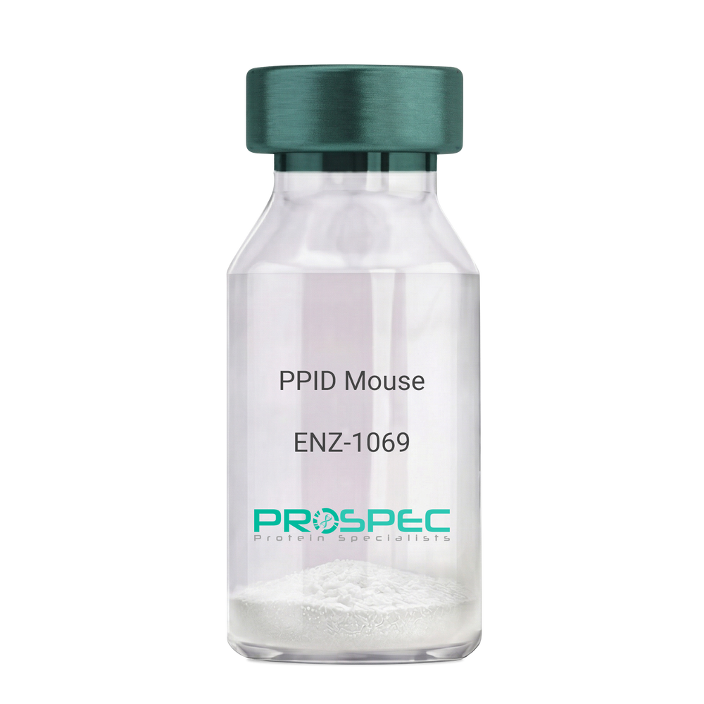
    PPID Mouse

    Description:

    Peptidylprolyl Isomerase D Mouse Recombinant

    Peptidyl-prolyl cis-trans isomerase D, PPIase D, 40 kDa peptidyl-prolyl cis-trans isomerase, Cyclophilin-40, CYP-40, Cyclophilin-related protein, CYP40, CYPD, PPID, Peptidylprolyl Isomerase D.

    Product # :

    ENZ-1069

    Price :

    Quantity :

    Shipping Method :

    Ice Icon

    Shipped with Ice Packs

    Add To Cart

    More Info

    • description
    • source
    • formulation
    • purity
    • biological activity
    • More Info

    Description

    PPID Mouse Recombinant produced in E.Coli is a single, non-glycosylated polypeptide chain containing 395 amino acids (1-370a.a.) and having a molecular mass of 43.4kDa. PPID is fused to a 25 amino acid His-tag at N-terminus & purified by proprietary chromatographic techniques.

    Source

    Escherichia Coli.

    Formulation

    PPID protein solution (1mg/ml) containing 20mM Tris-Hcl buffer (pH8.0), 10% glycerol and 1mM DTT.

    Purity

    Greater than 90.0% as determined by SDS-PAGE.

    Biological Activity

    Specific activity is > 700nmol/min/mg, and is defined as the amount of enzyme that cleaves 1umole of suc-AAFP-PNA per minute at 37°C in Tris–HCl pH 8.0 using chymotrypsin.

    More Info

    • Introduction

      Cyclophilin-D is a member of the peptidyl-prolyl cis-trans isomerase (PPIase) family. PPIases catalyze the cis-trans isomerization of proline imidic peptide bonds in oligopeptides and speeds up the protein folding. Cyclophilin-D possess PPIase activity and binds to the immunosuppressant cyclosporin-A. Cyclophilin-D is very well known that its overexpression suppresses the apoptosis in cancer cell. Cyclophilin-D suppresses apoptotic cell death by the use of mitochondrial hexokinase-2 dependent mechanism in cancer cells.

    • Synonyms

      Peptidyl-prolyl cis-trans isomerase D, PPIase D, 40 kDa peptidyl-prolyl cis-trans isomerase, Cyclophilin-40, CYP-40, Cyclophilin-related protein, CYP40, CYPD, PPID, Peptidylprolyl Isomerase D.

    • Physical Appearance

      Sterile Filtered colorless solution.

    • Stability

      Store at 4°C if entire vial will be used within 2-4 weeks. Store, frozen at -20°C for longer periods of time. For long term storage it is recommended to add a carrier protein (0.1% HSA or BSA). Please prevent freeze-thaw cycles.

    • Amino Acid Sequence

      MGSSHHHHHH SSGLVPRGSH MGSEFMSHAS PAAKPSNSKN PRVFFDVDIG GERVGRIVLE LFADIVPKTA ENFRALCTGE KGTGSTTGKP LHFKGCPFHR IIKKFMIQGG DFSNQNGTGG ESIYGEKFED ENFHYKHDRE GLLSMANAGP NTNGSQFFIT TVPTPHLDGK HVVFGQVIKG LGVARTLENV EVNGEKPAKL CVIAECGELK EGDDWGIFPK DGSGDSHPDF PEDADIDLKD VDKILLISED LKNIGNTFFK SQNWEMAIKK YAKVLRYVDS SKAVIEKADR SRLQPIALSC VLNIGACKLK MSNWQGAIDS CLEALEMDPS NTKALYRKAQ GWQGLKEYDQ ALADLKKAQE IAPGDKAIQA ELLKVKQMIK AQKDKEKAVY AKMFA.

    ProSpec's products are furnished for LABORATORY RESEARCH USE ONLY. The product may not be used as drugs, agricultural or pesticidal products, food additives or household chemicals.

    Ppid Mouse
  • View Data Sheet

    Name :

    EIF4EBP1 Human

    Description:

    Eukaryotic translation initiation factor 4E-binding protein 1 Human Recombinant

    Eukaryotic translation initiation factor 4E-binding protein 1, eIF4E-binding protein 1, 4E-BP1, PHAS-I, EIF4EBP1, BP-1, 4EBP1, MGC4316.

    Product # :

    PRO-532

    Price :

    Quantity :

    Shipping Method :

    Ice Icon

    Shipped with Ice Packs

    Add To Cart

    More Info

    • description
    • source
    • formulation
    • purity
    • More Info

    Description

    EIF4EBP1 Human Recombinant fused with a 20 amino acid His tag at N-terminus produced in E.Coli is a single, non-glycosylated, polypeptide chain containing 138 amino acids (1-118 a.a.) and having a molecular mass of 14.7kDa (molecular weight on SDS-PAGE will appear higher).The EIF4EBP1 is purified by proprietary chromatographic techniques.

    Source

    Escherichia Coli.

    Formulation

    The EIF4EBP1 solution (1mg/ml) contains 20mM Tris-HCl buffer (pH8.0) and 10% glycerol.

    Purity

    Greater than 95.0% as determined by SDS-PAGE.

    More Info

    • Introduction

      EIF4EBP1 (eukaryotic translation initiation factor 4E-binding protein 1) belongs to a family of translation repressor proteins. EIF4EBP1 regulates eIF4E (eukaryotic translation initiation factor 4E) activity by preventing its assembly into the eIF4F complex and mediates the regulation of protein translation by hormones, growth factors and other stimuli that signal through the MAP kinase and mTORC1 pathways. EIF4EBP1 is phosphorylated in response to various signals including UV irradiation, resulting in its dissociation from eIF4E and activation of mRNA translation. EIF4EBP1 C-terminus has domains which control function and phosphorylation. EIF4EBP1 has a role in progression of breast neoplasms through cell signaling.

    • Synonyms

      Eukaryotic translation initiation factor 4E-binding protein 1, eIF4E-binding protein 1, 4E-BP1, PHAS-I, EIF4EBP1, BP-1, 4EBP1, MGC4316.

    • Physical Appearance

      Sterile Filtered colorless solution.

    • Stability

      Store at 4°C if entire vial will be used within 2-4 weeks. Store, frozen at -20°C for longer periods of time. For long term storage it is recommended to add a carrier protein (0.1% HSA or BSA).Avoid multiple freeze-thaw cycles.

    • Amino Acid Sequence

      MGSSHHHHHH SSGLVPRGSH MSGGSSCSQT PSRAIPATRR VVLGDGVQLP PGDYSTTPGG TLFSTTPGGT RIIYDRKFLM ECRNSPVTKT PPRDLPTIPG VTSPSSDEPP MEASQSHLRN SPEDKRAGGE ESQFEMDI.

    ProSpec's products are furnished for LABORATORY RESEARCH USE ONLY. The product may not be used as drugs, agricultural or pesticidal products, food additives or household chemicals.

    Eif4Ebp1 Human
  • View Data Sheet

    Name :

    VEGF Human

    Description:

    Vascular Endothelial Growth Factor Human Recombinant

    Vascular endothelial growth factor A, VEGF-A, Vascular permeability factor, VPF, VEGF, MGC70609.

    Product # :

    CYT-241

    Price :

    Quantity :

    Shipping Method :

    Room Temp Icon

    Shipped at Room temp

    Add To Cart

    More Info

    • description
    • source
    • formulation
    • purity
    • biological activity
    • More Info

    Description

    Vascular Endothelial Growth Factor Human Recombinant produced in E.Coli is a double, non-glycosylated, polypeptide chain containing 165 amino acids and having a molecular mass of 38.2kDa.The VEGF is purified by proprietary chromatographic techniques.

    Source

    Escherichia Coli.

    Formulation

    The VEGF protein was lyophilized from a concentrated (1mg/ml) solution with no additives.

    Purity

    Greater than 95.0% as determined by:
    (a) Analysis by RP-HPLC.
    (b) Analysis by SDS-PAGE.

    Biological Activity

    Determined by the dose-dependent stimulation of the proliferation of human umbilical vein endothelial cells (HUVEC) using a concentration range of 3.7-5.6 ng/ml, corresponding to a Specific Activity of 178,570-270,270IU/mg.

    More Info

    • Introduction

      Vascular endothelial growth factor (VEGF) is an important signaling protein involved in vessel formation As its name implies, VEGF activity has been mostly studied on cells of the vascular endothelium, although it does have effects on a number of other cell types (e.g. stimulation monocyte/macrophage migration, neurons, cancer cells, kidney epithelial cells ). VEGF mediates increased vascular permeability, induces vasculogenesis and endothelial cell production, promotes cell migration, and inhibits apoptosis. In vitro, VEGF has been shown to stimulate endothelial cell mitogenesis and cell migration. VEGF is also a vasodilator and increases microvascular permeability and was originally referred to as vascular permeability factor. VEGF is located in normal cartilage though only osteoarthritic cartilage expresses the VEGF receptors, NP1, VEGFR1 and VEGFR2. The VEGF level in the culture media from OA chondrocytes was more than 3 folds higher than in media from normal chondrocytes

    • Synonyms

      Vascular endothelial growth factor A, VEGF-A, Vascular permeability factor, VPF, VEGF, MGC70609.

    • Physical Appearance

      Sterile Filtered White lyophilized (freeze-dried) powder.

    • Stability

      Lyophilized VEGF although stable at room temperature for 3 weeks, should be stored desiccated below -18°C. Upon reconstitution VEGF should be stored at 4°C between 2-7 days and for future use below -18°C.For long term storage it is recommended to add a carrier protein (0.1% HSA or BSA).Please prevent freeze-thaw cycles.

    • Solubility

      It is recommended to reconstitute the lyophilized VEGF in sterile 18M-cm H2O not less than 100µg/ml, which can then be further diluted to other aqueous solutions.

    • Amino Acid Sequence

      APMAEGGGQN HHEVVKFMDV YQRSYCHPIE TLVDIFQEYP DEIEYIFKPS CVPLMRCGGC CNDEGLECVP TEESNITMQI MRIKPHQGQH IGEMSFLQHN KCECRPKKDR ARQENPCGPC SERRKHLFVQ DPQTCKCSCK NTDSRCKARQ LELNERTCRC DKPRR

    • Protein content

      VEGF protein quantitation was carried out by two independent methods:1. UV spectroscopy at 280 nm using the absorbency value of 0.2875 as the extinction coefficient for a 0.1% (1mg/ml) solution. This value is calculated by the PC GENE computer analysis program of protein sequences (IntelliGenetics). 2. Analysis by RP-HPLC, using a calibrated solution of VEGF as a Reference Standard.

    ProSpec's products are furnished for LABORATORY RESEARCH USE ONLY. The product may not be used as drugs, agricultural or pesticidal products, food additives or household chemicals.

    Vegf Human
  • View Data Sheet

    Name :

    CK2a Zea Mays

    Description:

    Casein Kinase 2 alpha Zea Mays Recombinant

    Casein kinase II subunit alpha, EC 2.7.11.1, CK II, CK2-alpha, CK2?.

    Product # :

    PKA-210

    Price :

    Quantity :

    Shipping Method :

    Ice Icon

    Shipped with Ice Packs

    Add To Cart

    More Info

    • description
    • source
    • formulation
    • purity
    • More Info

    Description

    Casein Kinase 2 alpha Zea Mays Recombinant is a non-glycosylated polypeptide having a molecular mass of 39.2 kDa. Casein Kinase 2 is purified by proprietary chromatographic techniques.

    Source

    Escherichia Coli.

    Formulation

    CK2a is supplied in 50% glycerol.

    Purity

    Greater than 99% as determined by SDS-PAGE.

    More Info

    • Introduction

      The Casein kinase 2 (EC2.7.11.1) is a serine/threonine-selective protein kinasethat is a tetramer of two alpha subunits and two beta subunits. The alpha subunits have the catalytic kinase domain. Casein kinase 2 has been implicated in cell cyclecontrol, DNA repair, regulation of the circadian rhythmand other cellular processes.
      Casein kinase 2 activity has been reported to be activated following Wnt signaling pathwayactivation. A Pertussis toxin-sensitive G proteinand Disheveled appear to be an intermediary between Wnt-mediated activation of the Frizzled receptor and activation of casein kinase 2.

    • Synonyms

      Casein kinase II subunit alpha, EC 2.7.11.1, CK II, CK2-alpha, CK2?.

    • Stability

      Store at 4°C if entire vial will be used within 2-4 weeks. Store, frozen at -20°C for longer periods of time. Avoid multiple freeze-thaw cycles.

    • Unit Definition

      No protease activity detectable, specific activity > 1U/mg (1U = 1µmol/min at 37 degree C) using the synthetic peptide RRRDDDSDDD.

    ProSpec's products are furnished for LABORATORY RESEARCH USE ONLY. The product may not be used as drugs, agricultural or pesticidal products, food additives or household chemicals.

    Ck2A Zea Mays
  • View Data Sheet

    Name :

    XYLT2 Human

    Description:

    Xylosyltransferase 2 Human Recombinant

    Xylosyltransferase 2, Peptide O-xylosyltransferase 1, Xylosyltransferase II, XT-II, XylT-II, XYLT2, XT2.

    Product # :

    ENZ-1086

    Price :

    Quantity :

    Shipping Method :

    Room Temp Icon

    Shipped at Room temp

    Add To Cart

    More Info

    • description
    • source
    • formulation
    • purity
    • More Info

    Description

    XYLT2 Human Recombinant is a single, glycosylated polypeptide chain containing 839 amino acids (Gly37-Arg865, luminal domain, isoform 1, natural variant with Thr305) and having a molecular mass of 94.0kDa. XYLT2 is fused to an N-terminal linker (2 extra a.a), C-terminal linker (2 extra a.a) and C-terminal His-tag (6 extra a.a).

    Source

    HEK293 Cells.

    Formulation

    XYLT2 filtered (0.4 µm) and lyophilized in 0.05 M PBS and 0.075 M NaCl, pH 7.4.

    Purity

    Greater than 95.0% as determined by SDS-PAGE.

    More Info

    • Introduction

      XYLT2 or Xylosyltransferase 2 is an enzyme which is expressed in ubiquitous and is part of the glycosyltransfe-rases family. XYLT2 promotes proteoglycans formation by attaching GAG chains to the substrate protein via transfer of xylose molecule from the donor (nucleoside diphosphate) to the protein’s serine residues. XYLT2 is present in the ER and the cis part of the Golgi, furthermore the protein is released to the extracellular matrix.

    • Synonyms

      Xylosyltransferase 2, Peptide O-xylosyltransferase 1, Xylosyltransferase II, XT-II, XylT-II, XYLT2, XT2.

    • Physical Appearance

      Filtered White lyophilized (freeze-dried) powder.

    • Stability

      Store lyophilized protein at -20°C. Aliquot the product after reconstitution to avoid repeated freezing/thawing cycles. Reconstituted protein can be stored at 4°C for a limited period of time; it does not show any change after two weeks at 4°C.

    • Solubility

      It is recommended to add deionized water to prepare a working stock solution of approximately 0.5mg/ml and let the lyophilized pellet dissolve completely. XYLT2 is not sterile! Please filter the product by an appropriate sterile filter before using it in the cell culture.

    • Amino Acid Sequence

      ASGLEEDEAG EKGRQRKPRP LDPGEGSKDT DSSAGRRGST GRRHGRWRGR AESPGVPVAK VVRAVTSRQR ASRRVPPAPP PEAPGRQNLS GAAAGEALVG AAGFPPHGDT GSVEGAPQPT DNGFTPKCEI VGKDALSALA RASTKQCQQE IANVVCLHQA GSLMPKAVPR HCQLTGKMSP GIQWDESQAQ QPMDGPPVRI AYMLVVHGRA IRQLKRLLKA VYHEQHFFYI HVDKRSDYLH REVVELAQGY DNVRVTPWRM VTIWGGASLL TMYLRSMRDL LEVPGWAWDF FINLSATDYP TRTNEELVAF LSKNRDKNFL KSHGRDNSRF IKKQGLDRLF HECDSHMWRL GERQIPAGIV VDGGSDWFVL TRSFVEYVVY TDDPLVAQLR QFYTYTLLPA ESFFHTVLEN SLACETLVDN NLRVTNWNRK LGCKCQYKHI VDWCGCSPND FKPQDFLRLQ QVSRPTFFAR KFESTVNQEV LEILDFHLYG SYPPGTPALK AYWENTYDAA DGPSGLSDVM LTAYTAFARL SLHHAATAAP PMGTPLCRFE PRGLPSSVHL YFYDDHFQGY LVTQAVQPSA QGPAETLEMW LMPQGSLKLL GRSDQASRLQ SLEVGTDWDP KERLFRNFGG LLGPLDEPVA VQRWARGPNL TATVVWIDPT YVVATSYDIT VDTETEVTQY KPPLSRPLRP GPWTVRLLQF WEPLGETRFL VLPLTFNRKL PLRKDDASWL HAGPPHNEYM EQSFQGLSSI LNLPQPELAE EAAQRHTQLT GPALEAWTDR ELSSFWSVAG LCAIGPSPCP SLEPCRLTSW SSLSPDPKSE LGPVKADGRL RKLHHHHHH.

    ProSpec's products are furnished for LABORATORY RESEARCH USE ONLY. The product may not be used as drugs, agricultural or pesticidal products, food additives or household chemicals.

    Xylt2 Human
  • View Data Sheet

    Name :

    FGF 21 Mouse

    Description:

    Fibroblast Growth Factor-21 Mouse Recombinant

    Fibroblast growth factor 21, FGF-21.

    Product # :

    CYT-339

    Price :

    Quantity :

    Shipping Method :

    Room Temp Icon

    Shipped at Room temp

    Add To Cart

    More Info

    • description
    • source
    • formulation
    • purity
    • More Info

    Description

    Fibroblast Growth Factor -21 Mouse Recombinant produced in E.Coli is a single, non-glycosylated, polypeptide chain containing 183 amino acids including N-terminal Methionin and having a molecular mass of 20.1 kDa. The FGF-21 is purified by proprietary chromatographic techniques.

    Source

    Escherichia Coli.

    Formulation

    Filtered (0.4µm) and lyophilized from 0.5mg/ml in 20mM TRIS, 20mM NaCl, pH 7.5.

    Purity

    Greater than 95.0% as determined by SDS-PAGE.

    More Info

    • Introduction

      The FGFs are a family of more than 20 small (~17–26 kDa) secreted peptides. The initial characterization of these proteins focused on their ability to stimulate fibroblast proliferation. This mitogenic activity was mediated through FGF receptors (FGFRs) 1, 2, or 3. A fourth closely related tyrosine kinase receptor (FGFR4) was able to bind the FGFs but did not lead to a mitogenic response.
      FGFs modulate cellular activity via at least 5 distinct subfamilies of high-affinity FGF receptors (FGFRs): FGFR-1, -2, -3, and -4, all with intrinsic tyrosine kinase activity and, except for FGFR-4, multiple splice isoforms, and FGFR-5, which lacks an intracellular kinase domain. There is growing evidence that FGFRs can be important for regulation of glucose and lipid homeostasis. The overexpression of a dominant negative form of FGFR-1 in ? cells leads to diabetes in mice, which thus implies that proper FGF signaling is required for normal ? cell function and glycemia maintenance. FGFR-2 appears to be a key molecule during pancreatic development. Moreover, FGFR-4 has been implicated in cholesterol metabolism and bile acid synthesis.
      FGF-19, has been shown to cause resistance to diet-induced obesity desensitization and to improve glucose, and lipid profiles in diabetic rodents. Since these effects, at least in part, are mediated through the observed changes in metabolic rates, FGF-19 can be considered as a regulator of energy expenditure.
      FGF-21 is preferentially expressed in liver, but an exact knowledge of FGF-21 bioactivity and its mode of action have been lacking to date. FGF-21 is a potent activator of glucose uptake on adipocytes, protects animals from diet-induced obesity when overexpressed in transgenic mice, and lowers blood glucose and triglyceride levels when therapeutically administered to diabetic rodents.

    • Synonyms

      Fibroblast growth factor 21, FGF-21.

    • Physical Appearance

      Filtered white lyophilized powder.

    • Stability

      Lyophilized FGF-21 Mouse Recombinant although stable at room temperature for 3 weeks, should be stored desiccated below -18°C. Upon reconstitution Fibroblast Growth Factor 21 should be stored at 4°C between 2-7 days and for future use below -18°C.For long term storage it is recommended to add a carrier protein (0.1% HSA or BSA).Please prevent freeze-thaw cycles.

    • Solubility

      It is recommended to add deionized water to prepare a working stock solution of approximately 0.5mg/ml and let the lyophilized pellet dissolve completely. Product is not sterile! Please filter the product by an appropriate sterile filter before using it in the cell culture. Add DTT (0.2mM) and NaCl (0.1-0.15M) before freezing to prevent potential aggregation.

    • Amino Acid Sequence

      MAY PIPDSSPLLQ FGGQVRQRYL YTDDDQDTEA HLEIREDGTV VGAAHRSPES LLELKALKPG VIQILGVKAS RFLCQQPDGA LYGSPHFDPE ACSFRELLLE DGYNVYQSEA HGLPLRLPQK DSPNQDATSW GPVRFLPMPG LLHEPQDQAG FLPPEPPDVG SSDPLSMVEP LQGRSPSYAS.

    • Background

      What is the molecular weight/Mw of FGF21 MOUSE Protein?
      FGF21 MOUSE Protein has a total Mw of 20.1kDa.

      What is the source or expression system of FGF21 MOUSE Protein?
      Escherichia Coli.

      What is the Purity of FGF21 MOUSE Protein?
      FGF21 MOUSE Protein is >95% pure as determined by SDS-PAGE.

      What is the Biological Activity of FGF21 MOUSE Protein?
      The biological functionality of FGF21 MOUSE Protein will be determined in the future.

      What is the amino acid sequence of FGF21 MOUSE Protein?
      MAY PIPDSSPLLQ FGGQVRQRYL YTDDDQDTEA HLEIREDGTV VGAAHRSPES LLELKALKPG VIQILGVKAS RFLCQQPDGA LYGSPHFDPE ACSFRELLLE DGYNVYQSEA HGLPLRLPQK DSPNQDATSW GPVRFLPMPG LLHEPQDQAG FLPPEPPDVG SSDPLSMVEP LQGRSPSYAS.

      What applications can FGF21 MOUSE Protein be used in?
      FGF21 MOUSE Protein can probably be used in western blot, ELISA and Lateral Flow.

      What is the endotoxin level for FGF21 MOUSE Protein?
      The endotoxin level is minimal, FGF21 MOUSE Protein was purified using conventional chromatography techniques.

    ProSpec's products are furnished for LABORATORY RESEARCH USE ONLY. The product may not be used as drugs, agricultural or pesticidal products, food additives or household chemicals.

    Fgf21 Mouse
  • View Data Sheet

    Name :

    FGF2 Human, Thermostable

    Description:

    Fibroblast Growth Factor-basic Human Recombinant, Thermostable

    HBGH-2, HBGF-2, FGF-2, FGF-b, HBGH2, HBGF2, FGF2, FGFb, FGF2 Thermostable, FGF-2 Thermostable.

    Product # :

    CYT-943

    Price :

    Quantity :

    Shipping Method :

    Room Temp Icon

    Shipped at Room temp

    Add To Cart

    More Info

    • description
    • source
    • formulation
    • purity
    • biological activity
    • More Info

    Description

    FGF2 Thermostable is a stabilized form of FGF2 growth factor which enables a novel method to produce FGF2-dependent cell cultures more efficiently, having less media changes. FGF2 Thermostable can maintain its biological activity even after five days at 37°C. The increase in the stability of FGF2 in cell-culture enables a more homogenous, undifferentiated stem cell culture, while saving scientists crucial time and money, as frequent supplementation of FGF-basic and the everyday medium change is not necessary. Thermostable FGF-2 is a hyperstable protein. The Thermal stability of the protein is increased by 15°C compared to the wild-type FGF-2. Thermostable FGF-2 is more than 5-times prolonged half-life in human cell culture incubated at 37°C. The FGF-2 Thermostable protein is engineered with fully retained biological function and has no harmful stabilizing additives.Thermostable FGF2 Human Recombinant produced in E.Coli is a single, non-glycosylated, polypeptide chain containing 154 amino acids and having a total calculated molecular mass is 17.2kDa.

    Source

    Escherichia Coli.

    Formulation

    FGF Basic Thermostable was lyophilized from 20mM Tris-HCl, 150mM NaCl & 5% Trehalose, pH-7.6.

    Purity

    Purity is greater than 95% as determined by SDS-PAGE.

    Biological Activity

    The ED50 as calculated by the dose-dependent proliferation of murine balb/c 3T3 cells is less than 0.05ng/ml, corresponding to a specific activity of >20,000,000 units/mg

    More Info

    • Synonyms

      FGF2 Thermostable, FGF-basic Thermostable.

    • Physical Appearance

      Filtered White lyophilized (freeze-dried) powder.

    • Stability

      Lyophilized Thermostable FGF2 although stable at room temperature for 3 weeks, should be stored desiccated below -18°C. Upon reconstitution FGF2 Thermostable should be stored at 4°C between 2-7 days and for future use below -18°C.For long term storage it is recommended to add a carrier protein (0.1% HSA or BSA).Please prevent freeze-thaw cycles.

    • Solubility

      It is recommended to reconstitute the lyophilized FGF2 thermostable protein in sterile 18MΩ-cm H2O not less than 100µg/ml, which can then be further diluted to other aqueous solutions.

    • Amino Acid Sequence

      AAGSITTLPA LPEDGGSGAF PPGHFKDPKL LYCKNGGFFL RIHPDGRVDG TRDKSDPFIK LQLQAEERGV VSIKGVCANR YLAMKEDGRL YAIKNVTDEC FFFERLEENN YNTYRSRKYP SWYVALKRTG QYKLGSKTGP GQKAILFLPM SAKS

    • Background

      Why Choose FGF2 Thermostable?
      FGF2 can lose much of its activity after 1-2 days at 37°C due to unfolding and degradation. Thermostable FGF2 is engineered with amino acid substitutions that increase structural stability without significantly altering receptor binding or biological function.

      What are the advantages of FGF2 Protein?
      *Better maintenance of pluripotency markers
      *Higher colony quality
      *Faster expansion rates
      *Reduced spontaneous differentiation
      *Greater viability after passaging

      What is the source or expression system of FGF2 Protein?
      Escherichia Coli.

      What is the Purity of FGF2 Protein?

      FGF2 Protein is >95% pure as determined by SDS-PAGE.

      What is the molecular weight / Mw of FGF2 Protein?
      FGF2 Protein having a total Mw of 17.2kDa.

      What is the Biological Activity of FGF2 Protein?
      The ED50 as calculated by the dose-dependent proliferation of murine balb/c 3T3 cells is less than 0.05ng/ml, corresponding to a specific activity of >20,000,000 units/mg.

      What is the endotoxin level for FGF2 Protein?
      The endotoxin level is minimal, FGF2 Protein was purified using conventional chromatography techniques.

      What is the amino acid sequence of FGF2 Protein?
      AAGSITTLPA LPEDGGSGAF PPGHFKDPKL LYCKNGGFFL RIHPDGRVDG TRDKSDPFIK LQLQAEERGV VSIKGVCANR YLAMKEDGRL YAIKNVTDEC FFFERLEENN YNTYRSRKYP SWYVALKRTG QYKLGSKTGP GQKAILFLPM SAKS

      Is FGF2 Protein conjugated to a Tag?
      FGF2 Protein is not conjugated to a tag.

      What applications can FGF2 Protein be used in?
      FGF2 Protein can probably be used in western blot, ELISA and Lateral Flow.

    ProSpec's products are furnished for LABORATORY RESEARCH USE ONLY. The product may not be used as drugs, agricultural or pesticidal products, food additives or household chemicals.

    Fgf2 Human Thermostable
  • View Data Sheet

    Name :

    EGF Rat Protein

    Description:

    Epidermal Growth Factor Rat

    Urogastrone, URG, EGF.

    Product # :

    CYT-556

    Price :

    Quantity :

    Shipping Method :

    Room Temp Icon

    Shipped at Room temp

    Add To Cart

    More Info

    • description
    • source
    • formulation
    • purity
    • More Info

    Description

    Epidermal Growth Factor Rat purified from submandibular gland is a single, glycosylated, polypeptide chain having a molecular mass of 6.15 kDa.The EGF is purified by proprietary chromatographic techniques.

    Source

    Adult Male Rat Submandibular Glands.

    Formulation

    The protein was lyophilized from a concentrated (1mg/ml) solution containing 0.01M sodium acetate buffer.

    Purity

    Greater than 99.0% as determined by(a) Analysis by RP-HPLC.
    (b) Analysis by SDS-PAGE.

    More Info

    • Introduction

      Epidermal growth factor has a profound effect on the differentiation of specific cells in vivo and is a potent mitogenic factor for a variety of cultured cells of both ectodermal and mesodermal origin. The EGF precursor is believed to exist as a membrane-bound molecule which is proteolytically cleaved to generate the 53-amino acid peptide hormone that stimulates cells to divide.
      EGF stimulates the growth of various epidermal and epithelial tissues in vivo and in vitro and of some fibroblasts in cell culture.

    • Synonyms

      Urogastrone, URG, EGF.

    • Physical Appearance

      Sterile Filtered White lyophilized (freeze-dried) powder.

    • Stability

      Lyophilized Epidermal Growth Factor Recombinant although stable at room temperature for 3 weeks, should be stored desiccated below -18°C. Upon reconstitution EGF should be stored at 4°C between 2-7 days and for future use below -18°C.For long term storage it is recommended to add a carrier protein (0.1% HSA or BSA).Please prevent freeze-thaw cycles.

    • Solubility

      It is recommended to reconstitute the lyophilized Epidermal Growth Factor in sterile 18MΩ-cm H2O not less than 100µg/ml, which can then be further diluted to other aqueous solutions.

    • Background

      Illuminating Epidermal Growth Factor Rat Recombinant: Deciphering Cellular Signaling and Therapeutic Potential

      Abstract:

      This research paper delves into the enigmatic realm of Epidermal Growth Factor Rat Recombinant (EGF-RR), unraveling its intricate molecular attributes, signaling cascades, and therapeutic prospects. By employing cutting-edge methodologies encompassing protein expression, receptor binding assays, and bioinformatics analyses, this study sheds light on the multifaceted interplay between EGF-RR and cellular responses, offering novel avenues for therapeutic interventions.

      Introduction:

      Epidermal Growth Factor (EGF) is pivotal in cellular regulation. This paper navigates the complexities of Epidermal Growth Factor Rat Recombinant (EGF-RR), focusing on its unique molecular properties and potential therapeutic applications.

      Protein Expression and Purification:

      The study embarks on precise gene optimization to enhance EGF-RR expression. Purification techniques like affinity chromatography yield purified EGF-RR, primed for subsequent analyses.

      Receptor Binding Assays and Ligand Interaction:

      Employing advanced receptor binding assays, the paper deciphers EGF-RR's engagement with its cognate receptor. Quantitative assessments uncover binding kinetics, shedding light on the intricacies of EGF-RR's molecular interaction.

      Cellular Signaling Pathways and Responses:

      In vitro cellular assays unveil the signaling cascades ignited by EGF-RR. Through quantitative phosphoproteomic profiling, the study unravels phosphorylation events triggered by EGF-RR, delineating its role in cellular proliferation, migration, and differentiation.

      Bioinformatics Insights and Structural Modeling:

      Bioinformatics tools facilitate molecular dynamics simulations, offering insights into EGF-RR's receptor interactions and downstream signaling pathways. Structural modeling captures EGF-RR's conformational changes during signaling cascades.

      Therapeutic Implications and Future Prospects:

      EGF-RR's intricate signaling dynamics open avenues for therapeutic exploration. Harnessing its potential in wound healing, tissue regeneration, and cancer modulation emerges as a promising avenue for precision medicine.

      Challenges and Future Directions:

      Challenges, including context-specific responses, beckon further investigation. Future research should delve into cross-talk between signaling pathways and EGF-RR's contributions to diverse disease contexts.

      Conclusion:

      A fusion of advanced methodologies and visionary insights unveils Epidermal Growth Factor Rat Recombinant as an intriguing subject. Its molecular intricacies and complex cellular interplay ignite prospects for therapeutic breakthroughs, ushering in a new era of precision medicine.

      What is the molecular weight/Mw of EGF RAT Protein?
      EGF RAT Protein has a total Mw of 6.15kDa.

      What is the source or expression system of EGF RAT Protein?
      Adult Male Rat Submandibular Glands.

      What is the Purity of EGF RAT Protein?
      EGF RAT Protein is >99% pure as determined by SDS-PAGE.

      What is the Biological Activity of EGF RAT Protein?
      The biological functionality of EGF RAT Protein will be determined in the future.

      What is the amino acid sequence of EGF RAT Protein?
      EGF RAT Protein is composed from 53 amino acids.

      What applications can EGF RAT Protein be used in?
      EGF RAT Protein can probably be used in western blot, ELISA and Lateral Flow.

      What is the endotoxin level for EGF RAT Protein?
      The endotoxin level is minimal, EGF RAT Protein was purified using conventional chromatography techniques.

    ProSpec's products are furnished for LABORATORY RESEARCH USE ONLY. The product may not be used as drugs, agricultural or pesticidal products, food additives or household chemicals.

    Egf Rat
  • View Data Sheet

    Name :

    G CSF Human, CHO

    Description:

    Granulocyte-Colony Stimulating Factor Human Recombinant, CHO

    CSF-3, MGI-1G, GM-CSF beta, Pluripoietin, Filgrastim, Lenograstim, G-CSF, MGC45931, GCSF.

    Product # :

    CYT-329

    Price :

    Quantity :

    Shipping Method :

    Room Temp Icon

    Shipped at Room temp

    Add To Cart

    More Info

    • description
    • source
    • formulation
    • purity
    • biological activity
    • More Info

    Description

    Granulocyte Colony Stimulating Factor Human Recombinant produced in CHO cells is a single, glycosylated, polypeptide chain containing 174 amino acids and having a molecular mass of approximately 18 kDa.G-CSF is purified by proprietary chromatographic techniques.

    Source

    Chinese Hamster Ovary Cells (CHO).

    Formulation

    G-CSF was lyophilized from a concentrated (1mg/ml) solution containing 10mM Hydrochloric Acid pH=6.5, 0.4mg tween 20, 100mg mannitol, 160mg L-arginine, 40mg phenylalanine and 4mg methionine.

    Purity

    Greater than 97.0% as determined by SDS-PAGE.

    Biological Activity

    The ED50, calculated by the dose-dependant proliferation of murine NFS-60 indicator cells (measured by 3H-thymidine uptake) is < 0.07 ng/ml, corresponding to a Specific Activity of 1.27 x 10⁸ IU/mg.

    More Info

    • Introduction

      Granulocyte Colony Stimulating Factor is a growth factor and/or cytokine produced by the endothelium, macrophages and a number of other immune cells. GCSF stimulates the bone marrow to produce granulocytes and also to stimulate the survival, proliferation, differentiation and function of neutrophil granulocyte progenator cells and mature neutrophils.

    • Synonyms

      CSF-3, MGI-1G, GM-CSF beta, Pluripoietin, Filgrastim, Lenograstim, G-CSF, MGC45931, GCSF.

    • Physical Appearance

      Sterile Filtered White lyophilized (freeze-dried) powder.

    • Stability

      Lyophilized Granulocyte Colony Stimulating Factor although stable at room temperature for 3 weeks, should be stored desiccated below -18°C. Upon reconstitution G-CSF should be stored at 4°C between 2-7 days and for future use below -18°C.For long term storage it is recommended to add a carrier protein (0.1% HSA or BSA).Please prevent freeze-thaw cycles.

    • Solubility

      It is recommended to reconstitute the lyophilized Granulocyte Colony Stimulating Factor in sterile 18MΩ-cm H2O not less than 100µg/ml, which can then be further diluted to other aqueous solutions.

    • Amino Acid Sequence

      TPLGPASSLP QSFLLKCLEQ VRKIQGDGAA LQEKLCATYK LCHPEELVLL GHSLGIPWAP LSSCPSQALQ LAGCLSQLHS GLFLYQGLLQ ALEGISPELG PTLDTLQLDV ADFATTIWQQ MEELGMAPAL QPTQGAMPAF ASAFQRRAGG VLVASHLQSF LEVSYRVLRH LAQP.

    • Background

      What is the molecular weight/Mw of G CSF Protein?
      G CSF Protein has a total Mw of 18kDa.

      What is the source or expression system of G CSF Protein?
      Chinese Hamster Ovary Cells (CHO).

      What is the Purity of G CSF Protein?
      G CSF Protein is >97% pure as determined by SDS-PAGE.

      What is the Biological Activity of G CSF Protein?
      The ED50, calculated by the dose-dependant proliferation of murine NFS-60 indicator cells (measured by 3H-thymidine uptake) is < 0.07 ng/ml, corresponding to a Specific Activity of 1.27 x 10⁸ IU/mg.

      What is the amino acid sequence of G CSF Protein?
      TPLGPASSLP QSFLLKCLEQ VRKIQGDGAA LQEKLCATYK LCHPEELVLL GHSLGIPWAP LSSCPSQALQ LAGCLSQLHS GLFLYQGLLQ ALEGISPELG PTLDTLQLDV ADFATTIWQQ MEELGMAPAL QPTQGAMPAF ASAFQRRAGG VLVASHLQSF LEVSYRVLRH LAQP.

      What applications can G CSF Protein be used in?
      G CSF Protein can probably be used in western blot, ELISA and Lateral Flow.

      What is the endotoxin level for G CSF Protein?
      The endotoxin level is minimal, G CSF Protein was purified using conventional chromatography techniques.

    ProSpec's products are furnished for LABORATORY RESEARCH USE ONLY. The product may not be used as drugs, agricultural or pesticidal products, food additives or household chemicals.

    G Csf Human Cho
  • 1
  • …
  • 14
  • 15
  • 16
  • 17
  • 18
  • …
  • 42
Your browser does not support the video tag.

Products

  • Cytokines
  • Growth Factors
  • Chemokines
  • CD Antigens
  • Neurotrophins
  • Hormones
  • Enzymes
  • Viral Antigens
  • Recombinant Proteins
  • Natural Proteins
  • Monoclonal Antibodies
  • Polyclonal Antibodies
  • Thermal Packaging

About

  • Company
  • Founder
  • Contribution
  • Contact Us
  • My Account

Ordering

  • Ordering Method
  • Ordering - Credit Card
  • Ordering PO
  • Shipping Fees
  • FAQ's

Legal

  • Terms & Conditions
  • Privacy
  • Site Map
  • info@prospecbio.com
  • Facebook
  • Instagram
  • Linkedin Profile

© 2026 ProSpec-Tany TechnoGene Ltd. All Rights Reserved.

  • Choosing a selection results in a full page refresh.
  • Opens in a new window.