Skip to content

High-quality research proteins.

  • 1-800-805-6531

  • Mon - Sat 8.00-18.00, Sun - Closed

  • info@prospecbio.com

  • PO Box 6591 East Brunswick NJ 08816

  • Products
    • Cytokines
    • Growth factors
    • Chemokines
    • Cd antigens
    • Neurotrophins
    • Hormones
    • Enzymes
    • Viral antigens
    • Recombinant proteins
    • Natural proteins
    • Monoclonal antibodies
    • Polyclonal antibodies
    • Thermal Packaging
  • About
    • Company
    • Founder
    • Contribution
  • Customers
    • Eastern Asia
    • Eastern Europe
    • North America
    • South America
    • South East Asia
    • Southern Asia
    • Western Europe
  • Ordering
    • Ordering Method
    • Ordering - Credit Card
    • Ordering - PO
    • Shipping Fees
    • FAQs
  • Services
    • DNA Cloning
    • Protein Expression
    • Monoclonal Antibody
    • Fermentation
    • Technology License
  • Contact us
Log in

Country/region

  • USD $ | Albania
  • USD $ | Andorra
  • USD $ | Argentina
  • USD $ | Armenia
  • USD $ | Australia
  • USD $ | Austria
  • USD $ | Azerbaijan
  • USD $ | Belarus
  • USD $ | Belgium
  • USD $ | Bolivia
  • USD $ | Brazil
  • USD $ | Bulgaria
  • USD $ | Cambodia
  • USD $ | Canada
  • USD $ | Chile
  • USD $ | China
  • USD $ | Colombia
  • USD $ | Costa Rica
  • USD $ | Croatia
  • USD $ | Cyprus
  • USD $ | Czechia
  • USD $ | Denmark
  • USD $ | Estonia
  • USD $ | Finland
  • USD $ | France
  • USD $ | Georgia
  • USD $ | Germany
  • USD $ | Greece
  • USD $ | Hong Kong SAR
  • USD $ | Hungary
  • USD $ | Iceland
  • USD $ | India
  • USD $ | Ireland
  • USD $ | Israel
  • USD $ | Italy
  • USD $ | Japan
  • USD $ | Kazakhstan
  • USD $ | Latvia
  • USD $ | Liechtenstein
  • USD $ | Lithuania
  • USD $ | Madagascar
  • USD $ | Malta
  • USD $ | Mexico
  • USD $ | Moldova
  • USD $ | Monaco
  • USD $ | Netherlands
  • USD $ | New Zealand
  • USD $ | North Macedonia
  • USD $ | Norway
  • USD $ | Panama
  • USD $ | Paraguay
  • USD $ | Peru
  • USD $ | Philippines
  • USD $ | Poland
  • USD $ | Portugal
  • USD $ | Romania
  • USD $ | Singapore
  • USD $ | Slovakia
  • USD $ | Slovenia
  • USD $ | South Africa
  • USD $ | South Korea
  • USD $ | Spain
  • USD $ | Sri Lanka
  • USD $ | Sweden
  • USD $ | Switzerland
  • USD $ | Taiwan
  • USD $ | Thailand
  • USD $ | Türkiye
  • USD $ | Ukraine
  • USD $ | United Kingdom
  • USD $ | United States
  • USD $ | Uruguay
  • USD $ | Vatican City
  • USD $ | Venezuela
  • USD $ | Vietnam
  • Facebook
  • Instagram
logoProSpec - Recombinant Protein Manufacturer for academic and R&D industry
Become a distributor
Account Log in
Wishlist
Cart Cart(0)
  • Products
    +
    • Cytokines
    • Growth factors
    • Chemokines
    • Cd antigens
    • Neurotrophins
    • Hormones
    • Enzymes
    • Viral antigens
    • Recombinant proteins
    • Natural proteins
    • Monoclonal antibodies
    • Polyclonal antibodies
    • Thermal Packaging
  • About
    +
    • Company
    • Founder
    • Contribution
  • Customers
    +
    • Eastern Asia
    • Eastern Europe
    • North America
    • South America
    • South East Asia
    • Southern Asia
    • Western Europe
  • Ordering
    +
    • Ordering Method
    • Ordering - Credit Card
    • Ordering - PO
    • Shipping Fees
    • FAQs
  • Services
    +
    • DNA Cloning
    • Protein Expression
    • Monoclonal Antibody
    • Fermentation
    • Technology License
  • Contact us

Item added to your cart

View cart
View All
  • Cytokines
  • Growth factors
  • Chemokines
  • Cd antigens
  • Neurotrophins
  • Hormones
  • Enzymes
  • Viral antigens
  • Recombinant proteins
  • Natural proteins
  • Monoclonal antibodies
  • Polyclonal antibodies
  • Thermal Packaging
  • Tumor Necrosis Factor

    Tumor Necrosis Factor

    View All
  • 4-1BB

    4-1BB

    View All
  • Adiponectin

    Adiponectin

    View All
  • AIF1

    AIF1

    View All
  • Other Cytokines

    Other Cytokines

    View All
  • AITRL

    AITRL

    View All
  • Angiopoietin

    Angiopoietin

    View All
  • Apolipoprotein

    Apolipoprotein

    View All
  • B-Cell Activating Factor

    B-Cell Activating Factor

    View All
  • Beta Defensin

    Beta Defensin

    View All
  • Bone Morphogenetic Protein

    Bone Morphogenetic Protein

    View All
  • B type Natriuretic Peptide

    B type Natriuretic Peptide

    View All
  • BST

    BST

    View All
  • Betacellulin

    Betacellulin

    View All
  • Cardiotrophin

    Cardiotrophin

    View All
  • Activin

    Activin

    View All
  • Fibroblast Growth Factor

    Fibroblast Growth Factor

    View All
  • Other Growth Factors

    Other Growth Factors

    View All
  • CSF

    CSF

    View All
  • CTGF

    CTGF

    View All
  • IGFBP

    IGFBP

    View All
  • VEGF Protein

    VEGF Protein

    View All
  • EGF

    EGF

    View All
  • Epigen

    Epigen

    View All
  • Erythropoietin

    Erythropoietin

    View All
  • Insulin-Like Growth Factor

    Insulin-Like Growth Factor

    View All
  • Growth Hormone

    Growth Hormone

    View All
  • HDGF

    HDGF

    View All
  • Hepatocyte Growth Factor

    Hepatocyte Growth Factor

    View All
  • Insulin

    Insulin

    View All
  • BCA-1/ BLC (CXCL13)

    BCA-1/ BLC (CXCL13)

    View All
  • BRAK (CXCL14)

    BRAK (CXCL14)

    View All
  • C-10 (CCL6)

    C-10 (CCL6)

    View All
  • Other Chemokines

    Other Chemokines

    View All
  • MEC (CCL28)

    MEC (CCL28)

    View All
  • LD78-beta (CCL3L1)

    LD78-beta (CCL3L1)

    View All
  • CTACK (CCL27)

    CTACK (CCL27)

    View All
  • CXCL16

    CXCL16

    View All
  • CXCL17

    CXCL17

    View All
  • Platelet Factor-4 (CXCL4)

    Platelet Factor-4 (CXCL4)

    View All
  • ENA-78 (CXCL5)

    ENA-78 (CXCL5)

    View All
  • Eotaxin (CCL11,24,26)

    Eotaxin (CCL11,24,26)

    View All
  • Exodus-2 (CCL21)

    Exodus-2 (CCL21)

    View All
  • Fractalkine (CX3CL1)

    Fractalkine (CX3CL1)

    View All
  • CXCL6

    CXCL6

    View All
  • Other CD Antigens

    Other CD Antigens

    View All
  • CD14

    CD14

    View All
  • CD164

    CD164

    View All
  • CD1

    CD1

    View All
  • CD2

    CD2

    View All
  • CD200

    CD200

    View All
  • CD207

    CD207

    View All
  • CD226

    CD226

    View All
  • CD23

    CD23

    View All
  • CD244

    CD244

    View All
  • CD247

    CD247

    View All
  • CD27

    CD27

    View All
  • CD274

    CD274

    View All
  • CD300

    CD300

    View All
  • CD33

    CD33

    View All
  • Other Neurotrophins

    Other Neurotrophins

    View All
  • Beta-NGF

    Beta-NGF

    View All
  • BDNF

    BDNF

    View All
  • CDNF

    CDNF

    View All
  • CNTF

    CNTF

    View All
  • GDNF

    GDNF

    View All
  • Glia Maturation Factor

    Glia Maturation Factor

    View All
  • MANF

    MANF

    View All
  • Midkine

    Midkine

    View All
  • Neuroglobin

    Neuroglobin

    View All
  • Neurotrophic factors

    Neurotrophic factors

    View All
  • Neuregulin

    Neuregulin

    View All
  • Neuropilin

    Neuropilin

    View All
  • Pigment Epithelium-Derived Factor

    Pigment Epithelium-Derived Factor

    View All
  • Pleiotrophin

    Pleiotrophin

    View All
  • Peptide Hormones

    Peptide Hormones

    View All
  • Other Hormones

    Other Hormones

    View All
  • HCG

    HCG

    View All
  • Endothelin

    Endothelin

    View All
  • Exendin

    Exendin

    View All
  • FSH

    FSH

    View All
  • Glucagon

    Glucagon

    View All
  • GHRP

    GHRP

    View All
  • GLP

    GLP

    View All
  • Thyrostimulin

    Thyrostimulin

    View All
  • Inhibin A

    Inhibin A

    View All
  • LHRH

    LHRH

    View All
  • Melanotan

    Melanotan

    View All
  • Procalcitonin

    Procalcitonin

    View All
  • PTH

    PTH

    View All
  • Peroxiredoxin

    Peroxiredoxin

    View All
  • Peptidase

    Peptidase

    View All
  • Synthetase

    Synthetase

    View All
  • Transferase

    Transferase

    View All
  • Hydrolase

    Hydrolase

    View All
  • Dehydrogenase

    Dehydrogenase

    View All
  • ACE2

    ACE2

    View All
  • Esterase

    Esterase

    View All
  • Protein Kinases

    Protein Kinases

    View All
  • Other Enzymes

    Other Enzymes

    View All
  • Phosphatase

    Phosphatase

    View All
  • Deaminase

    Deaminase

    View All
  • Oxygenase

    Oxygenase

    View All
  • Adenylate Kinase

    Adenylate Kinase

    View All
  • Lyase

    Lyase

    View All
  • Chagas

    Chagas

    View All
  • SARS

    SARS

    View All
  • Influenza

    Influenza

    View All
  • ASFV

    ASFV

    View All
  • Astrovirus

    Astrovirus

    View All
  • Borrelia

    Borrelia

    View All
  • Helicobacter Pylori

    Helicobacter Pylori

    View All
  • Chikungunya

    Chikungunya

    View All
  • Chlamydia

    Chlamydia

    View All
  • Cytomegalo

    Cytomegalo

    View All
  • Dengue

    Dengue

    View All
  • EBV

    EBV

    View All
  • Ebola

    Ebola

    View All
  • Feline Leukemia Virus

    Feline Leukemia Virus

    View All
  • Hepatitis A

    Hepatitis A

    View All
  • Other

    Other

    View All
  • Actin

    Actin

    View All
  • ADAM

    ADAM

    View All
  • Complement Component

    Complement Component

    View All
  • Ag85

    Ag85

    View All
  • Eukaryotic Translation Initiation Factor

    Eukaryotic Translation Initiation Factor

    View All
  • Anterior Gradient Protein

    Anterior Gradient Protein

    View All
  • Heat Shock Protein

    Heat Shock Protein

    View All
  • Alpha-2-HS-Glycoprotein

    Alpha-2-HS-Glycoprotein

    View All
  • Hemoglobin

    Hemoglobin

    View All
  • Allergy

    Allergy

    View All
  • Anaplasma

    Anaplasma

    View All
  • Angiogenin

    Angiogenin

    View All
  • Ankyrin Repeat Domain

    Ankyrin Repeat Domain

    View All
  • Annexin

    Annexin

    View All
  • Other Natural Proteins

    Other Natural Proteins

    View All
  • Aprotinin

    Aprotinin

    View All
  • Transferrin

    Transferrin

    View All
  • Avidin

    Avidin

    View All
  • Anti Coagulation Factors

    Anti Coagulation Factors

    View All
  • Natural Albumin

    Natural Albumin

    View All
  • Natural Coagulation Factors

    Natural Coagulation Factors

    View All
  • Fibronectin

    Fibronectin

    View All
  • Thrombin

    Thrombin

    View All
  • Anti Human Enzyme

    Anti Human Enzyme

    View All
  • Other Monoclonal Antibodies

    Other Monoclonal Antibodies

    View All
  • Anti Human Cytokine

    Anti Human Cytokine

    View All
  • Anti Human Heat Shock Protein

    Anti Human Heat Shock Protein

    View All
  • Anti Mouse Lymphocyte

    Anti Mouse Lymphocyte

    View All
  • Anti Human Chemokine

    Anti Human Chemokine

    View All
  • Anti Human Lymphocyte

    Anti Human Lymphocyte

    View All
  • Anti Viral Monoclonal

    Anti Viral Monoclonal

    View All
  • Anti Mouse Cytokine

    Anti Mouse Cytokine

    View All
  • Anti-GST Monoclonal

    Anti-GST Monoclonal

    View All
  • Other Polyclonal Antibodies

    Other Polyclonal Antibodies

    View All
  • Anti Viral

    Anti Viral

    View All
  • Anti-GST Monoclonal

    Anti-GST Monoclonal

    View All
Back

Search results

406 results found for “lymphotactin (xcl1)”

Name

Description

Product #

Price

Quantity

Shipping Method

  • View Data Sheet

    Name :

    C1R Human

    Description:

    Complement C1r Human

    Complement C1r subcomponent, Complement component 1 subcomponent r, C1R.

    Product # :

    ENZ-1163

    Price :

    Quantity :

    Shipping Method :

    Ice Icon

    Shipped with Ice Packs

    Add To Cart

    More Info

    • description
    • source
    • formulation
    • purity
    • More Info
    • Citations

    Description

    Human Complement C1r produced in Human plasma having a molecular mass of 92 kDa.

    Source

    Human Plasma.

    Formulation

    (1mg/ml) 140mM NaCl, 10mM Imidazole and 8mM EDTA, pH 7.4.

    Purity

    Greater than 90.0% as determined by SDS-PAGE.

    Citations

    More Info

    • Introduction

      C1r enzyme is the activated form of C1r proenzyme which is inactive zymogen until C1 is activated.C1r is a subunit of the C1 complex which is the first complement component in the classical pathway of complement. C1 is a non-covalent calcium-dependent complex containing1 C1q, 2 C1r and 2 C1s molecules.Each C1q bindsthe Fc domains of IgG or IgM by using2 or more of its six arms. The binding of multiple arms to immune complexes causesthe production of 2 proteases that cleave and activate the 2 C1s protease zymogens in the complex by the C1r proteins. The activation of C1r results from cleavage of C1r into 2 fragments of 57,000 and 35,000 Dalton.

    • Synonyms

      Complement C1r subcomponent, Complement component 1 subcomponent r, C1R.

    • Physical Appearance

      Sterile Filtered solution.

    • Stability

      Human C1r is stable at 4°C if entire vial will be used within 2-4 weeks.Store, frozen below -20°C for longer periods of time.For long term storage it is recommended to add a carrier protein (0.1% HSA or BSA).Avoid multiple freeze-thaw cycles

    • Human Virus Test

      Plasma from each donor has been tested and found negative for antibody to HIV-1, HIV-2, HCV, HTLV-I/II, STS and HBSAG.

    ProSpec's products are furnished for LABORATORY RESEARCH USE ONLY. The product may not be used as drugs, agricultural or pesticidal products, food additives or household chemicals.

    C1R Human
  • View Data Sheet

    Name :

    S.Typhi OMP 52kDa

    Description:

    Salmonella Typhi Outer Membrane Protein 52kDa Recombinant

    Product # :

    STY-003

    Price :

    Quantity :

    Shipping Method :

    Room Temp Icon

    Shipped at Room temp

    Add To Cart

    More Info

    • description
    • source
    • formulation
    • purity
    • More Info
    • Citations

    Description

    Recombinant S.Typhi OMP produced in E.coli is a non-glycosylated polypeptide chain having a molecular mass of 52 kDa and fused to a His tag at C-terminus.

    Source

    Escherichia Coli.

    Formulation

    Lyophilized from 1mg/ml in 20mM sodium carbonate pH-10.

    Purity

    Greater than 95.0% as determined by SDS-PAGE.

    Citations

    More Info

    • Introduction

      Salmonella Typhi is a pathogen causing typhoid fever, affecting over 17 million people with approximately 600,000 deaths annually worldwide. If untreated, typhoid fever cases result in mortality rates ranging from 12-30%.

    • Physical Appearance

      Sterile Filtered White lyophilized (freeze-dried) powder.

    • Stability

      Store the lyophilized S.Typhi OMP between 2-8°C, do not freeze. Upon reconstitution S.Typhi OMP should be stored at 4°C between 2-7 days and for future use below -18°C. Please prevent freeze-thaw cycles.

    • Solubility

      It is recommended to reconstitute the lyophilized S.Typhi OMP in sterile 18M-cm H2O not less than 100µg/ml, which can then be further diluted to other aqueous solutions.

    ProSpec's products are furnished for LABORATORY RESEARCH USE ONLY. The product may not be used as drugs, agricultural or pesticidal products, food additives or household chemicals.

    Styphi Omp 52Kda
  • View Data Sheet

    Name :

    MX1 Bovine

    Description:

    Myxovirus Resistance 1 Bovine Recombinant

    Interferon-induced GTP-binding protein Mx1, Myxoma resistance protein 1, Myxovirus resistance protein 1, MX1, Interferon-Induced Protein P78, IFI-78K, IFI78, MxA.

    Product # :

    PRO-1662

    Price :

    Quantity :

    Shipping Method :

    Room Temp Icon

    Shipped at Room temp

    Add To Cart

    More Info

    • description
    • source
    • formulation
    • purity
    • More Info
    • Citations

    Description

    MX1 Bovine Recombinant fused with a 20 amino acid His tag at N-terminus produced in E.Coli is a single, non-glycosylated, polypeptide chain (1-648 a.a) containing a total of 668 amino acids and having a molecular mass of 77kDa. The MX1 is purified by proprietary chromatographic techniques.

    Source

    Escherichia Coli.

    Formulation

    The MX1 protein was lyophilized from a (1mg/ml) 0.2µm filtered solution containing 20mM Tris-HCl, pH7.9, 500mM NaCl, 0.5mM Imidazole, 0.1mM DTT and 6M urea.

    Purity

    Greater than 90.0% as determined by:
    (a) Analysis by RP-HPLC.
    (b) Analysis by SDS-PAGE.

    Citations

    More Info

    • Introduction

      Myxoma Resistance Protein 1 (MX1) is a member of the dynamin family and contains 1 GED domain. MX1 is an Interferon-induced dynamin-like GTPase with antiviral activity against rabies virus (RABV), vesicular stomatitis virus (VSV) and murine pneumonia virus (MPV). MX1 is ubiquitously expressed. MX1 is induced by type I and type III interferons.

    • Synonyms

      Interferon-induced GTP-binding protein Mx1, Myxoma resistance protein 1, Myxovirus resistance protein 1, MX1, Interferon-Induced Protein P78, IFI-78K, IFI78, MxA.

    • Physical Appearance

      Sterile Filtered White lyophilized (freeze-dried) powder.

    • Stability

      Lyophilized MX1 although stable at room temperature for 3 weeks, should be stored desiccated below -18°C. Upon reconstitution MX1 should be stored at 4°C between 2-7 days and for future use below -18°C.For long term storage it is recommended to add a carrier protein (0.1% HSA or BSA).Please prevent freeze-thaw cycles.

    • Solubility

      It is recommended to reconstitute the lyophilized MX1 in sterile 18M-cm H2O not less than 100µg/ml, which can then be further diluted to other aqueous solutions.

    • Amino Acid Sequence

      MGSSHHHHHHSSGLVPRGSHMVHSDLGIEELDSPESSLNGSEDMESKSNLYSQYEEKVRPCID
      LIDSLRSLGVEQDLALPAIAVIGDQSSGKSSVLEALSGVALPRGSGIVTRCPLVLRLKKLGNE
      DEWKGKVSFLDKEIEIPDASQVEKEISEAQIAIAGEGTGISHELISLEVSSPHVPDLTLIDLP
      GITRVAVGNQPPDIEYQIKSLIRKYILRQETINLVVVPANVDIATTEALRMAQEVDPQGDRTI
      GILTKPDLVDKGTEDKVVDVVRNLVFHLKKGYMIVKCRGQQDIKHRMSLDKALQRERIFFEDH
      AHFRDLLEEGKATIPCLAERLTSELIMHICKTLPLLENQIKETHQRITEELQKYGKDIPEEES
      EKMFCLIEKIDTFNKEIISTIEGEEFVEQYDSRLFTKVRAEFSKWSAVVEKNFEKGYEAIRKE
      IKQFENRYRGRELPGFVNYKTFETIIKKQVRVLEEPAVDMLHTVTDIIRNTFTDVSGKHFNEF
      FNLHRTAKSKIEDIRLEQENEAEKSIRLHFQMEQLVYCQDQVYRRALQQVREKEAEEEKNKKS
      NHYFQSQVSEPSTDEIFQHLTAYQQEVSTRISGHIPLIIQFFVLRTYGEQLKKSMLQLLQDKD
      QYDWLLKERTDTRDKRKFLKERLERLTRARQRLAKFPG.

    ProSpec's products are furnished for LABORATORY RESEARCH USE ONLY. The product may not be used as drugs, agricultural or pesticidal products, food additives or household chemicals.

    Mx1 Bovine
  • View Data Sheet

    Name :

    THYN1 Human

    Description:

    Thymocyte Nuclear Protein 1 Human Recombinant

    MY105, THY28, Thymocyte Protein Thy28, Cyte Nuclear Protein 1.

    Product # :

    PRO-270

    Price :

    Quantity :

    Shipping Method :

    Ice Icon

    Shipped with Ice Packs

    Add To Cart

    More Info

    • description
    • source
    • formulation
    • purity
    • More Info
    • Citations

    Description

    THYN1 produced in E.Coli is a single, non-glycosylated polypeptide chain containing 245 amino acids (1-225a.a.) and having a molecular mass of 27.8kDa.THYN1 is fused to a 20 amino acid His-tag at N-terminus & purified by proprietary chromatographic techniques.

    Source

    Escherichia Coli.

    Formulation

    The THYN1 protein solution (0.5mg/1ml) is formulated in 20mM Tris-HCl buffer (pH 8.0) 1mM DTT, 100mM NaCl and 20% glycerol.

    Purity

    Greater than 95% as determined by SDS-PAGE.

    Citations

    More Info

    • Introduction

      THYN1, is greatly conserved among vertebrates and plant types. TYHN1 takes part in the induction of apoptosis. THYN1 in the thymocytes was primarily found in the nucleus, as shown in lymphoma cells, demonstrating that the THYN1 is localized in the nucleus, regardless of the cyclic or resting stage of the cell cycle.

    • Synonyms

      MY105, THY28, Thymocyte Protein Thy28, Cyte Nuclear Protein 1.

    • Physical Appearance

      Sterile Filtered clear solution.

    • Stability

      Store at 4°C if entire vial will be used within 2-4 weeks. Store, frozen at -20°C for longer periods of time.Please avoid freeze thaw cycles.

    • Amino Acid Sequence

      MGSSHHHHHH SSGLVPRGSH MSRPRKRLAG TSGSDKGLSG KRTKTENSGE ALAKVEDSNP QKTSATKNCL KNLSSHWLMK SEPESRLEKG VDVKFSIEDL KAQPKQTTCW DGVRNYQARN FLRAMKLGEE AFFYHSNCKE PGIAGLMKIV KEAYPDHTQF EKNNPHYDPS SKEDNPKWSM VDVQFVRMMK RFIPLAELKS YHQAHKATGG PLKNMVLFTR QRLSIQPLTQ EEFDFVLSLE EKEPS

    ProSpec's products are furnished for LABORATORY RESEARCH USE ONLY. The product may not be used as drugs, agricultural or pesticidal products, food additives or household chemicals.

    Thyn1 Human
  • View Data Sheet

    Name :

    TNFSF7 Human

    Description:

    CD70 Human Recombinant

    CD70 Molecule, TNFSF7, CD27L, Tumor Necrosis Factor (Ligand) Superfamily, Member 7, Tumor Necrosis Factor Ligand Superfamily Member 7, CD70 Antigen, CD27 Ligand, CD27LG, CD27-L, Surface Antigen CD70, Ki-24 Antigen, CD70 antigen.

    Product # :

    CYT-880

    Price :

    Quantity :

    Shipping Method :

    Ice Icon

    Shipped with Ice Packs

    Add To Cart

    More Info

    • description
    • source
    • formulation
    • purity
    • More Info
    • Citations

    Description

    TNFSF7 Human Recombinant produced in E.Coli is a single, non-glycosylated polypeptide chain containing 178 amino acids (39-193 a.a) and having a molecular mass of 19.5kDa. TNFSF7 is fused to a 23 amino acid His-tag at N-terminus & purified by proprietary chromatographic techniques.

    Source

    Escherichia Coli.

    Formulation

    TNFSF7 protein solution (1mg/ml) containing 20mM Tris-HCl buffer (pH 8.0) and 10% glycerol.

    Purity

    Greater than 80% as determined by SDS-PAGE.

    Citations

    More Info

    • Introduction

      CD70 also known as TNFSF7 is a cytokine which binds to CD27. TNFSF7 takes part in T-cell activation as well as induces the proliferation of costimulated T-cells. Moreover, TNFSF7 enhances the generation of cytolytic T-cells. Among the diseases which are associated with TNFSF7: Include acute myocarditis & Myocarditis.

    • Synonyms

      CD70 Molecule, TNFSF7, CD27L, Tumor Necrosis Factor (Ligand) Superfamily, Member 7, Tumor Necrosis Factor Ligand Superfamily Member 7, CD70 Antigen, CD27 Ligand, CD27LG, CD27-L, Surface Antigen CD70, Ki-24 Antigen, CD70 antigen.

    • Physical Appearance

      Sterile filtered colorless solution.

    • Stability

      Store at 4°C if entire vial will be used within 2-4 weeks. Store, frozen at -20°C for longer periods of time. For long term storage it is recommended to add a carrier protein (0.1% HSA or BSA).Avoid multiple freeze-thaw cycles.

    • Amino Acid Sequence

      MGSSHHHHHH SSGLVPRGSH MGSQRFAQAQ QQLPLESLGW DVAELQLNHT GPQQDPRLYW QGGPALGRSF LHGPELDKGQ LRIHRDGIYM VHIQVTLAIC SSTTASRHHP TTLAVGICSP ASRSISLLRL SFHQGCTIAS QRLTPLARGD TLCTNLTGTL LPSRNTDETF FGVQWVRP.

    ProSpec's products are furnished for LABORATORY RESEARCH USE ONLY. The product may not be used as drugs, agricultural or pesticidal products, food additives or household chemicals.

    Tnfsf7 Human
  • View Data Sheet

    Name :

    CMV Pp28

    Description:

    Cytomegalo Virus Pp28 (UL99) Recombinant

    Product # :

    CMV-212

    Price :

    Quantity :

    Shipping Method :

    Ice Icon

    Shipped with Ice Packs

    Add To Cart

    More Info

    • description
    • source
    • formulation
    • purity
    • More Info
    • Citations

    Description

    The E.Coli derived recombinant protein contains the CMV Pp28 (UL99) immunodominant regions, 130-160 amino acids.

    Source

    Escherichia Coli.

    Formulation

    50mM Tris-Hcl pH 7.2, 1mM EDTA and 50% glycerol.

    Purity

    CMV Pp28 protein is >95% pure as determined by 10% PAGE (coomassie staining).

    Citations

    More Info

    • Introduction

      The human cytomegalovirus UL99-encoded pp28 is a myristylated phosphoprotein that is a constituent of the virion. The pp28 protein is positioned within the tegument of the virus particle, a protein structure that resides between the capsid and envelope. In the infected cell, pp28 is found in a cytoplasmic compartment derived from the Golgi apparatus, where the virus buds into vesicles to acquire its final membrane.

    • Stability

      CMV Pp28 protein although stable at 4°C for 1 week, should be stored below -18°C. Please prevent freeze thaw cycles.

    • Applications

      CMV Pp28 antigen is suitable for ELISA and Western blots, excellent antigen for detection of CMV with minimal specificity problems.

    • Specificity

      Immunoreactive with sera of CMV-infected individuals.

    • Purification Method

      Purified by GS-4B Sepharose-Affinity Purification.

    ProSpec's products are furnished for LABORATORY RESEARCH USE ONLY. The product may not be used as drugs, agricultural or pesticidal products, food additives or household chemicals.

    Cmv Pp28
  • View Data Sheet

    Name :

    Noggin Human, HEK

    Description:

    Noggin Human Recombinant, HEK

    Noggin, Symphalangism 1 (Proximal), Synostoses (Multiple) Syndrome 1, SYNS1A, SYNS1, SYM1.

    Product # :

    CYT-977

    Price :

    Quantity :

    Shipping Method :

    Ice Icon

    Shipped with Ice Packs

    Add To Cart

    More Info

    • description
    • source
    • formulation
    • purity
    • More Info
    • Citations

    Description

    Noggin produced in HEK293 cells is a polypeptide chain containing 211 amino acids (28-232a.a.) and having a molecular mass of 23.8kDa. (Molecular size on SDS-PAGE will appear at approximately 28-40kDa).Noggin is expressed with a 6 amino acid His tag at C-Terminus and purified by proprietary chromatographic techniques.

    Source

    HEK293 Cells.

    Formulation

    Recombinant Human Noggin hek293 derived protein is provided as a solution (0.25mg/ml) containing 50mM MES (pH 6.5) and 30% glycerol.

    Purity

    Greater than 90.0% as determined by SDS-PAGE.

    Citations

    More Info

    • Introduction

      The secreted polypeptide noggin, encoded by the NOG gene, binds and inactivates members of the transforming growth factor-beta (TGF-beta) superfamily signaling proteins, such as bone morphogenetic protein-4 (BMP4). By diffusing through extracellular matrices more efficiently than members of the TGF-beta superfamily, noggin may have a principal role in creating morphogenic gradients. Noggin appears to have pleiotropic effect, both early in development as well as in later stages. It was originally isolated from Xenopus based on its ability to restore normal dorsal-ventral body axis in embryos that had been artificially ventralized by UV treatment. The results of the mouse knockout of noggin suggest that it is involved in numerous developmental processes, such as neural tube fusion and joint formation. Recently, several dominant human NOG mutations in unrelated families with proximal symphalangism (SYM1) and multiple synostoses syndrome (SYNS1) were identified; both SYM1 and SYNS1 have multiple joint fusion as their principal feature, and map to the same region (17q22) as NOG. All NOG mutations altered evolutionarily conserved amino acid residues. The amino acid sequence of human noggin is highly homologous to that of Xenopus, rat and mouse.

    • Synonyms

      Noggin, Symphalangism 1 (Proximal), Synostoses (Multiple) Syndrome 1, SYNS1A, SYNS1, SYM1.

    • Physical Appearance

      Sterile filtered colorless solution.

    • Stability

      Store at 4°C if entire vial will be used within 2-4 weeks.Store, frozen at -20°C for longer periods of time. For long term storage it is recommended to add a carrier protein (0.1% HSA or BSA).Avoid multiple freeze-thaw cycles.

    • Amino Acid Sequence

      QHYLHIRPAP SDNLPLVDLI EHPDPIFDPK EKDLNETLLR SLLGGHYDPG FMATSPPEDR PGGGGGAAGG AEDLAELDQL LRQRPSGAMP SEIKGLEFSE GLAQGKKQRL SKKLRRKLQM WLWSQTFCPV LYAWNDLGSR FWPRYVKVGS CFSKRSCSVP EGMVCKPSKS VHLTVLRWRC QRRGGQRCGW IPIQYPIISE CKCSCHHHHH H.

    ProSpec's products are furnished for LABORATORY RESEARCH USE ONLY. The product may not be used as drugs, agricultural or pesticidal products, food additives or household chemicals.

    Noggin Human Sf9
  • View Data Sheet

    Name :

    ITGB3BP Human

    Description:

    Integrin Beta 3 Binding Protein Human Recombinant

    CENP-R, CENPR, HSU37139, NRIF3, TAP20, Centromere protein R, Beta-3-endonexin, Integrin beta-3-binding protein, Nuclear receptor-interacting factor 3, ITGB3BP.

    Product # :

    PRO-1435

    Price :

    Quantity :

    Shipping Method :

    Ice Icon

    Shipped with Ice Packs

    Add To Cart

    More Info

    • description
    • source
    • formulation
    • purity
    • More Info
    • Citations

    Description

    ITGB3BP Human Recombinant produced in E. coli is a single polypeptide chain containing 239 amino acids (1-216) and having a molecular mass of 27.1kDa. ITGB3BP is fused to a 23 amino acid His-tag at N-terminus & purified by proprietary chromatographic techniques.

    Source

    E.coli.

    Formulation

    The ITGB3BP solution (0.5mg/ml) contains 20mM Tris-HCl buffer (pH 8.0), 0.4M Urea and 10% glycerol.

    Purity

    Greater than 90% as determined by SDS-PAGE.

    Citations

    More Info

    • Introduction

      Integrin Beta 3 Binding Protein (ITGB3BP1) is a broadly expressed protein which contains a DD1 or RepD1 domain and an LXXLL motif. ITGB3BP1 functions as a nuclear receptor coactivator while induced by estrogen. ITGB3BP1 is also relates with the CENP-A-CAD complex that is engaged in mitotic progression, the assembly of kinetochore proteins and chromosome segregation. ITGB3BP1 behave s as a transcriptional corepressor via its interaction with the NFKB1 NF-kappa-B subunit by interfering with the transactivation domain of NFKB1.

    • Synonyms

      CENP-R, CENPR, HSU37139, NRIF3, TAP20, Centromere protein R, Beta-3-endonexin, Integrin beta-3-binding protein, Nuclear receptor-interacting factor 3, ITGB3BP.

    • Physical Appearance

      Sterile Filtered colorless solution.

    • Stability

      Store at 4°C if entire vial will be used within 2-4 weeks. Store, frozen at -20°C for longer periods of time. For long term storage it is recommended to add a carrier protein (0.1% HSA or BSA).Avoid multiple freeze-thaw cycles.

    • Amino Acid Sequence

      MGSSHHHHHH SSGLVPRGSH MGSMPFAPVA QARVQWHDFR SLQHLLPAFK RFSCLSLGSS WDYSVKRSLK LDGLLEENSF DPSKITRKKS VITYSPTTGT CQMSLFASPT SSEEQKHRNG LSNEKRKKLN HPSLTESKES TTKDNDEFMM LLSKVEKLSE EIMEIMQNLS SIQALEGSRE LENLIGISCA SHFLKREMQK TKELMTKVNK QKLFEKSTGL PHKASRHLDS YEFLKAILN.

    ProSpec's products are furnished for LABORATORY RESEARCH USE ONLY. The product may not be used as drugs, agricultural or pesticidal products, food additives or household chemicals.

    Itgb3Bp Human
  • View Data Sheet

    Name :

    FLT1 D4 Human

    Description:

    Vascular Endothelial Growth Factor Receptor-1 D4 Human Recombinant

    FLT-1, FLT1, Tyrosine-protein kinase receptor FLT, Flt-1, Tyrosine-protein kinase FRT, Fms-like tyrosine kinase 1, VEGFR-1.

    Product # :

    PKA-235

    Price :

    Quantity :

    Shipping Method :

    Room Temp Icon

    Shipped at Room temp

    Add To Cart

    More Info

    • description
    • source
    • formulation
    • purity
    • biological activity
    • More Info
    • Citations

    Description

    Soluble FLT1 D1-4 Human Recombinant produced in baculovirus is monomeric, glycosylated, polypeptide containing 457 amino acids and having a molecular mass of 55 kDa. The soluble receptor protein contains only the first 4 extracellular domains, which contain all the information necessary for binding of VEGF.The VEGFR1 is purified by proprietary chromatographic techniques.

    Source

    Insect Cells.

    Formulation

    FLT1 D1-4 was lyophilized from a concentrated (1mg/ml) sterile solution containing no additives.

    Purity

    Greater than 90.0% as determined by(a)Analysis by RP-HPLC.
    (b)Analysis by SDS-PAGE.

    Biological Activity

    The activity of FLT1D1-4 was determined by its ability to abolish the binding of iodinated VEGF to solid surfaces or cell surfaces, and in Far-Western and cross-linking experiments with iodinated VEGF.

    Citations

    More Info

    • Introduction

      Endothelial cells express three different vascular endothelial growth factor (VEGF) receptors, belonging to the family of receptor tyrosine kinases (RTKs). They are named VEGFR-1 (Flt-1), VEGFR-2 (KDR/Flk-1), VEGFR-3 (Flt-4). Their expression is almost exclusively restricted to endothelial cells, but VEGFR-1 can also be found on monocytes, dendritic cells and on trophoblast cells. The flt-1 gene was first described in 1990. The receptor contains seven immunoglobulin-like extracellular domains, a single transmembrane region and an intracellular splited tyrosine kinase domain. Compared to VEGFR-2 the Flt-1 receptor has a higher affinity for VEGF but a weaker signaling activity. VEGFR-1 thus leads not to proliferation of endothelial cells, but mediates signals for differentiation. Interestingly a naturally occuring soluble variant of VEGFR-1 (sVEGFR-1) was found in HUVE supernatants in 1996, which is generated by alternative splicing of the flt-1 mRNA. The biological functions of sVEGFR-1 still are not clear, but it seems to be an endogenous regulator of angiogenesis, binding VEGF with the same affinity as the full-length receptor.

    • Synonyms

      FLT-1, FLT1, Tyrosine-protein kinase receptor FLT, Flt-1, Tyrosine-protein kinase FRT, Fms-like tyrosine kinase 1, VEGFR-1.

    • Physical Appearance

      Sterile Filtered White lyophilized (freeze-dried) powder.

    • Stability

      Lyophilized FLT-1 although stable at room temperature for 3 weeks, should be stored desiccated below -18°C. Upon reconstitution FLT1 should be stored at 4°C between 2-7 days and for future use below -18°C.For long term storage it is recommended to add a carrier protein (0.1% HSA or BSA).Please prevent freeze-thaw cycles.

    • Solubility

      It is recommended to reconstitute the lyophilized FLT1 D4 in sterile water not less than 100 µg/ml, which can then be further diluted to other aqueous solutions.

    ProSpec's products are furnished for LABORATORY RESEARCH USE ONLY. The product may not be used as drugs, agricultural or pesticidal products, food additives or household chemicals.

    Flt1 D4 Human
  • View Data Sheet

    Name :

    IL1F10 Human His

    Description:

    Interleukin 1 Family, Member 10 Human Recombinant, His Tag

    Interleukin-1 family member 10, IL-1F10, FIL1 theta, Interleukin-1 HY2, IL-1HY2, Interleukin-1 theta, IL-1 theta, IL1F10, FIL1T, IL1HY2, FKSG75, MGC119831, MGC119832, MGC119833, FIL1-theta.

    Product # :

    CYT-783

    Price :

    Quantity :

    Shipping Method :

    Ice Icon

    Shipped with Ice Packs

    Add To Cart

    More Info

    • description
    • source
    • formulation
    • purity
    • More Info
    • Citations

    Description

    IL1F10 Human Recombinant produced in E.coli is a single, non-glycosylated polypeptide chain containing 172 amino acids (1-152) and having a molecular mass of 19.1kDa.IL1F10 is fused to a 20 amino acid His-tag at N-terminus & purified by proprietary chromatographic techniques.

    Source

    Escherichia Coli.

    Formulation

    The IL1F10 solution contains 20mM Tris-HCl buffer (pH 8.0), 0.15M NaCl, 1mM DTT and 20% glycerol.

    Purity

    Greater than 85% as determined by SDS-PAGE.

    Citations

    More Info

    • Introduction

      Human interleukin family 1, member 10 (IL1F10) belongs to the interleukin 1 cytokine family. IL1F10 is expressed in the fetal skin, spleen and tonsil, generally in the basal epithelia of skin and in proliferating B-cells of the tonsil. IL1F10 binds soluble IL1 receptor type 1 and may be implicated in the regulation of adapted and innate immune responses.

    • Synonyms

      Interleukin-1 family member 10, IL-1F10, FIL1 theta, Interleukin-1 HY2, IL-1HY2, Interleukin-1 theta, IL-1 theta, IL1F10, FIL1T, IL1HY2, FKSG75, MGC119831, MGC119832, MGC119833, FIL1-theta.

    • Physical Appearance

      Sterile filtered colorless solution.

    • Stability

      Store at 4°C if entire vial will be used within 2-4 weeks. Store, frozen at -20°C for longer periods of time. For long term storage it is recommended to add a carrier protein (0.1% HSA or BSA).Avoid multiple freeze-thaw cycles.

    • Amino Acid Sequence

      MGSSHHHHHH SSGLVPRGSH MCSLPMARYY IIKYADQKAL YTRDGQLLVG DPVADNCCAE KICTLPNRGL DRTKVPIFLG IQGGSRCLAC VETEEGPSLQ LEDVNIEELY KGGEEATRFT FFQSSSGSAF RLEAAAWPGW FLCGPAEPQQ PVQLTKESEP SARTKFYFEQ SW

    ProSpec's products are furnished for LABORATORY RESEARCH USE ONLY. The product may not be used as drugs, agricultural or pesticidal products, food additives or household chemicals.

    Il1F10 Human His
  • View Data Sheet

    Name :

    HSV-1 gG

    Description:

    Herpes Simplex Virus-1 gG Recombinant

    Product # :

    HSV-224

    Price :

    Quantity :

    Shipping Method :

    Ice Icon

    Shipped with Ice Packs

    Add To Cart

    More Info

    • description
    • formulation
    • purity
    • More Info
    • Citations

    Description

    The E.Coli derived recombinant protein contains the HSV-1 gG immunodominant regions, 84-175 amino acids and fused to a GST-Tag at C-terminus.

    Formulation

    25mM Tris-HCl pH 7.2, 1mM EDTA, and 50% glycerol.

    Purity

    HSV-1 gG protein is >95% pure as determined by 10% PAGE (coomassie staining).

    Citations

    More Info

    • Introduction

      Entry of HSV into the host cell involves interactions of several viral glycoproteins with cell surface receptors. The virus particle is covered by an envelope which, when bound to specific receptors on the cell surface, will fuse with the cell membrane and create an opening, or pore, through which the virus enters the host cell. The sequential stages of HSV entry are analagous to those of other viruses. At first, complementary receptors on the virus and cell surface bring the two membranes into proximity. In an intermediate state, the two membranes begin to merge, forming a hemifusion state. Finally, a stable entry pore is formed through which the viral envelope contents are introduced to the host cell.

    • Physical Appearance

      Sterile Filtered clear solution.

    • Stability

      HSV-1 gG protein although stable at 4°C for 1 week, should be stored below -18°C. Please prevent freeze thaw cycles.

    • Specificity

      Immunoreactive with sera of HSV-infected individuals.

    • Purification Method

      HSV-1 gG was purified by Sepharose-Derived Purification.

    ProSpec's products are furnished for LABORATORY RESEARCH USE ONLY. The product may not be used as drugs, agricultural or pesticidal products, food additives or household chemicals.

    Hsv 1 Gg
  • View Data Sheet

    Name :

    VZV ORF26

    Description:

    Varicella Zoster Virus ORF26 Recombinant

    Product # :

    VZV-233

    Price :

    Quantity :

    Shipping Method :

    Ice Icon

    Shipped with Ice Packs

    Add To Cart

    More Info

    • description
    • source
    • formulation
    • purity
    • More Info
    • Citations

    Description

    The E.Coli derived recombinant protein contains the VZV ORF26 immunodominant regions, amino acids 9-33, 184-208.

    Source

    Escherichia Coli.

    Formulation

    25mM Tris-Hcl pH 7.2, 1mM EDTA and 50% glycerol.

    Purity

    Varicella protein is >95% pure as determined by 10% PAGE (coomassie staining).

    Citations

    More Info

    • Introduction

      VZV is closely related to the herpes simplex viruses(HSV), sharing much genomehomology. The known envelope glycoproteins (gB, gC, gE, gH, gI, gK, gL) correspond with those in HSV, however there is no equivalent of HSV gD. VZV virons are spherical and 150-200 nm in diameter. Their lipidenvelope encloses the nucleocapsid of 162 capsomeres arranged in a hexagonalform. Its DNAis a single, linear, double-stranded molecule, 125,000 nt long. The virus is very susceptible to disinfectants, notably sodium hypochlorite. Within the body it can be treated by a number of drugs and therapeutic agents including aciclovir, zoster-immune globulin(ZIG), and vidarabine.

    • Stability

      Varicella protein although stable at 4°C for 1 week, should be stored below -18°C. Please prevent freeze thaw cycles.

    • Specificity

      Immunoreactive with sera of VZV-infected individuals.

    • Purification Method

      Varicella was purified by proprietary chromatographic technique.

    ProSpec's products are furnished for LABORATORY RESEARCH USE ONLY. The product may not be used as drugs, agricultural or pesticidal products, food additives or household chemicals.

    Vzv Orf26
  • View Data Sheet

    Name :

    H1N1 Beijing

    Description:

    H1N1 Influenza-A Virus Beijing/262/95

    Product # :

    IHA-002

    Price :

    Quantity :

    Shipping Method :

    Ice Icon

    Shipped with Ice Packs

    Add To Cart

    More Info

    • description
    • formulation
    • purity
    • More Info
    • Citations

    Description

    Allantoic fluid of 10 days old embryonated eggs, inoculated with influenza A virus, strain A/Beijing/262/95. The Influenza Virus was purified by Ultracentrifugation with 10-40 % sucrose gradient.

    Formulation

    The H1N1 A/Beijing/262/95 solution contains STE, 0.09% sodium azide (NaN3) and 0.005% thimerosal.

    Stir thoroughly prior to its use.

    Purity

    Greater than 90.0% as determined by SDS-PAGE.

    Citations

    More Info

    • Introduction

      H1N1 is a subtype specie of Influenza A virus. H1N1 Influenza Virus has mutated into various strains such as the Spanish Flustrain, mild humanflu strains, endemic pigstrains, and various strains found in birds. The Influenza A Virus is a globular particle about 100nm in diameter, sheathed in a lipid bilayer derived from the plasma membrane of its host. Studded in the lipid bilayer are two integral membrane proteins some 500 molecules of hemagglutinin ("H") and some 100 molecules of neuraminidase ("N"). Within the lipid bilayer are 3000 molecules of matrix protein and 8 pieces of RNA. Each of the 8 RNA molecules is associated with many copies of a nucleoprotein, several molecules of the three subunits of its RNA polymerase some "non-structural" protein molecules of uncertain function.

    • Physical Appearance

      Opaque suspension.

    • Stability

      H1N1 A/Beijing/262/95 although stable 4°C for 4 weeks, should be stored below -18°C. Please prevent freeze-thaw cycles.

    • Inactivation

      Thimerosal and beta propiolactone treatmentThis product has been treated in a manner consistent with methods of inactivation. Generally accepted good laboratory practices appropriate to microbiological/viral safe handling practices and techniques are required at work.

    • Immunological Activity

      Serological studies of influenza A virus, immunogen for antibody production.Tested with anti-influenza A monoclonal antibodies in ELISA.

    ProSpec's products are furnished for LABORATORY RESEARCH USE ONLY. The product may not be used as drugs, agricultural or pesticidal products, food additives or household chemicals.

    Beijing 262 95
  • View Data Sheet

    Name :

    CD3 Antibody, Biotin

    Description:

    CD3, Mouse Anti-Human, Biotin

    Product # :

    ANT-138

    Price :

    Quantity :

    Shipping Method :

    Room Temp Icon

    Shipped at Room temp

    Add To Cart

    More Info

    • formulation
    • More Info
    • Citations

    Formulation

    1mg/ml in PBS (after reconstitution).

    Citations

    More Info

    • Introduction

      CD3 has four peptide chains (gamma, delta, epsilon and zeta) that form CD3. Defects in CD3 are associated with T cell immunodeficiency CD3 complex mediates signal transduction.

    • Solubility

      Reconstitute with of H20. Mix gently, wash the sides of the vial and wait 30-60 seconds before use.

    • Immunogen

      Purified human PBL T cells.

    • Ig Subclass

      Mouse IgG2a.

    • Clone

      hCD3.

    • Applications

      Blocking, staining and activating antibody. For staining, use 10µl/1,000,000 cells. Titer for blocking and activating T cells should be determined by the investigator.

    • Available Conjugates

      This antibody is also available conjugated to FITC.

    • Type

      Mouse Anti Human Monoclonal.

    • Storage Procedures

      Lyophilized: store at 4oC. After reconstitution, if not intended for use within a month, aliquot and store at -20oC.

    • Purification Method

      Ion exchange column.

    ProSpec's products are furnished for LABORATORY RESEARCH USE ONLY. The product may not be used as drugs, agricultural or pesticidal products, food additives or household chemicals.

    Cd3 Biotin Antibody
  • View Data Sheet

    Name :

    1F8 Chagas

    Description:

    1F8 Chagas Recombinant

    Product # :

    CCH-002

    Price :

    Quantity :

    Shipping Method :

    Room Temp Icon

    Shipped at Room temp

    Add To Cart

    More Info

    • description
    • source
    • formulation
    • purity
    • More Info
    • Citations

    Description

    Recombinant Chagas 1F8 protein (Epitope of the flagellar calcium-binding protein FCABP of Trypanosoma cruzi) produced in E.Coli, is a polypeptide chain having a total of 234 a.a (includes his tag). The protein is fused to a 6-His tag having a total Mw of 25.8kDa and a theoretical pI of 5.39.

    Source

    Escherichia Coli.

    Formulation

    1F8 Chagas antigen was lyophilized from 20mM Hepes, 300mM NaCl, pH-8, and varying 284mM imidazole.

    Purity

    Protein is >90% pure as determined by SDS PAGE.

    Citations

    More Info

    • Introduction

      Flagellar calcium-binding protein (Protein FcaBP) is a protein which may be involved in swift motility of Trypanosoma, having an imperative role in flagellar structure and calcium metabolism, as it exhibits 4 EF-hand type (helix-loop-helix) structural domains, of which 3 may contain calcium-binding sites. Calcium has a vital role in the motility of Trypanosoma. The subcellular localization of Protein FcaBP is in the flagella of T. cruzi.

    • Stability

      Store lyophilized 1F8 Chagas antigen at 2-8°C. After reconstitution, store at -20°C. Prevent freeze/thaw cycles.

    • Solubility

      It is recommended to reconstitute the lyophilized 1F8 Chagas protein in sterile distilled pyrogen free water at 100µg/ml, which can then be further diluted to other aqueous solutions.

    • Applications

      Reacts with sera from infected patients, Useful for immunoassays.

    ProSpec's products are furnished for LABORATORY RESEARCH USE ONLY. The product may not be used as drugs, agricultural or pesticidal products, food additives or household chemicals.

    1F8 Chagas
  • View Data Sheet

    Name :

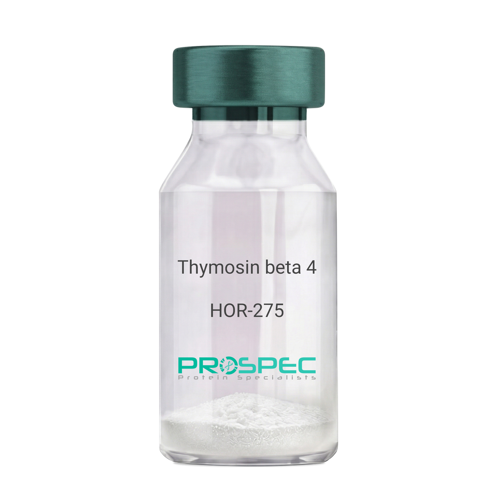
    Thymosin beta 4

    Description:

    Thymosin β4

    Thymosin beta-4. TB500, TB-500

    Product # :

    HOR-275

    Price :

    Quantity :

    Shipping Method :

    Room Temp Icon

    Shipped at Room temp

    Add To Cart

    More Info

    • description
    • formulation
    • purity
    • More Info
    • Citations

    Description

    Thymosin b4 is a 43 amino acid peptide which is regarded as the main intracellular G-actin sequestering peptide. It has a molecular weight of 4963.55 Da, and its molecular formula is: C212H350N56O78S1. Extracellular Thymosin b4 may contribute to physiological processes such as angiogenesis, wound healing, and regulation of inflammation.

    Formulation

    The protein (1mg/ml) was lyophilized with no additives.

    Purity

    Greater than 98.0% as determined by RP-HPLC.

    Citations

    More Info

    • Introduction

      Thymosin is a hormone secreted from the thymus. Its primary function is to stimulate the production of T cells, which are an important part of the immune system. Thymosin also assists in the development of B cells to plasma cells to produce antibodies. The predominant form of thymosin, thymosin b4, is a member of a highly conserved family of actin monomer-sequestering proteins. b-thymosins are the primary regulators of unpolymerized actin, and are essential for maintaining the small cytoplasmic pool of free G-actin monomers required for rapid filament elongation and allowing for the flux of monomers between the thymosin-bound pool and F-actin.

    • Synonyms

      Thymosin beta-4. TB500, TB-500

    • Physical Appearance

      Sterile Filtered White lyophilized (freeze-dried) powder.

    • Stability

      Lyophilized Thymosin b4 although stable at room temperature for 3 weeks, should be stored desiccated below -18°C. Upon reconstitution T beta 4 should be stored at 4°C between 2-7 days and for future use below -18°C.For long term storage it is recommended to add a carrier protein (0.1% HSA or BSA).Please prevent freeze-thaw cycles.

    • Solubility

      It is recommended to reconstitute the lyophilized Thymosin beta-4 in sterile 18MΩ-cm H2O not less than 100 µg/ml, which can then be further diluted to other aqueous solutions.

    • Amino Acid Sequence

      Thymosin b4 has an a.a. sequence of Ac-Ser-Asp-Lys-Pro-Asp-Met-Ala-Glu-Ile-Glu-Lys-Phe-Asp-Lys-Ser-Lys-Leu-Lys-Lys-Thr-Glu-Thr-Gln-Glu-Lys-Asn-Pro-Leu-Pro-Ser-Lys-Glu-Thr-Ile-Glu-Gln-Glu-Lys-Gln-Ala-Gly-Glu-Ser-OH.

    • Background

      What is the molecular weight/Mw of THYMOSIN BETA 4 Protein?
      THYMOSIN BETA 4 Protein has a total Mw of 4.96kDa.

      What is the Purity of THYMOSIN BETA 4 Protein?
      THYMOSIN BETA 4 Protein is >98% pure as determined by SDS-PAGE.

      What is the Biological Activity of THYMOSIN BETA 4 Protein?
      The biological functionality of THYMOSIN BETA 4 Protein will be determined in the future.

      What is the amino acid sequence of THYMOSIN BETA 4 Protein?
      Thymosin b4 has an a.a. sequence of Ac-Ser-Asp-Lys-Pro-Asp-Met-Ala-Glu-Ile-Glu-Lys-Phe-Asp-Lys-Ser-Lys-Leu-Lys-Lys-Thr-Glu-Thr-Gln-Glu-Lys-Asn-Pro-Leu-Pro-Ser-Lys-Glu-Thr-Ile-Glu-Gln-Glu-Lys-Gln-Ala-Gly-Glu-Ser-OH.
      What applications can THYMOSIN BETA 4 Protein be used in?
      THYMOSIN BETA 4 Protein can probably be used in western blot, ELISA and Lateral Flow.

      What is the endotoxin level for THYMOSIN BETA 4 Protein?
      The endotoxin level is minimal, THYMOSIN BETA 4 Protein was purified using conventional chromatography techniques.

      Who supplies THYMOSIN BETA 4 that is in stock and available to order?
      ProSpec has the protein in stock and available for immediate delivery.

      Where can I order bulk mg quantities of THYMOSIN BETA 4?
      ProSpec supplies bulk mg quantities at affordable prices that meets research and academic budgets.

    ProSpec's products are furnished for LABORATORY RESEARCH USE ONLY. The product may not be used as drugs, agricultural or pesticidal products, food additives or household chemicals.

    Thymosin Beta 4
  • View Data Sheet

    Name :

    IL 12p40 Antibody

    Description:

    Interleukin-12 p40, Rat Anti-Mouse

    NKSF2, CTL maturation factor (TCMF), Cytotoxic lymphocyte maturation factor 40 kDa subunit (CLMF p40), TSF, Edodekin-alpha, IL-12 p40, IL-12B, IL-12 subunit p40, NK cell stimulatory factor chain 2.

    Product # :

    ANT-114

    Price :

    Quantity :

    Shipping Method :

    Room Temp Icon

    Shipped at Room temp

    Add To Cart

    More Info

    • More Info
    • Citations

    Citations

    More Info

    • Introduction

      Active IL-12 is a p70 disulphide-linked dimer composed of p35 and p40 subunits. The protein is a pleiotropic cytokine produced primarily by antigen presenting cells and has multiple effects on T lymphocytes and natural killer cells in terms of stimulating cytotoxicity, proliferation, production of other cytokines and Th1 subset differentiation.

    • Synonyms

      NKSF2, CTL maturation factor (TCMF), Cytotoxic lymphocyte maturation factor 40 kDa subunit (CLMF p40), TSF, Edodekin-alpha, IL-12 p40, IL-12B, IL-12 subunit p40, NK cell stimulatory factor chain 2.

    • Solubility

      Reconstitute with sterile H20. Mix gently, wash the sides of the vial and wait 30-60 seconds before use.

    • Immunogen

      r.Murine IL-12 p40.

    • Ig Subclass

      Rat IgG2a.

    • Clone

      NYRmIL-12p40.

    • Applications

      Direct ELISA, Western Blot, Immuneprecipitation.

    • Titer

      By direct ELISA, 1:5,000 dilution will yield 0.5 O.D using alkaline phosphatase conjugated rabbit anti-mouse Ig (Jackson Laboratories).

    • Shipping Conditions

      Antibody is shipped lyophilized at ambient temperature.

    • Type

      Rat Anti Mouse Monoclonal.

    • Storage Procedures

      In lyophilized form, for long periods, store at 4oC in a dry environment. After reconstitution, if not intended for use within a month, aliquot and store at -20oC.

    • Purification Method

      Ion exchange.

    ProSpec's products are furnished for LABORATORY RESEARCH USE ONLY. The product may not be used as drugs, agricultural or pesticidal products, food additives or household chemicals.

    Il 12P40 Antibody
  • View Data Sheet

    Name :

    CRABP1 Human

    Description:

    Cellular Retinoic Acid binding Protein 1 Human Recombinant

    Cellular retinoic acid-binding protein 1, Cellular retinoic acid-binding protein I, CRABP-I, CRABP1, RBP5, CRABP, CRABPI.

    Product # :

    PRO-710

    Price :

    Quantity :

    Shipping Method :

    Ice Icon

    Shipped with Ice Packs

    Add To Cart

    More Info

    • description
    • source
    • formulation
    • purity
    • More Info
    • Citations

    Description

    CRABP1 Human Recombinant produced in E.Coli is a single, non-glycosylated polypeptide chain containing 137 amino acids and having a molecular mass of 15.5kDa. The CRABP1 is purified by proprietary chromatographic techniques.

    Source

    Escherichia Coli.

    Formulation

    The CRABP1 (1mg/ml) protein solution contains 20mM Tris-HCl buffer (pH8.0) and 10% glycerol.

    Purity

    Greater than 95.0% as determined by SDS-PAGE.

    Citations

    More Info

    • Introduction

      CRABP1 is a member of special carrier proteins for members of the vitamin A family. It is believed that CRABP1 has an essential role in retinoic acid-mediated differentiation and proliferation processes. Though, CRABP1 is structurally similar to the cellular retinol-binding proteins, it binds only retinoic acid at specific sites within the nucleus, which may contribute to vitamin A-directed differentiation in epithelial tissue. CRABP1 is constitutively expressed and is thought to have different functions in the cell than the related CRABP2. CRABP1 forms a beta-barrel structure which accommodates hydrophobic ligands in its interior.
      Loss of CRABP1 function as a result of hypermethylation of its promoter leads to pathogenesis of papillary thyroid carcinoma. Furthermore, frequent methylation-associated silencing of CRABP1 is linked to esophageal squamous-cell carcinoma.

    • Synonyms

      Cellular retinoic acid-binding protein 1, Cellular retinoic acid-binding protein I, CRABP-I, CRABP1, RBP5, CRABP, CRABPI.

    • Physical Appearance

      Sterile Filtered colorless solution.

    • Stability

      Store at 4°C if entire vial will be used within 2-4 weeks. Store, frozen at -20°C for longer periods of time. For long term storage it is recommended to add a carrier protein (0.1% HSA or BSA).Avoid multiple freeze-thaw cycles.

    • Amino Acid Sequence

      MPNFAGTWKM RSSENFDELL KALGVNAMLR KVAVAAASKP HVEIRQDGDQ FYIKTSTTVR TTEINFKVGE GFEEETVDGR KCRSLATWEN ENKIHCTQTL LEGDGPKTYW TRELANDELI LTFGADDVVC TRIYVRE.

    ProSpec's products are furnished for LABORATORY RESEARCH USE ONLY. The product may not be used as drugs, agricultural or pesticidal products, food additives or household chemicals.

    Crabp1 Human
  • View Data Sheet

    Name :

    CoV-2-S1 (319-541), Biotin

    Description:

    Coronavirus 2019 Spike Glycoprotein-S1 Receptor Binding Domain (319-541 a.a), Biotinylated Recombinant

    Product # :

    SARS-030

    Price :

    Quantity :

    Shipping Method :

    Room Temp Icon

    Shipped at Room temp

    Add To Cart

    More Info

    • description
    • source
    • formulation
    • purity
    • More Info
    • Citations

    Description

    The HEK293 derived Biotinylated recombinant protein contains the Coronavirus 2019 Spike Glycoprotein S1 Receptor Binding Domain [ RBD ], Wuhan-Hu-1 strain, amino acids 319-541 fused to His tag & AVI tag at C-terminal and having a molecular mass of 28.7kDa.

    Source

    HEK293 Cells.

    Formulation

    CoV-2 S1 RBD protein is lyophilized from 1x PBS pH-7.4 + 10% trehalose.

    Purity

    Protein is >90% pure as determined SDS-PAGE.

    Citations

    More Info

    • Introduction

      A human infecting coronavirus (viral pneumonia) called 2019 novel coronavirus, 2019-nCoV was found in the fish market at the city of Wuhan, Hubei province of China on December 2019.

      The 2019-nCoV shares an 87% identity to the 2 bat-derived severe acute respiratory syndrome 2018 SARS-CoV-2 located in Zhoushan of eastern China. 2019-nCoV has an analogous receptor-BD-structure to that of 2018 SARS-CoV, even though there is a.a. diversity so thus the 2019-nCoV might bind to ACE2 receptor protein (angiotensin-converting enzyme 2) in humans.

      While bats are possibly the host of 2019-nCoV, researchers suspect that animal from the ocean sold at the seafood market was an intermediate host. RSCU analysis proposes that the 2019-nCoV is a recombinant within the viral spike glycoprotein between the bat coronavirus and an unknown coronavirus.

    • Physical Appearance

      Lyophilized freezed dried powder.

    • Stability

      Lyophilized Cov-2 S1 Glycoprotein RBD although stable at room temperature for 3 weeks, should be stored desiccated below -18°C. Upon reconstitution CoV2 Spike protein should be stored at 4°C between 2-7 days and for future use below -18°C.For long term storage it is recommended to add a carrier protein (0.1% HSA or BSA).Please prevent freeze-thaw cycles.

    • Solubility

      It is recommended to reconstitute the lyophilized CoV-2 S1 protein in sterile 18M-cm H2O at aconcentration of 0.2mg/ml and not less than 0.1mg/ml, which can then be further diluted to other aqueous solutions.

    • Purification Method

      Purified by Metal-Afinity chromatographic technique.

    ProSpec's products are furnished for LABORATORY RESEARCH USE ONLY. The product may not be used as drugs, agricultural or pesticidal products, food additives or household chemicals.

    product_image.jpg
  • View Data Sheet

    Name :

    SCGB1A1 Human

    Description:

    Uteroglobin Human Recombinant

    Uteroglobin, Clara cell phospholipid-binding protein, CCPBP, Clara cells 10 kDa secretory protein, CC10, Secretoglobin family 1A member 1, Urinary protein 1, UP-1, UP1, Urine protein 1, SCGB1A1, CCSP, UGB, CC16.

    Product # :

    CYT-743

    Price :

    Quantity :

    Shipping Method :

    Room Temp Icon

    Shipped at Room temp

    Add To Cart

    More Info

    • description
    • source
    • formulation
    • purity
    • biological activity
    • More Info
    • Citations

    Description

    Uteroglobin Human Recombinant produced in E.coli is a single, non-glycosylated polypeptide chain containing 70 amino acids and having a molecular mass of 15.8kDa.The SCGB1A1 is purified by proprietary chromatographic techniques.

    Source

    Escherichia Coli.

    Formulation

    The Uteroglobin protein was lyophilized from a 0.2µm filtered concentrated solution in PBS, pH 7.4.

    Purity

    Greater than 98.0% as determined by:
    (a) Analysis by RP-HPLC.
    (b) Analysis by SDS-PAGE.

    Biological Activity

    The ED50 as determined by the ability of the immobilized protein to support the adhesion of the A549 human lung carcinoma cells is less than 5.0µg/ml, corresponding to a specific activity of > 200 IU/mg.

    Citations

    More Info

    • Introduction

      Uteroglobin (SCGB1A1) which belongs to the Secretoglobin (SCGBs) superfamily, is a multifunctional protein that exerts anti-inflammatory and anti-tumorigenic effects by binding small hydrophobic molecules such as phospholipids and prostaglandins. Uteroglobin is involved in numerous functions including anti-inflammation, inhibition of phospholipase A2 and the sequestering of hydrophobic ligands. SCGB1A1 is expressed by Clara cells, the non-ciliated, non-mucous secretory cells predominant in lung bronchioles, and by other epithelia which communicate with the external environment. On top of sequestering pro-inflammatory mediators and carcinogens, Uteroglobin is implicated in the inhibition of cell migration and invasion, platelet aggregation, and T cell differentiation. SCGB1A1 gene defects are associated with a susceptibility to asthma.

    • Synonyms

      Uteroglobin, Clara cell phospholipid-binding protein, CCPBP, Clara cells 10 kDa secretory protein, CC10, Secretoglobin family 1A member 1, Urinary protein 1, UP-1, UP1, Urine protein 1, SCGB1A1, CCSP, UGB, CC16.

    • Physical Appearance

      Sterile Filtered White lyophilized (freeze-dried) powder.

    • Stability

      Lyophilized Uteroglobin although stable at room temperature for 3 weeks, should be stored desiccated below -18°C. Upon reconstitution SCGB1A1 should be stored at 4°C between 2-7 days and for future use below -18°C. For long term storage it is recommended to add a carrier protein (0.1% HSA or BSA). Please prevent freeze-thaw cycles.

    • Solubility

      It is recommended to reconstitute the lyophilized SCGB1A1 in sterile 18M-cm H2O not less than 100µg/ml, which can then be further diluted to other aqueous solutions.

    • Amino Acid Sequence

      EICPSFQRVI ETLLMDTPSS YEAAMELFSP DQDMREAGAQ LKKLVDTLPQ KPRESIIKLM EKIAQSSLCN.

    ProSpec's products are furnished for LABORATORY RESEARCH USE ONLY. The product may not be used as drugs, agricultural or pesticidal products, food additives or household chemicals.

    Scgb1A1 Human
  • View Data Sheet

    Name :

    IGF1 Rat

    Description:

    IGF-1 Rat Recombinant

    Somatomedin C, IGF-I, IGFIA, IGF1.

    Product # :

    CYT-289

    Price :

    Quantity :

    Shipping Method :

    Room Temp Icon

    Shipped at Room temp

    Add To Cart

    More Info

    • description
    • source
    • formulation
    • purity
    • biological activity
    • More Info
    • Citations

    Description

    IGF-1 Rat Recombinant produced in E.Coli is a single, non-glycosylated, polypeptide chain containing 70 amino acids and having a molecular mass of 7.7kDa. IGF-I is purified by proprietary chromatographic techniques.

    Source

    Escherichia Coli.

    Formulation

    The protein was lyophilized with a 0.2µm filtered concentrated solution in 20mM PBS, pH 7.0.

    Purity

    Greater than 98.0% as determined by SDS-PAGE.

    Biological Activity

    The ED50 as determined by a cell proliferation assay using FDC-P1 cells is less than 2.0ng/ml, corresponding to a specific activity of >500,000units/mg.

    Citations

    More Info

    • Introduction

      The somatomedins, or IGFs, comprise a family of peptides that play important roles in mammalian growth and development. IGF1 mediates many of the growth-promoting effects of GH. Early studies showed that GH did not directly stimulate the incorporation of sulfate into cartilage, but rather acted through a serum factor, termed 'sulfation factor,' which later became known as 'somatomedin' (Daughaday et al., 1972). Three main somatomedins have been characterized: somatomedin C (IGF1), somatomedin A (IGF2; MIM 147470), and somatomedin B (MIM 193190) (Rotwein, 1986; Rosenfeld, 2003).

    • Synonyms

      Somatomedin C, IGF-I, IGFIA, IGF1.

    • Physical Appearance

      Sterile Filtered White lyophilized (freeze-dried) powder.

    • Stability

      Lyophilized IGF-1 although stable at room temperature for 3 weeks, should be stored desiccated below -18°C. Upon reconstitution IGFI should be stored at 4°C between 2-7 days and for future use below -18°C.For long term storage it is recommended to add a carrier protein (0.1% HSA or BSA).Please prevent freeze-thaw cycles.

    • Solubility

      It is recommended to reconstitute the lyophilized IGF1 in sterile 18MΩ-cm H2O not less than 100µg/ml, which can then be further diluted to other aqueous solutions.

    • Amino Acid Sequence

      GPETLCGAEL VDALQFVCGP RGFYFNKPTG YGSSIRRAPQ TGIVDECCFR SCDLRRLEMY CAPLKPTKSA.

    • Background

      What is the molecular weight/Mw of IGF1 RAT Protein?
      IGF1 RAT Protein has a total Mw of 7.7kDa.

      What is the source or expression system of IGF1 RAT Protein?
      Escherichia Coli.

      What is the Purity of IGF1 RAT Protein?
      IGF1 RAT Protein is >98% pure as determined by SDS-PAGE.

      What is the Biological Activity of IGF1 RAT Protein?
      The ED50 as determined by a cell proliferation assay using FDC-P1 cells is less than 2.0ng/ml, corresponding to a specific activity of >500,000units/mg.

      What is the amino acid sequence of IGF1 RAT Protein?
      GPETLCGAEL VDALQFVCGP RGFYFNKPTG YGSSIRRAPQ TGIVDECCFR SCDLRRLEMY CAPLKPTKSA.

      What applications can IGF1 RAT Protein be used in?
      IGF1 RAT Protein can probably be used in western blot, ELISA and Lateral Flow.

      What is the endotoxin level for IGF1 RAT Protein?
      The endotoxin level is minimal, IGF1 RAT Protein was purified using conventional chromatography techniques.

    ProSpec's products are furnished for LABORATORY RESEARCH USE ONLY. The product may not be used as drugs, agricultural or pesticidal products, food additives or household chemicals.

    Igf 1 Rat
  • View Data Sheet

    Name :

    IL 15 Antibody

    Description:

    Interleukin-15, Mouse Anti-Human

    IL-15, MGC9721.

    Product # :

    ANT-115

    Price :

    Quantity :

    Shipping Method :

    Room Temp Icon

    Shipped at Room temp

    Add To Cart

    More Info

    • formulation
    • More Info
    • Citations

    Formulation

    1mg/ml in PBS (after reconstitution).

    Citations

    More Info

    • Introduction

      The protein encoded by this gene is a cytokine that regulates T and natural killer cell activation and proliferation. This cytokine and interleukine 2 share many biological activities. They are found to bind common hematopoietin receptor subunits, and may compete for the same receptor, and thus negatively regulate each other's activity. The number of CD8+ memory cells is shown to be controlled by a balance between this cytokine and IL2. This cytokine induces the activation of JAK kinases, as well as the phosphorylation and activation of transcription activators STAT3, STAT5, and STAT6. Studies of the mouse counterpart suggested that this cytokine may increase the expression of apoptosis inhibitor BCL2L1/BCL-x(L), possibly through the transcription activation activity of STAT6, and thus prevent apoptosis. Two alternatively spliced transcript variants of this gene encoding the same protein have been reported.

    • Synonyms

      IL-15, MGC9721.

    • Solubility

      Reconstitute with sterile H20. Mix gently, wash the sides of the vial and wait 30-60 seconds before use.

    • Immunogen

      r.Human IL-15.

    • Ig Subclass

      Mouse IgG1.

    • Clone

      YNR-HIL15.

    • Applications

      Direct ELISA, Immuneprecipitation, Immunohistochemistry.

    • Titer

      By direct ELISA, 1:20,000 dilution will yield >1.0 O.D using alkaline phosphatase conjugated rabbit anti-mouse Ig (Jackson Laboratories).

    • Shipping Conditions

      Antibody is shipped lyophilized at ambient temperature.

    • Type

      Mouse Anti Human Monoclonal.

    • Storage Procedures

      In lyophilized form, for long periods, store at 4oC in a dry environment. After reconstitution, if not intended for use within a month, aliquot and store at -20oC.

    • Purification Method

      Ion exchange.

    ProSpec's products are furnished for LABORATORY RESEARCH USE ONLY. The product may not be used as drugs, agricultural or pesticidal products, food additives or household chemicals.

    Il 15 Antibody
  • View Data Sheet

    Name :

    CD80 Antibody

    Description:

    B7-1 (CD80), Rat Anti-Mouse

    LAB7, CD28LG, CD28LG1, CD28LG1, B71.

    Product # :

    ANT-133

    Price :

    Quantity :

    Shipping Method :

    Room Temp Icon

    Shipped at Room temp

    Add To Cart

    More Info

    • formulation
    • More Info
    • Citations

    Formulation

    1mg/ml in PBS (after reconstitution).

    Citations

    More Info

    • Introduction

      The B-lymphocyte activation antigen B7-1 provides regulatory signals for T lymphocytes as a consequence of binding to the CD28 and CTLA4 ligands of T cells CD-80 is involved in the costimulatory signal essential for t- lymphocyte activation. t-cell proliferation and cytokine production is induced by the binding of cd28 or ctla-4 to this receptor.

    • Synonyms

      LAB7, CD28LG, CD28LG1, CD28LG1, B71.

    • Solubility

      Reconstitute with of H20. Mix gently, wash the sides of the vial and wait 30-60 seconds before use.

    • Immunogen

      Purified mouse LPS-activated B cells.

    • Ig Subclass

      Rat IgG2a.

    • Clone

      mB7-1.

    • Applications

      Blocking and staining antibody. For staining, use 10µl/1,000,000 cells. Titer for blocking T cell activation should be determined by the investigator.

    • Available Conjugates

      This antibody is only available as purified antibody.

    • Type

      Rat Anti Mouse Monoclonal.

    • Storage Procedures

      Lyophilized: store at 4oC. After reconstitution, if not intended for use within a month, aliquot and store at -20oC.

    • Purification Method

      Ion exchange column.

    ProSpec's products are furnished for LABORATORY RESEARCH USE ONLY. The product may not be used as drugs, agricultural or pesticidal products, food additives or household chemicals.

    Cd80 Antibody
  • View Data Sheet

    Name :

    NBL1 Antibody

    Description:

    Neuroblastoma 1, Mouse Anti Human

    D1S1733E, DAN, DAND1, NB, NO3, Neuroblastoma suppressor of tumorigenicity 1, DAN domain family member 1, Zinc finger protein DAN, NBL1.

    Product # :

    ANT-488

    Price :

    Quantity :

    Shipping Method :

    Ice Icon

    Shipped with Ice Packs

    Add To Cart

    More Info

    • formulation
    • More Info
    • Citations

    Formulation

    1mg/ml containing PBS, pH-7.4, 10% Glycerol and 0.02% Sodium Azide.

    Citations

    More Info

    • Introduction

      Neuroblastoma suppressor of tumorigenicity 1 (NBL1) is a part of the evolutionarily conserved CAN (Cerberus and DAN) family of proteins, which contain a domain resembling the CTCK (C-terminal cystine knot-like) motif found in several signaling molecules. NBL1 is a is a tumor suppressor of neuroblastoma and takes part in preventing cells from entering the final stage (G1/S) of the transformation process. NBL1 is produced in small neurons of the dorsal root ganglion. The expression of NBL1 is triggered by MATH-1.

    • Synonyms

      D1S1733E, DAN, DAND1, NB, NO3, Neuroblastoma suppressor of tumorigenicity 1, DAN domain family member 1, Zinc finger protein DAN, NBL1.

    • Physical Appearance

      Sterile filtered colorless solution.

    • Immunogen

      Anti-human NBL1 mAb, clone PAT38G8AT, is derived from hybridization of mouse F0 myeloma cells with spleen cells from BALB/c mice immunized with a recombinant human NBL1 protein 18-181 amino acids purified from E. coli.

    • Ig Subclass

      Mouse IgG1 heavy chain and Kappa light chain.

    • Clone

      PAT38G8AT.

    • Applications

      The antibody has been tested by ELISA, Western blot analysis to assure specificity and reactivity. Since application varies, however, each investigation should be titrated by the reagent to obtain optimal results. Recommended starting dilution is 1:1000.

    • Type

      Mouse Anti Human Monoclonal.

    • Storage Procedures

      For periods up to 1 month store at 4°C, for longer periods of time, store at -20°C. Prevent freeze thaw cycles.

    • Purification Method

      NBL1 antibody was purified from mouse ascitic fluids by protein-A affinity chromatography.

    ProSpec's products are furnished for LABORATORY RESEARCH USE ONLY. The product may not be used as drugs, agricultural or pesticidal products, food additives or household chemicals.

    Nbl1 Antibody
  • 1
  • …
  • 14
  • 15
  • 16
  • 17
Your browser does not support the video tag.

Products

  • Cytokines
  • Growth Factors
  • Chemokines
  • CD Antigens
  • Neurotrophins
  • Hormones
  • Enzymes
  • Viral Antigens
  • Recombinant Proteins
  • Natural Proteins
  • Monoclonal Antibodies
  • Polyclonal Antibodies
  • Thermal Packaging

About

  • Company
  • Founder
  • Contribution
  • Contact Us
  • My Account

Ordering

  • Ordering Method
  • Ordering - Credit Card
  • Ordering PO
  • Shipping Fees
  • FAQ's

Legal

  • Terms & Conditions
  • Privacy
  • Site Map
  • info@prospecbio.com
  • Facebook
  • Instagram
  • Linkedin Profile

© 2026 ProSpec-Tany TechnoGene Ltd. All Rights Reserved.

  • Choosing a selection results in a full page refresh.
  • Opens in a new window.