Search results
1000 results found for “epigen”
Name
Description
Product #
Price
Quantity
Shipping Method
- View Data Sheet
Name :
Latexin HumanDescription:
Latexin Human Recombinant
LXN, TCI, ECI, Latexin, Endogenous carboxypeptidase inhibitor, Tissue carboxypeptidase inhibitor, MUM.
Product # :
ENZ-407Price :
Quantity :
Shipping Method :
Shipped with Ice Packs
More Info
- description
- source
- formulation
- purity
- More Info
Description
Recombinant Human Latexin produced in E.Coli is a single, non-glycosylated polypeptide chain containing 222 amino acids and having a molecular mass of 25.7kDa. Latexin is purified by conventional chromatography techniques.
Source
Escherichia Coli.
Formulation
The Latexin protein solution contains 20mM Tris-HCl, pH-7.5, 50mM NaCl and 10% Glycerol.
Purity
Greater than 95.0% as determined by SDS-PAGE.
More Info
-
Introduction
Latexin enzyme is a carboxypeptidase A inhibitor that is highly expressed in the heart, prostate, ovary, kidney, pancrease, brain and colon. Latexin has no noticeable sequence resemblance with plant and parasite inhibitors, however it is related to a human putative tumor suppressor protein, TIG1. Latexin is down-regulated in the presenilin-1-deficient mouse brain, thus putatively playing a role in Alzheimer's disease.
-
Synonyms
LXN, TCI, ECI, Latexin, Endogenous carboxypeptidase inhibitor, Tissue carboxypeptidase inhibitor, MUM.
-
Physical Appearance
Sterile filtered colorless solution.
-
Stability
Store at 4°C if entire vial will be used within 2-4 weeks. Store, frozen at -20°C for longer periods of time. For long term storage it is recommended to add a carrier protein (0.1% HSA or BSA). Avoid multiple freeze-thaw cycles.
-
Amino Acid Sequence
MEIPPTNYPA SRAALVAQNY INYQQGTPHR VFEVQKVKQA SMEDIPGRGH KYRLKFAVEE IIQKQVKVNC TAEVLYPSTG QETAPEVNFTFEGETGKNPD EEDNTFYQRL KSMKEPLEAQ NIPDNFGNVS PEMTLVLHLA WVACGYIIWQ NSTEDTWYKM VKIQTVKQVQ RNDDFIELDYTILLHNIASQ EIIPWQMQVL WHPQYGTKVK HNSRLPKEVQ LE.
ProSpec's products are furnished for LABORATORY RESEARCH USE ONLY. The product may not be used as drugs, agricultural or pesticidal products, food additives or household chemicals.
- View Data Sheet
Name :
TRIM21 HumanDescription:
Tripartite Motif Containing 21 (RO52) Human Recombinant
52 kDa Ro protein, Sjoegren syndrome type A antigen, SS-A, Ro(SS-A), 52 kDa ribonucleoprotein autoantigen Ro/SS-A, Tripartite motif-containing protein 21, RING finger protein 81, TRIM21, RNF81, RO52, SSA1, SSA, RO-52.
Product # :
PRO-328Price :
Quantity :
Shipping Method :
Shipped with Ice Packs
More Info
- description
- source
- formulation
- purity
- More Info
Description
TRIM21 Human Recombinant produced in SF9 is a polypeptide chain having a calculated molecular mass of 52 kDa. TRIM21 is expressed with a -6x His tag at N-terminus and purified by proprietary chromatographic techniques.
Source
Sf9 insect cells.
Formulation
TRIM21 solution is supplied in 20mM HEPES pH-7.5, 0.01mM EDTA and 0.02% SDS.
Purity
Greater than 95% as determined by SDS-PAGE.
More Info
-
Introduction
TRIM21 is a member of the tripartite motif (TRIM) family. The TRIM motif includes three zinc-binding domains, a RING, a B-box type 1 and a B-box type 2, and a coiled-coil region. The 52 kDa Ro protein is part of the RoSSA ribonucleoprotein, which includes a single polypeptide and one of four small RNA molecules. The RoSSA particle localizes to both the cytoplasm and the nucleus. Ro/SSA interacts with autoantigens in patients with Sjogren syndrome and systemic lupus erythematosus. Ribonucleoprotein particle is composed of a single polypeptide and one of four small RNA molecules. The RoSSA is present in all mammalian cells studied but has no known function. At least 2 isoforms are present in nucleated and red blood cells, and tissue specific differences in Ro/SSA proteins were identified.
-
Synonyms
52 kDa Ro protein, Sjoegren syndrome type A antigen, SS-A, Ro(SS-A), 52 kDa ribonucleoprotein autoantigen Ro/SS-A, Tripartite motif-containing protein 21, RING finger protein 81, TRIM21, RNF81, RO52, SSA1, SSA, RO-52.
-
Physical Appearance
Sterile Filtered clear solution.
-
Stability
Store at 4°C if entire vial will be used within 2-4 weeks. Store, frozen at -20°C for longer periods of time. Avoid multiple freeze-thaw cycles.
ProSpec's products are furnished for LABORATORY RESEARCH USE ONLY. The product may not be used as drugs, agricultural or pesticidal products, food additives or household chemicals.
- View Data Sheet
Name :
Leptin Receptor ChickenDescription:
Leptin Receptor Chicken Recombinant
OB Protein, Obesity Protein, OBS, Obesity factor, Leptin Receptor.
Product # :
CYT-509Price :
Quantity :
Shipping Method :
Shipped with Ice Packs
More Info
- description
- source
- formulation
- purity
- More Info
Description
Leptin Binding Domain Chicken Recombinant also called Leptin Receptor produced in E.Coli is a single, non-glycosylated, polypeptide chain containing 208 amino acids and having a molecular mass of 24.5 kDa. Chicken Leptin Receptor consists of the cytokine binding domain of leptin receptor amino acids 420-626 of chicken leptin receptor.The Leptin Binding Domain is purified by proprietary chromatographic techniques.
Source
Escherichia Coli.
Formulation
The protein was filter sterilized and stored at 4°C (0.2 to 0.5 mg/ml) solution of Tris-HCl buffer, pH 9.0 with 150mM NaCl.
Purity
Greater than 99.0% as determined by:
(a) Analysis by SEC-HPLC.
(b) Analysis by SDS-PAGE.More Info
-
Introduction
Leptin Receptor is a part of the gp130 family of cytokine receptors that stimulate gene transcription by activating cytosolic STAT proteins. Leptin Receptor plays a role in the regulation of fat metabolism and in novel hematopoietic pathway that is obligatory for normal lymphopoiesis. Leptin Receptorparticipates in the regulation of counter-regulatory response to hypoglycemia by inhibiting neurons of the parabrachial nucleus.Leptin Receptoraffectsspecifically on T lymphocyte responses.
-
Synonyms
OB Protein, Obesity Protein, OBS, Obesity factor, Leptin Receptor.
-
Physical Appearance
Sterile Filtered colorless solution at a concentration of 0.4 mg/ml.
-
Stability
Sterile solutions at 0.5mg/ml or less are stable at 4°C for several months.
-
Amino Acid Sequence
The sequence of the first six N-terminal amino acids was determined and was found to be Ala-Ile-Asp-Val-Asn-Ile Biological ActivityBiological Activity is evidenced by high affinity binding of mammalian leptins at 1:1 molar ratio.
-
Protein content
Protein quantitation was carried out by two independent methods1. UV spectroscopy at 280 nm using the absorbency value of 2.45 as the extinction coefficient for a 0.1% (1mg/ml) solution. This value is calculated by the PC GENE computer analysis program of protein sequences (IntelliGenetics). 2. Analysis by RP-HPLC, using a calibrated solution of Leptin Binding Domain as a Reference Standard.
ProSpec's products are furnished for LABORATORY RESEARCH USE ONLY. The product may not be used as drugs, agricultural or pesticidal products, food additives or household chemicals.
- View Data Sheet
Name :
H2AFZ HumanDescription:
H2A Histone Family, Member Z Human Recombinant
H2A Histone Family, Member Z, H2AZ, H2A/z, H2A.Z-1, H2A.z, H2AZ Histone, Histone H2A.Z.
Product # :
PRO-1908Price :
Quantity :
Shipping Method :
Shipped with Ice Packs
More Info
- description
- source
- formulation
- purity
- More Info
Description
H2AFZ Human Recombinant produced in E.Coli is a single, non-glycosylated polypeptide chain containing 151 amino acids (1-128 a.a) and having a molecular mass of 15.9kDa.H2AFZ is fused to a 23 amino acid His-tag at N-terminus & purified by proprietary chromatographic techniques.
Source
Escherichia Coli.
Formulation
H2AFZ protein solution (0.25mg/ml) containing 20mM Tris-HCl buffer (pH 8.0), 0.4M Urea and 10% glycerol.
Purity
Greater than 85% as determined by SDS-PAGE.
More Info
-
Introduction
Histone H2A.Z (H2AFZ) belongs to the histone H2A family. Histones are basic nuclear proteins which are responsible for the nucleosome structure of the chromosomal fiber in eukaryotes. Nucleosomes consist of approximately 146 bp of DNA wrapped around a histone octamer composed of pairs of each of the four core histones (H2A, H2B, H3, and H4). H2AFZ is implicated in the formation of constitutive heterochromatin and vital for chromosome segregation during cell division. Moreover, variant Histone H2A, which takes the place of conventional H2A in a subset of nucleosomes. Studies in mice have demonstrated that this particular histone is essential for embryonic development and show that lack of functional histone H2A leads to embryonic lethality.
-
Synonyms
H2A Histone Family, Member Z, H2AZ, H2A/z, H2A.Z-1, H2A.z, H2AZ Histone, Histone H2A.Z.
-
Physical Appearance
Sterile Filtered clear solution.
-
Stability
Store at 4°C if entire vial will be used within 2-4 weeks. Store, frozen at -20°C for longer periods of time.For long term storage it is recommended to add a carrier protein (0.1% HSA or BSA).Please avoid freeze thaw cycles.
-
Amino Acid Sequence
MGSSHHHHHH SSGLVPRGSH MGSMAGGKAG KDSGKAKTKA VSRSQRAGLQ FPVGRIHRHL KSRTTSHGRV GATAAVYSAA ILEYLTAEVL ELAGNASKDL KVKRITPRHL QLAIRGDEEL DSLIKATIAG GGVIPHIHKS LIGKKGQQKT V
ProSpec's products are furnished for LABORATORY RESEARCH USE ONLY. The product may not be used as drugs, agricultural or pesticidal products, food additives or household chemicals.
- View Data Sheet
Name :
HAPLN1 HumanDescription:
Hyaluronan And Proteoglycan Link Protein 1 Human Recombinant
Hyaluronan and proteoglycan link protein 1 precursor, CRTL1, Cartilage-linking protein 1, Cartilage-link protein, Proteoglycan link protein, HAPLN1.
Product # :
PRO-2012Price :
Quantity :
Shipping Method :
Shipped with Ice Packs
More Info
- description
- source
- formulation
- purity
- More Info
Description
HAPLN1 Human Recombinant produced in E.Coli is a single, non-glycosylated polypeptide chain containing 362 amino acids (16-354 a.a.) and having a molecular mass of 40.9kDa.HAPLN1 is fused to a 23 amino acid His-tag at N-terminus & purified by proprietary chromatographic techniques.
Source
Escherichia Coli.
Formulation
HAPLN1 protein solution (1mg/ml) containing 20mM Tris-HCl buffer (pH 8.0), 0.4M Urea and 10% glycerol.
Purity
Greater than 80.0% as determined by SDS-PAGE.
More Info
-
Introduction
Hyaluronan And Proteoglycan Link Protein 1, which is also known as HAPLN1 is a part of the HAPLN family.HAPLN1 contains one Ig-like V-type domain and two Link domains. In the extracellular cartilage matrix, HAPLN1 stabilizes the aggregates of proteoglycan monomers by means of hyaluronic acid .
-
Synonyms
Hyaluronan and proteoglycan link protein 1 precursor, CRTL1, Cartilage-linking protein 1, Cartilage-link protein, Proteoglycan link protein, HAPLN1.
-
Physical Appearance
Sterile Filtered colorless solution.
-
Stability
Store at 4°C if entire vial will be used within 2-4 weeks. Store, frozen at -20°C for longer periods of time. For long term storage it is recommended to add a carrier protein (0.1% HSA or BSA).Avoid multiple freeze-thaw cycles.
-
Amino Acid Sequence
MGSSHHHHHH SSGLVPRGSH MGSDHLSDNY TLDHDRAIHI QAENGPHLLV EAEQAKVFSH RGGNVTLPCK FYRDPTAFGS GIHKIRIKWT KLTSDYLKEV DVFVSMGYHK KTYGGYQGRV FLKGGSDSDA SLVITDLTLE DYGRYKCEVI EGLEDDTVVV ALDLQGVVFP YFPRLGRYNL NFHEAQQACL DQDAVIASFD QLYDAWRGGL DWCNAGWLSD GSVQYPITKP REPCGGQNTV PGVRNYGFWD KDKSRYDVFC FTSNFNGRFY YLIHPTKLTY DEAVQACLND GAQIAKVGQI FAAWKILGYD RCDAGWLADG SVRYPISRPR RRCSPTEAAV RFVGFPDKKH KLYGVYCFRA YN.
ProSpec's products are furnished for LABORATORY RESEARCH USE ONLY. The product may not be used as drugs, agricultural or pesticidal products, food additives or household chemicals.
- View Data Sheet
Name :
BSG HumanDescription:
Basigin Human Recombinant
Basigin isoform 2, 5F7, CD147, EMMPRIN, OK, TCSF, Leukocyte activation antigen M6, OK blood group antigen.
Product # :
PRO-2516Price :
Quantity :
Shipping Method :
Shipped with Ice Packs
More Info
- description
- source
- formulation
- purity
- More Info
Description
BSG produced in Sf9 Baculovirus cells is a single, glycosylated polypeptide chain containing 428 amino acids (22-207a.a.) and having a molecular mass of 47.5kDa. (Molecular size on SDS-PAGE will appear at approximately 40-57kDa).BSG is expressed with a 242 amino acid hIgG-His tag at C-Terminus and purified by proprietary chromatographic techniques.
Source
Sf9, Baculovirus cells.
Formulation
BSG protein solution (0.5mg/ml) contains phosphate buffered saline (pH7.4) and 10% glycerol.
Purity
Greater than 90.0% as determined by SDS-PAGE.
More Info
-
Introduction
Basigin Isoform 2 or BSG is a part of the immunoglobulin superfamily, it is a type 1 transmembrane protein which has a variety of Nand O- glycosylation. BSG has several ligands, such as integrins, cyclophilin proteins Cyp-A & CyP-B. The protein has a crucial role in intercellular recognition, it takes part in many immunologic processes, differentiation and cell development. Also, it regulates numerus cell processes such as production and release of expression of the mono-carboxylate transporter, spermatogenesis, etc. Basigin Isoform 2 promotes invasion, metastasis, growth and survival of malignant cells by producing and releasing matrix metalloproteinases nearby mesenchymal cells and tumor cells.
-
Synonyms
Basigin isoform 2, 5F7, CD147, EMMPRIN, OK, TCSF, Leukocyte activation antigen M6, OK blood group antigen.
-
Physical Appearance
Sterile Filtered colorless solution.
-
Stability
Store at 4°C if entire vial will be used within 2-4 weeks. Store, frozen at -20°C for longer periods of time. For long term storage it is recommended to add a carrier protein (0.1% HSA or BSA).Avoid multiple freeze-thaw cycles.
-
Amino Acid Sequence
ADPAAGTVFT TVEDLGSKIL LTCSLNDSAT EVTGHRWLKG GVVLKEDALP GQKTEFKVDS DDQWGEYSCV FLPEPMGTAN IQLHGPPRVK AVKSSEHINE GETAMLVCKS ESVPPVTDWA WYKITDSEDK ALMNGSESRF FVSSSQGRSE LHIENLNMEA DPGQYRCNGT SSKGSDQAII
TLRVRSHLAL EPKSCDKTHT CPPCPAPELL GGPSVFLFPP KPKDTLMISR TPEVTCVVVD VSHEDPEVKF NWYVDGVEVH NAKTKPREEQ YNSTYRVVSV LTVLHQDWLN GKEYKCKVSN KALPAPIEKT ISKAKGQPRE PQVYTLPPSR DELTKNQVSL TCLVKGFYPS DIAVEWESNG
QPENNYKTTP PVLDSDGSFF LYSKLTVDKS RWQQGNVFSC SVMHEALHNH YTQKSLSLSP GKHHHHHH.
ProSpec's products are furnished for LABORATORY RESEARCH USE ONLY. The product may not be used as drugs, agricultural or pesticidal products, food additives or household chemicals.
- View Data Sheet
Name :
LGALS4 MouseDescription:
Galectin-4 Mouse Recombinant
gal-4 , Galectin-4, Lactose-binding lectin 4, lectin galactoside-binding soluble 4.
Product # :
CYT-187Price :
Quantity :
Shipping Method :
Shipped with Ice Packs
More Info
- description
- source
- formulation
- purity
- biological activity
- More Info
- SDS-PAGE
Description
LGALS4 Mouse Recombinant produced in E.Coli is a single, non-glycosylated polypeptide chain containing 349 amino acids (1-326a.a) and having a molecular mass of 38.8kDa.LGALS4 is fused to a 23 amino acid His-tag at N-terminus & purified by proprietary chromatographic techniques.
Source
Escherichia Coli.
Formulation
LGALS4 protein solution (0.5mg/ml) containing 20mM Tris-HCl buffer (pH 8.0), 0.1M NaCl, 10% glycerol and 1mM DTT.
Purity
Greater than 90.0% as determined by SDS-PAGE.
Biological Activity
The ED50 was measured by its ability to agglutinate human red blood cells and was found to be <5 ug/ml.
SDS-PAGE
More Info
-
Introduction
Galectin-4 is a member of the subfamily of galectins composed of two carbohydrate recognition domains having similar peptide chains. The galectins are a family of beta-galactoside-binding proteins having a role in modulating cell-cell and cell-matrix interactions, which inhibits chronic inflammations, GVHD, and allergic responses. LGALS4 expression is limited to small intestine, colon, and rectum, and it is underexpressed in colorectal cancer. LGALS4 binds as an endogenous ligand to glycosphingolipids having 3-O-sulfated Gal residues and bind as well to cholesterol-3-sulfate. LGALS4 takes part in cell adhesion. LGALS4 plays a role in crosslinking the lateral cell membranes of the surface-lining epithelial cells, thus supporting epithelial integrity against mechanical stress exerted by the bowel lume. LGALS4 is in charge of intestinal inflammation via selective regulation of peripheral and mucosal T-cell cell cycle, in addition to cell death by apoptosis of T-cells by a pathway independent of the activation of caspases. LGALS4 blockade decreases TNF-alpha inhibitor induced T-cell death. LGALS4 decreases pro-inflammatory cytokine secretion including IL-6 & IL-17.
-
Synonyms
gal-4 , Galectin-4, Lactose-binding lectin 4, lectin galactoside-binding soluble 4.
-
Physical Appearance
Sterile filtered colorless solution.
-
Stability
Store at 4°C if entire vial will be used within 2-4 weeks. Store, frozen at -20°C for longer periods of time. For long term storage it is recommended to add a carrier protein (0.1% HSA or BSA).Avoid multiple freeze-thaw cycles.
-
Amino Acid Sequence
MGSSHHHHHH SSGLVPRGSH MGSMAYVPAP GYQPTYNPTL PYKRPIPGGL SVGMSVYIQG MAKENMRRFH VNFAVGQDDG ADVAFHFNPR FDGWDKVVFN TMQSGQWGKE EKKKSMPFQK GKHFELVFMV MPEHYKVVVN GNSFYEYGHR LPVQMVTHLQ VDGDLELQSI NFLGGQPAAA PYPGAMTIPA YPAGSPGYNP PQMNTLPVMT GPPVFNPRVP YVGALQGGLT VRRTIIIKGY VLPTARNFVI NFKVGSSGDI ALHLNPRIGD SVVRNSFMNG SWGAEERKVA YNPFGPGQFF DLSIRCGMDR FKVFANGQHL FDFSHRFQAF QMVDTLEING DITLSYVQI.
-
Background
What is the molecular weight/Mw of LGALS4 MOUSE Protein?
LGALS4 MOUSE Protein has a total Mw of 38.8kDa.
What is the source or expression system of LGALS4 MOUSE Protein?
Escherichia Coli.
What is the Purity of LGALS4 MOUSE Protein?
LGALS4 MOUSE Protein is >90% pure as determined by SDS-PAGE.
What is the Biological Activity of LGALS4 MOUSE Protein?
The ED50 was measured by its ability to agglutinate human red blood cells and was found to be <5 ug/ml.
What is the amino acid sequence of LGALS4 MOUSE Protein?
MGSSHHHHHH SSGLVPRGSH MGSMAYVPAP GYQPTYNPTL PYKRPIPGGL SVGMSVYIQG MAKENMRRFH VNFAVGQDDG ADVAFHFNPR FDGWDKVVFN TMQSGQWGKE EKKKSMPFQK GKHFELVFMV MPEHYKVVVN GNSFYEYGHR LPVQMVTHLQ VDGDLELQSI NFLGGQPAAA PYPGAMTIPA YPAGSPGYNP PQMNTLPVMT GPPVFNPRVP YVGALQGGLT VRRTIIIKGY VLPTARNFVI NFKVGSSGDI ALHLNPRIGD SVVRNSFMNG SWGAEERKVA YNPFGPGQFF DLSIRCGMDR FKVFANGQHL FDFSHRFQAF QMVDTLEING DITLSYVQI.
What applications can LGALS4 MOUSE Protein be used in?
LGALS4 MOUSE Protein can probably be used in western blot, ELISA and Lateral Flow.
What is the endotoxin level for LGALS4 MOUSE Protein?
The endotoxin level is minimal, LGALS4 MOUSE Protein was purified using conventional chromatography techniques.
ProSpec's products are furnished for LABORATORY RESEARCH USE ONLY. The product may not be used as drugs, agricultural or pesticidal products, food additives or household chemicals.
- View Data Sheet
Name :
C-JUN HumanDescription:
Jun Proto-Oncogene Human Recombinant
Transcription factor AP-1, Activator protein 1, AP1, Proto-oncogene c-jun, V-jun avian sarcoma virus 17 oncogene homolog, p39, c-Jun.
Product # :
PKA-323Price :
Quantity :
Shipping Method :
Shipped at Room temp
More Info
- description
- source
- formulation
- purity
- biological activity
- More Info
Description
C-JUN amino acids 1-81 produced in E.coli, is a non-glycosylated, polypeptide chain having a molecular mass of 52 kDa.C-JUN is a maltose binding protein (MBP) fusion protein with an amino-terminal polyhistidine tag and purified by proprietary chromatographic techniques.
Source
Escherichia Coli.
Formulation
C-JUN is supplied as lyophilized powder containing no additives.
Purity
Greater than 95% as determined by SDS-PAGE.
Biological Activity
C-Jun is phosphorylatable in vitro, using either recombinant active JNK1 or JNK2, or with JNK immunoprecipitated from stimulated cells. This phosphorylation can be monitored by Western blot analysis using an antibody directed to c-Jun [pS73], in conjunction with chemiluminescence detection methods. Optimization of the cell stimulation protocol, cell lysis procedure, and reaction conditions may be required for each specific application.More Info
-
Introduction
C-JUN is a gene which, in combination with c-Fos, forms the AP-1early response transcription factor. C-JUN is activated by the JNKpathway. C-JUN is the putative transforming gene of avian sarcoma virus 17. C-JUN is a protein which is highly similar to the viral protein, and which interacts directly with specific target DNA sequences to regulate gene expression. The C-JUN gene is intronless and is mapped to 1p32-p31, a chromosomal region involved in both translocations and deletions in human malignancies.
-
Synonyms
Transcription factor AP-1, Activator protein 1, AP1, Proto-oncogene c-jun, V-jun avian sarcoma virus 17 oncogene homolog, p39, c-Jun.
-
Physical Appearance
Sterile Filtered White lyophilized (freeze-dried) powder.
-
Stability
Store at 4°C if entire vial will be used within 1-2 weeks. Store, frozen at -20°C for longer periods of time. Avoid multiple freeze-thaw cycles.
-
Solubility
It is recommended to centrifuge the vial prior to opening in order to bring the contents to the bottom. The reconstitution of the lyophilized c-Jun is recommended in 40mM Tris, pH 7.5, to a concentration of 0.2-1.0 mg/ml.
-
Note
Kinase activity may vary depending on the substrate and reaction conditions.
ProSpec's products are furnished for LABORATORY RESEARCH USE ONLY. The product may not be used as drugs, agricultural or pesticidal products, food additives or household chemicals.
- View Data Sheet
Name :
HIF1AN HumanDescription:
Hypoxia-Inducible Factor-1 Alpha Inhibitor Human Recombinant
FIH1, FIH-1, HIF1AN, Factor Inhibiting HIF1A, Hypoxia-inducible factor 1-alpha inhibitor, Hypoxia-inducible factor asparagine hydroxylase, Factor inhibiting HIF-1, FLJ20615, FLJ22027, DKFZp762F1811.
Product # :
PRO-662Price :
Quantity :
Shipping Method :
Shipped with Ice Packs
More Info
- description
- source
- formulation
- purity
- More Info
Description
HIF1AN Human Recombinant produced in E.Coli is a single, non-glycosylated polypeptide chain containing 349 amino acids (1-349) and having a molecular mass of 40.2kDa. The HIF1AN is purified by proprietary chromatographic techniques.
Source
Escherichia Coli.
Formulation
The protein solution (1mg/ml) contains 20mM Tris-HCl pH-8.
Purity
Greater than 90% as determined by SDS-PAGE.
More Info
-
Introduction
Overexpression of the HIF1AN is linked with tumor aggressiveness in pancreatic endocrine tumors. HIF1AN hydroxylates Notch ICD at two residues that are crucial for the function of Notch ICD as a transactivator within cells and during neurogenesis and myogenesis. HIF1AN is commonly expressed in invasive breast carcinoma. The hypoxic response and survival recommends that tumour regulation of HIF1AN is an additional important mechanism for HIF pathway activation. HIF1AN is an asparaginyl hydroxylase enzyme that controls the transcriptional activity of hypoxia-inducible factor. FIH1 is a part of the Fe2+ and 2-oxoglutarate-dependent dioxygenase superfamily, FIH1 is protein that hydoxylates a specific asparagine residue (Asn-803) within the HIF1A C-terminal transactivation domain. In normoxia, the HIF1AN-mediated hydroxylation of the HIF1? transactivation domain which results in blockage of the HIF1A-p300/CBP interaction and represses transcriptional activity of HIF1A targeted genes.
-
Synonyms
FIH1, FIH-1, HIF1AN, Factor Inhibiting HIF1A, Hypoxia-inducible factor 1-alpha inhibitor, Hypoxia-inducible factor asparagine hydroxylase, Factor inhibiting HIF-1, FLJ20615, FLJ22027, DKFZp762F1811.
-
Physical Appearance
Sterile Filtered colorless solution.
-
Stability
Store at 4°C if entire vial will be used within 2-4 weeks.Store, frozen at -20°C for longer periods of time. For long term storage it is recommended to add a carrier protein (0.1% HSA or BSA).Avoid multiple freeze-thaw cycles.
-
Amino Acid Sequence
MAATAAEAVA SGSGEPREEA GALGPAWDES QLRSYSFPTR PIPRLSQSDP RAEELIENEE PVVLTDTNLV YPALKWDLEY LQENIGNGDF SVYSASTHKF LYYDEKKMAN FQNFKPRSNR EEMKFHEFVE KLQDIQQRGG EERLYLQQTL NDTVGRKIVM DFLGFNWNWI NKQQGKRGWG QLTSNLLLIG MEGNVTPAHY DEQQNFFAQI KGYKRCILFP PDQFECLYPY PVHHPCDRQS QVDFDNPDYE RFPNFQNVVG YETVVGPGDV LYIPMYWWHH IESLLNGGIT ITVNFWYKGA PTPKRIEYPL KAHQKVAIMR NIEKMLGEAL GNPQEVGPLL NTMIKGRYN.
ProSpec's products are furnished for LABORATORY RESEARCH USE ONLY. The product may not be used as drugs, agricultural or pesticidal products, food additives or household chemicals.
- View Data Sheet
Name :
SIRPA Human, HEKDescription:
Signal-Regulatory Protein Alpha Human Recombinant, HEK
Tyrosine-protein phosphatase non-receptor type substrate 1 isoform 1, SHP substrate 1, SHPS-1, Brain Iglike molecule with tyrosine-based activation motifs, Bit, CD172 antigen-like family member A, MYD1, PTPNS1, SHPS1,SIRP, Inhibitory receptorSHPS-1, Macrophage fusion receptor, MyD-1 antigen, Signal-regulatory protein alpha-1, Sirp-alpha-1, Signalregulatory protein alpha-2, Sirp-alpha-2, Signal-regulatory protein alpha-3, Sirp-alpha-3, p84, CD172a, BIT, MFR.
Product # :
PRO-2755Price :
Quantity :
Shipping Method :
Shipped with Ice Packs
More Info
- description
- source
- formulation
- purity
- biological activity
- More Info
Description
SIRPA Human Recombinant produced in HEK293 cells is a single, glycosylated polypeptide chain (27-373a.a) containing 356 amino acids and having a molecular mass of 39kDa.SIRPA is fused to a 6 amino acid His-tag at C-terminus,and is purified by proprietary chromatographic techniques.
Source
HEK293 Cells.
Formulation
The SIRPA solution (1mg/ml) contains Phosphate-Buffered Saline (pH 7.4) and 10% glycerol.
Purity
Greater than 95% as determined by SDS-PAGE.
Biological Activity
Measured by its binding ability in a functional ELISA with Human CD47.
More Info
-
Introduction
Signal-Regulatory Protein Alpha, SIRPA belongs to the signal-regulatory-protein (SIRP) family, as well as the immunoglobulin super family. The members of the SIRP family are receptor-type transmembrane glycoproteins which are involved in the negative regulation of receptor tyrosine kinase-coupled signaling processes.
SIRPA can be phosphorylated by tyrosine kinases. The phospho-tyrosine residues of this PTP have been shown to recruit SH2 domain containing tyrosine phosphatases (PTP), and perform as substrates of PTPs. SIRPA take part in signal transduction mediated by a variety of growth factor receptors. CD47 has been shown to be a ligand for SIRPA. -
Synonyms
Tyrosine-protein phosphatase non-receptor type substrate 1 isoform 1, SHP substrate 1, SHPS-1, Brain Iglike molecule with tyrosine-based activation motifs, Bit, CD172 antigen-like family member A, MYD1, PTPNS1, SHPS1,SIRP, Inhibitory receptor
SHPS-1, Macrophage fusion receptor, MyD-1 antigen, Signal-regulatory protein alpha-1, Sirp-alpha-1, Signalregulatory protein alpha-2, Sirp-alpha-2, Signal-regulatory protein alpha-3, Sirp-alpha-3, p84, CD172a, BIT, MFR. -
Physical Appearance
Sterile Filtered clear solution.
-
Stability
Store at 4°C if entire vial will be used within 2-4 weeks. Store, frozen at -20°C for longer periods of time. For long term storage it is recommended to add a carrier protein (0.1% HSA or BSA).Avoid multiple freeze-thaw cycles.
-
Amino Acid Sequence
DGSGVAGEEE LQVIQPDKSV LVAAGETATL RCTATSLIPV GPIQWFRGAG PGRELIYNQK EGHFPRVTTV SDLTKRNNMD FSIRIGNITP ADAGTYYCVK FRKGSPDDVE FKSGAGTELS VRAKPSAPVV SGPAARATPQ HTVSFTCESH GFSPRDITLK WFKNGNELSD FQTNVDPVGE SVSYSIHSTA KVVLTREDVH SQVICEVAHV TLQGDPLRGT ANLSETIRVP PTLEVTQQPV RAENQVNVTC QVRKFYPQRL QLTWLENGNV SRTETASTVT ENKDGTYNWM SWLLVNVSAH RDDVKLTCQV EHDGQPAVSK SHDLKVSAHP KEQGSNTAAE NTGSNERNIY HHHHHH.
ProSpec's products are furnished for LABORATORY RESEARCH USE ONLY. The product may not be used as drugs, agricultural or pesticidal products, food additives or household chemicals.
- View Data Sheet
Name :
CD105 (27-581) MouseDescription:
Endoglin (27-581) Mouse Recombinant
Endoglin, Cell surface MJ7/18 antigen, CD105, Eng, Edg.
Product # :
CYT-972Price :
Quantity :
Shipping Method :
Shipped with Ice Packs
More Info
- description
- source
- formulation
- purity
- More Info
Description
Endoglin Mouse Recombinant produced in Sf9 Baculovirus cells is a single, glycosylated polypeptide chain containing 563 amino acids (27-581 a.a.) and having a molecular mass of 60.9kDa (Migrates at 50-70kDa on SDS-PAGE under reducing conditions).Endoglin is expressed with an 8 amino acid His tag at C-Terminus and purified by proprietary chromatographic techniques.
Source
Sf9, Baculovirus cells.
Formulation
Endoglin protein solution (0.5mg/ml) containing Phosphate Buffered Saline (pH 7.4) and 10% glycerol.
Purity
Greater than 95.0% as determined by SDS-PAGE.
More Info
-
Introduction
Endoglin is a type I membrane glycoprotein located on cell surfaces and is part of the TGF beta receptor complex.
The Endoglin protein consists of a homodimer of 180 kDA with disulfide links. Endoglin has been found on endothelial cells, activated macrophages, fibroblasts, and smooth muscle cells. Furthermore, Endoglin has been found to be part of the TGF-beta1 receptor complex. Endoglin thus may be involved in the binding of TGF-beta1, TGF-beta3, activin-A, BMP-2, and BMP-7. Beside TGF-beta signaling endoglin may have other functions. It has been postulated that endoglin is involved in the cytoskeletal organization affecting cell morphology and migration. Endoglin has a role in the development of the cardiovascular system and in vascular remodeling. Endoglin expression is regulated during heart development . Experimental mice without the endoglin gene die due to cardiovascular abnormalities. -
Synonyms
Endoglin, Cell surface MJ7/18 antigen, CD105, Eng, Edg.
-
Physical Appearance
Sterile Filtered colorless solution.
-
Stability
Store at 4°C if entire vial will be used within 2-4 weeks. Store, frozen at -20°C for longer periods of time. For long term storage it is recommended to add a carrier protein (0.1% HSA or BSA). Avoid multiple freeze-thaw cycles.
-
Amino Acid Sequence
ERVGCDLQPV DPTRGEVTFT TSQVSEGCVA QAANAVREVH VLFLDFPGML SHLELTLQAS KQNGTETQEV FLVLVSNKNV FVKFQAPEIP LHLAYDSSLV IFQGQPRVNI TVLPSLTSRK QILDWAATKG AITSIAALDD PQSIVLQLGQ DPKAPFLCLP EAHKDMGATL EWQPRAQTPV QSCRLEGVSG HKEAYILRIL PGSEAGPRTV TVMMELSCTS GDAILILHGP PYVSWFIDIN HSMQILTTGE YSVKIFPGSK VKGVELPDTP QGLIAEARKL NASIVTSFVE LPLVSNVSLR ASSCGGVFQT TPAPVVTTPP KDTCSPVLLM SLIQPKCGNQ VMTLALNKKH VQTLQCTITG LTFWDSSCQA EDTDDHLVLS SAYSSCGMKV TAHVVSNEVI ISFPSGSPPL RKKVQCIDMD SLSFQLGLYL SPHFLQASNT IELGQQAFVQ VSVSPLTSEV TVQLDSCHLD LGPEGDMVEL IQSRTAKGSC VTLLSPSPEG DPRFSFLLRV YMVPTPTAGT LSCNLALRPS TLSQEVYKTV SMRLNIVSPD LSGKGLEHHH HHH
-
Background
What is the molecular weight/Mw of CD105 Protein?
CD105 Protein has a total Mw of 60.9kDa.
What is the source or expression system of CD105 Protein?
Sf9, Baculovirus cells.
What is the Purity of CD105 Protein?
CD105 Protein is >95% pure as determined by SDS-PAGE.
What is the Biological Activity of CD105 Protein?
The biological functionality of CD105 Protein will be determined in the future.
What is the amino acid sequence of CD105 Protein?
ERVGCDLQPV DPTRGEVTFT TSQVSEGCVA QAANAVREVH VLFLDFPGML SHLELTLQAS KQNGTETQEV FLVLVSNKNV FVKFQAPEIP LHLAYDSSLV IFQGQPRVNI TVLPSLTSRK QILDWAATKG AITSIAALDD PQSIVLQLGQ DPKAPFLCLP EAHKDMGATL EWQPRAQTPV QSCRLEGVSG HKEAYILRIL PGSEAGPRTV TVMMELSCTS GDAILILHGP PYVSWFIDIN HSMQILTTGE YSVKIFPGSK VKGVELPDTP QGLIAEARKL NASIVTSFVE LPLVSNVSLR ASSCGGVFQT TPAPVVTTPP KDTCSPVLLM SLIQPKCGNQ VMTLALNKKH VQTLQCTITG LTFWDSSCQA EDTDDHLVLS SAYSSCGMKV TAHVVSNEVI ISFPSGSPPL RKKVQCIDMD SLSFQLGLYL SPHFLQASNT IELGQQAFVQ VSVSPLTSEV TVQLDSCHLD LGPEGDMVEL IQSRTAKGSC VTLLSPSPEG DPRFSFLLRV YMVPTPTAGT LSCNLALRPS TLSQEVYKTV SMRLNIVSPD LSGKGLEHHH HHH
What applications can CD105 Protein be used in?
CD105 Protein can probably be used in western blot, ELISA and Lateral Flow.
What is the endotoxin level for CD105 Protein?
The endotoxin level is minimal, CD105 Protein was purified using conventional chromatography techniques.
ProSpec's products are furnished for LABORATORY RESEARCH USE ONLY. The product may not be used as drugs, agricultural or pesticidal products, food additives or household chemicals.
- View Data Sheet
Name :
HAPLN1 Human, HEKDescription:
Hyaluronan And Proteoglycan Link Protein 1 Human Recombinant, HEK
Hyaluronan and proteoglycan link protein 1 precursor, CRTL1, Cartilage-linking protein 1, Cartilage-link protein, Proteoglycan link protein, HAPLN1.
Product # :
PRO-2776Price :
Quantity :
Shipping Method :
Shipped with Ice Packs
More Info
- description
- source
- formulation
- purity
- biological activity
- More Info
Description
HAPLN1 Human Recombinant produced in HEK293 Cells.is a single, glycosylated polypeptide chain containing 345 amino acids (16-354 a.a.) and having a molecular mass of 39.3kDa. HAPLN1 is fused to a 6 amino acid His-tag at C-terminus and is purified by proprietary chromatographic techniques.
Source
HEK293 Cells.
Formulation
HAPLN1 protein solution (0.25mg/ml) containing 20% Glycerol and Phosphate-Buffered Saline (pH 7.4).
Purity
Greater than 95.0% as determined by SDS-PAGE.
Biological Activity
The ED50 range is ≤ 1 ug/ml and is measured by its binding ability in a functional ELISA with Hyaluronic acid.
More Info
-
Synonyms
Hyaluronan and proteoglycan link protein 1 precursor, CRTL1, Cartilage-linking protein 1, Cartilage-link protein, Proteoglycan link protein, HAPLN1.
-
Physical Appearance
Sterile Filtered colorless solution.
-
Stability
Store at 4°C if entire vial will be used within 2-4 weeks. Store, frozen at -20°C for longer periods of time. For long term storage it is recommended to add a carrier protein (0.1% HSA or BSA).Avoid multiple freeze-thaw cycles.
-
Amino Acid Sequence
DHLSDNYTLD HDRAIHIQAE NGPHLLVEAE QAKVFSHRGG NVTLPCKFYR DPTAFGSGIH KIRIKWTKLT SDYLKEVDVF VSMGYHKKTY GGYQGRVFLK GGSDSDASLV ITDLTLEDYG RYKCEVIEGL EDDTVVVALD LQGVVFPYFP RLGRYNLNFH EAQQACLDQD AVIASFDQLY DAWRGGLDWC NAGWLSDGSV QYPITKPREP CGGQNTVPGV RNYGFWDKDK SRYDVFCFTS NFNGRFYYLI HPTKLTYDEA VQACLNDGAQ IAKVGQIFAA WKILGYDRCD AGWLADGSVR YPISRPRRRC SPTEAAVRFV GFPDKKHKLY GVYCFRAYNH HHHHH.
-
Background
Bibliography:
Armingol, E., Officer, A., Harismendy, O., & Lewis, N. E. (2020). Deciphering cell-cell interactions and communication from gene expression. Nature Reviews Genetics, 21(2), 71-88.
Bonnans, C., Chou, J., & Werb, Z. (2014). Remodelling the extracellular matrix in development and disease. Nature Reviews Molecular Cell Biology, 15(12), 786-801.
Bönnemann, C. G. (2011). The collagen VI-related myopathies: muscle meets its matrix. Nature Reviews Neurology, 7(7), 379-390.
Choi, H., Lee, R. H., Bazhanov, N., Oh, J. Y., & Prockop, D. J. (2011). Anti-inflammatory protein TSG-6 secreted by activated MSCs attenuates zymosan-induced mouse peritonitis by decreasing TLR2/NF-κB signaling in resident macrophages. Blood, 118(2), 330-338.
Lesley, J., Hyman, R., & Kincade, P. W. (1993). CD44 and its interaction with extracellularmatrix. Advances in Immunology, 54, 271-335.
Sherman, L. S., Rizvi, T. A., Karyala, S., & Ratner, N. (2000). CD44 enhances neuregulin signaling by Schwann cells. The Journal of Cell Biology, 150(5), 1071-1084.
Toole, B. P. (2004). Hyaluronan: from extracellular glue to pericellular cue. Nature Reviews Cancer, 4(7), 528-539.
Yamada, Y., Itano, N., Narimatsu, H., Kudo, T., Morozumi, K., Hirohashi, S., ... & Kimata, K. (2004). Elevated transcript level of hyaluronan synthase1 gene correlates with poor prognosis of human colon cancer. Clinical & Experimental Metastasis, 21(1), 57-63.
ProSpec's products are furnished for LABORATORY RESEARCH USE ONLY. The product may not be used as drugs, agricultural or pesticidal products, food additives or household chemicals.
- View Data Sheet
Name :
LGALS3 Human, HisDescription:
Galectin-3 Human Recombinant, His Tag
Galectin-3, GAL3, MAC2, CBP35, GALB, GALIG, LGALS2, LGALS3, Galactose-specific lectin 3, Mac-2 antigen, IgE-binding protein, 35 kDa lectin, Carbohydrate-binding protein 35, CBP 35, Laminin-binding protein, Lectin L-29, L-31, Galactoside-binding protein, GALBP.
Product # :
CYT-693Price :
Quantity :
Shipping Method :
Shipped with Ice Packs
More Info
- description
- source
- formulation
- purity
- biological activity
- More Info
- sds-page
Description
LGALS3 Human Recombinant produced in E.Coli is a single, non-glycosylated, polypeptide chain containing 270 amino acids (1-250 a.a.) and having a molecular mass of 28.3 kDa. The LGALS3 is fused to a 20 amino acid His-Tag at N-terminus and purified by proprietary chromatographic techniques.
Source
Escherichia Coli.
Formulation
The Galectin-3 protein solution (0.5mg/ml) contains 20mM Tris-HCl buffer (pH8.0), 0.1M NaCl, 1mM DTT and 10% Glycerol.
Purity
Greater than 95.0% as determined by SDS-PAGE.
Biological Activity
The ED50 for this effect is < 15ug/ml as measured by its ability to agglutinate human red blood cells.sds-page
More Info
-
Introduction
Galectin-3 mediates with the alpha-3, beta-1 integrin the stimulation by cspg4 of endothelial cells migration. Galectin-3 plays an necessary part during the acquisition of vasculogenic mimicry and angiogenic properties associated with melanoma progression. LGALS3 overexpression is highly expressed in early stages of papillary carcinoma, and its expression intensity declines during tumor progression. Serum levels of LGALS3 are high in patients with thyroid malignancy but there is considerable overlap in serum LGALS3 concentrations between those with benign and malignant nodular thyroid disease. LGLAS3 takes part as an immune regulator to inhibit T-cell immune responses and promote tumor growth, as a result providing a new mechanism for tumor immune tolerance.
-
Synonyms
Galectin-3, GAL3, MAC2, CBP35, GALB, GALIG, LGALS2, LGALS3, Galactose-specific lectin 3, Mac-2 antigen, IgE-binding protein, 35 kDa lectin, Carbohydrate-binding protein 35, CBP 35, Laminin-binding protein, Lectin L-29, L-31, Galactoside-binding protein, GALBP.
-
Physical Appearance
Sterile Filtered colorless solution.
-
Stability
Store at 4°C if entire vial will be used within 2-4 weeks. Store, frozen at -20°C for longer periods of time. For long term storage it is recommended to add a carrier protein (0.1% HSA or BSA).Avoid multiple freeze-thaw cycles.
-
Amino Acid Sequence
MGSSHHHHHH SSGLVPRGSH MADNFSLHDA LSGSGNPNPQ GWPGAWGNQP AGAGGYPGAS YPGAYPGQAP PGAYPGQAPP GAYPGAPGAY PGAPAPGVYP GPPSGPGAYP SSGQPSATGA YPATGPYGAP AGPLIVPYNL PLPGGVVPRM LITILGTVKP NANRIALDFQ RGNDVAFHFN PRFNENNRRV IVCNTKLDNN WGREERQSVF PFESGKPFKI QVLVEPDHFK VAVNDAHLLQ YNHRVKKLNE ISKLGISGDI DLTSASYTMI.
-
Background
What is the molecular weight/Mw of LGALS3 HUMAN, HIS Protein?
LGALS3 HUMAN, HIS Protein has a total Mw of 28.3kDa.
What is the source or expression system of LGALS3 HUMAN, HIS Protein?
Escherichia Coli.
What is the Purity of LGALS3 HUMAN, HIS Protein?
LGALS3 HUMAN, HIS Protein is >95% pure as determined by SDS-PAGE.
What is the Biological Activity of LGALS3 HUMAN, HIS Protein?
The ED50 for this effect is < 2.5ug/ml as measured by its ability to agglutinate human red blood cells.
What is the amino acid sequence of LGALS3 HUMAN, HIS Protein?
MGSSHHHHHH SSGLVPRGSH MADNFSLHDA LSGSGNPNPQ GWPGAWGNQP AGAGGYPGAS YPGAYPGQAP PGAYPGQAPP GAYPGAPGAY PGAPAPGVYP GPPSGPGAYP SSGQPSATGA YPATGPYGAP AGPLIVPYNL PLPGGVVPRM LITILGTVKP NANRIALDFQ RGNDVAFHFN PRFNENNRRV IVCNTKLDNN WGREERQSVF PFESGKPFKI QVLVEPDHFK VAVNDAHLLQ YNHRVKKLNE ISKLGISGDI DLTSASYTMI.
What applications can LGALS3 HUMAN, HIS Protein be used in?
LGALS3 HUMAN, HIS Protein can probably be used in western blot, ELISA and Lateral Flow.
What is the endotoxin level for LGALS3 HUMAN, HIS Protein?
The endotoxin level is minimal, LGALS3 HUMAN, HIS Protein was purified using conventional chromatography techniques.
ProSpec's products are furnished for LABORATORY RESEARCH USE ONLY. The product may not be used as drugs, agricultural or pesticidal products, food additives or household chemicals.
- View Data Sheet
Name :
MoeXDescription:
Mycobacterium Tuberculosis MoeX Recombinant
Product # :
PRO-1919Price :
Quantity :
Shipping Method :
Shipped with Ice Packs
More Info
- description
- source
- formulation
- purity
- More Info
Description
Recombinant Mycobacterium Tuberculosis MoeX produced in E.Coli is a single, non-glycosylated, polypeptide chain having a molecular mass of 38 kDa. The MoeX is fused to a His tag at C-terminus and purified by proprietary chromatographic techniques.
Source
Escherichia Coli.
Formulation
MoeX protein solution 0.75mg/ml contains 1xPBS, 25mM arginine and 0.02% NaN3.
Purity
Protein is >95% pure as determined by 12% PAGE (coomassie staining).
More Info
-
Physical Appearance
Sterile Filtered clear solution.
-
Stability
MoeX Recombinant although stable at 4°C for 1 week, should be stored below -18°C. Please prevent freeze thaw cycles.
ProSpec's products are furnished for LABORATORY RESEARCH USE ONLY. The product may not be used as drugs, agricultural or pesticidal products, food additives or household chemicals.
- View Data Sheet
Name :
Ebola Sudan ProteinDescription:
Ebola Sudan Recombinant Protein
Product # :
EVD-079Price :
Quantity :
Shipping Method :
Shipped with Ice Packs
More Info
- description
- source
- formulation
- purity
- More Info
Description
Nucleoprotein (NP) of Ebola virus has strong antigenicity in immune reactions. C-terminal of EBO-S nucleoprotein was expressed and purified from E. coli, it migrated at 15kDa on SDS-PAGE and pI is 4.52. Ebola Sudan was purified by a proprietary chromatographic technique.
Source
Escherichia Coli.
Formulation
Ebola Sudan protein solution is supplied in Phosphate buffer and 0.02% sodium azide.
Purity
>95% pure as determined by 12% SDS-PAGE (Coomassie blue stain).
More Info
-
Introduction
Ebolavirus (EVD) belongs to the Filoviridae family of proteins which is comprised of a single-strand, non-infectious RNA genome. EVD genome is about 19,000 base pairs long and covers 7 genes in the order 3'-UTR-NP-VP35-VP40-GP-VP30-VP24-L-5'-UTR. There are 4 different ebolaviruses such as: Zaire (EBO-Z), Sudan (EBO-S), Cote d’Ivoire (EBO-CI) and Reston (EBO-R) that differ in amino acid sequence and location of where the gene overlaps. Similar to filoviruses and ebolavirions, EVD is a filamentous element that could appear in 3 forms: shepherd's crook, U shape or a 6 shape. Ebolaviruses may be coiled, toroid, or branched. Most ebolavirions are 80nm wideand 14,000nm long.
-
Stability
Store at 4°C if entire vial will be used within 2-4 weeks. Store, frozen at -20°C for longer periods of time. For long term storage it is recommended to add a carrier protein (0.1% HSA or BSA).Avoid multiple freeze-thaw cycles.
-
Applications
Immunoassay.
ProSpec's products are furnished for LABORATORY RESEARCH USE ONLY. The product may not be used as drugs, agricultural or pesticidal products, food additives or household chemicals.
- View Data Sheet
Name :
Leptin Human, HisDescription:
Leptin Human Recombinant, His Tag
OB Protein, Obesity Protein, OBS, Obesity factor.
Product # :
CYT-287Price :
Quantity :
Shipping Method :
Shipped at Room temp
More Info
- description
- source
- formulation
- purity
- More Info
Description
Leptin Human Recombinant produced in E.Coli is a single, non-glycosylated, polypeptide chain containing amino acids 48-167 and having a total molecular mass of 19 kDa including the 4 kDa His tag.The Leptin is purified by proprietary chromatographic techniques.
Source
Escherichia Coli.
Formulation
The protein was lyophilized from a concentrated (1mg/ml) solution with 1X PBS, 0.1% SDS and 1mM DTT.
Purity
Greater than 90.0% as determined by SDS-PAGE.
More Info
-
Introduction
Leptin is a protein hormone with important effects in regulating body weight, metabolism and reproductive function. The protein is approximately~16 kDa in mass and encoded by the obese (ob)gene. leptin is expressed predominantly by adipocytes, which fits with the idea that body weight is sensed as the total mass of fat in the body. Smaller amounts of leptin are also secreted by cellsin the epithelium of the stomach and in the placenta. Leptin receptors are highly expressed in areas of the hypothalamus known to be important in regulating body weight, as well as in T lymphocytes and vascular endothelial cells.
-
Synonyms
OB Protein, Obesity Protein, OBS, Obesity factor.
-
Physical Appearance
Sterile Filtered White lyophilized (freeze-dried) powder.
-
Stability
Lyophilized Leptin although stable at room temperature, should be stored desiccated below 0°C. Reconstituted Leptin is best stored refrigerated at 4°C.Please avoid freeze-thaw cycles.
-
Solubility
The lyophilized Leptin is very soluble in water and most aqueous buffers below and above the isoelectric point.
ProSpec's products are furnished for LABORATORY RESEARCH USE ONLY. The product may not be used as drugs, agricultural or pesticidal products, food additives or household chemicals.
- View Data Sheet
Name :
Epoetin Fc HumanDescription:
Erythropoietin-Alpha Fc-Chimera Human Recombinant
EPO-a, EPO-alpha, Epoetin, EP, MGC138142.
Product # :
CYT-325Price :
Quantity :
Shipping Method :
Shipped at Room temp
More Info
- description
- source
- formulation
- purity
- biological activity
- More Info
Description
Erythropoietin-alpha Fc-Chimera Human Recombinant is produced in Chinese hamster ovary (CHO) cells by recombinant DNA technology is a dimeric, glycosilated, polypeptide chain consisting of two mature human EPO molecules linked to the Fc portion of human IgG1. The Fc component contains the CH2 domain, the CH3 domain and hinge region, but not the CH1 domain of IgG1. As a result of glycosylation, the recombinant protein migrates with an apparent molecular mass of 140 kDa in non-reducing SDS-PAGE.
Source
Chinese Hamster Ovary Cells(CHO).
Formulation
Each mg of lyophilized powder contains 1x PBS pH-7.4.
Purity
Greater than 98.0% as determined by:
(a) Analysis by RP-HPLC.
(b) Analysis by SDS-PAGE.Biological Activity
The ED50 as determined by the dose-dependent stimulation of human megakaryoblastic leukemia cells is less than 2.0 ng/ml, corresponding to a Specific Activity of 5.0 x 10⁵ IU/mg.More Info
-
Introduction
This gene is a member of the EPO/TPO family and encodes a secreted, glycosylated cytokine composed of four alpha helical bundles. The protein is found in the plasma and regulates red cell production by promoting erythroid differentiation and initiating hemoglobin synthesis. This protein also has neuroprotective activity against a variety of potential brain injuries and antiapoptotic functions in several tissue types.
-
Synonyms
EPO-a, EPO-alpha, Epoetin, EP, MGC138142.
-
Physical Appearance
Sterile Filtered White lyophilized (freeze-dried) powder.
-
Stability
Lyophilized Erythropoietin-a although stable at room temperature for 3 weeks, should be stored desiccated below -18°C. Upon reconstitution EPO-alpha should be stored at 4°C between 2-7 days and for future use below -18°C.For long term storage it is recommended to add a carrier protein (0.1% HSA or BSA).Please prevent freeze-thaw cycles.
-
Solubility
It is recommended to reconstitute the lyophilized Erythropoietin in sterile 18MΩ-cm H2O not less than 100µg/ml, which can then be further diluted to other aqueous solutions.
-
Background
What is the molecular weight/Mw of EPOETIN Protein?
EPOETIN Protein has a total Mw of 140kDa.
What is the source or expression system of EPOETIN Protein?
Chinese Hamster Ovary Cells(CHO).
What is the Purity of EPOETIN Protein?
EPOETIN Protein is >98% pure as determined by SDS-PAGE.
What is the Biological Activity of EPOETIN Protein?
The ED50 as determined by the dose-dependent stimulation of human megakaryoblastic leukemia cells is less than 2.0 ng/ml, corresponding to a Specific Activity of 5.0 x 10⁵ IU/mg.
What applications can EPOETIN Protein be used in?
EPOETIN Protein can probably be used in western blot, ELISA and Lateral Flow.
What is the endotoxin level for EPOETIN Protein?
The endotoxin level is minimal, EPOETIN Protein was purified using conventional chromatography techniques.
ProSpec's products are furnished for LABORATORY RESEARCH USE ONLY. The product may not be used as drugs, agricultural or pesticidal products, food additives or household chemicals.
- View Data Sheet
Name :
Eotaxin Human, HisDescription:
Eotaxin Human Recombinant (CCL11), His Tag
Small inducible cytokine A11, CCL11, Eosinophil chemotactic protein, chemokine (C-C motif) ligand 11, SCYA11, MGC22554.
Product # :
CHM-344Price :
Quantity :
Shipping Method :
Shipped with Ice Packs
More Info
- description
- source
- formulation
- purity
- More Info
Description
Eotaxin His Tag Human Recombinant produced in E.Coli is a single, non-glycosylated polypeptide chain containing 74 amino acids fragment (24-87) corresponding to the mature Eotaxin protein and having a molecular mass of 8345.9 Dalton with an amino-terminal hexahistidine tag.The Eotaxin-His is purified by proprietary chromatographic techniques.
Source
Escherichia Coli.
Formulation
Eotaxin-His is supplied liquid In Phosphate Buffered Saline pH7.4 containing 10% glycerol.
Purity
Greater than 90.0% as determined by SDS-PAGE
More Info
-
Introduction
Chemokine (C-C motif) ligand 11 (CCL11) is a small cytokine belonging to the CC chemokine family that is also known as eotaxin. CCL11 selectively recruits eosinophils by inducing their chemotaxis, and therefore, is implicated in allergic responses. The effects of CCL11 are mediated by its binding to a G-protein-linked receptor known as a chemokine receptor. Chemokine receptors for which CCL11 is a ligand include CCR2, CCR3 and CCR5. The gene for human CCL11 (scya11) is encoded on three exons and is located on chromosome 17.
-
Synonyms
Small inducible cytokine A11, CCL11, Eosinophil chemotactic protein, chemokine (C-C motif) ligand 11, SCYA11, MGC22554.
-
Physical Appearance
Sterile Filtered clear solution.
-
Stability
Store at 4°C if entire vial will be used within 2-4 weeks. Store, frozen at -20°C for longer periods of time. Please avoid freeze thaw cycles.
-
Amino Acid Sequence
MGSSHHHHHH SSGLVPRGSH MGPASVPTTC CFNLANRKIP LQRLESYRRI TSGKCPQKAV IFKTKLAKDI CADPKKKWVQ DSMKYLDQKS PTPKP
-
Background
What is the molecular weight/Mw of EOTAXIN HUMAN, HIS Protein?
EOTAXIN HUMAN, HIS Protein has a total Mw of 8.34kDa.
What is the source or expression system of EOTAXIN HUMAN, HIS Protein?
Escherichia Coli.
What is the Purity of EOTAXIN HUMAN, HIS Protein?
EOTAXIN HUMAN, HIS Protein is >90% pure as determined by SDS-PAGE.
What is the Biological Activity of EOTAXIN HUMAN, HIS Protein?
The biological functionality of EOTAXIN HUMAN, HIS Protein will be determined in the future.
What is the amino acid sequence of EOTAXIN HUMAN, HIS Protein?
MGSSHHHHHH SSGLVPRGSH MGPASVPTTC CFNLANRKIP LQRLESYRRI TSGKCPQKAV IFKTKLAKDI CADPKKKWVQ DSMKYLDQKS PTPKP
What applications can EOTAXIN HUMAN, HIS Protein be used in?
EOTAXIN HUMAN, HIS Protein can probably be used in western blot, ELISA and Lateral Flow.
What is the endotoxin level for EOTAXIN HUMAN, HIS Protein?
The endotoxin level is minimal, EOTAXIN HUMAN, HIS Protein was purified using conventional chromatography techniques.
ProSpec's products are furnished for LABORATORY RESEARCH USE ONLY. The product may not be used as drugs, agricultural or pesticidal products, food additives or household chemicals.
- View Data Sheet
Name :
LGALS3 Mouse, ActiveDescription:
Galectin-3 Mouse Recombinant, BioActive
Lectin galactose binding soluble 3, Lectin, galactose binding, soluble 3, CBP35, GAL3, GALBP, GALIG, LGALS2, MAC2.
Product # :
CYT-1151Price :
Quantity :
Shipping Method :
Shipped with Ice Packs
More Info
- description
- source
- formulation
- purity
- biological activity
- More Info
- sds-page
Description
LGALS3 Mouse Recombinant produced in E.Coli is a single, non-glycosylated polypeptide chain containing 287 amino acids ( 1-264 a.a) and having a molecular mass of 29.8kDa.LGALS3 is fused to a 23 amino acid His-tag at N-terminus & purified by proprietary chromatographic techniques.
Source
E.coli.
Formulation
LGALS3 protein (0.5mg/ml) contains 20mM Tris-HCl buffer (pH 8.0), 0.15M NaCl, 50% glycerol,1mM DTT and 2mM EDTA.
Purity
Greater than 95% as determined by SDS-PAGE.
Biological Activity
Measured by its ability to agglutinate human red blood cells. The ED50 for this effect is ≥ 25ug/ml.
sds-page
More Info
-
Introduction
Galectin 3, or LGALS3, is a protein which belongs to the animal lectins family, that binds betagalactoside residues selectively. LGALS3 is originated and leaves cells through ectocytosis. The protein is capable of inhibition apoptosis and the development of cancer. Galectin 3 is found in epithelial tissues in organisms, it can be located in dendritic cells, Kupffer cells, macrophages etc. LGALS3 levels elevated when inflammation is generating, cell proliferation, trans-activation by viral proteins and cell differentiation.
-
Synonyms
Lectin galactose binding soluble 3, Lectin, galactose binding, soluble 3, CBP35, GAL3, GALBP, GALIG, LGALS2, MAC2.
-
Physical Appearance
Sterile Filtered colorless solution.
-
Stability
Store at 4°C if entire vial will be used within 2-4 weeks. Store, frozen at -20°C for longer periods of time. For long term storage it is recommended to add a carrier protein (0.1% HSA or BSA).Avoid multiple freeze-thaw cycles.
-
Amino Acid Sequence
MGSSHHHHHH SSGLVPRGSH MGSMADSFSL NDALAGSGNP NPQGYPGAWG NQPGAGGYPG AAYPGAYPGQ APPGAYPGQA PPGAYPGQAP PSAYPGPTAP GAYPGPTAPG AYPGSTAPGA FPGQPGAPGA YPSAPGGYPA AGPYGVPAGP LTVPYDLPLP GGVMPRMLIT IMGTVKPNAN RIVLDFRRGN DVAFHFNPRF NENNRRVIVC NTKQDNNWGK EERQSAFPFE SGKPFKIQVL VEADHFKVAV NDAHLLQYNH RMKNLREISQ LGISGDITLT SANHAMI.
-
Background
What is the molecular weight/Mw of LGALS3 MOUSE Protein?
LGALS3 MOUSE Protein has a total Mw of 29.8kDa.
What is the source or expression system of LGALS3 MOUSE Protein?
Escherichia Coli.
What is the Purity of LGALS3 MOUSE Protein?
LGALS3 MOUSE Protein is >95% pure as determined by SDS-PAGE.
What is the Biological Activity of LGALS3 MOUSE Protein?
Measured by its ability to agglutinate human red blood cells. The ED50 for this effect is ≥ 25ug/ml.
What is the amino acid sequence of LGALS3 MOUSE Protein?
MGSSHHHHHH SSGLVPRGSH MGSMADSFSL NDALAGSGNP NPQGYPGAWG NQPGAGGYPG AAYPGAYPGQ APPGAYPGQA PPGAYPGQAP PSAYPGPTAP GAYPGPTAPG AYPGSTAPGA FPGQPGAPGA YPSAPGGYPA AGPYGVPAGP LTVPYDLPLP GGVMPRMLIT IMGTVKPNAN RIVLDFRRGN DVAFHFNPRF NENNRRVIVC NTKQDNNWGK EERQSAFPFE SGKPFKIQVL VEADHFKVAV NDAHLLQYNH RMKNLREISQ LGISGDITLT SANHAMI.
What applications can LGALS3 MOUSE Protein be used in?
LGALS3 MOUSE Protein can probably be used in western blot, ELISA and Lateral Flow.
ProSpec's products are furnished for LABORATORY RESEARCH USE ONLY. The product may not be used as drugs, agricultural or pesticidal products, food additives or household chemicals.
- View Data Sheet
Name :
S100A9 HumanDescription:
S100 Calcium Binding Protein A9 Human Recombinant
Calgranulin B, 60B8AG, CAGB, CFAG, CGLB, L1AG, LIAG, MAC387, MIF, MRP14, NIF, P14, Protein S100-A9, S100 calcium-binding protein A9.
Product # :
PRO-814Price :
Quantity :
Shipping Method :
Shipped with Ice Packs
More Info
- description
- source
- formulation
- purity
- More Info
Description
S100A9 Human Recombinant produced in E.Coli is a single, non-glycosylated, polypeptide chain containing 122 amino acids (1-114 a.a.) and having a molecular mass of 14.3kDa. S100A9 protein is fused to an 8 amino acid His-Tag at C-terminus and purified by standard chromatography.
Source
Escherichia Coli.
Formulation
S100A9 Human solution containing 20mM Tris HCl pH-8, 0.1M NaCl and 10% glycerol.
Purity
Greater than 90% as determined by SDS-PAGE.
More Info
-
Introduction
S100A9 is part of the S100 family of proteins containing 2 EF-hand calcium-binding motifs. S100A9 protein is localized in the cytoplasm and/or nucleus of a wide range of cells, and participates in the regulation of several cellular processes such as cell cycle progression and differentiation. S100 genes include no less than 13 proteins which are localized as a cluster on chromosome 1q21. S100A9 is involved in the inhibition of casein kinase and altered expression of this protein is associated with the disease cystic fibrosis.
-
Synonyms
Calgranulin B, 60B8AG, CAGB, CFAG, CGLB, L1AG, LIAG, MAC387, MIF, MRP14, NIF, P14, Protein S100-A9, S100 calcium-binding protein A9.
-
Physical Appearance
Sterile filtered colorless solution.
-
Stability
Store at 4°C if entire vial will be used within 2-4 weeks. Store, frozen at -20°C for longer periods of time. For long term storage it is recommended to add a carrier protein (0.1% HSA or BSA).Avoid multiple freeze-thaw cycles.
-
Amino Acid Sequence
MTCKMSQLER NIETIINTFH QYSVKLGHPD TLNQGEFKEL VRKDLQNFLK KENKNEKVIE HIMEDLDTNA DKQLSFEEFI MLMARLTWAS HEKMHEGDEG PGHHHKPGLG EGTPLEHHHH HH.
ProSpec's products are furnished for LABORATORY RESEARCH USE ONLY. The product may not be used as drugs, agricultural or pesticidal products, food additives or household chemicals.
- View Data Sheet
Name :
CoV-2-Spike (1-260)Description:
Coronavirus 2019 Spike (1-260 a.a.), Recombinant
Product # :
SARS-022Price :
Quantity :
Shipping Method :
Shipped with Ice Packs
More Info
- description
- source
- formulation
- purity
- More Info
Description
The HEK293 derived recombinant protein contains the Coronavirus 2019-Spike N-Terminal Domain, Wuhan-Hu-1 strain, amino acids 1-260 fused to Fc tag at C-terminal.
Source
HEK293
Formulation
CoV-2 spike protein solution is supplied in DPBS.
Purity
Protein is >95% pure as determined SDS-PAGE.
More Info
-
Introduction
A human infecting coronavirus (viral pneumonia) called 2019 novel coronavirus, 2019-nCoV was found in the fish market at the city of Wuhan, Hubei province of China on December 2019.
The 2019-nCoV shares an 87% identity to the 2 bat-derived severe acute respiratory syndrome 2018 SARS-CoV-2 located in Zhoushan of eastern China. 2019-nCoV has an analogous receptor-BD-structure to that of 2018 SARS-CoV, even though there is a.a. diversity so thus the 2019-nCoV might bind to ACE2 receptor protein (angiotensin-converting enzyme 2) in humans.
While bats are possibly the host of 2019-nCoV, researchers suspect that animal from the ocean sold at the seafood market was an intermediate host. RSCU analysis proposes that the 2019-nCoV is a recombinant within the viral spike glycoprotein between the bat coronavirus and an unknown coronavirus.
-
Physical Appearance
Sterile Filtered clear solution.
-
Stability
CoV-2 Spike Protein is shipped on ice packs. Upon arrival, Store at -20°C.
-
Purification Method
Purified by Protein-G chromatographic technique.
ProSpec's products are furnished for LABORATORY RESEARCH USE ONLY. The product may not be used as drugs, agricultural or pesticidal products, food additives or household chemicals.
- View Data Sheet
Name :
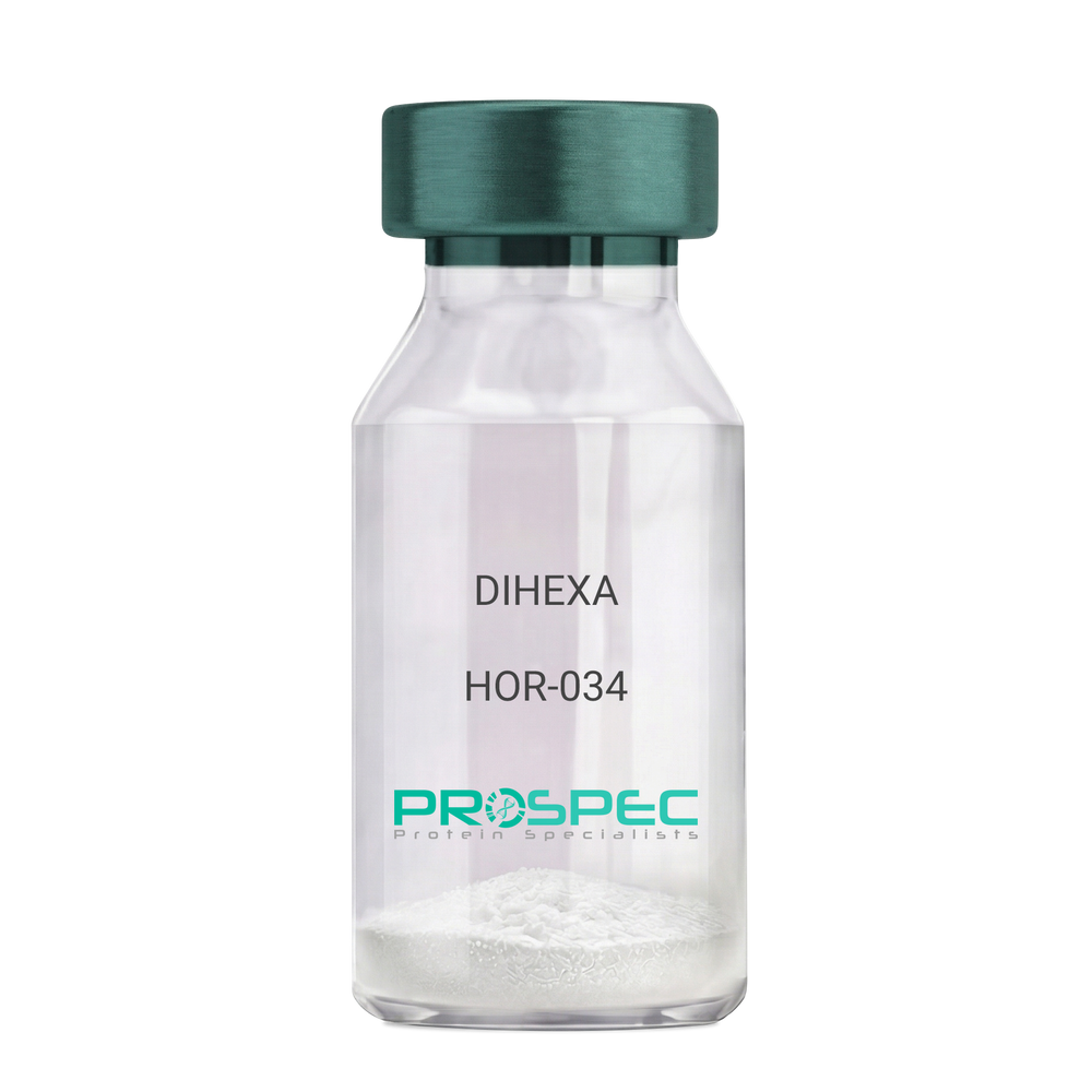
DIHEXADescription:
DIHEXA
Product # :
HOR-034Price :
Quantity :
Shipping Method :
Shipped at Room temp
More Info
- description
- formulation
- purity
- More Info
Description
DIHEXA Synthetic is a single, non-glycosylated polypeptide chain containing 3 amino acids, having a molecular mass of 504.28 Dalton and a Molecular formula of C27H44N4O5.
Formulation
The protein was lyophilized with no additives.
Purity
Greater than 97.0% as determined by analysis by RP-HPLC.
More Info
-
Physical Appearance
Sterile Filtered White lyophilized (freeze-dried) powder.
-
Stability
Lyophilized DIHEXA although stable at room temperature for 3 weeks, should be stored desiccated below -18°C. Upon reconstitution DIHEXA should be stored at 4°C between 2-7 days and for future use below -18°C. For long term storage it is recommended to add a carrier protein (0.1% HSA or BSA). Please prevent freeze-thaw cycles.
-
Solubility
It is recommended to reconstitute the lyophilized DIHEXA in sterile 18MΩ-cm H2O not less than 100 µg/ml, which can then be further diluted to other aqueous solutions.
-
Amino Acid Sequence
Hexanoyl-Tyr-Ile-Ahx-NH2.
-
Background
Dihexa, also known as N-hexanoic-Tyr-Ile-(6) aminohexanoic amide, is a potent and orally active small peptide that has been the focus of significant research due to its potential neurogenic and neuroprotective effects. This compound, derived from angiotensin IV, has been shown to possess a wide range of biological activities, including enhancing cognitive function, promoting neurogenesis, and potentially mitigating the effects of neurodegenerative diseases.
Dihexa's primary mechanism of action involves its interaction with hepatocyte growth factor (HGF) and its receptor, c-Met. By mimicking the effects of HGF, Dihexa can stimulate the c-Met receptor, leading to a cascade of events that promote neurogenesis and synaptic plasticity. Studies by Benoist et al. (2014) have demonstrated that Dihexa can enhance cognitive function in rats, suggesting potential applications in cognitive enhancement and the treatment of cognitive disorders.
In addition to its neurogenic effects, Dihexa has been shown to possess neuroprotective properties. Research by Kawas et al. (2017) found that Dihexa could protect neurons from apoptosis, suggesting potential applications in the treatment of neurodegenerative diseases such as Alzheimer's and Parkinson's disease.
Given its neurogenic and neuroprotective effects, Dihexa has been proposed as a potential therapeutic agent for a variety of conditions, including cognitive disorders, neurodegenerative diseases, and stroke. For instance, a study by Harding et al. (2018) found that Dihexa could improve outcomes in animal models of stroke, indicating its potential as a therapeutic agent in stroke recovery.
While research on Dihexa is promising, it is important to note that most studies have been conducted in animals or in vitro. More research is needed to fully understand the potential effects and applications of Dihexa in humans. However, the existing body of research suggests that Dihexa could be a promising tool in the treatment of cognitive disorders and neurodegenerative diseases.
ProSpec's products are furnished for LABORATORY RESEARCH USE ONLY. The product may not be used as drugs, agricultural or pesticidal products, food additives or household chemicals.
- View Data Sheet
Name :
SNAP25 C.elegansDescription:
Synaptosomal-associated protein 25kDa C.elegans Recombinant
Super-Protein, SUP, RIC4, SEC9, SNAP, RIC-4, SNAP25, SNAP-25, Synaptosomal-associated protein 25, Synaptosomal-associated 25 kDa protein, FLJ23079, bA416N4.2, dJ1068F16.2.
Product # :
PRO-396Price :
Quantity :
Shipping Method :
Shipped with Ice Packs
More Info
- description
- source
- formulation
- purity
- More Info
Description
Recombinant C.elegans SNAP-25 produced in E.Coli is a single, non-glycosylated polypeptide chain containing 207 amino acids and having a molecular mass of 23kDa. SNAP-25 gene was amplified by PCR from C.elegans and cloned into an E. coli expression vector. SNAP-25 was purified by using conventional chromatography techniques.
Source
Escherichia Coli.
Formulation
The protein contains 20mM Tris-HCl pH7.5, 50mM NaCl, 5mM DTT, 1mM EDTA and 10% Glycerol.
Purity
Greater than 95.0% as determined by SDS-PAGE.
More Info
-
Introduction
Synaptic vesicle membrane docking and fusion is mediated by SNAREs (soluble N-ethylmaleimide-sensitive factor attachment protein receptors) located on the vesicle membrane (v-SNAREs) and the target membrane (t-SNAREs). The assembled v-SNARE/t-SNARE complex consists of a bundle of four helices, one of which is supplied by v-SNARE and the other three by t-SNARE. For t-SNAREs on the plasma membrane, the protein syntaxin supplies one helix and the protein encoded by this gene contributes the other two. Therefore, SNAP25 product is a presynaptic plasma membrane protein involved in the regulation of neurotransmitter release. The synaptosomal-associated protein (SNAP-25) is an essential component of the core complex that mediates presynaptic vesicle trafficking. Thus, SNAP-25 is directly involved in the release of neurotransmitters.
-
Synonyms
Super-Protein, SUP, RIC4, SEC9, SNAP, RIC-4, SNAP25, SNAP-25, Synaptosomal-associated protein 25, Synaptosomal-associated 25 kDa protein, FLJ23079, bA416N4.2, dJ1068F16.2.
-
Physical Appearance
Sterile filtered colorless solution.
-
Stability
Store at 4°C if entire vial will be used within 2-4 weeks. Store, frozen at -20°C for longer periods of time. For long term storage it is recommended to add a carrier protein (0.1% HSA or BSA). Avoid multiple freeze-thaw cycles.
-
Amino Acid Sequence
MSGDDDIPEG LEAINLKMNA TTDDSLESTR RMLALCEESK EAGIKTLVML DDQGEQLERCEGALDTINQD MKEAEDHLKG MEKCCGLCVL PWNKTDDFEK TEFAKAWKKD DDGGVISDQPRITVGDSSMG PQGGYITKIT NDAREDEMDE NVQQVSTMVG NLRNMAIDMS TEVSNQNRQL DRIHDKAQSN EVRVESANKR AKNLITK.
ProSpec's products are furnished for LABORATORY RESEARCH USE ONLY. The product may not be used as drugs, agricultural or pesticidal products, food additives or household chemicals.
- View Data Sheet
Name :
KAAG1 HumanDescription:
Kidney Associated Antigen 1 Human Recombinant
Kidney-associated antigen 1, RU2 antisense gene protein, KAAG1, RU2AS, MGC78738.
Product # :
PRO-192Price :
Quantity :
Shipping Method :
Shipped with Ice Packs
More Info
- description
- source
- formulation
- purity
- More Info
Description
KAAG1 Human Recombinant fused with a 20 amino acid His tag at N-terminus produced in E.Coli is a single, non-glycosylated, polypeptide chain containing 104 amino acids (1-84 a.a.) and having a molecular mass of 11.1kDa. The KAAG1 is purified by proprietary chromatographic techniques.
Source
Escherichia Coli.
Formulation
The KAAG1 solution (0.5 mg/ml) contains 20mM Tris-HCl buffer (pH 8.0), 1mM DTT, 10% glycerol and 100mM NaCl.
Purity
Greater than 90.0% as determined by SDS-PAGE.
More Info
-
Introduction
Kidney associated antigen 1 (KAAG1) is expressed in testis and kidney, and, at lower levels, in urinary bladder and liver. KAAG1 is expressed by numerous tumors of various histologic origin, including melanomas, sarcomas and colorectal carcinomas.
-
Synonyms
Kidney-associated antigen 1, RU2 antisense gene protein, KAAG1, RU2AS, MGC78738.
-
Physical Appearance
Sterile Filtered colorless solution.
-
Stability
Store at 4°C if entire vial will be used within 2-4 weeks. Store, frozen at -20°C for longer periods of time. For long term storage it is recommended to add a carrier protein (0.1% HSA or BSA).Avoid multiple freeze-thaw cycles.
-
Amino Acid Sequence
MGSSHHHHHH SSGLVPRGSH MDDDAAPRVE GVPVAVHKHA LHDGLRQVAG PGAAAAHLPR WPPPQLAASR REAPPLSQRP HRTQGAGSPP ETNEKLTNPQ VKEK.
ProSpec's products are furnished for LABORATORY RESEARCH USE ONLY. The product may not be used as drugs, agricultural or pesticidal products, food additives or household chemicals.