Search results
1000 results found for “GRO (CXCL1,2,3)”
Name
Description
Product #
Price
Quantity
Shipping Method
- View Data Sheet
Name :
IL 1RA AntibodyDescription:
Interleukin-1 Receptor Antagonist, Mouse Anti-Human
IRAP, IL1F3, IL1RA, IL-1ra3, ICIL-1RA, IL1RN, IL1 inhibitor, IL-1ra, MGC10430.
Product # :
ANT-238Price :
Quantity :
Shipping Method :
Shipped at Room temp
More Info
- formulation
- More Info
Formulation
1mg/ml in PBS (after reconstitution)
More Info
-
Introduction
Interleukin-1 ra is a member of the interleukin 1 cytokine family. This protein inhibits the activities of interleukin 1, alpha (IL1A) and interleukin 1, beta (IL1B), and modulates a variety of interleukin 1 related immune and inflammatory responses. This gene and five other closely related cytokine genes form a gene cluster spanning approximately 400 kb on chromosome 2. A polymorphism of this gene is reported to be associated with increased risk of osteoporotic fractures and gastric cancer. Four alternatively spliced transcript variants encoding distinct isoforms have been reported.
-
Synonyms
IRAP, IL1F3, IL1RA, IL-1ra3, ICIL-1RA, IL1RN, IL1 inhibitor, IL-1ra, MGC10430.
-
Solubility
Reconstitute with sterile H20. Mix gently, wash the sides of the vial and wait 30-60 seconds before use.
-
Immunogen
r.Human IL-1RA.
-
Ig Subclass
Mouse IgG2b.
-
Clone
NYR-hIL1RA.
-
Applications
Direct ELISA, Western Blot, Immuneprecipitation.
-
Titer
By direct ELISA, 1:10,000 dilution will yield 0.8 O.D using alkaline phosphatase conjugated rabbit anti-mouse Ig (Jackson Laboratories).
-
Shipping Conditions
Antibody is shipped lyophilized at ambient temperature.
-
Type
Mouse Anti Human Monoclonal.
-
Storage Procedures
In lyophilized form, for long periods, store at 4oC in a dry environment. After reconstitution, if not intended for use within a month, aliquot and store at -20oC.
-
Purification Method
Ion exchange.
ProSpec's products are furnished for LABORATORY RESEARCH USE ONLY. The product may not be used as drugs, agricultural or pesticidal products, food additives or household chemicals.
- View Data Sheet
Name :
sCD23 HumanDescription:
Soluble CD23 Human Recombinant
Low affinity immunoglobulin epsilon Fc receptor, Lymphocyte IgE receptor, Fc-epsilon-RII, BLAST-2, Immunoglobulin E-binding factor, CD23 antigen, FCER2, CD23, FCE2, CD23A, IGEBF, CLEC4J.
Product # :
PRO-1789Price :
Quantity :
Shipping Method :
Shipped at Room temp
More Info
- description
- source
- formulation
- purity
- biological activity
- More Info
Description
sCD23 Human Recombinant produced in E.Coli is a single, non-glycosylated, polypeptide chain containing 172 amino acids and having a molecular mass of 19.2kDa.The sCD23 is purified by proprietary chromatographic techniques.
Source
Escherichia Coli.
Formulation
Lyophilized from a 0.2µm filtered concentrated solution in PBS, pH 7.4.
Purity
Greater than 97.0% as determined by:
(a) Analysis by RP-HPLC.
(b) Analysis by SDS-PAGE.Biological Activity
The ED50 is determined by its ability to induce TNF-alpha production by human PBMCs.More Info
-
Introduction
CD23 is a 45kDa glycoprotein, which is present on a subpopulation of freshly isolated peripheral blood and tonsil B cells and strongly expressed on EBV-transformed B lymphoblasts. The CD23 molecule is identical to the low affinity IgE receptor found on B cells. Expression of CD23 has been detected in neoplastic cells from cases of B cell chronic lymphocyctic leukaemia and some cases of centroblastic/centrocytic lymphoma.
-
Synonyms
Low affinity immunoglobulin epsilon Fc receptor, Lymphocyte IgE receptor, Fc-epsilon-RII, BLAST-2, Immunoglobulin E-binding factor, CD23 antigen, FCER2, CD23, FCE2, CD23A, IGEBF, CLEC4J.
-
Physical Appearance
Sterile Filtered White lyophilized (freeze-dried) powder.
-
Stability
Lyophilized sCD23 although stable at room temperature for 3 weeks, should be stored desiccated below -18°C. Upon reconstitution sCD23 should be stored at 4°C between 2-7 days and for future use below -18°C. For long term storage it is recommended to add a carrier protein (0.1% HSA or BSA). Please prevent freeze-thaw cycles.
-
Solubility
It is recommended to reconstitute the lyophilized sCD23 in sterile 18M-cm H2O not less than 100µg/ml, which can then be further diluted to other aqueous solutions.
-
Amino Acid Sequence
MELQVSSGFV CNTCPEKWIN FQRKCYYFGK GTKQWVHARY ACDDMEGQLV SIHSPEEQDF LTKHASHTGS WIGLRNLDLK GEFIWVDGSH VDYSNWAPGE PTSRSQGEDC VMMRGSGRWN DAFCDRKLGA WVCDRLATCT PPASEGSAES MGPDSRPDPD GRLPTPSAPL HS
ProSpec's products are furnished for LABORATORY RESEARCH USE ONLY. The product may not be used as drugs, agricultural or pesticidal products, food additives or household chemicals.
- View Data Sheet
Name :
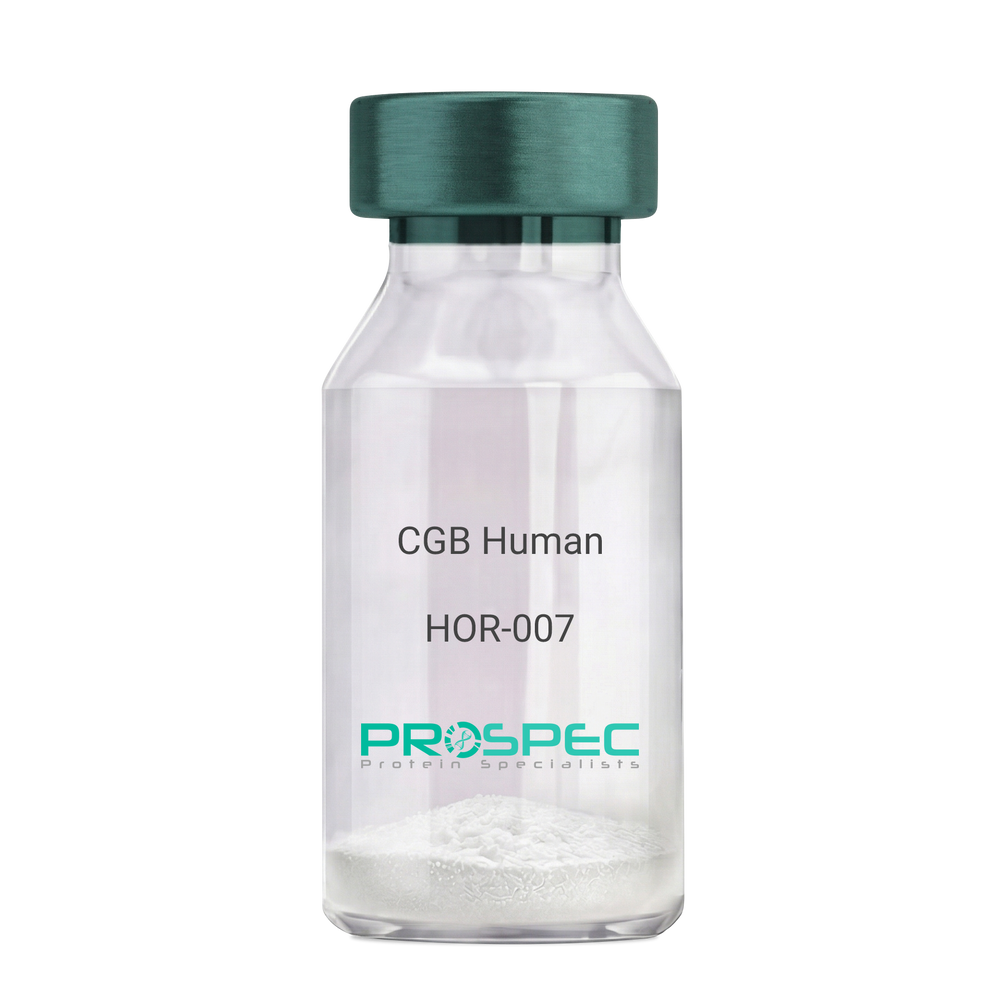
CGB HumanDescription:
CGB Human Recombinant
CGB3, CGB5, CGB7, CGB8, hCGB, CG-beta, CGB.
Product # :
HOR-007Price :
Quantity :
Shipping Method :
Shipped with Ice Packs
More Info
- description
- source
- formulation
- purity
- More Info
Description
CGB Human Recombinant produced in E.Coli is a single, non-glycosylated polypeptide chain containing 168 amino acids (21-165 a.a.) and having a molecular mass of 17.9kDa. CGB is fused to a 23 amino acid His-tag at N-terminus & purified by proprietary chromatographic techniques.
Source
Escherichia Coli.
Formulation
CGB protein solution (0.5mg/ml) containing 20mM Tris-HCl buffer (pH 8.0), 0.4M Urea and 10% glycerol.
Purity
Greater than 85.0% as determined by SDS-PAGE.
More Info
-
Introduction
Glycoprotein hormones are heterodimers consisting of a common alpha subunit and a unique beta subunit which confers biological specificity. CGB is a member of the glycoprotein hormone beta chain family and is the beta 3 subunit of (CG). CG is produced by the trophoblastic cells of the placenta and stimulates the ovaries. The beta subunit of CG is encoded by 6 genes which are arranged in tandem and inverted pairs on chromosome 19q13.3 and contiguous with the luteinizing hormone beta subunit gene.
-
Synonyms
CGB3, CGB5, CGB7, CGB8, hCGB, CG-beta, CGB.
-
Physical Appearance
Sterile filtered colorless solution.
-
Stability
Store at 4°C if entire vial will be used within 2-4 weeks. Store, frozen at -20°C for longer periods of time. For long term storage it is recommended to add a carrier protein (0.1% HSA or BSA).Avoid multiple freeze-thaw cycles.
-
Amino Acid Sequence
MGSSHHHHHH SSGLVPRGSH MGSSKEPLRP RCRPINATLA VEKEGCPVCI TVNTTICAGY CPTMTRVLQG VLPALPQVVC NYRDVRFESI RLPGCPRGVN PVVSYAVALS CQCALCRRST TDCGGPKDHP LTCDDPRFQD SSSSKAPPPS LPSPSRLPGP SDTPILPQ.
ProSpec's products are furnished for LABORATORY RESEARCH USE ONLY. The product may not be used as drugs, agricultural or pesticidal products, food additives or household chemicals.
- View Data Sheet
Name :
CHGA AntibodyDescription:
Chromogranin-A, Mouse Anti Human
CGA, CHGA, Vasostatin-2, Pituitary secretory protein I, SP-I.
Product # :
ANT-351Price :
Quantity :
Shipping Method :
Shipped with Ice Packs
More Info
- formulation
- More Info
Formulation
1mg/ml containing PBS, pH-7.4, & 0.1% Sodium Azide.
More Info
-
Introduction
Chromgranin-A is part of the neuroendocrine secretory protein family. CHGA is located in secretory vesicles of neurons and endocrine cells. Chromgranin-A is a precursor to three biologically active peptides; vasostatin, pancreastatin, and parastatin. These peptides act as autocrine or paracrine negative modulators of the neuroendocrine system. Other peptides, including chromostatin, beta-granin, WE-14 and GE-25, are also derived from the full-length protein. Chromgranin-A has numerous biological activities on some tissues and organs and exerts a large spectrum of homeostatic actions, including antifungal and antimicrobial effect, modulation of cell adhesion, and inhibition of parathyroid hormone secretion.
-
Synonyms
CGA, CHGA, Vasostatin-2, Pituitary secretory protein I, SP-I.
-
Physical Appearance
Sterile Filtered colorless solution.
-
Immunogen
Anti-human CHGA mAb is derived from hybridization of mouse F0 myeloma cells with spleen cells from BALB/c mice immunized with recombinant human CHGA amino acids 19-131 purified from E. coli.
-
Ig Subclass
Mouse IgG1 heavy chain and κ light chain.
-
Clone
P3A5AT.
-
Applications
CHGA antibody has been tested by ELISA, Western blot and immunohistochemistry analysis to assure specificity and reactivity. Since application varies, however, each investigation should be titrated by the reagent to obtain optimal results. Recommended dilution range for Western blot is 1:500 ~ 1:1,000 and immunohistochemistry analysis is 1:100~200. Recommended starting dilution for Western blot is 1:500 and Immunohistochemistry is 1:100.
-
Type
Mouse Anti Human Monoclonal.
-
Storage Procedures
For periods up to 1 month store at 4°C, for longer periods of time, store at -20°C. Prevent freeze thaw cycles.
-
Purification Method
CHGA antibody was purified from mouse ascitic fluids by protein-G affinity chromatography.
ProSpec's products are furnished for LABORATORY RESEARCH USE ONLY. The product may not be used as drugs, agricultural or pesticidal products, food additives or household chemicals.
- View Data Sheet
Name :
CCN1 HumanDescription:
Cysteine-Rich Angiogenic Inducer 61 Human Recombinant
CYR61, Protein CYR61, Cysteine-rich angiogenic inducer 61, IGF-binding protein 10, IGFBP-10, IBP-10, Protein GIG1, CCN family member 1, CCN1, GIG1, IGFBP10.
Product # :
CYT-164Price :
Quantity :
Shipping Method :
Shipped at Room temp
More Info
- description
- source
- formulation
- purity
- biological activity
- More Info
Description
CYR61 Human Recombinant produced in E.Coli is a single, non-glycosylated, polypeptide chain containing 357 amino acids and having a molecular mass of 39.5kDa.The CYR61 is purified by proprietary chromatographic techniques.
Source
Escherichia Coli.
Formulation
Lyophilized from a 0.2m filtered concentrated solution in PBS, pH 7.4.
Purity
Greater than 95.0% as determined by SDS-PAGE.
Biological Activity
The ED50 was determined by the proliferation of mouse 3T3 cells is < 2.0 ug/ml, corresponding to a specific activity of > 500 units/mg.More Info
-
Introduction
CYR61 is a growth factor-inducible, immediate-early gene that has multifaceted activities in various cancers. CYR61 is a secreted, cysteine-rich, binding protein which is encoded by a growth factor-inducible immediate-early gene. Acting as an extracellular, matrix-associated signaling molecule, CYR61 promotes the adhesion of endothelial cells through interaction with integrin and enhances growth factor-induced DNA synthesis in the same cell type.
-
Synonyms
CYR61, Protein CYR61, Cysteine-rich angiogenic inducer 61, IGF-binding protein 10, IGFBP-10, IBP-10, Protein GIG1, CCN family member 1, CCN1, GIG1, IGFBP10.
-
Physical Appearance
Sterile Filtered White lyophilized (freeze-dried) powder.
-
Stability
Lyophilized CYR61 Human although stable at room temperature for 3 weeks, should be stored desiccated below -18°C. Upon reconstitution CYR61 should be stored at 4°C between 2-7 days and for future use below -18°C.Please prevent freeze-thaw cycles.
-
Solubility
It is recommended to reconstitute the lyophilized CYR61 in sterile 18M-cm H2O not less than 100µg/ml, which can then be further diluted to other aqueous solutions.
-
Amino Acid Sequence
TCPAACHCPL EAPKCAPGVG LVRDGCGCCK VCAKQLNEDC SKTQPCDHTK GLECNFGASS TALKGICRAQ SEGRPCEYNS RIYQNGESFQ PNCKHQCTCI DGAVGCIPLC PQELSLPNLG CPNPRLVKVT GQCCEEWVCD EDSIKDPMED QDGLLGKELG FDASEVELTR NNELIAVGKG SSLKRLPVFG MEPRILYNPL QGQKCIVQTT SWSQCSKTCG TGISTRVTND NPECRLVKET RICEVRPCGQ PVYSSLKKGK KCSKTKKSPE PVRFTYAGCL SVKKYRPKYC GSCVDGRCCT
PQLTRTVKMR FRCEDGETFS KNVMMIQSCK CNYNCPHANE AAFPFYRLFN DIHKFRD -
Background
Title: Cysteine-Rich Angiogenic Inducer 61 Human Recombinant: A Potential Regulator of Angiogenesis
Abstract:
Cysteine-rich angiogenic inducer 61 (CYR61) is an important extracellular matrix-associated protein that plays a significant role in angiogenesis and cell adhesion. This research paper provides a comprehensive analysis of human recombinant CYR61, focusing on its production, characterization, and potential applications in regulating angiogenesis. The paper discusses the significance of CYR61 in physiological and pathological angiogenesis, including wound healing, tumor development, and cardiovascular diseases. Furthermore, it explores ongoing research and clinical trials investigating the therapeutic potential of recombinant CYR61 in various pathological conditions. The information presented in this paper aims to enhance our understanding of human recombinant CYR61 and its utility as a research tool and a potential regulator of angiogenesis.Introduction:
Cysteine-rich angiogenic inducer 61 (CYR61) is an extracellular matrix-associated protein that plays a crucial role in angiogenesis, the formation of new blood vessels from pre-existing ones. Human recombinant CYR61, produced through genetic engineering techniques, provides researchers with a valuable tool to study its biological functions and explore its therapeutic potential.Production and Characterization:
Recombinant CYR61 is typically generated using expression systems such as mammalian cells or bacteria. The protein is then purified and characterized to ensure its structural integrity and functional activity. Quality control measures are implemented to confirm the specificity and bioactivity of the recombinant CYR61.Role in Angiogenesis:
CYR61 is involved in various aspects of angiogenesis, including endothelial cell proliferation, migration, and tube formation. It interacts with integrins and other cell surface receptors to modulate signaling pathways involved in angiogenic processes. Recombinant CYR61 serves as a valuable tool for studying the mechanisms underlying angiogenesis and exploring its potential as a therapeutic target.Therapeutic Implications:
The dysregulation of angiogenesis is associated with several pathological conditions, including cancer, cardiovascular diseases, and chronic wounds. Recombinant CYR61 has shown promise as a potential regulator of angiogenesis and a therapeutic agent. It can be used to promote or inhibit angiogenesis, depending on the specific context. Ongoing research and clinical trials are investigating the therapeutic applications of recombinant CYR61 in various diseases, including cancer and ischemic disorders.Conclusion:
Human recombinant CYR61 is a valuable research tool and a potential regulator of angiogenesis. Its production, characterization, and applications in modulating angiogenic processes contribute to our understanding of angiogenesis and the development of novel therapeutic interventions. Continued research and clinical trials exploring the therapeutic potential of recombinant CYR61 offer promising prospects for improving outcomes in cancer, cardiovascular diseases, and wound healing.What is the molecular weight/Mw of CCN1 Protein?
CCN1 Protein has a total Mw of 39.5kDa.
What is the source or expression system of CCN1 Protein?
Escherichia Coli.
What is the Purity of CCN1 Protein?
CCN1 Protein is >95% pure as determined by SDS-PAGE.
What is the Biological Activity of CCN1 Protein?
The ED50 was determined by the proliferation of mouse 3T3 cells is < 2.0 ug/ml, corresponding to a specific activity of > 500 units/mg.
What is the amino acid sequence of CCN1 Protein?
TCPAACHCPL EAPKCAPGVG LVRDGCGCCK VCAKQLNEDC SKTQPCDHTK GLECNFGASS TALKGICRAQ SEGRPCEYNS RIYQNGESFQ PNCKHQCTCI DGAVGCIPLC PQELSLPNLG CPNPRLVKVT GQCCEEWVCD EDSIKDPMED QDGLLGKELG FDASEVELTR NNELIAVGKG SSLKRLPVFG MEPRILYNPL QGQKCIVQTT SWSQCSKTCG TGISTRVTND NPECRLVKET RICEVRPCGQ PVYSSLKKGK KCSKTKKSPE PVRFTYAGCL SVKKYRPKYC GSCVDGRCCT
PQLTRTVKMR FRCEDGETFS KNVMMIQSCK CNYNCPHANE AAFPFYRLFN DIHKFRD
What applications can CCN1 Protein be used in?
CCN1 Protein can probably be used in western blot, ELISA and Lateral Flow.
What is the endotoxin level for CCN1 Protein?
The endotoxin level is minimal, CCN1 Protein was purified using conventional chromatography techniques.
ProSpec's products are furnished for LABORATORY RESEARCH USE ONLY. The product may not be used as drugs, agricultural or pesticidal products, food additives or household chemicals.
- View Data Sheet
Name :
MIP 3a MouseDescription:
Macrophage Inflammatory Protein-3 alpha Mouse Recombinant (CCL20)
Small inducible cytokine A20 precursor, CCL20, Macrophage inflammatory protein 3 alpha, MIP-3-alpha, Liver and activation- regulated chemokine, CC chemokine LARC, Beta chemokine exodus-1, CKb4, LARC, ST38, MIP3A, MIP-3a, SCYA20, C-C motif chemokine 20, CC chemokine ST38, exodus-1.
Product # :
CHM-236Price :
Quantity :
Shipping Method :
Shipped at Room temp
More Info
- description
- source
- formulation
- purity
- biological activity
- More Info
Description
Macrophage Inflammatory Protein-3 alpha Mouse Recombinant produced in E.Coli is a single, non-glycosylated, polypeptide chain containing 70 amino acids and having a molecular mass of 7.9 kDa. The MIP-3a is purified by proprietary chromatographic techniques.
Source
Escherichia Coli.
Formulation
Lyophilized from a concentrated (1mg/ml) solution in water containing no additives.
Purity
Greater than 95.0% as determined by(a) Analysis by RP-HPLC.
(b) Analysis by SDS-PAGE.Biological Activity
The Activity is calculated by its ability to chemoattract primary human T cells at 2-20ng/ml, corresponding to a specific activity of 50,000-500,000 units/mg.
More Info
-
Introduction
CCL20 is a chemotactic factor that draws lymphocytes & neutrophils, rather than monocytes. MIP-3 alpha inhibits proliferation of myeloid progenitors in colony formation assays. MIP3A plays a role in the formation and function of the mucosal lymphoid tissues by attracting lymphocytes and dendritic cells towards epithelial cells. C-terminal processed forms have been shown to be equally chemotactically active for leukocytes. CCL20 holdes antibacterial activity e.coli atcc 25922 and s.aureus atcc 29213. CCL20 gene transcription is activated by H. pylori, which activates NF-kappaB through intracellular signal pathway which involves IkappaB kinase and NF-kappaB-inducing kinase. MIP-3 alpha is invloved in chemokine-mediated lymphocyte trafficking during gastric inflammation in Helicobacter infection. CCL20 expression is involved in the recruitment of CD45R0-positive T cell subsets into the intestinal lamina propria.
MIP-3A is in charge of the advancement of pulpal inflammation through the recruitment of C-C motif Receptor 6-expressing lymphocytes Vaginal epithelial cells respond to factors present in semen by secreting MIP-3 alpha, which increases langerhans cells recruitment during HIV transmission. -
Synonyms
Small inducible cytokine A20 precursor, CCL20, Macrophage inflammatory protein 3 alpha, MIP-3-alpha, Liver and activation- regulated chemokine, CC chemokine LARC, Beta chemokine exodus-1, CKb4, LARC, ST38, MIP3A, MIP-3a, SCYA20, C-C motif chemokine 20, CC chemokine ST38, exodus-1.
-
Physical Appearance
Sterile Filtered White lyophilized (freeze-dried) powder.
-
Stability
Lyophilized MIP-3a although stable at room temperature for 3 weeks, should be stored desiccated below -18°C. Upon reconstitution CCL-20 should be stored at 4°C between 2-7 days and for future use below -18°C.For long term storage it is recommended to add a carrier protein (0.1% HSA or BSA).Please prevent freeze-thaw cycles.
-
Solubility
It is recommended to reconstitute the lyophilized MIP-3a in sterile 18MΩ-cm H2O not less than 100µg/ml, which can then be further diluted to other aqueous solutions.
-
Amino Acid Sequence
ASNYDCCLSY IQTPLPSRAI VGFTRQMADE ACDINAIIFH TKKRKSVCAD PKQNWVKRAV NLLSLRVKKM.
ProSpec's products are furnished for LABORATORY RESEARCH USE ONLY. The product may not be used as drugs, agricultural or pesticidal products, food additives or household chemicals.
- View Data Sheet
Name :
CoV-2 S1 (16-685)Description:
Coronavirus 2019 Spike Glycoprotein-S1 (16-685 a.a.), Recombinant
Spike glycoprotein, S glycoprotein, E2, Peplomer protein, covid19, COVID-19, COVID-19 virus, HCoV-19, Human coronavirus 2019, SARS2, Spike protein S1, Severe acute respiratory syndrome coronavirus 2, 2019-nCoV, S, Peplomer protein
Product # :
SARS-026Price :
Quantity :
Shipping Method :
Shipped with Ice Packs
More Info
- description
- source
- formulation
- purity
- biological activity
- More Info
Description
The recombinant Coronavirus 2019 CoV-2 Spike Glycoprotein S1, Wuhan-Hu-1 strain containing a total of 680 amino acids (16-685) and having a calculated Mw of 76.2 kDa. CoV-2 S1 (16-685) is fused to a 6 amino acid His-tag at C-terminus,and is purified by proprietary chromatographic techniques.
Source
HEK293 Cells.
Formulation
The CoV-2 S1 (16-685) solution (0.25mg/ml) contains Phosphate-Buffered Saline (pH 7.4) and 10% Glycerol.
Purity
Greater than 85.0% as determined by SDS-PAGE.
Biological Activity
Measured by its binding ability in a functional ELISA with Human ACE-2 (enz-1125).
More Info
-
Introduction
A human infecting coronavirus (viral pneumonia) called 2019 novel coronavirus, 2019-nCoV was found in the fish market at the city of Wuhan, Hubei province of China on December 2019.
The 2019-nCoV shares an 87% identity to the 2 bat-derived severe acute respiratory syndrome 2018 SARS-CoV-2 located in Zhoushan of eastern China. 2019-nCoV has an analogous receptor-BD-structure to that of 2018 SARS-CoV, even though there is a.a. diversity so thus the 2019-nCoV might bind to ACE2 receptor protein (angiotensin-converting enzyme 2) in humans.
While bats are possibly the host of 2019-nCoV, researchers suspect that animal from the ocean sold at the seafood market was an intermediate host. RSCU analysis proposes that the 2019-nCoV is a recombinant within the viral spike glycoprotein between the bat coronavirus and an unknown coronavirus.
-
Synonyms
Spike glycoprotein, S glycoprotein, E2, Peplomer protein, covid19, COVID-19, COVID-19 virus, HCoV-19, Human coronavirus 2019, SARS2, Spike protein S1, Severe acute respiratory syndrome coronavirus 2, 2019-nCoV, S, Peplomer protein
-
Physical Appearance
Sterile Filtered White lyophilized (freeze-dried) powder.
-
Stability
Store at 4°C if entire vial will be used within 2-4 weeks. Store, frozen at -20°C for longer periods of time. For long term storage it is recommended to add a carrier protein (0.1% HSA or BSA). Avoid multiple freeze-thaw cycles.
-
Amino Acid Sequence
DGSMVNLTTR TQLPPAYTNS FTRGVYYPDK VFRSSVLHST QDLFLPFFSN VTWFHAIHVS GTNGTKRFDN PVLPFNDGVY FASTEKSNII RGWIFGTTLD SKTQSLLIVN NATNVVIKVC EFQFCNDPFL GVYYHKNNKS WMESEFRVYS SANNCTFEYV SQPFLMDLEG KQGNFKNLRE FVFKNIDGYF KIYSKHTPIN LVRDLPQGFS ALEPLVDLPI GINITRFQTL LALHRSYLTP GDSSSGWTAG AAAYYVGYLQ PRTFLLKYNE NGTITDAVDC ALDPLSETKC TLKSFTVEKG IYQTSNFRVQ PTESIVRFPN ITNLCPFGEV FNATRFASVY AWNRKRISNC VADYSVLYNS ASFSTFKCYG VSPTKLNDLC FTNVYADSFV IRGDEVRQIA PGQTGKIADY NYKLPDDFTG CVIAWNSNNL DSKVGGNYNY LYRLFRKSNL KPFERDISTE IYQAGSTPCN GVEGFNCYFP LQSYGFQPTN GVGYQPYRVV VLSFELLHAP ATVCGPKKST NLVKNKCVNF NFNGLTGTGV LTESNKKFLP FQQFGRDIAD TTDAVRDPQT LEILDITPCS FGGVSVITPG TNTSNQVAVL YQDVNCTEVP VAIHADQLTP TWRVYSTGSN VFQTRAGCLI GAEHVNNSYE CDIPIGAGIC ASYQTQTNSP RRARHHHHHH.
ProSpec's products are furnished for LABORATORY RESEARCH USE ONLY. The product may not be used as drugs, agricultural or pesticidal products, food additives or household chemicals.
- View Data Sheet
Name :
MIP 3 HumanDescription:
Macrophage Inflammatory Protein-3 Human Recombinant (CCL23)
C-C motif chemokine 23, Small-inducible cytokine A23, Macrophage inflammatory protein 3, Myeloid progenitor inhibitory factor 1, CK-beta-8, MIP-3, MPIF-1, CKB-8, CCL23, MIP3, MPIF1, SCYA23, CKb8, Ckb-8-1.
Product # :
CHM-358Price :
Quantity :
Shipping Method :
Shipped at Room temp
More Info
- description
- source
- formulation
- purity
- biological activity
- More Info
Description
MIP-3 Human Recombinant produced in E.Coli is a single, non-glycosylated, polypeptide chain containing 99 amino acids and having a molecular mass of 11.3kDa. The MIP-3 is purified by proprietary chromatographic techniques.
Source
Escherichia Coli.
Formulation
Filtered (0.2µm) and lyophilized from a concentrated (1mg/ml) solution in 20mM PB, pH 7.4, 150mM NaCl.
Purity
Greater than 97.0% as determined by:
(a) Analysis by RP-HPLC.
(b) Analysis by SDS-PAGE.Biological Activity
Determined by its ability to chemoattract human T cell population using a concentration range of 10-50ng/ml corresponding to a Specific Activity of 20,000-100,000IU/mg.More Info
-
Introduction
CCL23 (MIP-3) is a ligand for the CCR1chemokine receptor. CCL23 is one of several cytokine genes clustered on the q-arm of chromosome 17, in a locus containing several other CC chemokines. MIP-3 chemoattracts monocytes, resting T-lymphocytes and neutrophils, but not activated lymphocytes. Furthermore, it was shown that MIP-3 inhibits colony formation of bone marrow myeloid immature progenitors. MIP-3 is mainly expressed in lung and liver tissue, but can be also found in bone marrow and placenta, as well as in some cell lines of myeloid origin.
Alternative splicing of the CCL23 gene produces 2 mRNAs which encode a short (CK?8) and a long (CK?81) isoform of the MIP-3. CK?8 cDNA encodes a 120 amino acid residue precursor protein with a putative 21 a.a. residue signal peptide which is cleaved to generate a 99 a.a. residue mature CK?8 (a.a. 22-120). Further N-terminal processing of the 99 a.a. residue variant can produce a 75 a.a. residue CK?8 (a.a. 46-120) which is considerably more active than the 99 a.a. residue variant.
MIP-3 may be involved in the malignant progression of certain human cancer cells which overexpress ErbB2 through the transactivation of ErbB2 tyrosine kinase. MIP-3 may also be involved in angiogenesis via upregulation of matrix metalloproteinase MMP-2 expression. -
Synonyms
C-C motif chemokine 23, Small-inducible cytokine A23, Macrophage inflammatory protein 3, Myeloid progenitor inhibitory factor 1, CK-beta-8, MIP-3, MPIF-1, CKB-8, CCL23, MIP3, MPIF1, SCYA23, CKb8, Ckb-8-1.
-
Physical Appearance
Sterile Filtered White lyophilized (freeze-dried) powder.
-
Stability
Lyophilized MIP-3 although stable at room temperature for 3 weeks, should be stored desiccated below -18°C. Upon reconstitution CCL23 should be stored at 4°C between 2-7 days and for future use below -18°C.For long term storage it is recommended to add a carrier protein (0.1% HSA or BSA).Please prevent freeze-thaw cycles.
-
Solubility
It is recommended to reconstitute the lyophilized MIP-3 in sterile 18MΩ-cm H2O not less than 100µg/ml, which can then be further diluted to other aqueous solutions.
-
Amino Acid Sequence
RVTKDAETEF MMSKLPLENP VLLDRFHATS ADCCISYTPR SIPCSLLESYFETNSECSKP GVIFLTKKGR RFCANPSDKQ VQVCMRMLKL DTRIKTRKN.
ProSpec's products are furnished for LABORATORY RESEARCH USE ONLY. The product may not be used as drugs, agricultural or pesticidal products, food additives or household chemicals.
- View Data Sheet
Name :
MEMO1 AntibodyDescription:
Mediator of Cell Motility 1, Mouse Anti Human
Protein MEMO1, C21orf19-like protein, Hepatitis C virus NS5A-transactivated protein 7, HCV NS5A-transactivated protein 7, Mediator of ErbB2-driven cell motility 1, Mediator of cell motility 1, Memo-1, MEMO1, C2orf4, MEMO, NS5ATP7, CGI-27.
Product # :
ANT-090Price :
Quantity :
Shipping Method :
Shipped with Ice Packs
More Info
- formulation
- More Info
Formulation
1mg/ml containing PBS, pH-7.4, & 0.1% Sodium Azide.
More Info
-
Introduction
Mediator of ErbB2-driven cell motility 1 (MEMO1) is a member of the UPF0103 family. MEMO1 regulates cell migration by transmitting extracellular chemotactic signals to the microtubule cytoskeleton. Furthermore, MEMO1 controls the localization of APC and CLASP2 to the cell membrane, using the regulation of GSK3B activity. MEMO1 is essential for breast carcinoma cell migration, suggesting a key role in tumorigenesis. MEMO1 is also a mediator of ERBB2 signaling.
-
Synonyms
Protein MEMO1, C21orf19-like protein, Hepatitis C virus NS5A-transactivated protein 7, HCV NS5A-transactivated protein 7, Mediator of ErbB2-driven cell motility 1, Mediator of cell motility 1, Memo-1, MEMO1, C2orf4, MEMO, NS5ATP7, CGI-27.
-
Immunogen
Anti-human MEMO1 mAb, is derived from hybridization of mouse FO myeloma cells with spleen cells from BALB/c mice immunized with recombinant human MEMO1 amino acids 1-297 purified from E. coli.
-
Ig Subclass
Mouse IgG1 heavy chain and kappa light chain.
-
Clone
P1E9AT.
-
Type
Mouse Anti Human Monoclonal.
-
Storage Procedures
For periods up to 1 month store at 4°C, for longer periods of time, store at -20°C. Prevent freeze thaw cycles.
-
Purification Method
MEMO1 antibody was purified from mouse ascitic fluids by protein-G affinity chromatography.
ProSpec's products are furnished for LABORATORY RESEARCH USE ONLY. The product may not be used as drugs, agricultural or pesticidal products, food additives or household chemicals.
- View Data Sheet
Name :
Flt3 Ligand Mouse, Sf9Description:
Flt3 Ligand Mouse Recombinant, Sf9
Fms-related tyrosine kinase 3 ligand, Flt3 ligand, Flt3L, SL cytokine, lt3lgF, Flt3l.
Product # :
CYT-910Price :
Quantity :
Shipping Method :
Shipped with Ice Packs
More Info
- description
- source
- formulation
- purity
- More Info
Description
Flt3 Ligand produced in Sf9 Baculovirus cells is a single, glycosylated polypeptide chain (27-189 a.a.) and fused to a 6 aa His Tag at C-terminus containing a total of 169 amino acids and having a molecular mass of 19.3kDa.Flt3 Ligand shows multiple bands between 18-28kDa on SDS-PAGE, reducing conditions and purified by proprietary chromatographic techniques.
Source
Sf9, Baculovirus cells.
Formulation
Flt3 Ligand protein solution (0.5mg/ml) contains Phosphate buffered saline (pH7.4) and 10% glycerol.
Purity
Greater than 85.0% as determined by SDS-PAGE.
More Info
-
Introduction
FLT3 ligand is a receptor for the fl cytokine has a tyrosine-protein kinase activity & a growth factor that regulates proliferation of early hematopoietic cells. Flt3-Ligand synergizes with other CSFs and interleukins to induce growth and differentiation.
-
Synonyms
Fms-related tyrosine kinase 3 ligand, Flt3 ligand, Flt3L, SL cytokine, lt3lgF, Flt3l.
-
Physical Appearance
Sterile filtered colorless solution.
-
Stability
Store at 4°C if entire vial will be used within 2-4 weeks. Store, frozen at -20°C for longer periods of time. For long term storage it is recommended to add a carrier protein (0.1% HSA or BSA).Avoid multiple freeze-thaw cycles.
-
Amino Acid Sequence
GTPDCYFSHS PISSNFKVKF RELTDHLLKD YPVTVAVNLQ DEKHCKALWS LFLAQRWIEQ LKTVAGSKMQ TLLEDVNTEI HFVTSCTFQP LPECLRFVQT NISHLLKDTC TQLLALKPCI GKACQNFSRC LEVQCQPDSS TLLPPRSPIA LEATELPEPR PRQHHHHHH
-
Background
What is the molecular weight/Mw of FLT3 LIGAND MOUSE, SF9 Protein?
FLT3 LIGAND MOUSE, SF9 Protein has a total Mw of 19.3kDa.
What is the source or expression system of FLT3 LIGAND MOUSE, SF9 Protein?
Sf9, Baculovirus cells.
What is the Purity of FLT3 LIGAND MOUSE, SF9 Protein?
FLT3 LIGAND MOUSE, SF9 Protein is >85% pure as determined by SDS-PAGE.
What is the Biological Activity of FLT3 LIGAND MOUSE, SF9 Protein?
The biological functionality of FLT3 LIGAND MOUSE, SF9 Protein will be determined in the future.
What is the amino acid sequence of FLT3 LIGAND MOUSE, SF9 Protein?
GTPDCYFSHS PISSNFKVKF RELTDHLLKD YPVTVAVNLQ DEKHCKALWS LFLAQRWIEQ LKTVAGSKMQ TLLEDVNTEI HFVTSCTFQP LPECLRFVQT NISHLLKDTC TQLLALKPCI GKACQNFSRC LEVQCQPDSS TLLPPRSPIA LEATELPEPR PRQHHHHHH.
What applications can FLT3 LIGAND MOUSE, SF9 Protein be used in?
FLT3 LIGAND MOUSE, SF9 Protein can probably be used in western blot, ELISA and Lateral Flow.
What is the endotoxin level for FLT3 LIGAND MOUSE, SF9 Protein?
The endotoxin level is minimal, FLT3 LIGAND MOUSE, SF9 Protein was purified using conventional chromatography techniques.
ProSpec's products are furnished for LABORATORY RESEARCH USE ONLY. The product may not be used as drugs, agricultural or pesticidal products, food additives or household chemicals.
- View Data Sheet
Name :
CoV-2-S1 (319-541), FcDescription:
Coronavirus 2019 Spike Glycoprotein-S1 Receptor Binding Domain (319-541 a.a), Fc Recombinant
Product # :
SARS-029Price :
Quantity :
Shipping Method :
Shipped at Room temp
More Info
- description
- source
- formulation
- purity
- More Info
Description
The HEK293 derived recombinant protein contains the Coronavirus 2019 Spike Glycoprotein S1 Receptor Binding Domain [ RBD ], Wuhan-Hu-1 strain, amino acids 319-541 fused to Fc tag at C-terminal.
Source
HEK293 Cells.
Formulation
CoV-S1 RBD protein is lyophilized from 1x PBS pH-7.4 + 5% trehalose.
Purity
Protein is >95% pure as determined SDS-PAGE.
More Info
-
Introduction
A human infecting coronavirus (viral pneumonia) called 2019 novel coronavirus, 2019-nCoV was found in the fish market at the city of Wuhan, Hubei province of China on December 2019.
The 2019-nCoV shares an 87% identity to the 2 bat-derived severe acute respiratory syndrome 2018 SARS-CoV-2 located in Zhoushan of eastern China. 2019-nCoV has an analogous receptor-BD-structure to that of 2018 SARS-CoV, even though there is a.a. diversity so thus the 2019-nCoV might bind to ACE2 receptor protein (angiotensin-converting enzyme 2) in humans.
While bats are possibly the host of 2019-nCoV, researchers suspect that animal from the ocean sold at the seafood market was an intermediate host. RSCU analysis proposes that the 2019-nCoV is a recombinant within the viral spike glycoprotein between the bat coronavirus and an unknown coronavirus.
-
Physical Appearance
Lyophilized freezed dried powder.
-
Stability
Lyophilized Cov-2 S1 Glycoprotein RBD although stable at room temperature for 3 weeks, should be stored desiccated below -18°C. Upon reconstitution CoV2 Spike protein should be stored at 4°C between 2-7 days and for future use below -18°C.For long term storage it is recommended to add a carrier protein (0.1% HSA or BSA).Please prevent freeze-thaw cycles.
-
Purification Method
Purified by Protein-G chromatographic technique.
ProSpec's products are furnished for LABORATORY RESEARCH USE ONLY. The product may not be used as drugs, agricultural or pesticidal products, food additives or household chemicals.
- View Data Sheet
Name :
MIP 3b AntibodyDescription:
Macrophage Inflammatory-3 Beta CCL19, Mouse Anti-Human
Small inducible cytokine A19, CCL19, Macrophage inflammatory protein 3 beta, MIP-3- beta, EBI1-ligand chemokine, ELC, Beta chemokine exodus-3, CK beta-11, chemokine (C-C motif) ligand 19, CKb11, MIP3B, MIP-3b, SCYA19, MGC34433.
Product # :
ANT-213Price :
Quantity :
Shipping Method :
Shipped at Room temp
More Info
- formulation
- More Info
Formulation
1mg/ml in PBS (after reconstitution).
More Info
-
Introduction
Chemokine (C-C motif) ligand 19 (CCL19) is a small cytokine belonging to the CC chemokine family that is also known as EBI1 ligand chemokine (ELC) and macrophage inflammatory protein-3-beta (MIP-3-beta). CCL19 is expressed abundantly in thymus and lymph nodes, with moderate levels in trachea and colonand low levels in stomach, small intestine, lung, kidney and spleen. The gene for CCL19 is located on human chromosome 9. This chemokine elicits its effects on its target cells by binding to the chemokine receptor chemokine receptor CCR7. It attracts certain cells of the immune system, including dendritic cellsand antigen-engaged B cells.
-
Synonyms
Small inducible cytokine A19, CCL19, Macrophage inflammatory protein 3 beta, MIP-3- beta, EBI1-ligand chemokine, ELC, Beta chemokine exodus-3, CK beta-11, chemokine (C-C motif) ligand 19, CKb11, MIP3B, MIP-3b, SCYA19, MGC34433.
-
Solubility
Reconstitute with 0.5ml of H20. Mix gently, wash the sides of the vial and wait 30-60 seconds before use.
-
Immunogen
r.Human MIP-3b.
-
Ig Subclass
Mouse IgG1.
-
Clone
YNR-HMIP3b.
-
Titer
By direct ELISA, 1:10,000 dilution will yield 0.4 O.D using alkaline phosphatase conjugated rabbit anti-mouse Ig (Jackson Laboratories).
-
Type
Mouse Anti Human Monoclonal.
-
Storage Procedures
Lyophilized: store at 4 C. After reconstitution, if not intended for use within a month, aliquot and store at 20 C.
-
Purification Method
Protein A.
ProSpec's products are furnished for LABORATORY RESEARCH USE ONLY. The product may not be used as drugs, agricultural or pesticidal products, food additives or household chemicals.
- View Data Sheet
Name :
Norovirus Group-1 P-DomainDescription:
Norovirus Group-1 Capsid P-Domain Recombinant
Product # :
NRV-215Price :
Quantity :
Shipping Method :
Shipped with Ice Packs
More Info
- description
- source
- formulation
- purity
- More Info
Description
The Recombinant Norovirus Group-1 Capsid P-Domain, E.Coli derived, Norwalk strain contains a.a. from 225-520 having a Mw of 30kDa. The protein is fused to a 6xHis tag at N-terminal and purified by chromatography techniques. P-domain (225-520 a.a.) forms P1- P2-P1 structure. P-domain has a receptor binding region to recognize human histo-blood group antigens (HBGAs). P-domain expressed in bacteria can spontaneously form a P dime and a P particle aggregated by 12 P dimmers. P-particle displays an enhanced binding activity to HBGAs higher than virus-like particle (VLP) formed by the full length capsid.
Source
Escherichia Coli.
Formulation
Phosphate buffer and 10 mM K2CO3.
Purity
Protein is >95% pure as determined by 12% PAGE (coomassie staining).
More Info
-
Introduction
Human norovirus is classified into two groups, group 1& group 2. Norwalk virus is the species which belongs to group 1 and was discovered in 1968 at Ohio. Norovirus is a familiar virus which causes human gastroenteritis with the following symptoms vomiting, diarrhea and sickness. CDC report revealed that there are 19-21 million Americans infected by Nororvirus annually with 800 deaths, 1 in 15 people with infection. Around the world, this virus affects about 267 million people and causes over 200,000 deaths each year; these losses are mostly in less developed countries and in the very young, elderly and immuno-suppressed population, though, most cases are self-limited with a full recovery within just a few days. Norovirus is extremely contagious and can spread from human to human through infected food, water or contaminated surfaces. The outbreaks usually occur from November-April, while the peak is in January. Norovirus is a positive sense RNA virus with 7.5 kb nucleotides, encoding a major structural protein VP1 with 50-55kDa. The full length of VP1 capsid comprises the internal N-terminal, Hinge, shell (S) and protruding (P) domains. P domain from 225 to 520 forms P1- P2-P1 structure. Moreover, P domain has a receptor binding region which recognizes human histo-blood group antigens (HBGAs). P domain expressed in bacteria can spontaneously form a P dime as well as a P particle aggregated by 12 P dimmers. P particle displays an increased binding activity to HBGAs higher than virus-like particle (VLP) formed by the full-length capsid. For norovirus vaccine development, we consider P domain as a good candidate.
-
Physical Appearance
Sterile Filtered clear solution.
-
Stability
The Recombinant Norovirus Group-1 P-Domain although stable at 4°C for 1 week, should be stored below -18°C. Please prevent freeze thaw cycles.
ProSpec's products are furnished for LABORATORY RESEARCH USE ONLY. The product may not be used as drugs, agricultural or pesticidal products, food additives or household chemicals.
- View Data Sheet
Name :
CD3 AntibodyDescription:
CD3, Rat Anti-Mouse
Product # :
ANT-175Price :
Quantity :
Shipping Method :
Shipped at Room temp
More Info
- formulation
- More Info
Formulation
1mg/ml in PBS (after reconstitution).
More Info
-
Introduction
CD3 has four peptide chains (gamma, delta, epsilon and zeta) that form CD3. Defects in CD3 are associated with T cell immunodeficiency CD3 complex mediates signal transduction.
-
Solubility
Reconstitute with of H20. Mix gently, wash the sides of the vial and wait 30-60 seconds before use.
-
Immunogen
purified mouse LN T cells.
-
Ig Subclass
Rat IgG.
-
Clone
NYRmCD3.
-
Applications
Cytotoxic and staining antibody. For staining, use 10µl/1,000,000 cells. Titer for cytotoxicity should be determined by the investigator.
-
Available Conjugates
This antibody is also available conjugated to biotin and FITC.
-
Type
Rat Anti Mouse Monoclonal.
-
Storage Procedures
Lyophilized: store at 4°C. After reconstitution, if not intended for use within a month, aliquot and store at -20°C.
-
Purification Method
Ion exchange column.
ProSpec's products are furnished for LABORATORY RESEARCH USE ONLY. The product may not be used as drugs, agricultural or pesticidal products, food additives or household chemicals.
- View Data Sheet
Name :
GDF11 HumanDescription:
Growth and Differentiation factor 11 Human Recombinant
Growth Differentiation Factor 11, GDF-11, Bone Morphogenetic Protein 11, BMP11.
Product # :
CYT-402Price :
Quantity :
Shipping Method :
Shipped at Room temp
More Info
- description
- source
- formulation
- purity
- biological activity
- More Info
Description
GDF11 Human Recombinant produced in E.Coli is a non-glycosylated homodimer containing 2x10⁹ amino acids and having a total molecular mass of 25kDa.
Source
Escherichia Coli.
Formulation
Lyophilized from a concentrated (1mg/ml) solution containing 0.1% Trifluoroacetic Acid (TFA).
Purity
Greater than 95.0% as determined by SDS-PAGE.
Biological Activity
The ED50, determined by the ability to inhibit alkaline phosphatase activity in ATDC5 cells, is typically less than 1 ng/mL. This corresponds to a specific activity of 1x10⁶ units/mg.More Info
-
Introduction
GDF-11 belongs to the bone morphogenetic protein (BMP) family and the TGF-beta superfamily. GDF-11 is a central developmental factor which controls muscular and neural development. In adults, GDF-11 encourages cardiac hypertrophy reverse by the revival of cardiomyocytes.
-
Synonyms
Growth Differentiation Factor 11, GDF-11, Bone Morphogenetic Protein 11, BMP11.
-
Physical Appearance
Sterile Filtered White lyophilized (freeze-dried) powder.
-
Stability
Lyophilized GDF11 although stable at room temperature for 3 weeks, should be stored desiccated below -18°C. Upon reconstitution GDF11 should be stored at 4°C between 2-7 days and for future use below -18°C.For long term storage it is recommended to add a carrier protein (0.1% HSA or BSA).Please prevent freeze-thaw cycles.
-
Solubility
It is recommended to reconstitute the lyophilized GDF11 in sterile 18M-cm H2O not less than 100µg/ml, which can then be further diluted to other aqueous solutions.
-
Amino Acid Sequence
NLGLDCDEHS SESRCCRYPL TVDFEAFGWD WIIAPKRYKA NYCSGQCEYM FMQKYPHTHLVQQANPRGSA GPCCTPTKMS PINMLYFNDK QQIIYGKIPG MVVDRCGCS
-
Background
What is the molecular weight/Mw of GDF11 HUMAN Protein?
GDF11 HUMAN Protein has a total Mw of 25kDa.
What is the source or expression system of GDF11 HUMAN Protein?
Escherichia Coli.
What is the Purity of GDF11 HUMAN Protein?
GDF11 HUMAN Protein is >95% pure as determined by SDS-PAGE.
What is the Biological Activity of GDF11 HUMAN Protein?
The ED50, determined by the ability to inhibit alkaline phosphatase activity in ATDC5 cells, is typically less than 1 ng/mL. This corresponds to a specific activity of 1x10⁶ units/mg.
What is the amino acid sequence of GDF11 HUMAN Protein?
NLGLDCDEHS SESRCCRYPL TVDFEAFGWD WIIAPKRYKA NYCSGQCEYM FMQKYPHTHL
VQQANPRGSA GPCCTPTKMS PINMLYFNDK QQIIYGKIPG MVVDRCGCS.
What applications can GDF11 HUMAN Protein be used in?
GDF11 HUMAN Protein can probably be used in western blot, ELISA and Lateral Flow.
What is the endotoxin level for GDF11 HUMAN Protein?
The endotoxin level is minimal, GDF11 HUMAN Protein was purified using conventional chromatography techniques.
ProSpec's products are furnished for LABORATORY RESEARCH USE ONLY. The product may not be used as drugs, agricultural or pesticidal products, food additives or household chemicals.
- View Data Sheet
Name :
FGFR1OP Human (1-80)Description:
FGFR1 Oncogene Partner (1-80 a.a.) Human Recombinant
FGFR1 oncogene partner, FGFR1OP, FOP.
Product # :
PKA-138Price :
Quantity :
Shipping Method :
Shipped at Room temp
More Info
- description
- source
- formulation
- purity
- More Info
Description
The FGFR1O PHuman is created as a recombinant protein with a 4kda N-terminal fusion of His Tag. The FGFR1OP His-Tagged Fusion Protein, produced in E. coli, is a 12kDa protein containing 80 amino acid residues of the FGFR1OP Human, 1-80 amino acids.
Source
Escherichia Coli.
Formulation
Each mg was lyophilized with 1xPBS, 0.4% SDS and 4mM DTT.
Purity
Greater than 90% as determined by SDS-PAGE.
More Info
-
Synonyms
FGFR1 oncogene partner, FGFR1OP, FOP.
-
Physical Appearance
Filtered White lyophilized (freeze-dried) powder.
-
Stability
Store lyophilized FGFR1OP at -20°C. Aliquot the product after reconstitution to avoid repeated freezing/thawing cycles. Reconstituted protein can be stored at 4°C for a limited period of time; it does not show any change after two weeks at 4°C.
-
Solubility
It is recommended to add deionized water to prepare a working stock solution of approximately 0.5mg/ml and let the lyophilized pellet dissolve completely. Product is not sterile! Please filter the product by an appropriate sterile filter before using it on cell culture.
-
Amino Acid Sequence
L MAATAAAVVA EEDTELRDLL VQTLENSGVL NRIKAELRAA VFLALEEQEK VENKTPLVNE SLRKFLNTKD GRLVASLVA
-
Background
FGFR1 oncogene partner also known as FGFR1OP is a leucine-rich member of the FGFR1OP family. The ensuing chimeric protein contains the N-terminal leucine-rich region of the FGFR1OP protein fused to the catalytic domain of FGFR1. The FGFR1OP plays a main role in normal proliferation and differentiation of the erythroid lineage.
ProSpec's products are furnished for LABORATORY RESEARCH USE ONLY. The product may not be used as drugs, agricultural or pesticidal products, food additives or household chemicals.
- View Data Sheet
Name :
IFN g MouseDescription:
IFN-Gamma Mouse Recombinant
Immune IFN, type II IFN, T cell IFN, MAF, IFNG, IFG, IFI, IFN-gamma.
Product # :
CYT-358Price :
Quantity :
Shipping Method :
Shipped at Room temp
More Info
- description
- source
- formulation
- purity
- biological activity
- More Info
Description
IFN-gamma Mouse Recombinant produced in E.Coli is a single, non-glycosylated, polypeptide chain containing 134 amino acids and having a molecular mass of 15.6kDa.The IFN-gamma is purified by proprietary chromatographic techniques.
Source
Escherichia Coli.
Formulation
Lyophilized from a 0.2µm filtered concentrated (1mg/ml) solution in PBS, pH 7.4 and 5% trehalose.
Purity
Greater than 95.0% as determined by:
(a) Analysis by RP-HPLC.
(b) Analysis by SDS-PAGE.Biological Activity
The specific activity as determined in a viral resistance assay is < 0.1 ng/ml, corresponding to a specific activity of 10,000,000 IU/mg
More Info
-
Introduction
IFN-gamma produced by lymphocytes activated by specific antigens or mitogens.
IFN-gamma, in addition to having antiviral activity, has important immunoregulatory functions, it is a potent activator of macrophages, and has antiproliferative effects on transformed cells and it can potentiate the antiviral and antitumor effects of the type I IFNs. -
Synonyms
Immune IFN, type II IFN, T cell IFN, MAF, IFNG, IFG, IFI, IFN-gamma.
-
Physical Appearance
Sterile Filtered White lyophilized (freeze-dried) powder.
-
Stability
Lyophilized IFN-gamma although stable at room temperature for 3 weeks, should be stored desiccated below -18°C. Upon reconstitution IFN-gamma should be stored at 4°C between 2-7 days and for future use below -18°C.For long term storage it is recommended to add a carrier protein (0.1% HSA or BSA).Please prevent freeze-thaw cycles.
-
Solubility
It is recommended to reconstitute the lyophilized IFN-gamma in sterile distilled water or 20mM AcOH at concentrations ranging between 0.1mg-0.5mg/ml, which can then be further diluted to other aqueous solutions.
-
Amino Acid Sequence
MHGTVIESLE SLNNYFNSSG IDVEEKSLFL DIWRNWQKDG DMKILQSQII SFYLRLFEVL KDNQAISNNI SVIESHLITT FFSNSKAKKD AFMSIAKFEV NNPQVQRQAF NELIRVVHQL LPESSLRKRK RSRC.
ProSpec's products are furnished for LABORATORY RESEARCH USE ONLY. The product may not be used as drugs, agricultural or pesticidal products, food additives or household chemicals.
- View Data Sheet
Name :
TGFB1 (113 a.a.) HumanDescription:
Transforming Growth Factor-Beta 1 (113 a.a.) Human Recombinant
Transforming growth factor beta-1, TGF-beta-1, CED, DPD1, TGFB, TGF-b 1.
Product # :
CYT-679Price :
Quantity :
Shipping Method :
Shipped with Ice Packs
More Info
- description
- source
- formulation
- purity
- More Info
Description
TGF-b 1 Human Recombinant produced in E.Coli is a single, non-glycosylated, polypeptide chain containing 113 amino acids (279-390 a.a.) and having a total molecular mass of 12.9 kDa. TGF-b 1 (113 a.a.) is purified by proprietary chromatographic techniques.
Source
Escherichia Coli.
Formulation
The TGF-b 1 solution contains 10mM Sodium Citrate (pH3.5) and 10% glycerol.
Purity
Greater than 95.0% as determined by SDS-PAGE.
More Info
-
Introduction
Transforming growth factor betas (TGFBetas) mediate many cell-cell interactions that occur during embryonic development. Three TGFBetas have been identified in mammals. TGFBeta1, TGFBeta2 and TGFBeta3 are each synthesized as precursor proteins that are very similar in that each is cleaved to yield a 112 amino acid polypeptide that remains associated with the latent portion of the molecule.
-
Synonyms
Transforming growth factor beta-1, TGF-beta-1, CED, DPD1, TGFB, TGF-b 1.
-
Physical Appearance
Sterile Filtered colorless solution.
-
Stability
Store at 4°C if entire vial will be used within 2-4 weeks. Store, frozen at -20°C for longer periods of time. For long term storage it is recommended to add a carrier protein (0.1% HSA or BSA).Avoid multiple freeze-thaw cycles.
-
Amino Acid Sequence
MALDTNYCFS STEKNCCVRQ LYIDFRKDLG WKWIHEPKGY HANFCLGPCP YIWSLDTQYS KVLALYNQHN PGASAAPCCV PQALEPLPIVYYVGRKPKVE QLSNMIVRSC KCS.
-
Background
Title: Transforming Growth Factor-Beta 1 (113 a.a.) Human Recombinant: A Key Regulator of Cellular Processes with Therapeutic Potential
Abstract:
Transforming Growth Factor-Beta 1 (TGF-β1) is a multifunctional cytokine that plays a crucial role in various cellular processes, including cell growth, differentiation, and immune modulation. The development of TGF-β1 human recombinant proteins has provided valuable tools for studying its biological functions and therapeutic applications. This research paper explores the production process, characteristics, and potential therapeutic uses of TGF-β1 human recombinant, highlighting its importance and clinical significance.Introduction:
TGF-β1 is a pivotal cytokine involved in numerous physiological and pathological processes, such as embryonic development, tissue repair, and immune regulation. Harnessing the therapeutic potential of TGF-β1 has been facilitated by the development of TGF-β1 human recombinant proteins using recombinant DNA technology. These recombinant proteins have become valuable tools for investigating the biological functions of TGF-β1 and exploring its therapeutic applications.Production Process and Characteristics:
TGF-β1 human recombinant proteins are produced using recombinant DNA technology, allowing for the expression of the TGF-β1 gene in different host systems. The resulting recombinant proteins possess similar structural and functional characteristics to native TGF-β1. They exhibit the ability to bind to the TGF-β receptor, initiate intracellular signaling pathways, and modulate various cellular responses.Therapeutic Applications:
TGF-β1 human recombinant proteins have shown promise in a wide range of therapeutic applications. They have been investigated for their potential in tissue regeneration and wound healing, as TGF-β1 plays a crucial role in promoting cell proliferation and extracellular matrix production. Additionally, TGF-β1 has been studied in the context of fibrotic diseases, such as pulmonary fibrosis and liver fibrosis, where it is implicated in the fibrotic cascade. Furthermore, TGF-β1 has been explored as a potential target for antitumor therapies due to its involvement in tumor progression and immune evasion.Advantages and Challenges:
The use of TGF-β1 human recombinant proteins offers several advantages, including the ability to study and manipulate its biological functions in a controlled manner. Recombinant proteins also provide a consistent and reproducible source of TGF-β1, overcoming the challenges associated with sourcing native TGF-β1 from biological samples. However, challenges remain in optimizing production processes, ensuring correct protein folding, and maintaining protein stability.Conclusion:
TGF-β1 human recombinant proteins have emerged as valuable tools for studying the biological functions of TGF-β1 and exploring its therapeutic applications. The production of TGF-β1 recombinant proteins using recombinant DNA technology allows for the investigation of its diverse roles in cellular processes. The therapeutic potential of TGF-β1 human recombinant proteins extends to tissue regeneration, fibrotic diseases, and cancer research. Continued research and development efforts are essential to further optimize production processes, address challenges, and fully exploit the clinical benefits of TGF-β1 human recombinant proteins.
ProSpec's products are furnished for LABORATORY RESEARCH USE ONLY. The product may not be used as drugs, agricultural or pesticidal products, food additives or household chemicals.
- View Data Sheet
Name :
GAD2 HumanDescription:
Glutamate Decarboxylase 2 Human Recombinant
Glutamate Decarboxylase 2 (Pancreatic Islets And Brain, 65kDa), Glutamate Decarboxylase 65 KDa Isoform, 65 KDa Glutamic Acid Decarboxylase, EC 4.1.1.15, GAD-65, GAD65, Glutamate Decarboxylase 2 (Pancreatic Islets And Brain, 65kD), Glutamate Decarboxylase-2 (Pancreas), EC 4.1.1, GAD2.
Product # :
ENZ-937Price :
Quantity :
Shipping Method :
Shipped with Ice Packs
More Info
- description
- source
- formulation
- purity
- More Info
Description
Recombinant Human GAD2 produced in SF9 is a glycosylated, polypeptide chain having a calculated molecular mass of 61 kDa. GAD2 is expressed with a 6xHis tag at N-terminus and purified by proprietary chromatographic techniques.
Source
Sf9 insect cells.
Formulation
GAD2 protein solution is supplied in 20mM Sodium phosphate pH-7.4, 150mM NaCl, 0.016mM Pyridoxal-5’-Phosphate, 0.05% Tergitol 15-S-9 and 20% glycerol.
Purity
Greater than 95.0% as determined by SDS-PAGE.
More Info
-
Introduction
Glutamate Decarboxylase 2 (GAD2) is one of several forms of glutamic acid decarboxylase, identified as a main autoantigen in type II diabetes. The GAD2 enzyme is responsible for catalyzing the production of gamma-aminobutyric acid from L-glutamic acid. A pathogenic role for the GAD2 enzyme has been characterized in the human pancreas since it has been identified as an autoantibody and an autoreactive T cell target in type II diabetes. GAD2 gene may also have a role in the stiff man syndrome. In addition, GAD2 catalyzes the production of GABA.
-
Synonyms
Glutamate Decarboxylase 2 (Pancreatic Islets And Brain, 65kDa), Glutamate Decarboxylase 65 KDa Isoform, 65 KDa Glutamic Acid Decarboxylase, EC 4.1.1.15, GAD-65, GAD65, Glutamate Decarboxylase 2 (Pancreatic Islets And Brain, 65kD), Glutamate Decarboxylase-2 (Pancreas), EC 4.1.1, GAD2.
-
Physical Appearance
Sterile Filtered clear solution.
-
Stability
Store at 4°C if entire vial will be used within 2-4 weeks. Store, frozen at -20°C for longer periods of time. Avoid multiple freeze-thaw cycles.
ProSpec's products are furnished for LABORATORY RESEARCH USE ONLY. The product may not be used as drugs, agricultural or pesticidal products, food additives or household chemicals.
- View Data Sheet
Name :
XAGE1A HumanDescription:
X Antigen Family, Member 1A Human Recombinant
X antigen family member 1, XAGE-1, Cancer/testis antigen 12.1, CT12.1, G antigen family D member 2, XAGE1A, GAGED2, XAGE1, CTP9, CT12.1C.
Product # :
PRO-1256Price :
Quantity :
Shipping Method :
Shipped with Ice Packs
More Info
- description
- source
- formulation
- purity
- More Info
Description
XAGE1A Human Recombinant produced in E.Coli is a single, non-glycosylated polypeptide chain containing 104 amino acids (1-81 a.a) and having a molecular mass of 11.5kDa.XAGE1A is fused to a 23 amino acid His-tag at N-terminus & purified by proprietary chromatographic techniques.
Source
Escherichia Coli.
Formulation
XAGE1A protein solution (0.25mg/ml) containing 20mM Tris-HCl buffer (pH 8.0), 0.2M NaCl, 20% glycerol and 2mM DTT.
Purity
Greater than 85.0% as determined by SDS-PAGE.
More Info
-
Introduction
G antigen family D member 2 (XAGE1A) belongs to the XAGE subfamily, which is a member of the GAGE family. The GAGE genes are expressed various tumors and in a number of fetal and reproductive tissues. XAGE1A contains a nuclear localization signal and shares a sequence similarity with other GAGE/PAGE proteins. XAGE1A is strongly expressed in Ewing's sarcoma, alveolar rhabdomyosarcoma and normal testis. Due to the expression pattern and the sequence similarity, the XAGE1A protein is also a member of a family of CT (cancer-testis) antigens.
-
Synonyms
X antigen family member 1, XAGE-1, Cancer/testis antigen 12.1, CT12.1, G antigen family D member 2, XAGE1A, GAGED2, XAGE1, CTP9, CT12.1C.
-
Physical Appearance
Sterile Filtered colorless solution.
-
Stability
Store at 4°C if entire vial will be used within 2-4 weeks. Store, frozen at -20°C for longer periods of time. For long term storage it is recommended to add a carrier protein (0.1% HSA or BSA).Avoid multiple freeze-thaw cycles.
-
Amino Acid Sequence
MGSSHHHHHH SSGLVPRGSH MGSMESPKKK NQQLKVGILH LGSRQKKIRI QLRSQCATWK VICKSCISQT PGINLDLGSG VKVKIIPKEE HCKMPEAGEE QPQV.
ProSpec's products are furnished for LABORATORY RESEARCH USE ONLY. The product may not be used as drugs, agricultural or pesticidal products, food additives or household chemicals.
- View Data Sheet
Name :
C3b RatDescription:
Complement C3b Rat
Complement C3, C3 and PZP-like alpha-2-macroglobulin domain-containing protein 1, C3, CPAMD1.
Product # :
PRO-2708Price :
Quantity :
Shipping Method :
Shipped with Ice Packs
More Info
- description
- source
- formulation
- purity
- More Info
Description
Rat Complement C3b produced in Rat plasma having a molecular weight of 175kDa.
Source
Rat Plasma.
Formulation
C3b solution contains contains phosphate Buffered Saline, pH 7.2.
Purity
Greater than 95.0% as determined by SDS-PAGE.
More Info
-
Introduction
C3b is derived from native C3 upon cleavage with the alternative pathway C3 convertase and release of C3a. C3b is essential to the function of all 3complement pathways. Initiation of each pathway generates proteolytic enzyme complexes (C3 convertases) which binds the target surface. These enzymes cleave a peptide bond in C3 releasing the anaphylatoxin C3a and activating C3b. Most of the C3b created during complement activation. Surface-bound C3b is needed in all 3 pathways for effective activation of C5 and formation of C5b-9 complexes which lyse the target cell membrane. Surface-bound C3b and its breakdown products iC3b and C3d are identifyid by various receptors on lymphoid and phagocytic cells which use the C3b ligand to stimulate antigen presentation to cells of the adaptive immune system. It results in an expansion of target-specific B-cell and T-cell populations.
-
Synonyms
Complement C3, C3 and PZP-like alpha-2-macroglobulin domain-containing protein 1, C3, CPAMD1.
-
Physical Appearance
Sterile filtered solution.
-
Stability
C3b Rat is stable at 4°C if entire vial will be used within 2-4 weeks.Store, frozen below -20°C for longer periods of time.For long term storage it is recommended to add a carrier protein (0.1% HSA or BSA).Avoid multiple freeze-thaw cycles.
ProSpec's products are furnished for LABORATORY RESEARCH USE ONLY. The product may not be used as drugs, agricultural or pesticidal products, food additives or household chemicals.
- View Data Sheet
Name :
IGF1 HumanDescription:
IGF-1 Human Recombinant
Somatomedin C, IGF-I, IGFI, IGF1, IGF-IA MGF.
Product # :
CYT-216Price :
Quantity :
Shipping Method :
Shipped at Room temp
More Info
- description
- source
- formulation
- purity
- biological activity
- More Info
Description
IGF1 Human Recombinant produced in E.Coli is a single, non-glycosylated, polypeptide chain containing 70 amino acids and having a molecular mass of 7.6kDa.IGF-1 is purified by proprietary chromatographic techniques.
Source
Escherichia Coli.
Formulation
The protein was lyophilized from a concentrated solution in PBS, pH 7.0.
Purity
Greater than 97.0% as determined by:
(a) Analysis by RP-HPLC.
(b) Analysis by SDS-PAGE.Biological Activity
The biological activity was determined by the cell proliferation assay using serum free human MCF-7 cells in <2ng/ml, corresponding to a Specific Activity of >5.0 x 10⁵IU/mg.
More Info
-
Introduction
The somatomedins, IGFs, comprise a family of peptides that play important roles in mammalian growth and development. IGF1 mediates many of the growth-promoting effects of GH. Early studies showed that GH did not directly stimulate the incorporation of sulfate into cartilage, but rather acted through a serum factor, termed 'sulfation factor,' which later became known as 'somatomedin. Three main somatomedins have been characterized: somatomedin C (IGF1), somatomedin A (IGF2; MIM 147470), and somatomedin B.
-
Synonyms
Somatomedin C, IGF-I, IGFI, IGF1, IGF-IA MGF.
-
Physical Appearance
Sterile Filtered White lyophilized (freeze-dried) powder.
-
Stability
Lyophilized IGF1 although stable at room temperature for 3 weeks, should be stored desiccated below -18°C. Upon reconstitution IGF1 should be stored at 4°C between 2-7 days and for future use below -18°C.For long term storage it is recommended to add a carrier protein (0.1% HSA or BSA).Please prevent freeze-thaw cycles.
-
Solubility
It is recommended to reconstitute the lyophilized IGF-1 in sterile 18MΩ-cm H2O not less than 100µg/ml, which can then be further diluted to other aqueous solutions.
-
Amino Acid Sequence
GPETLCGAEL VDALQFVCGD RGFYFNKPTG YGSSSRRAPQ TGIVDECCFR SCDLRRLEMY CAPLKPAKSA.
-
Background
IGF stands for INS like growth factors, which are proteins that have a high similarity to INS. They are part of a complicated process that uses cells to communication with the physiologic environment around them. IGF’s complex system is often called the ‘axis’. This consists of:
● Two cell-surface receptors (IGF1R and IGF2R)
● Two ligands (IGF-1 and IGF-2)
● A family of six high-affinity IGF-binding proteins.
● Proteases.The Effect IGF Has On The Body
Many unique tissue types express the IGF-1 receptor, and the effects can vary. It induces the survival of neurons, may catalyse skeletal muscle hypertrophy by inducing protein synthesis, and by blocking muscle atrophy. It works as a protector for cartilage cells, and may work to be an anabolic factor for the bones. When used in high concentrations, it can activate the INS receptor, and can even complement the effects that INS has on the body.Diseases And IGF
Diseases and IGF are closely related, and a number of them can be affected. The INS IGF axis is thought to have an effect on aging, with an increased life span shown in fruit flies when used in studies.
It is also important to note the crucial role that IGF plays in cancer and diabetes - IGF- 1 has been shown to stimulate growth in both prostate and breast cancer cells. The degree of risk that IGF-1 poses is up for debate, and many scientists are not in agreement. IGF has also shown to have the ability to decrease blood glucose levels, although not quite as effective as INS.How Was IGF Discovered?
Investigators began studying the effects of biological substances on cells and tissues outside the body when IGFs were discovered. The name is self explanatory in the fact that IGF performs INS actions in some tissues, but is less potent than INS at decreasing blood sugar. Its fundamental action is to stimulate growth, whether that be within the epidermal growth factor or the nerve growth factor.What’s The Difference Between IGF-1 And IGF-2?
The two types of IGF are IGF-1 and IGF-2. Although the names are similar, the specific actions that they take are different - they bind and activate completely difference receptors. The major actor in them both is the effect that they have on cell growth. Most of the actions of the pituitary GH are mediated by IGFs, but predominantly IGF-1. GH works to stimulate many tissues within the body, especially the liver which then secretes IGF-1. This then causes hypertrophy, or in layman's terms, an increase in cell size, as well as hyperplasia which is an increase in the number of cells.
The IGF-1 concentration will increase during childhood and hit peak during puberty, but will then decrease afterwards, as does the hormone secretion itself. It has been proven that children and adults with a deficiency of the GH have low serum IGF-1 concentrations when put in comparison with others in the same age range. Patients who have conditions like acromegaly have been shown to have increased serum IGF-1 concentrations. The production of IGF-2 is less dependent on the secretion of GH than IGF-1, and is much less important for stimulating linear growth.
ProSpec's products are furnished for LABORATORY RESEARCH USE ONLY. The product may not be used as drugs, agricultural or pesticidal products, food additives or household chemicals.
- View Data Sheet
Name :
CTHRC1 HumanDescription:
Collagen Triple Helix Repeat Containing 1 Human Recombinant
Collagen Triple Helix Repeat Containing 1, Protein NMTC1, CTHRC1.
Product # :
PRO-2027Price :
Quantity :
Shipping Method :
Shipped with Ice Packs
More Info
- description
- source
- formulation
- purity
- More Info
Description
CTHRC1 Human Recombinant produced in E.Coli is a single, non-glycosylated polypeptide chain containing 234 amino acids (31-243 a.a) and having a molecular mass of 25.3kDa.CTHRC1 is fused to a 21 amino acid His-tag at N-terminus & purified by proprietary chromatographic techniques.
Source
Escherichia Coli.
Formulation
CTHRC1 protein solution (1mg/ml) containing 20mM Tris-HCl (pH 8.0) and 10% glycerol.
Purity
Greater than 90.0% as determined by SDS-PAGE.
More Info
-
Introduction
Collagen triple helix repeat-containing protein 1 (CTHRC1) functions as a negative regulator of collagen matrix deposition. CTHRC1 is a secreted 28kDa protein which is glycosylated and highly conserved from lower chordates to mammals. CTHRC1 is highly connected with calcified tissues and cartilaginous matrix, but not with endothelial cells. CTHRC1 is detected qualitatively in plasma of healthy human subjects. CTHRC1 plasma levels are also significantly elevated during pregnancy, in diabetes, in inflammatory and infectious conditions, in subjects with acute myeloid leukemia but not in subjects with solid cancers. The hormonal functions of CTHRC1 include regulation of lipid storage and cellular glycogen levels with potentially far-reaching implications for cell metabolism and physiology. CTHRC1 gene deletion leads to fatty liver (steatosis) formation in mice while others exhibited inactivation of the CTHRC1 gene also results in low bone mass.
-
Synonyms
Collagen Triple Helix Repeat Containing 1, Protein NMTC1, CTHRC1.
-
Physical Appearance
Sterile filtered colorless solution.
-
Stability
Store at 4°C if entire vial will be used within 2-4 weeks. Store, frozen at -20°C for longer periods of time. For long term storage it is recommended to add a carrier protein (0.1% HSA or BSA).Avoid multiple freeze-thaw cycles.
-
Amino Acid Sequence
MGSSHHHHHH SSGLVPRGSH MSEIPKGKQK AQLRQREVVD LYNGMCLQGP AGVPGRDGSP GANGIPGTPG IPGRDGFKGE KGECLRESFE ESWTPNYKQC SWSSLNYGID LGKIAECTFT KMRSNSALRV LFSGSLRLKC RNACCQRWYF TFNGAECSGP LPIEAIIYLD QGSPEMNSTI NIHRTSSVEG LCEGIGAGLV DVAIWVGTCS DYPKGDASTG WNSVSRIIIE ELPK.
ProSpec's products are furnished for LABORATORY RESEARCH USE ONLY. The product may not be used as drugs, agricultural or pesticidal products, food additives or household chemicals.
- View Data Sheet
Name :
SH3BGRL HumanDescription:
SH3 Domain Binding Glutamic Acid-Rich Protein Like Human Recombinant
SH3 domain-binding glutamic acid-rich-like protein, SH3BGRL, SH3BGR.
Product # :
PRO-1159Price :
Quantity :
Shipping Method :
Shipped with Ice Packs
More Info
- description
- source
- formulation
- purity
- More Info
Description
SH3BGRL Human Recombinant produced in E.Coli is a single, non-glycosylated polypeptide chain containing 138 amino acids (1-114 a.a) and having a molecular mass of 15.3kDa.SH3BGRL is fused to a 24 amino acid His-tag at N-terminus & purified by proprietary chromatographic techniques.
Source
Escherichia Coli.
Formulation
SH3BGRL protein solution (1mg/ml) containing 20mM Tris-HCl buffer (pH8.0), 10% glycerol and 0.15M NaCl.
Purity
Greater than 95.0% as determined by SDS-PAGE.
More Info
-
Introduction
SH3 domain-binding glutamic acid-rich-like protein (SH3BGRL) belongs to the human SH3BGR family. SH3BGRL gene which is located to chromosome Xq13.3, encodes a small protein of 114 amino acids that is extensively expressed in various tissues including liver and blood. SH3BGRL protein contains a prolinerich sequence (PLPPQIF), which is comprsed of both the SH3 binding (PXXP)3 and the Homer EVH1 binding (PPXXF)4 motif.
-
Synonyms
SH3 domain-binding glutamic acid-rich-like protein, SH3BGRL, SH3BGR.
-
Physical Appearance
Sterile Filtered colorless solution.
-
Stability
Store at 4°C if entire vial will be used within 2-4 weeks. Store, frozen at -20°C for longer periods of time. For long term storage it is recommended to add a carrier protein (0.1% HSA or BSA).Avoid multiple freeze-thaw cycles.
-
Amino Acid Sequence
MGSSHHHHHH SSGLVPRGSH MGSHMVIRVY IASSSGSTAI KKKQQDVLGF LEANKIGFEE KDIAANEENR KWMRENVPEN SRPATGYPLP PQIFNESQYR GDYDAFFEAR ENNAVYAFLG LTAPPGSKEA EVQAKQQA.
ProSpec's products are furnished for LABORATORY RESEARCH USE ONLY. The product may not be used as drugs, agricultural or pesticidal products, food additives or household chemicals.