Search results
1000 results found for “syntaxin”
Name
Description
Product #
Price
Quantity
Shipping Method
- View Data Sheet
Name :
YWHAZ HumanDescription:
Tyr-3/Trp- 5 Monooxygenase Activation Protein Zeta Human Recombinant
YWHAZ, KCIP-1, MGC111427, MGC126532, MGC138156, 14-3-3 protein zeta/delta, Protein kinase C inhibitor protein 1, Tyr-3/Trp- 5 Monooxygenase Activation Protein Zeta, 14-3-3 Zeta.
Product # :
PKA-257Price :
Quantity :
Shipping Method :
Shipped with Ice Packs
More Info
- description
- source
- formulation
- purity
- More Info
Description
YWHAZ fused to 37 His Tag at N-terminus Human Recombinant produced in E.Coli is a single, non-glycosylated, polypeptide chain containing 282 amino acids (1-245) and having a molecular mass of 32kDa.YWHAZ is purified by proprietary chromatographic techniques.
Source
Escherichia Coli.
Formulation
YWHAZ solution containing 1xPBS pH-7.
Purity
Greater than 95.0% as determined by SDS-PAGE.
More Info
-
Introduction
YWHAZ accession number NP_ 663723 belongs to the 14-3-3 family of proteins which are in charge for checkpoint control, apoptotic & nutrient sensing pathways as well as signal transduction by binding to phosphoserine-containing proteins. The 14-3-3 protein family is found in both plants and mammals, and KCIP-1 protein is 99% identical to the mouse, rat and sheep orthologs. KCIP-1 interacts with IRS1 protein, signifying a role in regulating insulin. 14-3-3 proteins are highly conserved and ubiquitously expressed. YWHAZ function as an adapter protein involved in the regulation of a large spectrum of both general and specialized signaling pathway. YWHAZ binds to a large number of partners, usually by recognition of a phosphoserine or phosphothreonine motif. Binding generally results in the modulation of the activity of the binding partner.
-
Synonyms
YWHAZ, KCIP-1, MGC111427, MGC126532, MGC138156, 14-3-3 protein zeta/delta, Protein kinase C inhibitor protein 1, Tyr-3/Trp- 5 Monooxygenase Activation Protein Zeta, 14-3-3 Zeta.
-
Physical Appearance
Sterile filtered colorless solution.
-
Stability
YWHAZ Human Recombinant although stable at 4°C for 1 week, should be stored below -18°C. Please prevent freeze thaw cycles.
-
Amino Acid Sequence
MRGSHHHHHH GMASMTGGQQ MGRDLYDDDD KDRWGSHMDK NELVQKAKLA EQAERYDDMA ACMKSVTEQG AELSNEERNL LSVAYKNVVG ARRSSWRVVS SIEQKTEGAE KKQQMAREYR EKIETELRDI CNDVLSLLEK FLIPNASQAE SKVFYLKMKG DYYRYLAEVA AGDDKKGIVD QSQQAYQEAF EISKKEMQPT HPIRLGLALN FSVFYYEILN SPEKACSLAK TAFDEAIAEL DTLSEESYKD STLIMQLLRD NLTLWTSDTQ GDEAEAGEGG EN.
ProSpec's products are furnished for LABORATORY RESEARCH USE ONLY. The product may not be used as drugs, agricultural or pesticidal products, food additives or household chemicals.
- View Data Sheet
Name :
BLNK HumanDescription:
B-Cell Linker Human Recombinant
B-cell linker protein, B-cell adapter containing a SH2 domain protein, B-cell adapter containing a Src homology 2 domain protein, Cytoplasmic adapter protein, Src homology 2 domain-containing leukocyte protein of 65 kDa, SLP-65, BLNK, BASH, SLP65, AGM4, LY57, BLNK-S, MGC111051.
Product # :
PRO-102Price :
Quantity :
Shipping Method :
Shipped with Ice Packs
More Info
- description
- source
- formulation
- purity
- More Info
Description
BLNK Human Recombinant fused with a 20 amino acid His tag at N-terminus produced in E.Coli is a single, non-glycosylated, polypeptide chain containing 476 amino acids (1-456 a.a.) and having a molecular mass of 52.6kDa (Molecular weight on SDS-PAGE will appear higher). The BLNK is purified by proprietary chromatographic techniques.
Source
Escherichia Coli.
Formulation
The BLNK solution (1 mg/ml) contains 20mM Tris-HCl buffer (pH8.0), 20% glycerol, 0.1M NaCl, 1mM DTT and 0.1mM PMSF.
Purity
Greater than 90.0% as determined by SDS-PAGE.
More Info
-
Introduction
BLNK is a cytoplasmic linker or adaptor protein which has a significant role in B cell development. B-cell linker (BLNK) is essential for normal B-cell development. BLNK bridges B cell receptor-associated kinase activation with downstream signaling pathways, thus affecting different biological functions. BLNK associates with the effector proteins GRB2, Vav, NCK and PLC-g following activation of the B cell receptor. BLNK is phosphorylated by the Syk tyrosine kinase, which in turn permits activation of downstream effector proteins including GRB2 and PLC-g. Mutations in the BLNK gene cause hypoglobulinemia and absent B cells, a disease in which the pro- to pre-B-cell transition is developmentally blocked. Deficiency in the BLNK protein is seen in some cases of pre-B acute lymphoblastic leukemia.
-
Synonyms
B-cell linker protein, B-cell adapter containing a SH2 domain protein, B-cell adapter containing a Src homology 2 domain protein, Cytoplasmic adapter protein, Src homology 2 domain-containing leukocyte protein of 65 kDa, SLP-65, BLNK, BASH, SLP65, AGM4, LY57, BLNK-S, MGC111051.
-
Physical Appearance
Sterile Filtered colorless solution.
-
Stability
Store at 4°C if entire vial will be used within 2-4 weeks. Store, frozen at -20°C for longer periods of time. For long term storage it is recommended to add a carrier protein (0.1% HSA or BSA).Avoid multiple freeze-thaw cycles.
-
Amino Acid Sequence
MGSSHHHHHH SSGLVPRGSH MDKLNKITVP ASQKLRQLQK MVHDIKNNEG GIMNKIKKLK VKAPPSVPRR DYASESPADE EQQWSDDFDS DYENPDEHSD SEMYVMPAEE NADDSYEPPP VEQETRPVHP ALPFARGEYI DNRSSQRHSP PFSKTLPSKP SWPSEKARLT STLPALTALQ KPQVPPKPKG LLEDEADYVV PVEDNDENYI HPTESSSPPP EKAPMVNRST KPNSSTPASP PGTASGRNSG AWETKSPPPA APSPLPRAGK KPTTPLKTTP VASQQNASSV CEEKPIPAER HRGSSHRQEA VQSPVFPPAQ KQIHQKPIPL PRFTEGGNPT VDGPLPSFSS NSTISEQEAG VLCKPWYAGA CDRKSAEEAL HRSNKDGSFL IRKSSGHDSK QPYTLVVFFN KRVYNIPVRF IEATKQYALG RKKNGEEYFG SVAEIIRNHQ HSPLVLIDSQ NNTKDSTRLK YAVKVS.
ProSpec's products are furnished for LABORATORY RESEARCH USE ONLY. The product may not be used as drugs, agricultural or pesticidal products, food additives or household chemicals.
- View Data Sheet
Name :
SEPSECS MouseDescription:
Selenocysteinyl-tRNA(Sec) synthase Mouse Recombinant
AA986712, D5Ertd135e, SecS, SLA, SLA/LP autoantigen, SLA-p35, Selenocysteinyl-tRNA(Sec) synthase, Selenocysteine synthase, Liver-pancreas antigen, Soluble liver antigen, Sec synthase, UGA suppressor tRNA-associated protein, SepSecS.
Product # :
ENZ-1081Price :
Quantity :
Shipping Method :
Shipped with Ice Packs
More Info
- description
- source
- formulation
- purity
- More Info
Description
SEPSECS produced in E.Coli is a single, non-glycosylated polypeptide chain containing 527 amino acids (1-504 a.a.) and having a molecular mass of 57.7kDa.SEPSECS is fused to a 23 amino acid His-tag at N-terminus & purified by proprietary chromatographic techniques.
Source
Escherichia Coli.
Formulation
The SEPSECS protein solution (0.25 mg/ml) is formulated in 20mM Tris-HCl buffer (pH7.5) 1mM DTT, 0.2M NaCl and 50% glycerol.
Purity
Greater than 90% as determined by SDS-PAGE.
More Info
-
Introduction
SEPSECS catalyzes the last step of sec synthesis by converting O-phosphoseryl-tRNA(sec) to selenocysteinyl-tRNA(sec) using selenophosphate as the selenium donor. Furthermore, SEPSECS protein is considered a specific marker of autoimmune hepatitis.
-
Synonyms
AA986712, D5Ertd135e, SecS, SLA, SLA/LP autoantigen, SLA-p35, Selenocysteinyl-tRNA(Sec) synthase, Selenocysteine synthase, Liver-pancreas antigen, Soluble liver antigen, Sec synthase, UGA suppressor tRNA-associated protein, SepSecS.
-
Physical Appearance
Sterile Filtered clear solution.
-
Stability
Store at 4°C if entire vial will be used within 2-4 weeks. Store, frozen at -20°C for longer periods of time. For long term storage it is recommended to add a carrier protein (0.1% HSA or BSA).Avoid multiple freeze-thaw cycles
-
Amino Acid Sequence
MGSSHHHHHH SSGLVPRGSH MGSMNPESFA AGERRVSPAY VRQGCEARRA HEHLIRLLLE QGKCPEDGWD ESTLELFLHE LAVMDSNNFL GNCGVGEREG RVASALVARR HYRFIHGIGR SGDISAVQPK AAGSSLLNKI TNSLVLNVIK LAGVHSVASC FVVPMATGMS LTLCFLTLRH
KRPKAKYIIW PRIDQKSCFK SMVTAGFEPV VIENVLEGDE LRTDLKAVEA KIQELGPEHI LCLHSTTACF APRVPDRLEE LAVICANYDI PHVVNNAYGL QSSKCMHLIQ QGARVGRIDA FVQSLDKNFM VPVGGAIIAG FNEPFIQDIS KMYPGRASAS PSLDVLITLL SLGCSGYRKL
LKERKEMFVY LSTQLKKLAE AHNERLLQTP HNPISLAMTL KTIDGHHDKA VTQLGSMLFT RQVSGARAVP LGNVQTVSGH TFRGFMSHAD NYPCAYLNAA AAIGMKMQDV DLFIKRLDKC LNIVRKEQTR ASVVSGADRN KAEDADIEEM ALKLDDVLGD VGQGPAL.
ProSpec's products are furnished for LABORATORY RESEARCH USE ONLY. The product may not be used as drugs, agricultural or pesticidal products, food additives or household chemicals.
- View Data Sheet
Name :
CDC25A HumanDescription:
Cell Division Cycle 25A Human Recombinant
M-phase inducer phosphatase 1, Dual specificity phosphatase Cdc25A, CDC25A, CDC25A2.
Product # :
ENZ-091Price :
Quantity :
Shipping Method :
Shipped with Ice Packs
More Info
- description
- source
- formulation
- purity
- More Info
Description
CDC25A Human Recombinant fused with a 36 amino acid His tag at N-terminus produced in E.Coli is a single, non-glycosylated, polypeptide chain containing 560 amino acids (1-524 a.a.) and having a molecular mass of 63.2kDa. The CDC25A is purified by proprietary chromatographic techniques.
Source
Escherichia Coli.
Formulation
The CDC25A solution (0.25 mg/ml) contains 20mM Tris-HCl buffer (pH 8.0), 5mM DTT, 20% glycerol, 0.2M NaCl and 1mM EDTA.
Purity
Greater than 80.0% as determined by SDS-PAGE.
More Info
-
Introduction
M-phase inducer phosphatase 1 (CDC25A) belongs to the CDC25 family of phosphatases. CDC25A is essential for progression from G1 to the S phase of the cell cycle. CDC25A activates the cyclin-dependent kinase CDC2 by eliminating 2 phosphate groups. CDC25A is specifically degraded in reaction to DNA damage, which inhibits cells with chromosomal abnormalities from progressing in the course of cell division. CDC25A is an oncogene, though its exact function in oncogenesis has not been determined.
-
Synonyms
M-phase inducer phosphatase 1, Dual specificity phosphatase Cdc25A, CDC25A, CDC25A2.
-
Physical Appearance
Sterile Filtered colorless solution.
-
Stability
Store at 4°C if entire vial will be used within 2-4 weeks. Store, frozen at -20°C for longer periods of time. For long term storage it is recommended to add a carrier protein (0.1% HSA or BSA).Avoid multiple freeze-thaw cycles.
-
Amino Acid Sequence
MRGSHHHHHH GMASMTGGQQ MGRDLYDDDD KDRWGSMELG PEPPHRRRLL FACSPPPASQ PVVKALFGAS AAGGLSPVTN LTVTMDQLQG LGSDYEQPLE VKNNSNLQRM GSSESTDSGF CLDSPGPLDS KENLENPMRR IHSLPQKLLG CSPALKRSHS DSLDHDIFQL IDPDENKENE AFEFKKPVRP VSRGCLHSHG LQEGKDLFTQ RQNSAPARML SSNERDSSEP GNFIPLFTPQ SPVTATLSDE DDGFVDLLDG ENLKNEEETP SCMASLWTAP LVMRTTNLDN RCKLFDSPSL CSSSTRSVLK RPERSQEESP PGSTKRRKSM SGASPKESTN PEKAHETLHQ SLSLASSPKG TIENILDNDP RDLIGDFSKG YLFHTVAGKH QDLKYISPEI MASVLNGKFA NLIKEFVIID CRYPYEYEGG HIKGAVNLHM EEEVEDFLLK KPIVPTDGKR VIVVFHCEFS SERGPRMCRY VRERDRLGNE YPKLHYPELY VLKGGYKEFF MKCQSYCEPP SYRPMHHEDF KEDLKKFRTK SRTWAGEKSK REMYSRLKKL.
ProSpec's products are furnished for LABORATORY RESEARCH USE ONLY. The product may not be used as drugs, agricultural or pesticidal products, food additives or household chemicals.
- View Data Sheet
Name :
ASF1A HumanDescription:
ASF1 Anti-Silencing Function 1 Homolog A Human Recombinant
CGI-98, HSPC146, DKFZp547E2110, ASF1A, Histone chaperone ASF1A, Anti-silencing function protein 1 homolog A, hAsf1, hAsf1a, CCG1-interacting factor A, CIA, hCIA.
Product # :
PRO-682Price :
Quantity :
Shipping Method :
Shipped with Ice Packs
More Info
- description
- source
- formulation
- purity
- More Info
Description
ASF1A Human Recombinant produced in E.Coli is a single, non-glycosylated polypeptide chain containing 240 amino acids (1-204 a.a.) and having a molecular mass of 27kDa.ASF1A is fused to a 36 amino acid His Tag and purified by proprietary chromatographic techniques.
Source
Escherichia Coli.
Formulation
The ASF1A protein solution contains 20mM Tris-HCl, pH-8, 1mM DTT and 10% glycerol.
Purity
Greater than 90.0% as determined by SDS-PAGE.
More Info
-
Introduction
ASF1A is part of the H3/H4 family of histone chaperone proteins corresponding to the anti-silencing function-1 gene in yeast. ASF1A is an important element of the histone donor complex that functions in nucleosome assembly. ASF1A interacts with histones H3 and H4, and functions together with a chromatin assembly factor during DNA replication and repair. Deletion of ASF1A in yeast and Drosophila confers sensitivity to various DNA damaging agents and inhibitors of DNA replication, increases genomic instability and sister chromatid exchange, and activates the DNA damage checkpoint.
-
Synonyms
CGI-98, HSPC146, DKFZp547E2110, ASF1A, Histone chaperone ASF1A, Anti-silencing function protein 1 homolog A, hAsf1, hAsf1a, CCG1-interacting factor A, CIA, hCIA.
-
Physical Appearance
Sterile filtered colorless solution.
-
Stability
Store at 4°C if entire vial will be used within 2-4 weeks. Store, frozen at -20°C for longer periods of time. For long term storage it is recommended to add a carrier protein (0.1% HSA or BSA).Avoid multiple freeze-thaw cycles.
-
Amino Acid Sequence
MRGSHHHHHH GMASMTGGQQ MGRDLYDDDD KDRWGSMAKV QVNNVVVLDN PSPFYNPFQF EITFECIEDL SEDLEWKIIY VGSAESEEYD QVLDSVLVGP VPAGRHMFVF QADAPNPGLI PDADAVGVTV VLITCTYRGQ EFIRVGYYVN NEYTETELRE NPPVKPDFSK LQRNILASNPRVTRFHINWE DNTEKLEDAE SSNPNLQSLL STDALPSASK GWSTSENSLN VMLESHMDCM.
ProSpec's products are furnished for LABORATORY RESEARCH USE ONLY. The product may not be used as drugs, agricultural or pesticidal products, food additives or household chemicals.
- View Data Sheet
Name :
Exodus-2 Mouse, Sf9Description:
Exodus-2 (CCL21) Mouse Recombinant, Sf9
Ccl21a, C-C motif chemokine 21a, 6Ckine, Beta-chemokine exodus-2, Small-inducible cytokine A21a,Thymus-derived chemotactic agent 4, TCA4, Ccl21a, Scya21, Scya21a, 6CKBAC2, 6Ckine, ALP,AW987545, CKb9, plt, Scya21b, SLC.
Product # :
CHM-043Price :
Quantity :
Shipping Method :
Shipped with Ice Packs
More Info
- description
- source
- formulation
- purity
- More Info
Description
Exodus-2 Mouse Recombinant produced in Sf9 Baculovirus cells is a single, glycosylated polypeptide chain containing 119 amino acids (24-133aa) and having a molecular mass of 13.1kDa.Exodus-2 is fused to a 9 amino acid His tag at C-terminus and purified by proprietary chromatographic techniques.
Source
Sf9, Baculovirus cells.
Formulation
The Exodus-2 solution (0.5mg/ml) contains Phosphate-Buffered Saline (pH 7.4) and 10% glycerol.
Purity
Greater than 90.0% as determined by SDS-PAGE.
More Info
-
Introduction
Exodus-2 protein or CCL21 is a cytokine, part of the CC chemokines subgroup. Because of its 6 conserved residues of the cysteine amino acid (instead of the 4 residues we usually see in chemokines), the protein is also named 6Ckine and SLC (secondary lymphoid-tissue chemokine). The gene that codes for this protein is located on chromosome 9 in humans. The Exodus-2 is binding to the CCR7 receptor, which is a chemokine receptor located to the cell’s surface.
-
Synonyms
Ccl21a, C-C motif chemokine 21a, 6Ckine, Beta-chemokine exodus-2, Small-inducible cytokine A21a,Thymus-derived chemotactic agent 4, TCA4, Ccl21a, Scya21, Scya21a, 6CKBAC2, 6Ckine, ALP,AW987545, CKb9, plt, Scya21b, SLC.
-
Physical Appearance
Sterile Filtered colorless solution.
-
Stability
Store at 4°C if entire vial will be used within 2-4 weeks. Store, frozen at -20°C for longer periods of time. For long term storage it is recommended to add a carrier protein (0.1% HSA or BSA).Avoid multiple freeze-thaw cycles.
-
Amino Acid Sequence
ADPSDGGGQD CCLKYSQKKI PYSIVRGYRK QEPSLGCPIP AILFSPRKHS KPELCANPEE GWVQNLMRRL DQPPAPGKQS PGCRKNRGTS KSGKKGKGSK GCKRTEQTQP SRGHHHHHH
ProSpec's products are furnished for LABORATORY RESEARCH USE ONLY. The product may not be used as drugs, agricultural or pesticidal products, food additives or household chemicals.
- View Data Sheet
Name :
NPPC HumanDescription:
Natriuretic Peptide C Human Recombinant
Natriuretic Peptide Precursor C, C-Type Natriuretic Peptide, CNP2, CNP.
Product # :
CYT-760Price :
Quantity :
Shipping Method :
Shipped with Ice Packs
More Info
- description
- source
- formulation
- purity
- More Info
Description
NPPC Human Recombinant produced in E.coli is a single, non-glycosylated polypeptide chain containing 126 amino acids (24-126) and having a molecular mass of 13.2kDa.NPPC is fused to a 23 amino acid His-tag at N-terminus & purified by proprietary chromatographic techniques.
Source
Escherichia Coli.
Formulation
The NPPC solution (0.25mg/ml) contains 20mM Tris-HCl buffer (pH 8.0), 0.15M NaCl and 10% glycerol.
Purity
Greater than 85% as determined by SDS-PAGE.
More Info
-
Introduction
NPPC is proteolytically managed to create a secreted hormone of the natriuretic peptide family. NPPC is vasoactive and natriuretic and controls the evolution and differentiation of cartilaginous growth plate chondrocytes.
-
Synonyms
Natriuretic Peptide Precursor C, C-Type Natriuretic Peptide, CNP2, CNP.
-
Physical Appearance
Sterile Filtered clear solution.
-
Stability
Store at 4°C if entire vial will be used within 2-4 weeks. Store, frozen at -20°C for longer periods of time. For long term storage it is recommended to add a carrier protein (0.1% HSA or BSA).Avoid multiple freeze-thaw cycles.
-
Amino Acid Sequence
MGSSHHHHHH SSGLVPRGSH MGSKPGAPPK VPRTPPAEEL AEPQAAGGGQ KKGDKAPGGG GANLKGDRSR LLRDLRVDTK SRAAWARLLQ EHPNARKYKG ANKKGLSKGC FGLKLDRIGS MSGLGC
ProSpec's products are furnished for LABORATORY RESEARCH USE ONLY. The product may not be used as drugs, agricultural or pesticidal products, food additives or household chemicals.
- View Data Sheet
Name :
HistrelinDescription:
Histrelin
Product # :
HOR-244Price :
Quantity :
Shipping Method :
Shipped at Room temp
More Info
- description
- formulation
- purity
- More Info
Description
Histrelin has a molecular formula of C66H86N18O12, a.a. sequence of Pyr-His-Trp-Ser-Tyr-D-His(Bzl)-Leu-Arg-Pro-NHEt and having a Mw of 1323.32 Dalton.
Formulation
The Histrelin peptide was lyophilized with no additives.
Purity
Greater than 99.0% as determined by(a) Analysis by RP-HPLC.
(b) Analysis by SDS-PAGE.More Info
-
Introduction
Histrelin is a hormone similar to one normally released from the hypothalamus gland in the brain. Histrelin works by decreasing the amount of estrogen and testosterone in the blood. Suppressing estrogen can cause thinning of the bones or slowing of their growth. Histrelin acetate is a potent LHRH agonist which stimulates LH and FSH release and inhibits the actions of sex steroids on the male and female reproductive tracts. After a transient increase, continuous administration results in down regulation of LH and FSH levels followed by a suppression of ovarian and testicular steroid biosynthesis. Histrelin potency in vivo and in vitro is similar to that of the D-Trp6-containing analog. Especially because of its high water solubility and greater lipophilic character, it appears promising for clinical application.
-
Physical Appearance
Sterile Filtered White lyophilized (freeze-dried) powder.
-
Stability
Lyophilized Histrelin although stable at room temperature for 3 weeks, should be stored desiccated below -18°C. Upon reconstitution Histrelin should be stored at 4°C between 2-7 days and for future use below -18°C. For long term storage it is recommended to add a carrier protein (0.1% HSA or BSA).Please prevent freeze-thaw cycles.
-
Solubility
It is recommended to reconstitute the lyophilized Histrelin in sterile 18MΩ-cm H2O not less than 100 µg/ml, which can then be further diluted to other aqueous solutions.
ProSpec's products are furnished for LABORATORY RESEARCH USE ONLY. The product may not be used as drugs, agricultural or pesticidal products, food additives or household chemicals.
- View Data Sheet
Name :
PSMD11 PAT2C7AT AntibodyDescription:
26S proteasome non-ATPase regulatory subunit 11 clone PAT2C7AT Mouse Anti Human
26S proteasome non-ATPase regulatory subunit 11, 26S proteasome regulatory subunit S9, 26S proteasome regulatory subunit RPN6, 26S proteasome regulatory subunit p44.5, PSMD11, S9, Rpn6, p44.5, MGC3844.
Product # :
ANT-675Price :
Quantity :
Shipping Method :
Shipped with Ice Packs
More Info
- formulation
- More Info
Formulation
1mg/ml containing PBS, pH-7.4, 10% Glycerol and 0.02% Sodium Azide.
More Info
-
Introduction
The 26S proteasome is a multicatalytic proteinase complex with a well ordered structure composed of two complexes- a 20S core and a 19S regulator. PSMD11 is a non-ATPase subunit of the 19S regulator. The 20S core is composed of four rings of 28 non-identical subunits; two rings are composed of 7 alpha subunits and two rings are composed of 7 beta subunits. The 19S regulator is composed of a base that contains six ATPase subunits and two non-ATPase subunits, and a lid that contains up to ten non-ATPase subunits. Proteasomes are spread throughout eukaryotic cells at a high concentration and cleave peptides in an ATP/ubiquitin-dependent process in a non-lysosomal pathway. An indispensable function of a modified proteasome, the immunoproteasome, is the processing of class I MHC peptides.
-
Synonyms
26S proteasome non-ATPase regulatory subunit 11, 26S proteasome regulatory subunit S9, 26S proteasome regulatory subunit RPN6, 26S proteasome regulatory subunit p44.5, PSMD11, S9, Rpn6, p44.5, MGC3844.
-
Physical Appearance
Sterile filtered colorless solution.
-
Immunogen
Anti-human PSMD11 mAb, is derived from hybridization of mouse F0 myeloma cells with spleen cells from BALB/c mice immunized with recombinant human PSMD11 PAT2C7AT amino acids 1-422 purified from E. coli.
-
Ig Subclass
Mouse IgG2b heavy chain and ? light chain.
-
Clone
PAT2C7AT.
-
Applications
PSMD11 antibody has been tested by ELISA, Western blot analysis, Flow cytometry and ICC/IF to assure specificity and reactivity. Since application varies, however, each investigation should be titrated by the reagent to obtain optimal results.
-
Type
Mouse Anti Human Monoclonal.
-
Storage Procedures
For periods up to 1 month store at 4°C, for longer periods of time, store at -20°C. Prevent freeze thaw cycles.
-
Purification Method
PSMD11 antibody was purified from mouse ascitic fluids by protein-A affinity chromatography.
ProSpec's products are furnished for LABORATORY RESEARCH USE ONLY. The product may not be used as drugs, agricultural or pesticidal products, food additives or household chemicals.
- View Data Sheet
Name :
SSTDescription:
Somatostatin
Growth hormone release-inhibiting factor, SST, SMS, SMST, GHIH.
Product # :
HOR-299Price :
Quantity :
Shipping Method :
Shipped at Room temp
More Info
- description
- formulation
- purity
- More Info
Description
Somatostatin Synthetic is a single, non-glycosylated polypeptide chain containing 14 amino acids, having a molecular mass of 1637.9 Dalton and a Molecular formula of C76H104N18O19S2. The CAS# is 38916-34-6.
Formulation
The protein was lyophilized with no additives.
Purity
Greater than 98.0% as determined by:
(a) Analysis by RP-HPLC.
(b) Analysis by SDS-PAGE.More Info
-
Introduction
Somatostatin (also known as growth hormone inhibiting hormone (GHIH) or somatotropin release-inhibiting hormone (SRIF) is a peptide hormone that regulates the endocrine systemand affects neurotransmission and cell proliferation via interaction with G-protein-coupled somatostatin receptors and inhibition of the release of numerous secondary hormones. Somatostatin has two active forms produced by alternative cleavage of a single preproprotein: one of 14 amino acids, the other of 28 amino acids.
-
Synonyms
Growth hormone release-inhibiting factor, SST, SMS, SMST, GHIH.
-
Physical Appearance
Sterile Filtered White lyophilized (freeze-dried) powder.
-
Stability
Lyophilized SST although stable at room temperature for 3 weeks, should be stored desiccated below -18°C. Upon reconstitution Somatostatin should be stored at 4°C between 2-7 days and for future use below -18°C.For long term storage it is recommended to add a carrier protein (0.1% HSA or BSA).Please prevent freeze-thaw cycles.
-
Solubility
It is recommended to reconstitute the lyophilized Somatostatin in sterile 18MΩ-cm H2O not less than 100 µg/ml, which can then be further diluted to other aqueous solutions.
-
Amino Acid Sequence
Ala-Gly-Cys-Lys-Asn-Phe- Phe-Trp-Lys-Thr-Phe-Thr-Ser-Cys-OH.
ProSpec's products are furnished for LABORATORY RESEARCH USE ONLY. The product may not be used as drugs, agricultural or pesticidal products, food additives or household chemicals.
- View Data Sheet
Name :
ChitodextrinaseDescription:
Chitodextrinase Clostridium Botulinum Recombinant
Product # :
ENZ-032Price :
Quantity :
Shipping Method :
Shipped at Room temp
More Info
- description
- source
- formulation
- purity
- More Info
Description
Chitodextrinase Clostridium Botulinum Recombinant fused with a 13 amino acid His tag at N-terminus produced in E.Coli is a single, non-glycosylated, polypeptide chain containing 590 amino acids and having a molecular mass of 66.9kDa. The Chitodextrinase is purified by proprietary chromatographic techniques.
Source
Escherichia Coli.
Formulation
Chitodextrinase lyophilized from a 0.2µm filtered concentrated solution in PBS.
Purity
Greater than 95.0% as determined by SDS-PAGE.
More Info
-
Introduction
Chitodextrinase is a unique membrane-bound endoenzyme. The chitodextrinase enzyme cleaves soluble oligomers, but not chitin, to the di- and trisaccharides. Chitodextrinase is unable to solubilize chitin, but it can catalyze the hydrolysis of high to low molecular weight soluble chitin oligosaccharides.
-
Physical Appearance
Sterile Filtered White lyophilized (freeze-dried) powder.
-
Stability
Lyophilized Chitodextrinase although stable at room temperature for 3 weeks, should be stored desiccated below -18°C. Upon reconstitution Chitodextrinase should be stored at 4°C between 2-7 days and for future use below -18°C.For long term storage it is recommended to add a carrier protein (0.1% HSA or BSA).Please prevent freeze-thaw cycles.
-
Solubility
It is recommended to reconstitute the lyophilized Chitodextrinase in sterile 18MΩ-cm H2O not less than 100 µg/ml, which can then be further diluted to other aqueous solutions.
-
Amino Acid Sequence
HMRGSGSHHHHHHKEKFKTTKIKNSSELNRKLVGYFPEWAYSSEAQGYFNVTD
LQWDSLTHIQYSFAMVDPSTNKITLSNKHAAIEEDFSEFDLNYNGKKIELDPS
LPYKGHFNVLQTMKKNYPDVSLLISVGGWTGTRCFYTMIDTDNRINTFADSCV
DFIRKYGFDGVDIDFEYPSSTSQSGNPDDFDLSEPRRTKLNERYNILIKTLRE
KIDMASKEDGKEYLLTAAVTASPWVLGGISDNTYAKYLDFLSIMSYDYHGGWN
EYVEHLAGIYPNKEDRETVTQIMPTLCMDWAYRYYRGVLPAEKILMGIPYYTR
GWENVQGGINGLHGSSKTPASGKYNILGDDLNNDGVLEPDGANPLWHVLNLME
QDPNLKVYWDEISKVPYVWQNDKKVFVSFENEKSIDARLEYIQNKNLGGALIW
VMNGDYGLNPNYVEGSNKINEGKYTFGDTLTKRLSQGLKKMGVCNKTPDDLNI
SLEPINVDVKFNGKYDHPNYTYSIDITNYTDKEIKGGWNVSFDLPKSAVFKSS
WGGTYSVTDNGDFNTITLTSGAWQNIAPNSTITVQGMIGLCFSGIRNVTFNGM
NPIGNDK.
ProSpec's products are furnished for LABORATORY RESEARCH USE ONLY. The product may not be used as drugs, agricultural or pesticidal products, food additives or household chemicals.
- View Data Sheet
Name :
Ketohexokinase HumanDescription:
Ketohexokinase Human Recombinant
KHK, Hepatic Fructokinase, Ketohexokinase, Fructokinase.
Product # :
PKA-359Price :
Quantity :
Shipping Method :
Shipped with Ice Packs
More Info
- description
- source
- formulation
- purity
- More Info
Description
Ketohexokinase Human Recombinant produced in E.Coli is a single, non-glycosylated polypeptide chain containing 298 amino acids and having a molecular mass of 32.7 kDa.
Source
Escherichia Coli.
Formulation
The protein solution contains 1xPBS, pH 7.4 and 10% glycerol.
Purity
Greater than 90.0% as determined by SDS-PAGE.
More Info
-
Introduction
Ketohexokinase catalyzes the phosphorylation of fructose to produce fructose-1-phosphate, resulting in the utilization of ATP and creation of AMP. Ketohexokinase commences initial step in the metabolism of dietary fructose and is a significant regulator of hepatic glucose metabolism. Ketohexokinase is found in liver, renal cortex, and small intestine. Its deficiency causes the benign hereditary metabolic disorder essential fructosuria, leading to fructose being excreted in the urine. Ketohexokinase-dependent metabolism of fructose induces proinflammatory mediators in proximal tubular cells. ketohexokinase plays an unknown physiologic function that remains intact in essential fructosuria. Ketohexokinase expression is reduceed in human clear cell type of renal cell carcinoma.
-
Synonyms
KHK, Hepatic Fructokinase, Ketohexokinase, Fructokinase.
-
Physical Appearance
Sterile filtered colorless solution.
-
Stability
Store at 4°C if entire vial will be used within 2-4 weeks. Store, frozen at -20°C for longer periods of time. For long term storage it is recommended to add a carrier protein (0.1% HSA or BSA). Avoid multiple freeze-thaw cycles.
-
Amino Acid Sequence
MEEKQILCVG LVVLDVISLV DKYPKEDSEI RCLSQRWQRG GNASNSCTIL SLLGAPCAFM GSMAPGHVAD FVLDDLRRYS VDLRYTVFQT TGSVPIATVI INEASGSRTI LYYDRSLPDV SATDFEKVDL TQFKWIHIEG RNASEQVKML QRIDAHNTRQ PPEQKIRVSV EVEKPREELF QLFGYGDVVF VSKDVAKHLG FQSAEEALRG LYGRVRKGAV LVCAWAEEGA DALGPDGKLL HSDAFPPPRV VDTLGAGDTF NASVIFSLSQ GRSVQEALRF GCQVAGKKCG LQGFDGIV.
ProSpec's products are furnished for LABORATORY RESEARCH USE ONLY. The product may not be used as drugs, agricultural or pesticidal products, food additives or household chemicals.
- View Data Sheet
Name :
Resistin Human (64-110)Description:
Resistin (64-110) Human Recombinant
Cysteine-rich secreted protein FIZZ3, Adipose tissue-specific secretory factor, ADSF.
Product # :
CYT-1232Price :
Quantity :
Shipping Method :
Shipped at Room temp
More Info
- description
- source
- formulation
- purity
- More Info
Description
The Resistin Human is created as a recombinant protein with a 4kda N-terminal fusion of His Tag. The Resistin His-Tagged Fusion Protein, produced in E. coli, is a 12kDa protein containing 47 amino acid residues of the Resistin Human, 64-110 amino acids.
Source
Escherichia Coli.
Formulation
Each mg was lyophilized with 1xPBS, 0.4% SDS and 4mM DTT.
Purity
Greater than 80% as determined by SDS-PAGE.
More Info
-
Synonyms
Cysteine-rich secreted protein FIZZ3, Adipose tissue-specific secretory factor, ADSF.
-
Physical Appearance
Filtered White lyophilized (freeze-dried) powder.
-
Stability
Store lyophilized Resistin at -20°C. Aliquot the product after reconstitution to avoid repeated freezing/thawing cycles. Reconstituted protein can be stored at 4°C for a limited period of time; it does not show any change after two weeks at 4°C.
-
Solubility
It is recommended to add deionized water to prepare a working stock solution of approximately 0.5mg/ml and let the lyophilized pellet dissolve completely. Product is not sterile! Please filter the product by an appropriate sterile filter before using it on cell culture.
-
Background
Human resistin is an adipokine primarily secreted by adipose tissue, mainly in response to obesity and inflammatory conditions.
Resistin Function
Resistin takes part in insulin resistance, which can be the cause of the development of type 2 diabetes. Resistin can also affect glucose metabolism and insulin signalling.
Regulation
Levels of resistin are influenced by factors such as inflammation, obesity and certain hormones. It tends to increase in conditions associated with obesity and metabolic syndrome.
Clinical Relevance
Elevated levels of resistin have been associated with obesity-related conditions, cardiovascular diseases, and metabolic disorders. Resisting is considered as a potential biomarker for these conditions.
Resistin Mechanism
Resistin promotes insulin resistance through different pathways such as the modulation of inflammatory processes and the inhibition of insulin signaling in target tissues like liver and muscle.
Research
Ongoing studies are exploring resistin’s role in metabolic regulation, the exact mechanisms of action of resistin and its potential as a therapeutic target for treating metabolic diseases.
Overall, resistin is a critical factor in metabolic health, mainly in the context of diabetes and obesity.
ProSpec's products are furnished for LABORATORY RESEARCH USE ONLY. The product may not be used as drugs, agricultural or pesticidal products, food additives or household chemicals.
- View Data Sheet
Name :
SemaglutideDescription:
Semaglutide
Product # :
HOR-048Price :
Quantity :
Shipping Method :
Shipped at Room temp
More Info
- description
- formulation
- purity
- More Info
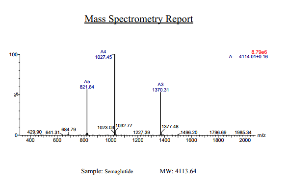
- HPLC, MS
Description
Semaglutide Synthetic is a single, non-glycosylated polypeptide chain containing 31 amino acids, having a molecular mass of 4113 Dalton and a Molecular formula of C187H291N45O59.
Formulation
The protein was lyophilized with no additives.
Purity
Greater than 97.0% as determined by analysis by RP-HPLC.
HPLC, MS
More Info
-
Physical Appearance
Sterile Filtered White lyophilized (freeze-dried) powder.
-
Stability
Lyophilized Semaglutide although stable at room temperature for 3 weeks, should be stored desiccated below -18°C. Upon reconstitution Semaglutide should be stored at 4°C between 2-7 days and for future use below -18°C. For long term storage it is recommended to add a carrier protein (0.1% HSA or BSA). Please prevent freeze-thaw cycles.
-
Solubility
It is recommended to reconstitute the lyophilized Semaglutide in sterile 18MΩ-cm H2O not less than 100 µg/ml, which can then be further diluted to other aqueous solutions.
-
Amino Acid Sequence
H-His-Aib-Glu-Gly-Thr-Phe-Thr-Ser-Asp-Val-Ser-Ser-Tyr-Leu-Glu-Gly-Gln-Ala-Ala-Lys(AEEAc-AEEAc-γ-Glu-17-carboxyheptadecanoyl)-Glu-Phe-Ile-Ala-Trp-Leu-Val-Arg-Gly-Arg-Gly-OH.
-
Background
Semaglutide, a glucagon-like peptide-1 receptor agonist (GLP-1 RA), has emerged as a breakthrough in the field of diabetes management and metabolic disorders. It is recognized for its potent glucose-lowering effects, weight management properties, and cardiovascular benefits. Beyond diabetes, semaglutide is under investigation for its potential applications in obesity treatment and other related conditions. This research aims to comprehensively explore semaglutide, shedding light on its multifaceted mechanisms of action and its broader therapeutic implications.
The primary objective of this research is to elucidate the mechanisms underlying the glucose-lowering and metabolic effects of semaglutide. In vitro and in vivo experiments will be conducted to investigate how semaglutide interacts with GLP-1 receptors, influences insulin secretion, and modulates glucose homeostasis. Understanding these mechanisms is crucial for harnessing the full therapeutic potential of semaglutide.
The second objective is to assess the clinical relevance of semaglutide in diabetes management. Clinical trials involving individuals with type 2 diabetes will be conducted to evaluate the efficacy, safety, and long-term outcomes of semaglutide treatment. These investigations may provide valuable insights into its use as a monotherapy or adjunct therapy in diabetes care.
The third objective is to explore the potential applications of semaglutide beyond diabetes. Research will investigate its role in obesity treatment, cardiovascular risk reduction, and non-alcoholic fatty liver disease (NAFLD) management. Understanding the multifaceted properties of semaglutide may open new avenues for therapeutic interventions in various metabolic and cardiovascular conditions.
By delving into the diverse functions of semaglutide, this research aims to expand our understanding of its therapeutic potential and clinical applications. The findings may contribute to improved treatment strategies for individuals affected by diabetes, obesity, and related metabolic disorders.
ProSpec's products are furnished for LABORATORY RESEARCH USE ONLY. The product may not be used as drugs, agricultural or pesticidal products, food additives or household chemicals.
- View Data Sheet
Name :
CRYAB HumanDescription:
Crystallin Alpha B Human Recombinant
CRYA2, CTPP2, HSPB5, Crystallin Alpha B, CRYAB.
Product # :
HSP-003Price :
Quantity :
Shipping Method :
Shipped with Ice Packs
More Info
- description
- source
- formulation
- purity
- More Info
Description
Recombinant CRYAB produced in E.Coli is a single,non-glycosylated polypeptide chain containing 175 amino acids and having a molecular mass of 20.1kDa. CRYAB is purified by proprietary chromatographic techniques.
Source
Escherichia Coli.
Formulation
The CRYAB protein (1mg/ml) solution contains 20mM Tris-HCl buffer pH-7.5, 50mM NaCl and 1mM EDTA.
Purity
Greater than 95.0% as determined by SDS-PAGE.
More Info
-
Introduction
Alpha crystallins are composed of two gene products ; alpha-A and alpha-B, for acidic and basic, respectively. Alpha crystallins can be induced by heat shock and are members of the small heat shock protein (sHSP also known as the HSP20). They act as molecular chaperones and hold them in in large soluble aggregates. These heterogeneous aggregates consist of 30-40 subunits; the alpha-A and alpha-B subunits have a 3:1 ratio, respectively. Two additional function of a-crystallins are an autokinase activity and participation in the intracellular architecture. Alpha-B is expressed widely in many tissues and organs and occurs in many neurological diseases.
-
Synonyms
CRYA2, CTPP2, HSPB5, Crystallin Alpha B, CRYAB.
-
Physical Appearance
Sterile filtered colorless solution.
-
Stability
Store at 4°C if entire vial will be used within 2-4 weeks. Store, frozen at -20°C for longer periods of time. For long term storage it is recommended to add a carrier protein (0.1% HSA or BSA).Avoid multiple freeze-thaw cycles.
-
Amino Acid Sequence
MDIAIHHPWI RRPFFPFHSP SRLFDQFFGE HLLESDLFPT STSLSPFYLR PPSFLRAPSWFDTGLSEMRL EKDRFSVNLD VKHFSPEELK VKVLGDVIEV HGKHEERQDE HGFISREFHRKYRIPADVDP LTITSSLSSD GVLTVNGPRK QVSGPERTIP ITREEKPAVT AAPKK.
ProSpec's products are furnished for LABORATORY RESEARCH USE ONLY. The product may not be used as drugs, agricultural or pesticidal products, food additives or household chemicals.
- View Data Sheet
Name :
DDT MouseDescription:
D-Dopachrome Tautomerase Mouse Recombinant
D-dopachrome decarboxylase (EC:4.1.1.84), D-dopachrome tautomerase, Ddt.
Product # :
ENZ-1073Price :
Quantity :
Shipping Method :
Shipped with Ice Packs
More Info
- description
- source
- formulation
- purity
- More Info
Description
DDT Mouse Recombinant produced in E.Coli is a single, non-glycosylated polypeptide chain containing 141 amino acids (1-118 a.a) and having a molecular mass of 15.5kDa. DDT is fused to a 23 amino acid His-tag at N-terminus & purified by proprietary chromatographic techniques.
Source
Escherichia Coli.
Formulation
DDT protein solution (1mg/ml) contains 20mM Tris-Hcl buffer (pH8.0) & 10% glycerol.
Purity
Greater than 95.0% as determined by SDS-PAGE.
More Info
-
Introduction
DDT is an enzyme that catayzes the tautomerization of D-dopachrome to give 5,6-dihydroxyindole (DHI). DDT is part of the family of lyases, specifically the carboxy-lyases, which cleave carbon-carbon bonds. DDT shares a homologous amino acid sequence (33% identical) with MIF and has similar tautomerase activity. DDT functions a proinflammatory cytokine.
-
Synonyms
D-dopachrome decarboxylase (EC:4.1.1.84), D-dopachrome tautomerase, Ddt.
-
Physical Appearance
Sterile Filtered clear solution.
-
Stability
Store at 4°C if entire vial will be used within 2-4 weeks. Store, frozen at -20°C for longer periods of time. For long term storage it is recommended to add a carrier protein (0.1% HSA or BSA).Avoid multiple freeze-thaw cycles.
-
Amino Acid Sequence
MGSSHHHHHH SSGLVPRGSH MGSMPFVELE TNLPASRIPA GLENRLCAAT ATILDKPEDR VSVTIRPGMT LLMNKSTEPC AHLLVSSIGV VGTAEQNRTH SASFFKFLTE ELSLDQDRIV IRFFPLEAWQ IGKKGTVMTF L
ProSpec's products are furnished for LABORATORY RESEARCH USE ONLY. The product may not be used as drugs, agricultural or pesticidal products, food additives or household chemicals.
- View Data Sheet
Name :
Shiga Like Toxin 1B AntibodyDescription:
Mouse Anti Shiga Like Toxin 1B
Product # :
ANT-660Price :
Quantity :
Shipping Method :
Shipped with Ice Packs
More Info
- description
- formulation
- purity
- More Info
Description
Shiga like toxin 1B monoclonal antibody IgG1 derived from mouse, immunized with recombinant Shiga-like toxin 1 subunit B ( E. coli O157:H7). The subunit B has nontoxic action and is a functional region which binds to the receptor. Shiga-like toxin 1 subunit B contains amino acid from 2 to 90 of Shiga-like toxin 1subunit B, it forms pentamer binding to the host cell receptor.
Formulation
1x PBS and 0.05% sodium nitrate.
Purity
Greater than 95% as determined by 12% PAGE (coomassie staining).
More Info
-
Introduction
Shiga-like toxin (verotoxin) is a toxin produced by some strains of Escherichia coli. Shiga-like toxin is named for its similarity to the AB5-type Shiga toxin produced by the bacteria Shigella dysenteriae. There are two known types-SLT1 and SLT2. The Shiga-like toxin is linked with hemolytic-uremic syndrome. Shiga-like toxin requires highly specific receptors on the cells'' surface in order to attach and enter the cell. Species such as cattle, swine, and deer which do not carry these receptors may harbor toxigenic bacteria without any ill effect, dropping them in their feces, from where they may be distributed to humans. Shiga Like Toxin-1 Subunit B has nontoxic action, it is the functional region which binds to the receptor. The Shiga Like Toxin-1 Subunit B can be useful in vaccine study, antibody test and other functional research.
-
Physical Appearance
Sterile Filtered clear colorless solution.
-
Stability
Shiga like toxin 1B monoclonal antibody although stable at 4°C for 1 week, should be stored below -18°C. For long term storage it is recommended to add a carrier protein (0.1% HSA or BSA). Please prevent freeze-thaw cycles.
-
Type
Mouse antibody Monoclonal.
ProSpec's products are furnished for LABORATORY RESEARCH USE ONLY. The product may not be used as drugs, agricultural or pesticidal products, food additives or household chemicals.
- View Data Sheet
Name :
TNF a MouseDescription:
Tumor Necrosis Factor-Alpha Mouse Recombinant
TNF-alpha, Tumor necrosis factor ligand superfamily member 2, TNF-a, Cachectin, DIF, TNFA, TNFSF2.
Product # :
CYT-252Price :
Quantity :
Shipping Method :
Shipped at Room temp
More Info
- description
- source
- formulation
- purity
- biological activity
- More Info
Description
Tumor Necrosis Factor-a Mouse Recombinant produced in E. coli is a single, non glycosylated, polypeptide chain containing 157 amino acids and having a molecular mass of 17301.32 Dalton. The TNF-alpha is purified by standard chromatographic techniques.
Source
Escherichia Coli.
Formulation
Lyophilized from a 0.2µm filtered concentrated solution in PBS, pH 7.2.
Purity
Greater than 97.0% as determined by:
(a) Analysis by RP-HPLC.
(c) Analysis by SDS-PAGE.Biological Activity
The ED50 as determined by the cytolysis of murine L929 cells in the presence of Actinomycin D is < 0.1ng/ml, corresponding to a Specific Activity of 10,000,000 Units/mg.More Info
-
Introduction
Tumor necrosis factor is a cytokine involved in systemic inflammation and is a member of a group of cytokines that all stimulate the acute phase reaction. TNF is mainly secreted by macrophages.
TNF causes apoptotic cell death, cellular proliferation, differentiation, inflammation, tumorigenesis and viral replication, TNF is also involved in lipid metabolism, and coagulation. TNF's primary role is in the regulation of immune cells.
Dysregulation and, in particular, overproduction of TNF have been implicated in a variety of human diseases- autoimmune diseases, insulin resistance, and cancer. -
Synonyms
TNF-alpha, Tumor necrosis factor ligand superfamily member 2, TNF-a, Cachectin, DIF, TNFA, TNFSF2.
-
Physical Appearance
Sterile Filtered White lyophilized (freeze-dried) powder.
-
Stability
Lyophilized Tumor Necrosis Factor-a although stable at room temperature for 3 weeks, should be stored desiccated below -18°C. Upon reconstitution TNF-a should be stored at 4°C between 2-7 days and for future use below -18°C.For long term storage it is recommended to add a carrier protein (0.1% HSA or BSA).Please prevent freeze-thaw cycles.
-
Solubility
It is recommended to reconstitute the lyophilized Tumor Necrosis Factor-alpha in sterile 18M-cm H2O not less than 100µg/ml, which can then be further diluted to other aqueous solutions.
-
Amino Acid Sequence
MLRSSSQNSS DKPVAHVVAN HQVEEQLEWL SQRANALLAN GMDLKDNQLV VPADGLYLVY SQVLFKGQGC PDYVLLTHTV SRFAISYQEK VNLLSAVKSP CPKDTPEGAE LKPWYEPIYL GGVFQLEKGD QLSAEVNLPK YLDFAESGQV YFGVIAL
-
Background
Tumor Necrosis Factor-alpha (TNF-α) is a pro-inflammatory cytokine that plays a critical role in the regulation of immune responses, inflammation, and cell survival. It is primarily produced by activated macrophages, but can also be secreted by other immune cells, such as T cells, natural killer cells, and mast cells. TNF-α is involved in a wide range of physiological and pathological processes, including the defense against infections, the development of autoimmune diseases, and the progression of cancer.
TNF-α exerts its effects by binding to two distinct cell surface receptors, TNF receptor 1 (TNFR1) and TNF receptor 2 (TNFR2). Upon binding to its receptors, TNF-α activates multiple signaling pathways, including the nuclear factor-kappa B (NF-κB) pathway, the mitogen-activated protein kinase (MAPK) pathway, and the apoptotic pathway. These signaling pathways regulate various cellular processes, such as inflammation, cell proliferation, differentiation, and apoptosis.
In the context of infections, TNF-α plays a crucial role in the body's defense against pathogens. It promotes the recruitment and activation of immune cells, enhances the production of other pro-inflammatory cytokines, and stimulates the expression of adhesion molecules on endothelial cells, facilitating the migration of immune cells to the site of infection. TNF-α also helps to induce fever, which is an important component of the body's immune response to infections.
However, excessive or prolonged production of TNF-α can contribute to the development of chronic inflammatory diseases, such as rheumatoid arthritis, inflammatory bowel disease, and psoriasis. In these conditions, elevated levels of TNF-α promote the infiltration of immune cells into the affected tissues, leading to tissue damage and the perpetuation of inflammation. The central role of TNF-α in the pathogenesis of these diseases has led to the development of anti-TNF-α therapies, which have revolutionized the treatment of chronic inflammatory diseases. These therapies include monoclonal antibodies, such as infliximab and adalimumab, and soluble TNF receptor fusion proteins, such as etanercept. Anti-TNF-α therapies have been shown to be effective in reducing inflammation, improving symptoms, and slowing disease progression in patients with chronic inflammatory diseases.
In the context of cancer, TNF-α has complex and context-dependent effects on tumor development and progression. On one hand, TNF-α can promote anti-tumor immunity by activating immune cells and stimulating the production of other pro-inflammatory cytokines. On the other hand, chronic inflammation driven by TNF-α can promote tumor growth, angiogenesis, and metastasis. Therefore, the role of TNF-α in cancer is still an area of active research, and the development of TNF-α-targeted therapies for cancer remains a challenge.
In conclusion, TNF-α is a pro-inflammatory cytokine that plays a critical role in the regulation of immune responses, inflammation, and cell survival. Its involvement in various physiological and pathological processes has made it an important target for the development of therapies for chronic inflammatory diseases and cancer. Anti-TNF-α therapies have revolutionized the treatment of chronic inflammatory diseases, but the complex role of TNF-α in cancer remains an area of ongoing research. Understanding the precise mechanisms by which TNF-α contributes to disease pathogenesis will be crucial for the development of more effective and targeted therapies.
-
Protein content
Protein quantitation was carried out by two independent methods1. UV spectroscopy at 280 nm using the absorbency value of 1.24 as the extinction coefficient for a 0.1% (1mg/ml) solution. This value is calculated by the PC GENE computer analysis program of protein sequences (IntelliGenetics). 2. Analysis by RP-HPLC, using a calibrated solution of TNF-a as a Reference Standard.
ProSpec's products are furnished for LABORATORY RESEARCH USE ONLY. The product may not be used as drugs, agricultural or pesticidal products, food additives or household chemicals.
- View Data Sheet
Name :
SDF 1a Human, HisDescription:
Stromal Cell-Derived Factor-1 alpha Human Recombinant, His Tag
SDF-1, CXCL12, Pre-B cell growth-stimulating factor, PBSF, hIRH, chemokine (C-X-C motif) ligand 12, SDF1, SDF1A, TPAR1, SCYB12, SDF-1a, TLSF-a.
Product # :
CHM-241Price :
Quantity :
Shipping Method :
Shipped at Room temp
More Info
- description
- source
- formulation
- purity
- More Info
Description
Stromal Cell-Derived Factor-1 alpha Human Recombinant produced in E.Coli is a non-glycosylated, Polypeptide chain containing 78 amino acids, having a molecular mass of 9.2 kDa. The SDF-1a is fused to 10 amino acids His-Tag at N-terminus and purified by proprietary chromatographic techniques.
Source
Escherichia Coli.
Formulation
The protein was filtered (0.4µm) and lyophilized from a concentrated (0.5mg/ml) solution containing 20mM Tris buffer pH-7.5 and 20mM sodium chloride.
Purity
Greater than 95.0% as determined SDS-PAGE.
More Info
-
Introduction
SDF-1 (stromal cell-derived factor-1) is small cytokine belonging to the chemokine family that is officially designated Chemokine (C-X-C motif) ligand 12 (CXCL12). It is produced in two forms, SDF-1?/CXCL12a and SDF-1?/CXCL12b, by alternate splicing of the same gene. Chemokines are characterized by the presence of four conserved cysteines, which form two disulfide bonds. The CXCL12 proteins belong to the group of CXC chemokines, whose initial pair of cysteines are separated by one intervening amino acid. CXCL12 is strongly chemotactic for lymphocytes and has been implicated as an important cell co-ordinator during development. During embryogenesis it directs the migration of hematopoietic cells from foetal liver to bone marrow. Mice which were knocked-out for CXCL12 gene were lethal before the birth or within just 1 hour of life. As another role, CXCL12a alters also the electrophysiology of neurons. CXCL12 was shown to be expressend in many tissues in mice (including brain, thymus, heart, lung, liver, kidney, spleen and bone marrow).
The receptor for this chemokine is CXCR4, which was previously called fusin. This CXCL12-CXCR4 interaction used to be considered exclusive (unlike for other chemokines and their receptors), but recently it was suggested that CXCL12 is also bound by CXCR7 receptor.
The gene for CXCL12 is located on human chromosome 10. In human and mouse both CXCL12 and CXCR4 show high identity of sequence: 99% and 90%, respectively. -
Synonyms
SDF-1, CXCL12, Pre-B cell growth-stimulating factor, PBSF, hIRH, chemokine (C-X-C motif) ligand 12, SDF1, SDF1A, TPAR1, SCYB12, SDF-1a, TLSF-a.
-
Physical Appearance
White lyophilized (freeze-dried) powder.
-
Stability
Lyophilized SDF1A although stable at room temperature for 3 weeks, should be stored desiccated below -18°C. Upon reconstitution CXCL12 should be stored at 4°C between 2-7 days and for future use below -18°C.For long term storage it is recommended to add a carrier protein (0.1% HSA or BSA).Please prevent freeze-thaw cycles.
-
Solubility
It is recommended to add deionized water to a working concentration approximately 0.5 mg/ml and let the lyophilized pellet dissolve completely. Product is not sterile! Please filter the product by appropriate sterile filter before using it in the cell culture.
-
Amino Acid Sequence
MKHHHHHHAS KPVSLSYRCP CRFFESHVAR ANVKHLKILN TPNCALQIVA RLKNNNRQVC IDPKLKWIQE YLEKALNK.
ProSpec's products are furnished for LABORATORY RESEARCH USE ONLY. The product may not be used as drugs, agricultural or pesticidal products, food additives or household chemicals.
- View Data Sheet
Name :
SDF 1a RatDescription:
Stromal Cell-Derived Factor-1 alpha Rat Recombinant (CXCL12)
SDF-1, CXCL12, Pre-B cell growth-stimulating factor, PBSF, hIRH, chemokine (C-X-C motif) ligand 12, SDF1, SDF1A, TPAR1, SCYB12, SDF-1a, TLSF-a, 12-O-tetradecanoylphorbol 13-acetate repressed protein 1, Thymic lymphoma cell-stimulating factor, TLSF.
Product # :
CHM-354Price :
Quantity :
Shipping Method :
Shipped at Room temp
More Info
- description
- source
- formulation
- purity
- biological activity
- More Info
Description
Stromal Cell-Derived Factor-1 alpha Rat Recombinant produced in E.Coli is a non-glycosylated, Polypeptide chain containing 68 amino acids and having a molecular mass of 7.9 kDa. The SDF-1a is purified by proprietary chromatographic techniques.
Source
Escherichia Coli.
Formulation
The CXCL12 protein was lyophilized from a concentrated (1 mg/ml) sterile solution containing 20mM Phosphate buffer pH-7.4 and 150mM NaCl.
Purity
Greater than 97.0% as determined by:
(a) Analysis by RP-HPLC.
(b) Analysis by SDS-PAGE.Biological Activity
The specific activity as determined by its ability to chemoattract human peripheral blood monocytes at 50-100ng/ml corresponding to a Specific Activity of 10,000-20,000IU/mg.More Info
-
Introduction
SDF-1 (stromal cell-derived factor-1) is small cytokine belonging to the chemokine family that is officially designated Chemokine (C-X-C motif) ligand 12 (CXCL12). It is produced in two forms, SDF-1?/CXCL12a and SDF-1?/CXCL12b, by alternate splicing of the same gene. Chemokines are characterized by the presence of four conserved cysteines, which form two disulfide bonds. The CXCL12 proteins belong to the group of CXC chemokines, whose initial pair of cysteines are separated by one intervening amino acid. CXCL12 is strongly chemotactic for lymphocytes and has been implicated as an important cell co-ordinator during development. During embryogenesis it directs the migration of hematopoietic cells from foetal liver to bone marrow. Mice which were knocked-out for CXCL12 gene were lethal before the birth or within just 1 hour of life. As another role, CXCL12a alters also the electrophysiology of neurons. CXCL12 was shown to be expressend in many tissues in mice (including brain, thymus, heart, lung, liver, kidney, spleen and bone marrow).
The receptor for this chemokine is CXCR4, which was previously called fusin. This CXCL12-CXCR4 interaction used to be considered exclusive (unlike for other chemokines and their receptors), but recently it was suggested that CXCL12 is also bound by CXCR7 receptor.
The gene for CXCL12 is located on human chromosome 10. In human and mouse both CXCL12 and CXCR4 show high identity of sequence: 99% and 90%, respectively. -
Synonyms
SDF-1, CXCL12, Pre-B cell growth-stimulating factor, PBSF, hIRH, chemokine (C-X-C motif) ligand 12, SDF1, SDF1A, TPAR1, SCYB12, SDF-1a, TLSF-a, 12-O-tetradecanoylphorbol 13-acetate repressed protein 1, Thymic lymphoma cell-stimulating factor, TLSF.
-
Physical Appearance
Sterile Filtered White lyophilized (freeze-dried) powder.
-
Stability
Lyophilized SDF-1a although stable at room temperature for 3 weeks, should be stored desiccated below -18°C. Upon reconstitution CXCL12 should be stored at 4°C between 2-7 days and for future use below -18°C.For long term storage it is recommended to add a carrier protein (0.1% HSA or BSA).Please prevent freeze-thaw cycles.
-
Solubility
It is recommended to reconstitute the lyophilized SDF-1a in sterile 18MΩ-cm H2O not less than 100µg/ml, which can then be further diluted to other aqueous solutions.
-
Amino Acid Sequence
KPVSLSYRCP CRFFESHVAR ANVKHLKILN TPNCALQIVA RLKSNNRQVC IDPKLKWIQE YLDKALNK.
ProSpec's products are furnished for LABORATORY RESEARCH USE ONLY. The product may not be used as drugs, agricultural or pesticidal products, food additives or household chemicals.
- View Data Sheet
Name :
ASS1 AntibodyDescription:
Argininosuccinate Synthase 1, Mouse Anti Human
ASS, CTLN1, EC 6.3.4.5, ASS1, Argininosuccinate Synthase 1.
Product # :
ANT-639Price :
Quantity :
Shipping Method :
Shipped with Ice Packs
More Info
- formulation
- More Info
Formulation
1mg/ml containing PBS, pH-7.4, 10% Glycerol and 0.02% Sodium Azide.
More Info
-
Introduction
ASS1 is involved in the urea cycle, which is a sequence of chemical reactions that is localized in liver cells. The urea cycle processes excess nitrogen that is generated as the body uses proteins. The surplus nitrogen is used to create a molecule called urea, which is excreted from the body in urine.
-
Synonyms
ASS, CTLN1, EC 6.3.4.5, ASS1, Argininosuccinate Synthase 1.
-
Physical Appearance
Sterile filtered colorless solution.
-
Immunogen
Anti-human ASS1 mAb, is derived from hybridization of mouse F0 myeloma cells with spleen cells from BALB/c mice immunized with recombinant human ASS1 amino acids 1-412 purified from E. coli.
-
Ig Subclass
Mouse IgG2a heavy chain and k light chain.
-
Clone
PAT1G11AT.
-
Applications
ASS1 antibody has been tested by ELISA, Western blot analysis, Flow cytometry and ICC/IF to assure specificity and reactivity. Since application varies, however, each investigation should be titrated by the reagent to obtain optimal results.
-
Type
Mouse Anti Human Monoclonal.
-
Storage Procedures
For periods up to 1 month store at 4°C, for longer periods of time, store at -20°C. Prevent freeze thaw cycles.
-
Purification Method
ASS1 antibody was purified from mouse ascitic fluids by protein-A affinity chromatography.
ProSpec's products are furnished for LABORATORY RESEARCH USE ONLY. The product may not be used as drugs, agricultural or pesticidal products, food additives or household chemicals.
- View Data Sheet
Name :
ATF HumanDescription:
Apo Transferrin Human
Serotransferrin, Transferrin, Siderophilin, Beta-1-metal-binding globulin, TF, PRO1557, PRO2086, DKFZp781D0156, Apo Transferrin, ATF.
Product # :
PRO-325Price :
Quantity :
Shipping Method :
Shipped at Room temp
More Info
- description
- source
- formulation
- purity
- More Info
Description
Human Apo Transferrin is a glycoprotein of approximately 77 kDa.
Source
Human serum.
Formulation
The protein was lyophilized from 20mM NH4HC03 solution. May contain traces of buffer salts.
Purity
Greater than 98.0% as determined by coomassie blue stained SDS-PAGE and Cellulose Acetate electrophoresis.
More Info
-
Introduction
Transferrin is the iron-transport protein of vertebrate serum and donates iron to cells through interaction with a specific membrane receptor, CD71. Transferrin appears to be indispensable for most cells growing in tissue culture.
It is referred to frequently as a growth factor because, in analogy to other growth factor-receptor interactions, proliferating cells express high numbers of transferrin receptors, and the binding of transferrin to their receptors is needed for cells to initiate and maintain their DNA synthesis. Apart from its role as an iron transport protein transferrin acts as a cytokine and has functions that may not be related to its iron-carrying capacity.
Human Transferrin is a crucial component for the cultivation of mammalian cells in-vitro. Human Transferrin is Critical for long-term cells growth in-vitro. Human Transferrin is used as detoxificant in media by binding contaminating metal ions. Human Transferrin is often used as a nutrient in fermentation media for recombinant protein and biopharmaceutical production. Additional common uses of Human Transferrin areMolecular weight, Affinity purification of anti-human transferrin antibodies and also as receptor mediated transfection of molecules such as DNA, into cells. -
Synonyms
Serotransferrin, Transferrin, Siderophilin, Beta-1-metal-binding globulin, TF, PRO1557, PRO2086, DKFZp781D0156, Apo Transferrin, ATF.
-
Physical Appearance
Sterile Filtered White lyophilized (freeze-dried) powder.
-
Stability
Store the lyophilized Apo Transferrin between 2-8°C, do not freeze. Upon reconstitution Apo Transferrin should be stored at 4°C between 2-7 days and for future use below -18°C.Please prevent freeze-thaw cycles.
-
Solubility
It is recommended to reconstitute the lyophilized Apo Transferrin in sterile 18MΩ-cm H2O not less than 100µg/ml, which can then be further diluted to other aqueous solutions.
-
Human Virus Test
Plasma from each donor has been tested and found negative for antibody to HIV-1, HIV-2, HCV, HBSAG and Syphilis.
-
Iron Content
The Iron content was estimated by ICP and was found to be <6 ppm.
ProSpec's products are furnished for LABORATORY RESEARCH USE ONLY. The product may not be used as drugs, agricultural or pesticidal products, food additives or household chemicals.
- View Data Sheet
Name :
TNF alpha humanDescription:
Tumor Necrosis Factor-Alpha Human Recombinant
TNF-alpha, Tumor necrosis factor ligand superfamily member 2, TNF-a, Cachectin, DIF, TNFA, TNFSF2.
Product # :
CYT-223Price :
Quantity :
Shipping Method :
Shipped at Room temp
More Info
- description
- source
- formulation
- purity
- biological activity
- More Info
Description
Tumor Necrosis Factor-a Human Recombinant produced in E.Coli is a single, non-glycosylated, polypeptide chain containing 158 amino acids (157 a.a. of the mature human TNF-alpha and an N-terminal methionine) and having a molecular mass of 17.5kDa. The TNF-alpha is purified by standard chromatographic techniques.
Source
Escherichia Coli.
Formulation
TNF-a Human was lyophilized from a concentrated 1mg/ml solution containing 20mM PB, pH-7.2, and 100mM NaCl.
Purity
Greater than 95.0% as determined by:
(a) Analysis by RP-HPLC.
(b) Analysis by SDS-PAGE.Biological Activity
The Specific Activity is >5.0×10⁷ IU/mg as determined by the cytolysis of murine L929 cells in the presence of Actinomycin D.More Info
-
Introduction
Tumor necrosis factor is a cytokine involved in systemic inflammation and is a member of a group of cytokines that all stimulate the acute phase reaction. TNF is mainly secreted by macrophages.
TNF causes apoptotic cell death, cellular proliferation, differentiation, inflammation, tumorigenesis and viral replication, TNF is also involved in lipid metabolism, and coagulation. TNF's primary role is in the regulation of immune cells.
Dysregulation and, in particular, overproduction of TNF have been implicated in a variety of human diseases- autoimmune diseases, INS resistance, and cancer. -
Synonyms
TNF-alpha, Tumor necrosis factor ligand superfamily member 2, TNF-a, Cachectin, DIF, TNFA, TNFSF2.
-
Physical Appearance
Sterile Filtered White lyophilized (freeze-dried) powder.
-
Stability
Lyophilized Tumor Necrosis Factor-a although stable at room temperature for 3 weeks, should be stored desiccated below -18°C. Upon reconstitution TNF-a should be stored at 4°C between 2-7 days and for future use below -18°C.For long term storage it is recommended to add a carrier protein (0.1% HSA or BSA).Please prevent freeze-thaw cycles.
-
Solubility
It is recommended to reconstitute the lyophilized Tumor Necrosis Factor-alpha in sterile 18MΩ-cm H2O not less than 100µg/ml, which can then be further diluted to other aqueous solutions.
-
Amino Acid Sequence
MVRSSSRTPS DKPVAHVVAN PQAEGQLQWL NRRANALLAN GVELRDNQLV VPSEGLYLIY SQVLFKGQGC PSTHVLLTHT ISRIAVSYQT KVNLLSAIKS PCQRETPEGA E AKPWYEPIY LGGVFQLEKG DRLSAEINRP DYLDFAESGQ VYFGIIAL.
-
Background
TNF Alpha Human: An Overview of Its Role and Importance in Immunology
TNF alpha human, also known as Tumor Necrosis Factor-alpha, is a critical cytokine in the immune system. Macrophages mainly produce this protein, and it plays a key role in inflammation and the acute phase reaction.
This protein is involved in various cellular functions, including cell death, differentiation, proliferation, and immune regulation.
Production and Properties
Tumor Necrosis Factor-alpha is produced recombinantly in E. coli and consists of a single, non-glycosylated polypeptide chain. It includes 157 amino acids of the mature human TNF-alpha and an N-terminal methionine, resulting in a molecular mass of approximately 17.5 kDa.
Furthermore, the protein is purified through standard chromatographic techniques to ensure high purity and biological activity.
Solubility and Usage
The lyophilized form of TNF appears as a sterile, white powder. It is recommended to reconstitute this powder in sterile water to achieve a solution of no less than 100µg/ml.
This solution can then be further diluted for various experimental applications. TNF alpha is used extensively in research, particularly for studying its effects on cell signaling and immune response.
Storage and Stability
For long-term storage, TNF should be kept desiccated below -18°C. Once reconstituted, it should be used within a week if stored at 4°C or kept below -18°C for future use. Avoiding freeze-thaw cycles is crucial to maintain the protein's functionality.
Biological Role and Implications
TNF alpha human is involved in the regulation of immune cells and is known for its role in inflammatory processes.
Dysregulation of TNF alpha production is linked to various diseases, such as autoimmune disorders, insulin resistance, and cancer. It is also a target for therapeutic interventions, particularly in conditions like rheumatoid arthritis and inflammatory bowel disease.
Mechanism of Action
TNF alpha can induce fever, apoptotic cell death, and can inhibit tumorigenesis and viral replication. Moreover, it is a potent mediator of the acute phase reaction, which influences the activity of various cells involved in systemic inflammation.
Research and Clinical Importance
Scientific research on TNF has provided insights into its complex role in disease mechanisms. Its interaction with receptors such as TNFRSF1A underscores its multifaceted effects across different organ systems, from liver function to brain activity.
Ongoing studies continue to explore its therapeutic potential, especially how it can be modulated to treat diseases without harmful side effects.
In essence, TNF alpha human is a versatile and powerful component of the immune system, important for both health and disease. Understanding its pathways and functions helps scientists develop better treatments for various inflammatory and autoimmune diseases.
ProSpec's products are furnished for LABORATORY RESEARCH USE ONLY. The product may not be used as drugs, agricultural or pesticidal products, food additives or household chemicals.
- View Data Sheet
Name :
DarbepoetinDescription:
Darbepoetin-Alpha Human Recombinant
Erythropoietin-Mutant, EPO-mutant, NESP, Novel Erythropoiesis Stimulating Protein, EPO Mutant.
Product # :
CYT-1263Price :
Quantity :
Shipping Method :
Shipped at Room temp
More Info
- description
- source
- formulation
- purity
- biological activity
- More Info
Description
Darbepoetin-alpha Human Recombinant is produced in Chinese hamster ovary (CHO) cells by recombinant DNA technology is a glycosylated polypeptide chain containing 165 amino acids and having a predicted molecular mass of 18,200 Dalton and apparent glycosylated molecular mass of 37-40kDa. Darbepoetin is purified by proprietary chromatographic techniques.
Source
Chinese Hamster Ovary Cells(CHO).
Formulation
Each mg of lyophilized Darbepoetin powder contains 20mM Phosphate Buffer, 0.15M NaCl pH-6.2 & 0.005% Tween-80.
Purity
Greater than 97.0% as determined by SDS-PAGE.
Biological Activity
The Specific Activity was measured by Normocyth -aemic mice and was found to be 1,000,000 IU/mg.More Info
-
Introduction
Darbepoetin is a mutant recombinant human EPO-Alpha protein re-engineered, containing 5 amino acid changes at N30, T32, V87, N88, T90. Darbepoetin exhibits longer half-life stimulating red blood cell production than Erythropoietin. Darbepoetin maintains hemoglobin more effectively compared to EPO-Alpha.
-
Synonyms
NESP, Novel Erythropoiesis Stimulating Protein, EPO Mutant.
-
Physical Appearance
Sterile Filtered White lyophilized (freeze-dried) powder.
-
Stability
Lyophilized Darbepoetin although stable at room temperature for 3 weeks, should be stored desiccated below -18°C. Upon reconstitution Darbepoetin should be stored at 4°C between 2-7 days and for future use below -18°C.For long term storage it is recommended to add a carrier protein (0.1% HSA or BSA).Please prevent freeze-thaw cycles.
-
Solubility
It is recommended to reconstitute the lyophilized Darbepoetin in sterile 18MΩ-cm H2O not less than 100µg/ml, which can then be further diluted to other aqueous solutions.
-
Amino Acid Sequence
APPRLICDSR VLERYLLEAK EAENITTGCN ETCSLNENIT VPDTKVNFYA WKRMEVGQQA VEVWQGLALL SEAVLRGQAL LVNSSQVNET LQLHVDKAVS GLRSLTTLLR ALGAQKEAIS PPDAASAAPL RTITADTFRK LFRVYSNFLR GKLKLYTGEA CRTGD.
-
Background
What is the molecular weight/Mw of DARBEPOETIN Protein?
DARBEPOETIN Protein has a total Mw of 38.5kDa.
What is the source or expression system of DARBEPOETIN Protein?
Chinese Hamster Ovary Cells(CHO).
What is the Purity of DARBEPOETIN Protein?
DARBEPOETIN Protein is >97% pure as determined by SDS-PAGE.
What is the Biological Activity of DARBEPOETIN Protein?
The Specific Activity was measured by Normocyth -aemic mice and was found to be 1,000,000 IU/mg.
What is the amino acid sequence of DARBEPOETIN Protein?
APPRLICDSR VLERYLLEAK EAENITTGCN ETCSLNENIT VPDTKVNFYA WKRMEVGQQA VEVWQGLALL SEAVLRGQAL LVNSSQVNET LQLHVDKAVS GLRSLTTLLR ALGAQKEAIS PPDAASAAPL RTITADTFRK LFRVYSNFLR GKLKLYTGEA CRTGD
What applications can DARBEPOETIN Protein be used in?
DARBEPOETIN Protein can probably be used in western blot, ELISA and Lateral Flow.
What is the endotoxin level for DARBEPOETIN Protein?
The endotoxin level is minimal, DARBEPOETIN Protein was purified using conventional chromatography techniques
ProSpec's products are furnished for LABORATORY RESEARCH USE ONLY. The product may not be used as drugs, agricultural or pesticidal products, food additives or household chemicals.