Search results
1000 results found for “inhibin a”
Name
Description
Product #
Price
Quantity
Shipping Method
- View Data Sheet
Name :
VAMP8 HumanDescription:
Endobrevin Human Recombinant
VAMP8, VAMP-8, Endobrevin, Vesicle-Associated Membrane Protein 8, EDB.
Product # :
PRO-660Price :
Quantity :
Shipping Method :
Shipped with Ice Packs
More Info
- description
- source
- formulation
- purity
- More Info
- Citations
Description
VAMP8 Human Recombinant produced in E.Coli is a single, non-glycosylated polypeptide chain containing 96 amino acids (1-76 a.a.) and having a molecular mass of 10.9 kDa. The VAMP8 is fused to a 20 amino acid His Tag at N-terminus and purified by proprietary chromatography techniques.
Source
Escherichia Coli.
Formulation
The VAMP8 protein solution (0.25mg/ml) contains 20mM Tris pH-8, 0.1mM PMSF, 0.2M NaCl and 50% glycerol.
Purity
Greater than 85.0% as determined by SDS-PAGE.
Citations
More Info
-
Introduction
VAMP8 also called endobrevin, is the main component of a SNARE complex involved in the docking and fusion of synaptic vesicles with the presynaptic membrane. VAMP8 protein is involved in the regulatation of enzyme secretion in pancreatic acinar cells and plays a part in the abscission of the midbody during cell division, which leads to completely separate daughter cells. VAMP8 is essential for dense-granule secretion in platelets. VAMP8 is related with the perinuclear vesicular structures of the early endocytic compartment. VAMP8 interacts particularly with the soluble NSF-attachment protein (alpha-SNAP), through an VAMP8-containing SNARE complex.
-
Synonyms
VAMP8, VAMP-8, Endobrevin, Vesicle-Associated Membrane Protein 8, EDB.
-
Physical Appearance
Sterile Filtered colorless solution.
-
Stability
Store at 4°C if entire vial will be used within 2-4 weeks. Store, frozen at -20°C for longer periods of time. For long term storage it is recommended to add a carrier protein (0.1% HSA or BSA).Please prevent freeze-thaw cycles.
-
Amino Acid Sequence
MGSSHHHHHH SSGLVPRGSH MEEASEGGGN DRVRNLQSEV EGVKNIMTQN VERILARGEN LEHLRNKTED LEATSEHFKT TSQKVARKFW WKNVKM.
ProSpec's products are furnished for LABORATORY RESEARCH USE ONLY. The product may not be used as drugs, agricultural or pesticidal products, food additives or household chemicals.
- View Data Sheet
Name :
p85a BovineDescription:
Phosphoinositide 3-kinase a, regulatory subunit Bovine Recombinant
Phosphatidylinositol 3-kinase regulatory subunit alpha, PI3-kinase p85 subunit alpha, PtdIns-3-kinase p85-alpha, PI3K, P85a.
Product # :
PKA-328Price :
Quantity :
Shipping Method :
Shipped with Ice Packs
More Info
- description
- source
- formulation
- purity
- More Info
- Citations
Description
Phosphoinositide 3-kinase subunit p85a Bovine Recombinant has a molecular weight of 83.5 kDa.
Source
Leishmania tarentolae.
Formulation
Supplied as a 1 mg/ml solution in 25mM HEPES, pH 8.0, 25mM NaCl, 2.5mM MgCl2 and 50% glycerol.
Purity
Greater than 90.0% as determined by SDS-PAGE.
Citations
More Info
-
Introduction
P85a functions as the regulatory subunit of the class IA PI3-kinase isoforms a, b, and d.
It contains two SH2 domains that bind to tyrosine-phosphorylated growth factor receptors or substrate adaptor proteins.
It also contains a BH (breakpoint cluster region homology) domain that shows GAP activity towards the small GTPases Rab4, Rab5, Cdc42, Rac1 and to a lesser extend towards Rab6 and Rab11.
It was shown that PI3Ka catalytic subunit mediated phosphorylation of the p85a adapter reduces the lipid kinase activity of the heterodimer and this gives hints for PI3K-dependent signaling events not requiring production of 3’-phosphorylated phosphoinositides.
PI3Ka protein kinase activity has been implicated in IRS-1 serine phosphorylation in insulin-treated adipocytes and in STAT3 and IRS-1 phosphorylation upon activation of the type 1 IFN receptor by IFN-a. -
Synonyms
Phosphatidylinositol 3-kinase regulatory subunit alpha, PI3-kinase p85 subunit alpha, PtdIns-3-kinase p85-alpha, PI3K, P85a.
-
Physical Appearance
Sterile filtered liquid formualtion.
-
Stability
Store at 4°C if entire vial will be used within 2-4 weeks. Store, frozen at -20°C for longer periods of time. For long term storage it is recommended to add a carrier protein (0.1% HSA or BSA).Avoid multiple freeze-thaw cycles.
ProSpec's products are furnished for LABORATORY RESEARCH USE ONLY. The product may not be used as drugs, agricultural or pesticidal products, food additives or household chemicals.
- View Data Sheet
Name :
HBsAgA AntibodyDescription:
Hepatitis B surface Antigen A, Polyclonal Goat Antibody
Product # :
ANT-163Price :
Quantity :
Shipping Method :
Shipped with Ice Packs
More Info
- description
- formulation
- More Info
- Citations
Description
Affinity-purified Goat antibodies against the Native Ad/AyHBsAg antigens.1mg/ml in PBS, was passed through a 0.2um Filter into a 50ml sterile conic Tube.
Formulation
(1ml) PBS.
Citations
More Info
-
Introduction
Hepatitis B is one of a few known non-retroviralviruses which employ reverse transcriptionas a part of its replication process. (HIV, a completely unrelated virus, also uses reverse transcription, but it is a retrovirus.) HBV invades the cell by binding to surface receptor and become internalized. The viral core particles then migrate to the hepatocyte nucleus and the partially double-stranded, relaxed circular genomes (RC-DNA) are repaired to form a covalently closed circular DNA (cccDNA), which is the template for viral genomic and sub-genomic RNAs by cellular RNA polymerase II. Of these, the pregenomic RNA (pgRNA is selectively packaged into progeny capsids and is then reverse-transcribed into new RC-DNA. The core can either bud into the endoplasmic reticulum to be enveloped or exported from the cell or recycled back into the genome for conversion to cccDNA.
-
Physical Appearance
Sterile Filtered solution.
-
Host
Rabbit.
-
Specificity
Immunoreactive with all HBsAg A antigens. Direct ELISA shows signal above cut-off at antibody levels of 100ng/test and above.
-
Type
Polyclonal Goat Antibody.
-
Storage Procedures
Store at -20°C.
ProSpec's products are furnished for LABORATORY RESEARCH USE ONLY. The product may not be used as drugs, agricultural or pesticidal products, food additives or household chemicals.
- View Data Sheet
Name :
CAMK2N1 MouseDescription:
Calcium/Calmodulin-Dependent Protein Kinase II Inhibitor 1 Mouse Recombinant
mCaMKIINalpha, calcium/calmodulin-dependent protein kinase II inhibitor alpha, Camk2n1.
Product # :
PKA-092Price :
Quantity :
Shipping Method :
Shipped with Ice Packs
More Info
- description
- source
- formulation
- purity
- More Info
- Citations
Description
CAMK2N1 Mouse Recombinant produced in E.Coli is a single, non-glycosylated polypeptide chain containing 101 amino acids (1-78 a.a) and having a molecular mass of 10.9kDa.CAMK2N1 is fused to a 23 amino acid His-tag at N-terminus & purified by proprietary chromatographic techniques.
Source
Escherichia Coli.
Formulation
CAMK2N1 protein solution (0.5mg/ml) containing 20mM Tris-HCl (pH8.5), 1mM DTT, 0.1M NaCl and 10% glycerol.
Purity
Greater than 90.0% as determined by SDS-PAGE.
Citations
More Info
-
Introduction
Calcium/Calmodulin Dependent Protein Kinase II Inhibitor 1 (CAMK2N1), which interacts with CAMK2B, is a part of the CAMK2N family. CAMK2N1 is also interacts with CAMK2A in a way which requires CAMK2A activation by Ca2+. CAMK2N1 is potent and specific inhibitor of CaM-kinase II (CAMK2).
-
Synonyms
mCaMKIINalpha, calcium/calmodulin-dependent protein kinase II inhibitor alpha, Camk2n1.
-
Physical Appearance
Sterile filtered colorless solution.
-
Stability
Store at 4°C if entire vial will be used within 2-4 weeks. Store, frozen at -20°C for longer periods of time. For long term storage it is recommended to add a carrier protein (0.1% HSA or BSA).Avoid multiple freeze-thaw cycles.
-
Amino Acid Sequence
MGSSHHHHHH SSGLVPRGSH MGSMSEVLPY GDEKLSPYGD GGDVGQIFSC RLQDTNNFFG AGQSKRPPKL GQIGRSKRVV IEDDRIDDVL KTMTDKAPPG V.
ProSpec's products are furnished for LABORATORY RESEARCH USE ONLY. The product may not be used as drugs, agricultural or pesticidal products, food additives or household chemicals.
- View Data Sheet
Name :
PRKACA Human, sf9Description:
c-AMP dependant Protein Kinase A catalytic subunit alpha Human Recombinant, Sf9
cAMP-dependent protein kinase alpha-catalytic subunit, EC 2.7.11.11, PKA C-alpha, PKACA, PRKACA, MGC48865, MGC102831.
Product # :
PKA-093Price :
Quantity :
Shipping Method :
Shipped with Ice Packs
More Info
- description
- source
- formulation
- purity
- More Info
- Citations
Description
PRKACA Human Recombinant produced in Sf9 Baculovirus cells is a single, glycosylated polypeptide chain containing 578 amino acids (1-351 a.a.) and having a molecular mass of 67kDa (Migrates at 50-70kDa on SDS-PAGE under reducing conditions). PRKACA is expressed with a 227 amino acid GST Tag at N-Terminus and purified by proprietary chromatographic techniques.
Source
Sf9, Baculovirus cells.
Formulation
PRKACA protein solution (0.25mg/ml) contains Phosphate Buffered Saline (pH 7.4) and 10% glycerol.
Purity
Greater than 85% as determined by SDS-PAGE.
Citations
More Info
-
Introduction
cAMP-dependent protein kinase catalytic subunit alpha isoform Calpha1 (PRKACA) belongs to the Ser/Thr protein kinase family. PRKACA is responsible for phosphorylating other proteins and substrates, changing their activity. The PRKACA protein is a member of the AGC kinase family, and contributes to the regulation of cellular processes which include glucose metabolism, cell division, and contextual memory.
-
Synonyms
cAMP-dependent protein kinase alpha-catalytic subunit, EC 2.7.11.11, PKA C-alpha, PKACA, PRKACA, MGC48865, MGC102831.
-
Physical Appearance
Sterile Filtered colorless solution.
-
Stability
Store at 4°C if entire vial will be used within 2-4 weeks. Store, frozen at -20°C for longer periods of time. For long term storage it is recommended to add a carrier protein (0.1% HSA or BSA).Avoid multiple freeze-thaw cycles.
-
Amino Acid Sequence
MSPILGYWKI KGLVQPTRLL LEYLEEKYEE HLYERDEGDK WRNKKFELGL EFPNLPYYID GDVKLTQSMA IIRYIADKHN MLGGCPKERA EISMLEGAVL DIRYGVSRIA YSKDFETLKV DFLSKLPEML KMFEDRLCHK TYLNGDHVTH PDFMLYDALD VVLYMDPMCL DAFPKLVCFK KRIEAIPQID KYLKSSKYIA WPLQGWQATF GGGDHPPKSD LVPRGSHMGN AAAAKKGSEQ ESVKEFLAKA KEDFLKKWES PAQNTAHLDQ FERIKTLGTG SFGRVMLVKH KETGNHYAMK ILDKQKVVKL KQIEHTLNEK RILQAVNFPF LVKLEFSFKD NSNLYMVMEY VPGGEMFSHL RRIGRFSEPH ARFYAAQIVL TFEYLHSLDL IYRDLKPENL LIDQQGYIQV TDFGFAKRVK GRTWTLCGTP EYLAPEIILS KGYNKAVDWW ALGVLIYEMA AGYPPFFADQ PIQIYEKIVS GKVRFPSHFS SDLKDLLRNL LQVDLTKRFG NLKNGVNDIK NHKWFATTDW IAIYQRKVEA PFIPKFKGPG DTSNFDDYEE EEIRVSINEK CGKEFSEF.
ProSpec's products are furnished for LABORATORY RESEARCH USE ONLY. The product may not be used as drugs, agricultural or pesticidal products, food additives or household chemicals.
- View Data Sheet
Name :
F9 AntibodyDescription:
Coagulation Factor-IX, Mouse Antibody
Product # :
ANT-229Price :
Quantity :
Shipping Method :
Shipped at Room temp
More Info
- formulation
- More Info
- Citations
Formulation
1mg/ml in PBS (after reconstitution).
Citations
More Info
-
Introduction
Factor IX is a glycoprotein, which is synthesized in the liver. The domain structure of factor IX is similar to that of the other vitamin K dependent coagulation factors. The NH2-terminal region contains 12 g-carboxyglutamic acid (gla) residues, which facilitate the calcium dependent binding of factor IX to negatively charged phospholipid surfaces. Two domains which are homologous to epidermal growth factor (EGF) span the region between the NH2-terminal gla domain and the activation peptide (Ala-146 to Arg-180). Factor IX is activated by either factor XIa or the factor VIIa/tissue factor/phospholipid complex. Cleavage at site A yields the intermediate IXa, which is subsequently converted to the fully active form IXab by cleavage at site B. The NH2-terminal light chain (GLA and EGF domains) remains covalently attached to the COOH-terminal heavy chain by a disulfide bond. The serine protease catalytic triad (Ser-365, His 221, Asp-269) is located in the heavy chain. Factor IXab is the catalytic component of the “intrinsic factor Xase complex” (factor VIIIa/IXa/Ca2+/phospholipid) which proteolytically activates factor X to factor Xa.
-
Solubility
Reconstitute with H20 to get a 1 mg/ml concentration. Mix gently, wash the sides of the vial and wait 30-60 seconds before use.
-
Immunogen
Human Factor-IX.
-
Ig Subclass
Mouse IgG1.
-
Clone
NYRhFIX.
-
Applications
Direct ELISA, Western Blot, imunohistochemistry. For W.B. use 1µg/ml of antibody.
-
Titer
By direct ELISA, 1:10,000 dilution will yield 0.5 O.D using alkaline phosphatase conjugated rabbit anti-mouse Ig (Jackson Laboratories).
-
Shipping Conditions
Antibody is shipped lyophilized at ambient temperature.
-
Type
Mouse Antibody Monoclonal.
-
Storage Procedures
In lyophilized form, for long periods, store at 4 C in a dry environment. After reconstitution, if not intended for use within a month, aliquot and store at -20 C.
-
Purification Method
Protein A.
ProSpec's products are furnished for LABORATORY RESEARCH USE ONLY. The product may not be used as drugs, agricultural or pesticidal products, food additives or household chemicals.
- View Data Sheet
Name :
CD11b Antibody, FITCDescription:
CD11b, Mouse Anti-Human, FITC
Integrin alpha-M, Cell surface glycoprotein MAC-1 subunit alpha, CR-3 alpha chain, Leukocyte adhesion receptor MO1, Neutrophil adherence receptor, CD11b antigen, ITGAM, CR3A, MO1A, CD11B, MAC-1, MAC1A, MGC117044.
Product # :
ANT-276Price :
Quantity :
Shipping Method :
Shipped at Room temp
More Info
- formulation
- More Info
- Citations
Formulation
1mg/ml in PBS (after reconstitution).
Citations
More Info
-
Introduction
Integrins are heterodimeric integral membrane proteins composed of an alpha chain and a beta chain. This I-domain containing alpha integrin combines with the beta 2 chain (ITGB2) to form a leukocyte-specific integrin referred to as macrophage receptor 1 ('Mac-1'), or inactivated-C3b (iC3b) receptor 3 ('CR3'). The alpha M beta 2 integrin is important in the adherence of neutrophils and monocytes to stimulated endothelium, and also in the phagocytosis of complement coated particles. Integrin alpha-m/beta-2 is implicated in various adhesive interactions of monocytes, macrophages and granulocytes as well as in mediating the uptake of complement-coated particles. CD-11b is identical with cr-3, the receptor for the ic3b fragment of the third complement component. CD-11b probably recognizes the r-g-d peptide in c3b. Integrin alpha-m/beta-2 is also a receptor for fibrinogen, factor x and icam1. it recognizes p1 and p2 peptides of fibrinogen gamma chain.
-
Synonyms
Integrin alpha-M, Cell surface glycoprotein MAC-1 subunit alpha, CR-3 alpha chain, Leukocyte adhesion receptor MO1, Neutrophil adherence receptor, CD11b antigen, ITGAM, CR3A, MO1A, CD11B, MAC-1, MAC1A, MGC117044.
-
Solubility
Reconstitute with of H20. Mix gently, wash the sides of the vial and wait 30-60 seconds before use.
-
Immunogen
Purified human PBL Monocytes.
-
Ig Subclass
Mouse IgG1.
-
Clone
hCD11b.
-
Applications
Blocking and staining antibody. For staining, use 10µl/1,000,000 cells. Titer for blocking LPS binding should be determined by the investigator.
-
Available Conjugates
This antibody is also available unconjugated and conjugated to Biotin. For staining with biotin or FITC-conjugated antibody use 5-10µl/1,000,000 cells.
-
Type
Mouse Anti Human Monoclonal.
-
Storage Procedures
Lyophilized: store at 4°C. After reconstitution, if not intended for use within a month, aliquot and store at -20°C.
-
Purification Method
Ion exchange column.
ProSpec's products are furnished for LABORATORY RESEARCH USE ONLY. The product may not be used as drugs, agricultural or pesticidal products, food additives or household chemicals.
- View Data Sheet
Name :
Vimentin Antibody, FITCDescription:
Vimentin, Mouse Anti Human Antibody, FITC
Product # :
ANT-247Price :
Quantity :
Shipping Method :
Shipped at Room temp
More Info
- formulation
- More Info
- Citations
Formulation
1mg/ml in PBS (after reconstitution).
Citations
More Info
-
Introduction
Vimentin expression in human malignant glioma cells depends on cellular density, algorithms of drug delivery and chemo/radio treatment. Vimentin and detyrosinated microtubules provide structural support for the extensive microtentacles observed in detached tumor cells and a mechanism to promote successful metastatic spread. Primary colorectal carcinomas display aberrant expression of vimentin, and have activated Notch and TGFbeta signaling pathways. Vimentin is a strong arterial substrate for transglutaminases. Transglutaminase-mediated vimentin dimerization results in a novel unifying pathway by which vasodilatory and remodeling responses may be regulated. Ablation of vimentin expression inhibits migration and invasion of colon and breast cancer cell lines.
-
Solubility
Reconstitute with H20. Mix gently, wash the sides of the vial and wait 30-60 seconds before use.
-
Immunogen
Purified Human Vimentin.
-
Ig Subclass
mouse IgG1.
-
Clone
hVimentin.
-
Applications
Immunohistochemistry.
-
Cross reactivity
Vimentin antibody will recognize most mammalian Vimentin except mouse and rat.
-
Titer
For intra-cellular staining, use 5-10µl/1,000,000 cells. Wash twice with PBS/Azide before adding the appropriate amount of secondary fluorescent reagent.
-
Shipping Conditions
Antibody is shipped lyophilized at ambient temperature.
-
Available Conjugates
This antibody is also available conjugated to Biotin and non conjugate form.
-
Type
Mouse Anti Human Monoclonal Antibody.
-
Storage Procedures
In lyophilized form, for long periods, store at 4°C in a dry environment. After reconstitution, if not intended for use within a month, aliquot and store at -20°C.
-
Purification Method
Ion exchange column.
ProSpec's products are furnished for LABORATORY RESEARCH USE ONLY. The product may not be used as drugs, agricultural or pesticidal products, food additives or household chemicals.
- View Data Sheet
Name :
IL1A Human, His ActiveDescription:
Interleukin-1 alpha Human Recombinant, His Tag Active
Interleukin-1 alpha, IL-1 alpha, Hematopoietin-1, Interleukin-1 alpha: Interleukin-1a, IL-1a, Interleukin-1 alpha, IL1A, interleukin 1 alpha, IL1F1, IL-1A, preinterleukin 1 alpha, pro-interleukin-1-alpha.
Product # :
CYT-1134Price :
Quantity :
Shipping Method :
Shipped with Ice Packs
More Info
- description
- source
- formulation
- purity
- biological activity
- More Info
- Citations
Description
IL1A Human Recombinant produced in E.Coli is a single, non-glycosylated polypeptide chain containing 197 amino acids (113-271 a.a) and having a molecular mass of 22.4kDa. IL1A is fused to a 38 amino acid His-tag at N-terminus & purified by proprietary chromatographic techniques.
Source
E.coli.
Formulation
IL1A protein solution (1mg/ml) contains 20mM Tris-HCl (pH 7.5) and 10% glycerol.
Purity
Greater than 90% as determined by SDS-PAGE.
Biological Activity
Measured in a cell proliferation assay using D10.G4.1 mouse helper T cell. The ED50 for this effect is less or equal to 0.04 ng/ml.
Citations
More Info
-
Introduction
IL-1 alpha is derived fromactivated macrophages. IL1A proteins take part in the inflammatory response, being identified as endogenous pyrogens, and stimulate the release of prostaglandin and collagenase from synovial cells.IL-1 alpha stimulates thymocyte proliferation by inducing il-2 release, b-cell maturation and proliferation, and fibroblast growth factor activity.
-
Synonyms
Interleukin-1 alpha, IL-1 alpha, Hematopoietin-1, Interleukin-1 alpha: Interleukin-1a, IL-1a, Interleukin-1 alpha, IL1A, interleukin 1 alpha, IL1F1, IL-1A, preinterleukin 1 alpha, pro-interleukin-1-alpha.
-
Physical Appearance
Sterile Filtered colorless solution.
-
Stability
Store at 4°C if entire vial will be used within 2-4 weeks. Store, frozen at -20°C for longer periods of time. For long term storage it is recommended to add a carrier protein (0.1% HSA or BSA).Avoid multiple freeze-thaw cycles.
-
Amino Acid Sequence
MRGSHHHHHH GMASMTGGQQ MGRDLYDDDD KDRWGSHMSA PFSFLSNVKY NFMRIIKYEF ILNDALNQSI IRANDQYLTA AALHNLDEAV KFDMGAYKSS KDDAKITVIL RISKTQLYVT AQDEDQPVLL KEMPEIPKTI TGSETNLLFF WETHGTKNYF TSVAHPNLFI ATKQDYWVCL AGGPPSITDF QILENQA.
-
Background
What is the molecular weight/Mw of IL1A HUMAN, HIS ACTIVE Protein?
IL1A HUMAN, HIS ACTIVE Protein has a total Mw of 22.4kDa.
What is the source or expression system of IL1A HUMAN, HIS ACTIVE Protein?
Escherichia Coli.
What is the Purity of IL1A HUMAN, HIS ACTIVE Protein?
IL1A HUMAN, HIS ACTIVE Protein is >90% pure as determined by SDS-PAGE.
What is the Biological Activity of IL1A HUMAN, HIS ACTIVE Protein?
Measured in a cell proliferation assay using D10.G4.1 mouse helper T cell. The ED50 for this effect is less or equal to 0.04 ng/ml.
What is the amino acid sequence of IL1A HUMAN, HIS ACTIVE Protein?
MRGSHHHHHH GMASMTGGQQ MGRDLYDDDD KDRWGSHMSA PFSFLSNVKY NFMRIIKYEF ILNDALNQSI IRANDQYLTA AALHNLDEAV KFDMGAYKSS KDDAKITVIL RISKTQLYVT AQDEDQPVLL KEMPEIPKTI TGSETNLLFF WETHGTKNYF TSVAHPNLFI ATKQDYWVCL AGGPPSITDF QILENQA.
What applications can IL1A HUMAN, HIS ACTIVE Protein be used in?
IL1A HUMAN, HIS ACTIVE Protein can probably be used in western blot, ELISA and Lateral Flow.
What is the endotoxin level for IL1A HUMAN, HIS ACTIVE Protein?
The endotoxin level is minimal, IL1A HUMAN, HIS ACTIVE Protein was purified using conventional chromatography techniques.
ProSpec's products are furnished for LABORATORY RESEARCH USE ONLY. The product may not be used as drugs, agricultural or pesticidal products, food additives or household chemicals.
- View Data Sheet
Name :
CSNK1A1 AntibodyDescription:
Casein kinase I isoform alpha, Mouse Anti Human
Casein kinase I isoform alpha, CKI-alpha, CK1, CSNK1A1, HLCDGP1, PRO2975.
Product # :
ANT-442Price :
Quantity :
Shipping Method :
Shipped with Ice Packs
More Info
- formulation
- More Info
- Citations
Formulation
1mg/ml containing PBS, pH-7.4, & 0.1% Sodium Azide.
Citations
More Info
-
Introduction
Caseine Kinase 1 alpha belongs is a member of the protein kinase superfamily, CK1 Ser/Thr protein kinase family, Casein kinase I subfamily. The CK1 isoforms - alpha, beta, gamma, delta, epsilon and their splice variants are involved in diverse cellular processes including membrane trafficking, circadian rhythm, cell cycle progression, chromosome segregation, apoptosis and cellular differentiation. Possibly CSNK1A1 can phosphorylate a large number of proteins and is involved in Wnt signaling where it phosphorylates CTNNB1 on Ser45. CSNK1A1 also interacts with the Axin complex.
-
Synonyms
Casein kinase I isoform alpha, CKI-alpha, CK1, CSNK1A1, HLCDGP1, PRO2975.
-
Immunogen
Anti-human CSNK1A1 mAb, is derived from hybridization of mouse F0 myeloma cells with spleen cells from BALB/c mice immunized with recombinant human CSNK1A1 amino acids 1-337 purified from E. coli.
-
Ig Subclass
Mouse IgG2b heavy chain and κ light chain.
-
Clone
PAT2E2AT.
-
Applications
CSNK1A1 antibody has been tested by ELISA and Western blot analysis to assure specificity and reactivity. Since application varies, however, each investigation should be titrated by the reagent to obtain optimal results. Recommended dilution range for Western blot analysis is 1:1000 ~ 2000. Recommended starting dilution is 1:1000.
-
Type
Mouse Anti Human Monoclonal.
-
Storage Procedures
For periods up to 1 month store at 4°C, for longer periods of time, store at -20°C. Prevent freeze thaw cycles.
-
Purification Method
CSNK1A1 antibody was purified from mouse ascitic fluids by protein-G affinity chromatography.
ProSpec's products are furnished for LABORATORY RESEARCH USE ONLY. The product may not be used as drugs, agricultural or pesticidal products, food additives or household chemicals.
- View Data Sheet
Name :
SERPINE1 AntibodyDescription:
Plasminogen Activator Inhibitor-1, Mouse Anti Human
Serpin Family E Member 1, Endothelial Plasminogen Activator Inhibitor, Serpin E1, PLANH1, PAI-1, PAI1, PAI, Plasminogen Activator Inhibitor Type I, Plasminogen Activator Inhibitor 1, SERPINE1.
Product # :
ANT-688Price :
Quantity :
Shipping Method :
Shipped with Ice Packs
More Info
- formulation
- More Info
- Citations
Formulation
1mg/ml containing PBS, pH-7.4, 10% Glycerol and 0.02% Sodium Azide.
Citations
More Info
-
Introduction
Plasminogen activator inhibitor-1 is the principal inhibitor of tissue plasminogen activator(tPA) and urokinase(uPA), the activators of plasminogenand hence fibrinolysis(the physiological breakdown of blood clots). It is a serine protease inhibitor(serpin) protein (SERPINE1). The other PAI, plasminogen activator inhibitor-2(PAI-2) is secreted by the placentaand only present in significant amounts during pregnancy. In addition, protease nexinacts as an inhibitor of tPA and urokinase. PAI-1, however, is the main inhibitor of the plasminogen activators.
-
Synonyms
Serpin Family E Member 1, Endothelial Plasminogen Activator Inhibitor, Serpin E1, PLANH1, PAI-1, PAI1, PAI, Plasminogen Activator Inhibitor Type I, Plasminogen Activator Inhibitor 1, SERPINE1.
-
Physical Appearance
Sterile filtered colorless solution.
-
Immunogen
Anti-human SERPINE1 mAb, is derived from hybridization of mouse F0 myeloma cells with spleen cells from BALB/c mice immunized with recombinant human SERPINE1 amino acids 24-402 purified from E. coli.
-
Ig Subclass
Mouse IgG1 heavy chain and k light chain.
-
Clone
PAT1C5AT.
-
Applications
SERPINE1 antibody has been tested by ELISA, Western blot analysis and ICC/IF to assure specificity and reactivity. Since application varies, however, each investigation should be titrated by the reagent to obtain optimal results.
-
Type
Mouse Anti Human Monoclonal.
-
Storage Procedures
For periods up to 1 month store at 4°C, for longer periods of time, store at -20°C. Prevent freeze thaw cycles.
-
Purification Method
SERPINE1 antibody was purified from mouse ascitic fluids by protein-A affinity chromatography.
ProSpec's products are furnished for LABORATORY RESEARCH USE ONLY. The product may not be used as drugs, agricultural or pesticidal products, food additives or household chemicals.
- View Data Sheet
Name :
NANS AntibodyDescription:
N-acetylneuraminic acid synthase, Mouse Anti Human
Sialic acid synthase, N-acetylneuraminate synthase, N-acetylneuraminate-9-phosphate synthase, N-acetylneuraminic acid phosphate synthase, N-acetylneuraminic acid synthase, NANS, SAS.
Product # :
ANT-054Price :
Quantity :
Shipping Method :
Shipped with Ice Packs
More Info
- formulation
- More Info
- Citations
Formulation
1mg/ml containing PBS, pH-7.4, & 0.1% Sodium Azide.
Citations
More Info
-
Introduction
NANS is a 359 amino acid protein that contains one AFP (antifreeze proteins)-like domain and functions in the biosynthesis of sialic acids. The ubiquitously expressed NANS enzymatically catalyzes the H2O-dependent formation of N-acetylneuraminic acid (Neu5Ac) and 2-keto-3-deoxy-D-glycero-D-galacto-nononic acid (KDN), both of which are sialic acids. NANS uses N-acetylmannosamine 6-phosphate as a substrate for Neu5Ac synthesis and mannose 6-phosphate as a substrate for KDN synthesis.
-
Synonyms
Sialic acid synthase, N-acetylneuraminate synthase, N-acetylneuraminate-9-phosphate synthase, N-acetylneuraminic acid phosphate synthase, N-acetylneuraminic acid synthase, NANS, SAS.
-
Immunogen
Anti-human NANS mAb, is derived from hybridization of mouse FO myeloma cells with spleen cells from BALB/c mice immunized with recombinant human NANS amino acids 1-359 purified from E. coli.
-
Ig Subclass
Mouse IgG1 heavy chain and k light chain.
-
Clone
PAT1G6AT.
-
Applications
NANS antibody has been tested by ELISA, Western blot analysis to assure specificity and reactivity. Since application varies, however, each investigation should be titrated by the reagent to obtain optimal results. Recommended dilution range for Western blot analysis is 1:1,000.
-
Type
Mouse Anti Human Monoclonal.
-
Storage Procedures
For periods up to 1 month store at 4°C, for longer periods of time, store at -20°C. Prevent freeze thaw cycles.
-
Purification Method
NANS antibody was purified from mouse ascitic fluids by protein-G affinity chromatography.
ProSpec's products are furnished for LABORATORY RESEARCH USE ONLY. The product may not be used as drugs, agricultural or pesticidal products, food additives or household chemicals.
- View Data Sheet
Name :
EDAR AntibodyDescription:
Ectodysplasin A Receptor, Mouse Anti Human
Ectodysplasin A Receptor, DL, Ectodysplasin 1, Anhidrotic Receptor, Anhidrotic Ectodysplasin Receptor 1, Ectodermal Dysplasia Receptor, Ectodysplasin-A Receptor, Downless Homolog, EDA-A1 Receptor, ECTD10A, ECTD10B, EDA3, HRM1, ED3, Tumor Necrosis Factor Receptor Superfamily Member EDAR, Downless, Mouse, Homolog Of, EDA-A1R, EDA1R, ED1R, ED5, Tumor necrosis factor receptor superfamily member EDAR.
Product # :
ANT-560Price :
Quantity :
Shipping Method :
Shipped with Ice Packs
More Info
- formulation
- More Info
- Citations
Formulation
1mg/ml containing PBS, pH-7.4, 10% Glycerol and 0.02% Sodium Azide.
Citations
More Info
-
Introduction
Ectodysplasin A Receptor, also known as EDAR belongs to the tumor necrosis factor receptor family. EDAR is a receptor for the soluble ligand ectodysplasin A, and is capable of activating the nuclear factor-kappaB, JNK, as well as caspase-independent cell death pathways. EDAR is necessary for the development of hair, teeth, and other ectodermal derivatives. Furthermore, mutations in EDAR resulted in autosomal dominant and recessive forms of hypohidrotic ectodermal dysplasia.
-
Synonyms
Ectodysplasin A Receptor, DL, Ectodysplasin 1, Anhidrotic Receptor, Anhidrotic Ectodysplasin Receptor 1, Ectodermal Dysplasia Receptor, Ectodysplasin-A Receptor, Downless Homolog, EDA-A1 Receptor, ECTD10A, ECTD10B, EDA3, HRM1, ED3, Tumor Necrosis Factor Receptor Superfamily Member EDAR, Downless, Mouse, Homolog Of, EDA-A1R, EDA1R, ED1R, ED5, Tumor necrosis factor receptor superfamily member EDAR.
-
Physical Appearance
Sterile filtered colorless solution.
-
Immunogen
Anti-human EDAR mAb, is derived from hybridization of mouse F0 myeloma cells with spleen cells from BALB/c mice immunized with a recombinant human EDAR protein 27-448 amino acids purified from E. coli.
-
Ig Subclass
Mouse IgG2a heavy chain and k light chain.
-
Clone
PAT19E8AT.
-
Applications
EDAR antibody has been tested by ELISA, Western blot analysis to assure specificity and reactivity. Since application varies, however, each investigation should be titrated by the reagent to obtain optimal results. Recommended starting dilution is 1:1000.
-
Type
Mouse Anti Human Monoclonal.
-
Storage Procedures
For periods up to 1 month store at 4°C, for longer periods of time, store at -20°C. Prevent freeze thaw cycles.
-
Purification Method
EDAR antibody was purified from mouse ascitic fluids by protein-A affinity chromatography.
ProSpec's products are furnished for LABORATORY RESEARCH USE ONLY. The product may not be used as drugs, agricultural or pesticidal products, food additives or household chemicals.
- View Data Sheet
Name :
EPHA2 AntibodyDescription:
EPH receptor A2, Mouse Anti Human
EPH receptor A2, Ephrin type-A receptor 2, Tyrosine-protein kinase receptor ECK, Epithelial cell kinase, EPHA2, ECK, ARCC2.
Product # :
ANT-425Price :
Quantity :
Shipping Method :
Shipped with Ice Packs
More Info
- formulation
- More Info
- Citations
Formulation
1mg/ml containing PBS, pH-7.4, & 0.1% Sodium Azide.
Citations
More Info
-
Introduction
EPHA2 is a member of the ephrin receptor subfamily of the protein-tyrosine kinase family. EPH and EPH-related receptors are implicated in mediating developmental events, particularly in the nervous system. Receptors in the EPH subfamily typically have a single kinase domain and an extracellular region containing a Cys-rich domain and 2 fibronectin type III repeats. The ephrin receptors are divided into two groups based on the similarity of their extracellular domain sequences and their affinities for binding ephrin-A and ephrin-B ligands. The EPHA2 gene encodes a protein that binds ephrin-A ligands. Mutations in the EPHA2 gene are the origin of certain genetically-related cataract disorders.
-
Synonyms
EPH receptor A2, Ephrin type-A receptor 2, Tyrosine-protein kinase receptor ECK, Epithelial cell kinase, EPHA2, ECK, ARCC2.
-
Immunogen
Anti-human EPHA2 mAb, is derived from hybridization of mouse F0 myeloma cells with spleen cells from BALB/c mice immunized with recombinant human EPHA2 amino acids 1-154 purified from E. coli.
-
Ig Subclass
Mouse IgG2b heavy chain and κ light chain.
-
Clone
P3D7AT.
-
Applications
EPHA2 antibody has been tested by ELISA, Western blot and immunohistochemistry analysis to assure specificity and reactivity. Since application varies, however, each investigation should be titrated by the reagent to obtain optimal results. Recommended dilution range for Western blot is 1:500 ~ 1:2,000 and immunohistochemistry analysis is 1:100~200. Recommended starting dilution for Western blot is 1:500 and Immunohistochemistry is 1:100.
-
Type
Mouse Anti Human Monoclonal.
-
Storage Procedures
For periods up to 1 month store at 4°C, for longer periods of time, store at -20°C. Prevent freeze thaw cycles.
-
Purification Method
EPHA2 antibody was purified from mouse ascitic fluids by protein-G affinity chromatography.
ProSpec's products are furnished for LABORATORY RESEARCH USE ONLY. The product may not be used as drugs, agricultural or pesticidal products, food additives or household chemicals.
- View Data Sheet
Name :
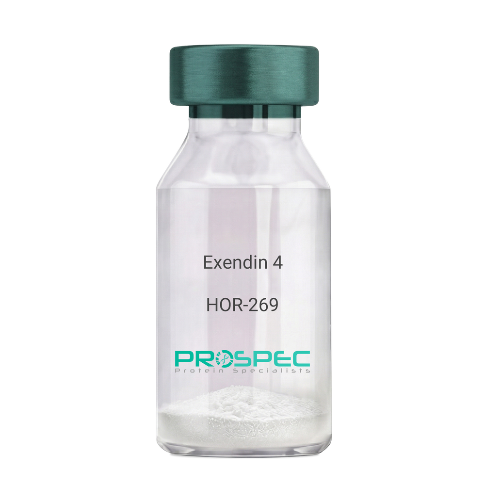
Exendin 4Description:
Exendin-4 Recombinant
Product # :
HOR-269Price :
Quantity :
Shipping Method :
Shipped at Room temp
More Info
- description
- source
- formulation
- purity
- biological activity
- More Info
- Citations
Description
Exendin-4 Recombinant produced in E.Coli is a non-glycosylated polypeptide chain containing 39 amino acids and having a molecular mass of approximately 4.2kDa. Exendin-4 is purified by proprietary chromatographic techniques.
Source
Escherichia Coli.
Formulation
Lyophilized from a 0.2μm filtered concentrated solution in PBS, pH 7.4.
Purity
Greater than 96.0% as determined by:
(a) Analysis by RP-HPLC.
(b) Analysis by SDS-PAGE.Biological Activity
1. Regulates Glucose levels rapidly
2. Reduces Insulin resistance
3. Reduces Glucagon4. Stimulates beta cell growth production.
Citations
More Info
-
Introduction
Exedin-4 Recombinant is a like peptide-1 receptor agonist. The native hormone is produced in the gut of Gila monster Heloderma suspectrum (a type of reptile found in the desert) that stimulates production without causing threateningly low blood sugar, which can occur after using some anti-diabetes products. Recently, researchers used extracted saliva from gila monsters to create an unprecedented breakthrough in Diabetes Type 2 treatment. Unlike other products taken for type 2 diabetes, Exedin-4 has not been linked with weight gain and actually resulted in weight loss, according to the researchers. Exedin-4 enhances glucose-dependant secretion, suppresses inappropriately elevated secretion and slows gastric emptying in vivo. It also promotes B-cell proliferation and neogenesis in vitro and in animal models. Exedin-4 stimulates an increase in acinar cAMP, without stimulating the release of amylase.
-
Physical Appearance
Sterile Filtered White lyophilized (freeze-dried) powder.
-
Stability
Lyophilized Exendin Recombinant although stable at room temperature for 3 weeks, should be stored desiccated below -18°C. Upon reconstitution Exendin-4 should be stored at 4°C between 2-7 days and for future use below -18°C. For long term storage it is recommended to add a carrier protein (0.1% HSA or BSA).Please prevent freeze-thaw cycles.
-
Solubility
It is recommended to reconstitute the lyophilized Exendin Recombinant in sterile 18MΩ-cm H2O not less than 100 µg/ml, which can then be further diluted to other aqueous solutions.
-
Amino Acid Sequence
HGEGTFTSDL SKQMEEEAVR LFIEWLKNGG PSSGAPPPS.
-
Background
What is the molecular weight/Mw of EXENDIN 4 Protein?
EXENDIN 4 Protein has a total Mw of 4.2kDa.
What is the source or expression system of EXENDIN 4 Protein?
Escherichia Coli.
What is the Purity of EXENDIN 4 Protein?
EXENDIN 4 Protein is > 96% pure as determined by SDS-PAGE.
What is the Biological Activity of EXENDIN 4 Protein?
1. Regulates Glucose levels rapidly
2. Reduces Insulin resistance
3. Reduces Glucagon
4. Stimulates beta cell growth production.
What is the amino acid sequence of EXENDIN 4 Protein?
HGEGTFTSDL SKQMEEEAVR LFIEWLKNGG PSSGAPPPS
What applications can EXENDIN 4 Protein be used in?
EXENDIN 4 Protein can probably be used in western blot, ELISA and Lateral Flow.
What is the endotoxin level for EXENDIN 4 Protein?
The endotoxin level is minimal, EXENDIN 4 Protein was purified using conventional chromatography techniques.
Who supplies recombinant EXENDIN 4 that is in stock and available to order?
ProSpec has the protein in stock and available for immediate delivery.
Where can I order bulk mg quantities of EXENDIN 4?
ProSpec supplies bulk mg quantities at affordable prices that meets research and academic budgets.
ProSpec's products are furnished for LABORATORY RESEARCH USE ONLY. The product may not be used as drugs, agricultural or pesticidal products, food additives or household chemicals.
- View Data Sheet
Name :
PDPN P56F7AT AntibodyDescription:
Podoplanin clone P56F7AT, Mouse Anti Human
Podoplanin, Glycoprotein 36, PA2.26 antigen, T1A, GP36, GP40, Gp38, OTS8, T1A2, HT1A-1, PA2.26, T1-alpha, PDPN.
Product # :
ANT-641Price :
Quantity :
Shipping Method :
Shipped with Ice Packs
More Info
- formulation
- More Info
- Citations
Formulation
1mg/ml containing PBS, pH-7.4, 10% Glycerol and 0.02% Sodium Azide.
Citations
More Info
-
Introduction
Podoplanin is a small mucin-like type-1 transmembrane protein, typically expressed in various specialized cell types throughout the body. Podoplanin is a type-I integral membrane glycoprotein with diverse distribution in human tissues. PDPN physiological function is related to its mucin-type character. The homologous protein in other species has been described as a differentiation antigen and influenza-virus receptor.
PDPN is expressed in lymphatic progenitor cells and afterwards during mouse development in lymphatic endothelial cells. Podoplanin is a specific marker for lymph vessel endothelial cells. Over-expression of podoplanin significantly elevates endothelial cell adhesion, migration, and tube formation. Inhibition of Podoplanin expression decreases cell adhesion in human dermal lymphatic endothelial cells. Podoplanin is used as a specific marker for lymphatic endothelium in histopathology.
Podoplanin expression is increased in nearly all human colon, rectum, and small intestine tumors. AGGRUS may serve as a diagnostic marker that distinguishes seminomas, the majority of which over express the protein, from embryonal carcinoma in testicular germ cell tumors. -
Synonyms
Podoplanin, Glycoprotein 36, PA2.26 antigen, T1A, GP36, GP40, Gp38, OTS8, T1A2, HT1A-1, PA2.26, T1-alpha, PDPN.
-
Physical Appearance
Sterile filtered colorless solution.
-
Immunogen
Anti-human PDPN mAb, is derived from hybridization of mouse F0 myeloma cells with spleen cells from BALB/c mice immunized with recombinant human PDPN amino acids 99-207 purified from E. coli.
-
Ig Subclass
Mouse IgG1 heavy chain and k light chain.
-
Clone
P56F7AT.
-
Applications
PDPN antibody has been tested by ELISA, Western blot analysis, Flow cytometry and ICC/IF to assure specificity and reactivity. Since application varies, however, each investigation should be titrated by the reagent to obtain optimal results.
-
Type
Mouse Anti Human Monoclonal.
-
Storage Procedures
For periods up to 1 month store at 4°C, for longer periods of time, store at -20°C. Prevent freeze thaw cycles.
-
Purification Method
PDPN antibody was purified from mouse ascitic fluids by protein-A affinity chromatography.
ProSpec's products are furnished for LABORATORY RESEARCH USE ONLY. The product may not be used as drugs, agricultural or pesticidal products, food additives or household chemicals.
- View Data Sheet
Name :
RAB7A AntibodyDescription:
Mouse Anti Human RAB7A, Member RAS Oncogene Family
RAB7A member RAS oncogene family, RAB7, ras-related protein Rab-7a, FLJ20819, PSN, CMT2B, PRO2706.
Product # :
ANT-702Price :
Quantity :
Shipping Method :
Shipped with Ice Packs
More Info
- formulation
- More Info
- Citations
Formulation
1mg/ml containing PBS, pH-7.4, 10% Glycerol and 0.02% Sodium Azide.
Citations
More Info
-
Introduction
RAB7A belongs to the Ras superfamily of small Rab GTPases. RAB7A connects with the RIPL effector protein to regulator membrane trafficking from early to late endosome and to lysosomes. RAB7A can also regulate growth receptor endocytic trafficking, degradation, maturation of phagosome and autophagic vacuoles.
-
Synonyms
RAB7A member RAS oncogene family, RAB7, ras-related protein Rab-7a, FLJ20819, PSN, CMT2B, PRO2706.
-
Physical Appearance
Sterile filtered colorless solution.
-
Immunogen
Anti-human RAB7A mAb, is derived from hybridization of mouse F0 myeloma cells with spleen cells from BALB/c mice immunized with a recombinant human RAB7A protein 1-207 amino acids purified from E. coli.
-
Ig Subclass
Mouse IgG2a heavy chain and k light chain.
-
Clone
PAT10E4AT.
-
Applications
RAB7A antibody has been tested by ELISA and Western blot analysis to assure specificity and reactivity. Since application varies, however, each investigation should be titrated by the reagent to obtain optimal results.
-
Type
Mouse Anti Human Monoclonal.
-
Storage Procedures
For periods up to 1 month store at 4°C, for longer periods of time, store at -20°C. Prevent freeze thaw cycles.
-
Purification Method
RAB7A antibody was purified from mouse ascitic fluids by protein-A affinity chromatography.
ProSpec's products are furnished for LABORATORY RESEARCH USE ONLY. The product may not be used as drugs, agricultural or pesticidal products, food additives or household chemicals.
- View Data Sheet
Name :
Visfatin AntibodyDescription:
Visfatin, Mouse Anti Human
PBEF, Pre-B cell colony-enhancing factor, Nicotinamide phosphoribosyltransferase NAmPRTase, Nampt, MGC117256, DKFZP666B131, 1110035O14Rik.
Product # :
ANT-368Price :
Quantity :
Shipping Method :
Shipped with Ice Packs
More Info
- formulation
- More Info
- Citations
Formulation
1mg/ml containing PBS, pH-7.4, 10% glycerol & 0.02% Sodium Azide.
Citations
More Info
-
Introduction
Excess adiposity is the most important risk in the development of insulin resistance and type 2 diabetes mellitus (T2DM). Adipose tissue produces several proteins (adipocytokines) such as leptin, adiponectin, resistin, tumor necrosis factor-α, and IL-6, that modulate insulin sensitivity and appear to play an important role in the pathogenesis of insulin resistance, diabetes, dyslipidemia, inflammation, and atherosclerosis. However, the mechanisms by which fat tissue induces insulin resistance and the role of adipocytokines in the pathogenesis of T2DM have not been well established. Visfatin, also known as pre-B cell colony-enhancing factor (PBEF), is a cytokine that is highly expressed in visceral fat and was originally isolated as a secreted factor that synergizes with IL-7 and stem cell factors to promote the growth of B cell precursors. Visfatin homologs have been identified in carp, invertebrate mollusks, and bacteria, as well as in vertebrates, including humans and the mouse. It h
-
Synonyms
PBEF, Pre-B cell colony-enhancing factor, Nicotinamide phosphoribosyltransferase NAmPRTase, Nampt, MGC117256, DKFZP666B131, 1110035O14Rik.
-
Immunogen
Anti-human Visfatin mAb, is derived from hybridization of mouse FO myeloma cells with spleen cells from BALB/c mice immunized with Recombinant human Visfatin amino acids 1-491 purified from E. coli.
-
Ig Subclass
Mouse IgG1heavy chain and κ light chain.
-
Clone
P4D5AT.
-
Applications
Visfatin antibody has been tested by ELISA and Western blot analysis to assure specificity and reactivity. Since application varies, however, each investigation should be titrated by the reagent to obtain optimal results.
-
Type
Mouse Anti Human Monoclonal.
-
Storage Procedures
For periods up to 1 month store at 4°C, for longer periods of time, store at -20°C. Prevent freeze thaw cycles.
-
Purification Method
Visfatin antibody was purified from mouse ascitic fluids by protein-G affinity chromatography.
ProSpec's products are furnished for LABORATORY RESEARCH USE ONLY. The product may not be used as drugs, agricultural or pesticidal products, food additives or household chemicals.
- View Data Sheet
Name :
ANXA8L1 HumanDescription:
Annexin A8 Like-1 Human Recombinant
Annexin A8-like protein 2, bA145E20.2, ANXA8, annexin A8L2.
Product # :
PRO-1138Price :
Quantity :
Shipping Method :
Shipped with Ice Packs
More Info
- description
- source
- formulation
- purity
- More Info
- Citations
Description
ANXA8L1 Human Recombinant produced in E. coli is a single polypeptide chain containing 351 amino acids (1-327) and having a molecular mass of 39.4 kDa.ANXA8L1 is fused to a 24 amino acid His-tag at N-terminus & purified by proprietary chromatographic techniques.
Source
E.coli.
Formulation
The ANXA8L1 solution (1mg/ml) contains 20mM Tris-HCl buffer (pH 8.0), 0.1M NaCl, 1mM DTT and 10% glycerol.
Purity
Greater than 90% as determined by SDS-PAGE.
Citations
More Info
-
Introduction
Annexin A8-like 2 (ANXA8L2) belongs to the annexin family of calcium-dependent phospholipid binding proteins. Annexin family members are regulators of such varied processes such as ion flux, endocytosis and exocytosis, and cellular adhesion. ANXA8L2 is a membrane binding protein with various properties, including voltage-sensitive calcium channel activity, ion selectivity and membrane fusion. ANXA8L2 overexpressionis linked to acute myelocytic leukemia. ANXA8L2 may act as an anticoagulant which indirectly inhibits the thromboplastin-specific complex.
-
Synonyms
Annexin A8-like protein 2, bA145E20.2, ANXA8, annexin A8L2.
-
Physical Appearance
Sterile Filtered colorless solution.
-
Stability
Store at 4°C if entire vial will be used within 2-4 weeks. Store, frozen at -20°C for longer periods of time. For long term storage it is recommended to add a carrier protein (0.1% HSA or BSA).Avoid multiple freeze-thaw cycles.
-
Amino Acid Sequence
MGSSHHHHHH SSGLVPRGSH MGSHMAWWKA WIEQEGVTVK SSSHFNPDPD AETLYKAMKG IGTNEQAIID VLTKRSNTQR QQIAKSFKAQ FGKDLTETLK SELSGKFERL IVALMYPPYR YEAKELHDAM KGLGTKEGVI IEILASRTKN QLREIMKAYE EDYGSSLEED IQADTSGYLE RILVCLLQGS RDDVSSFVDP ALALQDAQDL YAAGENIRGT DEMKFITILC TRSATHLLRV FEEYEKIANK SIEDSIKSET HGSLEEAMLT VVKCTQNLHS YFAERLYYAM KGAGTRDGTL IRNIVSRSEI DLNLIKCHFK KMYGKTLSSM IMEDTSGDYK NALLSLVGSD P
ProSpec's products are furnished for LABORATORY RESEARCH USE ONLY. The product may not be used as drugs, agricultural or pesticidal products, food additives or household chemicals.
- View Data Sheet
Name :
PPP1CA AntibodyDescription:
Protein Phosphatase 1 Catalytic Subunit Alpha, Mouse Anti Human
Serine/threonine-protein phosphatase PP1-alpha catalytic subunit, PP-1A, PPP1CA, PPP1A, protein phosphatase 1 catalytic subunit alpha isozyme, MGC1674, MGC15877, PP1alpha.
Product # :
ANT-415Price :
Quantity :
Shipping Method :
Shipped with Ice Packs
More Info
- formulation
- More Info
- Citations
Formulation
1mg/ml containing PBS, pH-7.4, & 0.1% Sodium Azide.
Citations
More Info
-
Introduction
PPP1CA is one of the 3 catalytic subunits of protein phosphatase 1 (PP1). PP1 is a serine/threonine specific protein phosphatase which is involved in the regulation of a variety of cellular processes, such as cell division, glycogen metabolism, muscle contractility, protein synthesis, and HIV-1 viral transcription. Increased PP1 activity is detected in the end stage of heart failure. Studies in both human and mice suggest that PP1 is a significant regulator of cardiac function. Mouse studies also propose that PP1 functions as a suppressor of learning and memory.
-
Synonyms
Serine/threonine-protein phosphatase PP1-alpha catalytic subunit, PP-1A, PPP1CA, PPP1A, protein phosphatase 1 catalytic subunit alpha isozyme, MGC1674, MGC15877, PP1alpha.
-
Physical Appearance
Sterile Filtered clear solution.
-
Immunogen
Anti-human PPP1CA mAb, is derived from hybridization of mouse FO myeloma cells with spleen cells from BALB/c mice immunized with recombinant human PPP1CA amino acids 30-299 purified from E. coli.
-
Ig Subclass
Mouse IgG2a heavy chain and κ light chain.
-
Clone
P4G3AT.
-
Applications
PPP1CA antibody has been tested by ELISA and Western blot analysis to assure specificity and reactivity. Since application varies, however, each investigation should be titrated by the reagent to obtain optimal results. Recommended dilution range for Western blot analysis is 1:500 ~ 2,000. Recommended starting dilution is 1:1,000.
-
Type
Mouse Anti Human Monoclonal.
-
Storage Procedures
For periods up to 1 month store at 4°C, for longer periods of time, store at -20°C. Prevent freeze thaw cycles.
-
Purification Method
PPP1CA antibody was purified from mouse ascitic fluids by protein-G affinity chromatography.
ProSpec's products are furnished for LABORATORY RESEARCH USE ONLY. The product may not be used as drugs, agricultural or pesticidal products, food additives or household chemicals.
- View Data Sheet
Name :
ACTB AntibodyDescription:
Actin Beta, Mouse Anti Human
Actin Beta, PS1TP5-Binding Protein 1, Beta Cytoskeletal Actin, Actin, Cytoplasmic 1, Beta-Actin, PS1TP5BP1, BRWS1, ACTB.
Product # :
ANT-687Price :
Quantity :
Shipping Method :
Shipped with Ice Packs
More Info
- formulation
- More Info
- Citations
Formulation
1mg/ml containing PBS, pH-7.4, 10% Glycerol and 0.02% Sodium Azide.
Citations
More Info
-
Introduction
Actin Beta (ACTB) is one of 6 different actin isoforms which have been identified in humans. ACTB is one of the 2 nonmuscle cytoskeletal actins. Actins are highly conserved proteins which are involved in cell motility, structure and integrity. Alpha actins are a key component of the contractile apparatus.
-
Synonyms
Actin Beta, PS1TP5-Binding Protein 1, Beta Cytoskeletal Actin, Actin, Cytoplasmic 1, Beta-Actin, PS1TP5BP1, BRWS1, ACTB.
-
Physical Appearance
Sterile filtered colorless solution.
-
Immunogen
Anti-human ACTB mAb, is derived from hybridization of mouse F0 myeloma cells with spleen cells from BALB/c mice immunized with recombinant human ACTB amino acids 1-375 purified from E. coli.
-
Ig Subclass
Mouse IgG1 heavy chain and κ light chain.
-
Clone
PAT4G6AT.
-
Applications
ACTB antibody has been tested by ELISA, Western blot analysis and ICC/IF to assure specificity and reactivity. Since application varies, however, each investigation should be titrated by the reagent to obtain optimal results.
-
Type
Mouse Anti Human Monoclonal.
-
Storage Procedures
For periods up to 1 month store at 4°C, for longer periods of time, store at -20°C. Prevent freeze thaw cycles.
-
Purification Method
ACTB antibody was purified from mouse ascitic fluids by protein-A affinity chromatography.
ProSpec's products are furnished for LABORATORY RESEARCH USE ONLY. The product may not be used as drugs, agricultural or pesticidal products, food additives or household chemicals.
- View Data Sheet
Name :
ANXA1 AntibodyDescription:
Annexin A1, Mouse Anti Human
ANX1, LPC1, ANXA1, Lipocortin I, Calpactin II, Chrombindin-9, p35, Annexin-1, Phospholipase A2 inhibitory protein, Annexin I, Annexin A1.
Product # :
ANT-562Price :
Quantity :
Shipping Method :
Shipped with Ice Packs
More Info
- formulation
- More Info
- Citations
Formulation
1mg/ml containing PBS, pH-7.4, 10% Glycerol and 0.02% Sodium Azide.
Citations
More Info
-
Introduction
ANXA1 is part of the family of Ca(2+)-dependent phospholipid binding proteins which have a Mw between 35kDa-40kDa and are situated on the cytosolic face of the plasma membrane. ANXA1 protein has a Mw of 40kDa, with phospholipase A2 inhibitory activity to bind from two to four calcium ions with high affinity. Since phospholipase A2 is necessary for the biosynthesis of the potent mediators of inflammation, prostaglandins and leukotrienes, ANXA1 might have potential anti-inflammatory activity. ANXA1 promotes membrane fusion and iplays a role in exocytosis. The recognition of ANXA1 protein by immunocytochemical leads a simple, highly sensitive and specific assay for diagnosis of hairy cell leukemia.
-
Synonyms
ANX1, LPC1, ANXA1, Lipocortin I, Calpactin II, Chrombindin-9, p35, Annexin-1, Phospholipase A2 inhibitory protein, Annexin I, Annexin A1.
-
Physical Appearance
Sterile filtered colorless solution.
-
Immunogen
Anti-human ANXA1 mAb, clone PAT2G5AT, is derived from hybridization of mouse F0 myeloma cells with spleen cells from BALB/c mice immunized with a recombinant human ANXA1 protein 1-346 amino acids purified from E. coli.
-
Ig Subclass
Mouse IgG1 heavy chain and k light chain.
-
Clone
PAT2G5AT
-
Applications
The antibody has been tested by ELISA, Western blot analysis to assure specificity and reactivity. Since application varies, however, each investigation should be titrated by the reagent to obtain optimal results.
-
Type
Mouse Anti Human Monoclonal.
-
Storage Procedures
For periods up to 1 month store at 4°C, for longer periods of time, store at -20°C. Prevent freeze thaw cycles.
-
Purification Method
ANXA1 antibody was purified from mouse ascitic fluids by protein-A affinity chromatography.
ProSpec's products are furnished for LABORATORY RESEARCH USE ONLY. The product may not be used as drugs, agricultural or pesticidal products, food additives or household chemicals.
- View Data Sheet
Name :
Clusterin AntibodyDescription:
Clusterin, Mouse Anti Human
CLI, AAG4, KUB1, SGP2, SGP-2, SP-40, TRPM2, MGC24903, Complement-associated protein SP-40,40, Complement cytolysis inhibitor, NA1/NA2, Apolipoprotein J, Apo-J, Testosterone-repressed prostate message 2, TRPM-2.
Product # :
ANT-314Price :
Quantity :
Shipping Method :
Shipped with Ice Packs
More Info
- formulation
- More Info
- Citations
Formulation
1mg/ml containing PBS, pH-7.4, 10% Glycerol & 0.02% Sodium Azide.
Citations
More Info
-
Introduction
Clusterin also named Apolipoprotein J (APO-J) is a 75-80 kD disulfide-linked heterodimeric protein containing about 30% of N-linked carbohydrate rich in sialic acid but truncated forms targeted to the nucleus have also been identified. The precursor polypeptide chain is cleaved proteolytically to remove the 22-mer secretory signal peptide and subsequently between residues 227/228 to generate the ? and ? chains. These are assembled in anti-parallel to give a heterodimeric molecule in which the cysteine-rich centers are linked by five disulfide bridges and are flanked by two predicted coiled-coil ? -helices and three predicted amphipathic a-helices. Across a broad range of species clusterin shows a high degree of sequence homology ranging from 70% to 80%. It is nearly ubiquitously expressed in most mammalian tissues and can be found in plasma, milk, urine, cerebrospinal fluid and semen. It is able to bind and form complexes with numerous partners such as immunoglobulins, lipids, heparin, b
-
Synonyms
CLI, AAG4, KUB1, SGP2, SGP-2, SP-40, TRPM2, MGC24903, Complement-associated protein SP-40,40, Complement cytolysis inhibitor, NA1/NA2, Apolipoprotein J, Apo-J, Testosterone-repressed prostate message 2, TRPM-2.
-
Physical Appearance
Sterile Filtered clear solution.
-
Immunogen
Anti-human Clusterin mAb, is derived from hybridization of mouse SP2/O myeloma cells with spleen cells from BALB/c mice immunized with Recombinant human Clusterin amino acids 1-333 purified from E. coli.
-
Ig Subclass
Mouse IgG1 heavy chain and κ light chain.
-
Clone
P1A11AT.
-
Applications
Clusterin antibody has been tested by ELISA, Western blot, ICC/IF, IHC and FACS analysis to assure specificity and reactivity. Since application varies, however, each investigation should be titrated by the reagent to obtain optimal results.
-
Type
Mouse Anti Human Monoclonal.
-
Storage Procedures
For periods up to 1 month store at 4°C, for longer periods of time, store at -20°C. Prevent freeze thaw cycles.
-
Purification Method
Clusterin antibody was purified from mouse ascitic fluids by protein-G affinity chromatography.
ProSpec's products are furnished for LABORATORY RESEARCH USE ONLY. The product may not be used as drugs, agricultural or pesticidal products, food additives or household chemicals.
- View Data Sheet
Name :
IL 1 Beta Antibody, BiotinDescription:
Interleukin-1b, Mouse Anti-Human Biotin
Catabolin, Lymphocyte-activating factor (LAF), Endogenous Pyrogen (EP), Leukocyte Endogenous Mediator (LEM), Mononuclear Cell Factor (MCF), IL1F2, IL-1 beta.
Product # :
ANT-249Price :
Quantity :
Shipping Method :
Shipped at Room temp
More Info
- formulation
- More Info
- Citations
Formulation
1 mg/ml in PBS (after reconstitution).
Citations
More Info
-
Introduction
Interleukin-1b is produced by activated macrophages, IL-1B stimulates thymocyte proliferation by inducing il-2 release, b-cell maturation and proliferation, and fibroblast growth factor activity. IL1B proteins are involved in the inflammatory response, being identified as endogenous pyrogens, and are reported to stimulate the release of prostaglandin and collagenase from synovial cells.
-
Synonyms
Catabolin, Lymphocyte-activating factor (LAF), Endogenous Pyrogen (EP), Leukocyte Endogenous Mediator (LEM), Mononuclear Cell Factor (MCF), IL1F2, IL-1 beta.
-
Solubility
Reconstitute with sterile H20. Mix gently, wash the sides of the vial and wait 30-60 seconds before use.
-
Immunogen
r.Human IL-1b.
-
Ig Subclass
Mouse IgG2b.
-
Clone
NYR-hIL1b.
-
Titer
For intra-cellular staining, 10µl per 1,000,000 cells. By direct ELISA, 1:10,000 dilution will yield >1.0 O.D using alkaline phosphatase conjugated rabbit anti-mouse Ig (Jackson Laboratories).
-
Shipping Conditions
Antibody is shipped lyophilized at ambient temperature.
-
Type
Mouse Anti Human Monoclonal.
-
Storage Procedures
In lyophilized form, for long periods, store at 4°C in a dry environment. After reconstitution, if not intended for use within a month, aliquot and store at -20°C.
-
Purification Method
Ion Exchange.
ProSpec's products are furnished for LABORATORY RESEARCH USE ONLY. The product may not be used as drugs, agricultural or pesticidal products, food additives or household chemicals.