Skip to content

High-quality research proteins.

  • 1-800-805-6531

  • Mon - Sat 8.00-18.00, Sun - Closed

  • info@prospecbio.com

  • PO Box 6591 East Brunswick NJ 08816

  • Products
    • Cytokines
    • Growth factors
    • Chemokines
    • Cd antigens
    • Neurotrophins
    • Hormones
    • Enzymes
    • Viral antigens
    • Recombinant proteins
    • Natural proteins
    • Monoclonal antibodies
    • Polyclonal antibodies
    • Thermal Packaging
  • About
    • Company
    • Founder
    • Contribution
  • Customers
    • Eastern Asia
    • Eastern Europe
    • North America
    • South America
    • South East Asia
    • Southern Asia
    • Western Europe
  • Ordering
    • Ordering Method
    • Ordering - Credit Card
    • Ordering - PO
    • Shipping Fees
    • FAQs
  • Services
    • DNA Cloning
    • Protein Expression
    • Monoclonal Antibody
    • Fermentation
    • Technology License
  • Contact us
Log in

Country/region

  • USD $ | Albania
  • USD $ | Andorra
  • USD $ | Argentina
  • USD $ | Armenia
  • USD $ | Australia
  • USD $ | Austria
  • USD $ | Azerbaijan
  • USD $ | Belarus
  • USD $ | Belgium
  • USD $ | Bolivia
  • USD $ | Brazil
  • USD $ | Bulgaria
  • USD $ | Cambodia
  • USD $ | Canada
  • USD $ | Chile
  • USD $ | China
  • USD $ | Colombia
  • USD $ | Costa Rica
  • USD $ | Croatia
  • USD $ | Cyprus
  • USD $ | Czechia
  • USD $ | Denmark
  • USD $ | Estonia
  • USD $ | Finland
  • USD $ | France
  • USD $ | Georgia
  • USD $ | Germany
  • USD $ | Greece
  • USD $ | Hong Kong SAR
  • USD $ | Hungary
  • USD $ | Iceland
  • USD $ | India
  • USD $ | Ireland
  • USD $ | Israel
  • USD $ | Italy
  • USD $ | Japan
  • USD $ | Kazakhstan
  • USD $ | Latvia
  • USD $ | Liechtenstein
  • USD $ | Lithuania
  • USD $ | Madagascar
  • USD $ | Malta
  • USD $ | Mexico
  • USD $ | Moldova
  • USD $ | Monaco
  • USD $ | Netherlands
  • USD $ | New Zealand
  • USD $ | North Macedonia
  • USD $ | Norway
  • USD $ | Panama
  • USD $ | Paraguay
  • USD $ | Peru
  • USD $ | Philippines
  • USD $ | Poland
  • USD $ | Portugal
  • USD $ | Romania
  • USD $ | Singapore
  • USD $ | Slovakia
  • USD $ | Slovenia
  • USD $ | South Africa
  • USD $ | South Korea
  • USD $ | Spain
  • USD $ | Sri Lanka
  • USD $ | Sweden
  • USD $ | Switzerland
  • USD $ | Taiwan
  • USD $ | Thailand
  • USD $ | Türkiye
  • USD $ | Ukraine
  • USD $ | United Kingdom
  • USD $ | United States
  • USD $ | Uruguay
  • USD $ | Vatican City
  • USD $ | Venezuela
  • USD $ | Vietnam
  • Facebook
  • Instagram
logoProSpec - Recombinant Protein Manufacturer for academic and R&D industry
Become a distributor
Account Log in
Wishlist
Cart Cart(0)
  • Products
    +
    • Cytokines
    • Growth factors
    • Chemokines
    • Cd antigens
    • Neurotrophins
    • Hormones
    • Enzymes
    • Viral antigens
    • Recombinant proteins
    • Natural proteins
    • Monoclonal antibodies
    • Polyclonal antibodies
    • Thermal Packaging
  • About
    +
    • Company
    • Founder
    • Contribution
  • Customers
    +
    • Eastern Asia
    • Eastern Europe
    • North America
    • South America
    • South East Asia
    • Southern Asia
    • Western Europe
  • Ordering
    +
    • Ordering Method
    • Ordering - Credit Card
    • Ordering - PO
    • Shipping Fees
    • FAQs
  • Services
    +
    • DNA Cloning
    • Protein Expression
    • Monoclonal Antibody
    • Fermentation
    • Technology License
  • Contact us

Item added to your cart

View cart
View All
  • Cytokines
  • Growth factors
  • Chemokines
  • Cd antigens
  • Neurotrophins
  • Hormones
  • Enzymes
  • Viral antigens
  • Recombinant proteins
  • Natural proteins
  • Monoclonal antibodies
  • Polyclonal antibodies
  • Thermal Packaging
  • Tumor Necrosis Factor

    Tumor Necrosis Factor

    View All
  • 4-1BB

    4-1BB

    View All
  • Adiponectin

    Adiponectin

    View All
  • AIF1

    AIF1

    View All
  • Other Cytokines

    Other Cytokines

    View All
  • AITRL

    AITRL

    View All
  • Angiopoietin

    Angiopoietin

    View All
  • Apolipoprotein

    Apolipoprotein

    View All
  • B-Cell Activating Factor

    B-Cell Activating Factor

    View All
  • Beta Defensin

    Beta Defensin

    View All
  • Bone Morphogenetic Protein

    Bone Morphogenetic Protein

    View All
  • B type Natriuretic Peptide

    B type Natriuretic Peptide

    View All
  • BST

    BST

    View All
  • Betacellulin

    Betacellulin

    View All
  • Cardiotrophin

    Cardiotrophin

    View All
  • Activin

    Activin

    View All
  • Fibroblast Growth Factor

    Fibroblast Growth Factor

    View All
  • Other Growth Factors

    Other Growth Factors

    View All
  • CSF

    CSF

    View All
  • CTGF

    CTGF

    View All
  • IGFBP

    IGFBP

    View All
  • VEGF Protein

    VEGF Protein

    View All
  • EGF

    EGF

    View All
  • Epigen

    Epigen

    View All
  • Erythropoietin

    Erythropoietin

    View All
  • Insulin-Like Growth Factor

    Insulin-Like Growth Factor

    View All
  • Growth Hormone

    Growth Hormone

    View All
  • HDGF

    HDGF

    View All
  • Hepatocyte Growth Factor

    Hepatocyte Growth Factor

    View All
  • Insulin

    Insulin

    View All
  • BCA-1/ BLC (CXCL13)

    BCA-1/ BLC (CXCL13)

    View All
  • BRAK (CXCL14)

    BRAK (CXCL14)

    View All
  • C-10 (CCL6)

    C-10 (CCL6)

    View All
  • Other Chemokines

    Other Chemokines

    View All
  • MEC (CCL28)

    MEC (CCL28)

    View All
  • LD78-beta (CCL3L1)

    LD78-beta (CCL3L1)

    View All
  • CTACK (CCL27)

    CTACK (CCL27)

    View All
  • CXCL16

    CXCL16

    View All
  • CXCL17

    CXCL17

    View All
  • Platelet Factor-4 (CXCL4)

    Platelet Factor-4 (CXCL4)

    View All
  • ENA-78 (CXCL5)

    ENA-78 (CXCL5)

    View All
  • Eotaxin (CCL11,24,26)

    Eotaxin (CCL11,24,26)

    View All
  • Exodus-2 (CCL21)

    Exodus-2 (CCL21)

    View All
  • Fractalkine (CX3CL1)

    Fractalkine (CX3CL1)

    View All
  • CXCL6

    CXCL6

    View All
  • Other CD Antigens

    Other CD Antigens

    View All
  • CD14

    CD14

    View All
  • CD164

    CD164

    View All
  • CD1

    CD1

    View All
  • CD2

    CD2

    View All
  • CD200

    CD200

    View All
  • CD207

    CD207

    View All
  • CD226

    CD226

    View All
  • CD23

    CD23

    View All
  • CD244

    CD244

    View All
  • CD247

    CD247

    View All
  • CD27

    CD27

    View All
  • CD274

    CD274

    View All
  • CD300

    CD300

    View All
  • CD33

    CD33

    View All
  • Other Neurotrophins

    Other Neurotrophins

    View All
  • Beta-NGF

    Beta-NGF

    View All
  • BDNF

    BDNF

    View All
  • CDNF

    CDNF

    View All
  • CNTF

    CNTF

    View All
  • GDNF

    GDNF

    View All
  • Glia Maturation Factor

    Glia Maturation Factor

    View All
  • MANF

    MANF

    View All
  • Midkine

    Midkine

    View All
  • Neuroglobin

    Neuroglobin

    View All
  • Neurotrophic factors

    Neurotrophic factors

    View All
  • Neuregulin

    Neuregulin

    View All
  • Neuropilin

    Neuropilin

    View All
  • Pigment Epithelium-Derived Factor

    Pigment Epithelium-Derived Factor

    View All
  • Pleiotrophin

    Pleiotrophin

    View All
  • Peptide Hormones

    Peptide Hormones

    View All
  • Other Hormones

    Other Hormones

    View All
  • HCG

    HCG

    View All
  • Endothelin

    Endothelin

    View All
  • Exendin

    Exendin

    View All
  • FSH

    FSH

    View All
  • Glucagon

    Glucagon

    View All
  • GHRP

    GHRP

    View All
  • GLP

    GLP

    View All
  • Thyrostimulin

    Thyrostimulin

    View All
  • Inhibin A

    Inhibin A

    View All
  • LHRH

    LHRH

    View All
  • Melanotan

    Melanotan

    View All
  • Procalcitonin

    Procalcitonin

    View All
  • PTH

    PTH

    View All
  • Peroxiredoxin

    Peroxiredoxin

    View All
  • Peptidase

    Peptidase

    View All
  • Synthetase

    Synthetase

    View All
  • Transferase

    Transferase

    View All
  • Hydrolase

    Hydrolase

    View All
  • Dehydrogenase

    Dehydrogenase

    View All
  • ACE2

    ACE2

    View All
  • Esterase

    Esterase

    View All
  • Protein Kinases

    Protein Kinases

    View All
  • Other Enzymes

    Other Enzymes

    View All
  • Phosphatase

    Phosphatase

    View All
  • Deaminase

    Deaminase

    View All
  • Oxygenase

    Oxygenase

    View All
  • Adenylate Kinase

    Adenylate Kinase

    View All
  • Lyase

    Lyase

    View All
  • Chagas

    Chagas

    View All
  • SARS

    SARS

    View All
  • Influenza

    Influenza

    View All
  • ASFV

    ASFV

    View All
  • Astrovirus

    Astrovirus

    View All
  • Borrelia

    Borrelia

    View All
  • Helicobacter Pylori

    Helicobacter Pylori

    View All
  • Chikungunya

    Chikungunya

    View All
  • Chlamydia

    Chlamydia

    View All
  • Cytomegalo

    Cytomegalo

    View All
  • Dengue

    Dengue

    View All
  • EBV

    EBV

    View All
  • Ebola

    Ebola

    View All
  • Feline Leukemia Virus

    Feline Leukemia Virus

    View All
  • Hepatitis A

    Hepatitis A

    View All
  • Other

    Other

    View All
  • Actin

    Actin

    View All
  • ADAM

    ADAM

    View All
  • Complement Component

    Complement Component

    View All
  • Ag85

    Ag85

    View All
  • Eukaryotic Translation Initiation Factor

    Eukaryotic Translation Initiation Factor

    View All
  • Anterior Gradient Protein

    Anterior Gradient Protein

    View All
  • Heat Shock Protein

    Heat Shock Protein

    View All
  • Alpha-2-HS-Glycoprotein

    Alpha-2-HS-Glycoprotein

    View All
  • Hemoglobin

    Hemoglobin

    View All
  • Allergy

    Allergy

    View All
  • Anaplasma

    Anaplasma

    View All
  • Angiogenin

    Angiogenin

    View All
  • Ankyrin Repeat Domain

    Ankyrin Repeat Domain

    View All
  • Annexin

    Annexin

    View All
  • Other Natural Proteins

    Other Natural Proteins

    View All
  • Aprotinin

    Aprotinin

    View All
  • Transferrin

    Transferrin

    View All
  • Avidin

    Avidin

    View All
  • Anti Coagulation Factors

    Anti Coagulation Factors

    View All
  • Natural Albumin

    Natural Albumin

    View All
  • Natural Coagulation Factors

    Natural Coagulation Factors

    View All
  • Fibronectin

    Fibronectin

    View All
  • Thrombin

    Thrombin

    View All
  • Anti Human Enzyme

    Anti Human Enzyme

    View All
  • Other Monoclonal Antibodies

    Other Monoclonal Antibodies

    View All
  • Anti Human Cytokine

    Anti Human Cytokine

    View All
  • Anti Human Heat Shock Protein

    Anti Human Heat Shock Protein

    View All
  • Anti Mouse Lymphocyte

    Anti Mouse Lymphocyte

    View All
  • Anti Human Chemokine

    Anti Human Chemokine

    View All
  • Anti Human Lymphocyte

    Anti Human Lymphocyte

    View All
  • Anti Viral Monoclonal

    Anti Viral Monoclonal

    View All
  • Anti Mouse Cytokine

    Anti Mouse Cytokine

    View All
  • Anti-GST Monoclonal

    Anti-GST Monoclonal

    View All
  • Other Polyclonal Antibodies

    Other Polyclonal Antibodies

    View All
  • Anti Viral

    Anti Viral

    View All
  • Anti-GST Monoclonal

    Anti-GST Monoclonal

    View All
Back

Search results

602 results found for “candida albicans”

Name

Description

Product #

Price

Quantity

Shipping Method

  • View Data Sheet

    Name :

    VZV ORF9

    Description:

    Varicella Zoster Virus ORF9 Recombinant

    Product # :

    VZV-232

    Price :

    Quantity :

    Shipping Method :

    Ice Icon

    Shipped with Ice Packs

    Add To Cart

    More Info

    • description
    • formulation
    • purity
    • More Info

    Description

    The E.Coli derived recombinant protein contains the VZV ORF9 immunodominant regions, amino acids 6-28, 76-100.

    Formulation

    25mM Tris-Hcl pH 8.0, 1mM EDTA and 50% glycerol.

    Purity

    Varicella protein is >95% pure as determined by 10% PAGE (coomassie staining).

    More Info

    • Introduction

      VZV is closely related to the herpes simplex viruses(HSV), sharing much genomehomology. The known envelope glycoproteins (gB, gC, gE, gH, gI, gK, gL) correspond with those in HSV, however there is no equivalent of HSV gD. VZV virons are spherical and 150-200 nm in diameter. Their lipidenvelope encloses the nucleocapsid of 162 capsomeres arranged in a hexagonalform. Its DNAis a single, linear, double-stranded molecule, 125,000 nt long. The virus is very susceptible to disinfectants, notably sodium hypochlorite. Within the body it can be treated by a number of drugs and therapeutic agents including aciclovir, zoster-immune globulin(ZIG), and vidarabine.

    • Stability

      Varicella protein although stable at 4°C for 1 week, should be stored below -18°C. Please prevent freeze thaw cycles.

    • Specificity

      Immunoreactive with sera of VZV-infected individuals.

    • Purification Method

      Varicella was purified by proprietary chromatographic technique.

    ProSpec's products are furnished for LABORATORY RESEARCH USE ONLY. The product may not be used as drugs, agricultural or pesticidal products, food additives or household chemicals.

    Vzv Orf9
  • View Data Sheet

    Name :

    Dengue-2 C-terminal

    Description:

    Dengue-2 Envelope Hydrophobic Domain Recombinant

    Product # :

    DEN-039

    Price :

    Quantity :

    Shipping Method :

    Ice Icon

    Shipped with Ice Packs

    Add To Cart

    More Info

    • description
    • source
    • formulation
    • purity
    • More Info

    Description

    The Recombinant Dengue 2 Envelope Hydrophobic Domain e.coli derived contains 225 amino acids (228-485) from dengue 2 envelope. The protein is fused to a His tag at C-terminal and purified by standard chromatography techniques.

    Source

    E.coli.

    Formulation

    PBS.

    Purity

    Protein is >95% pure as determined by 10% PAGE (coomassie staining).

    More Info

    • Introduction

      Caused by one of four closely related virus serotypes of the genus Flavivirus, family Flaviviridae, each serotype is sufficiently different that there is no cross-protection and epidemics caused by multiple serotypes (hyperendemicity) can occur. In cell culture experiments and mice Morpholino antisense oligos have shown specific activity against Dengue virus.

    • Physical Appearance

      Sterile Filtered clear solution.

    • Stability

      Dengue 2 Envelope Recombinant although stable at 4°C for 1 week, should be stored below -18°C. Please prevent freeze thaw cycles.

    • Applications

      Immunoassay.

    ProSpec's products are furnished for LABORATORY RESEARCH USE ONLY. The product may not be used as drugs, agricultural or pesticidal products, food additives or household chemicals.

    Dengue 2 C Terminal
  • View Data Sheet

    Name :

    Chlamydia W4-W5

    Description:

    Chlamydia Trachomatis W4-W5 Recombinant

    Product # :

    CHT-004

    Price :

    Quantity :

    Shipping Method :

    Ice Icon

    Shipped with Ice Packs

    Add To Cart

    More Info

    • description
    • source
    • formulation
    • purity
    • More Info

    Description

    The E.coli derived recombinant 6xHis fusion at C-terminus protein contains Chlamydia Trachomatis MOMP protein epitopes, 191-354 amino acids.

    Source

    Escherichia Coli.

    Formulation

    10mM Tris-HCl, pH 6.5, 100mM Sodium Phosphate and 8M urea.

    Purity

    Chlamydia W4-W5 protein is >95% pure as determined by 10% PAGE (coomassie staining) and RP-HPLC.

    More Info

    • Introduction

      Chlamydia is a common term for infection with any bacterium belonging to the phylum Chlamydiae. This term derives from the name of the bacterial genus Chlamydiain the family Chlamydiaceae, order Chlamydiales, class and phylum Chlamydiae. There are two genera in Chlamydiaceae: Chlamydia and Chlamydophila. The genus Chlamydia includes three species: C. trachomatis, C. muridarum, and C. suis.

    • Stability

      Chlamydia W4-W5 protein although stable at 4°C for 1 week, should be stored below -18°C. Please prevent freeze thaw cycles.

    • Applications

      Chlamydia W4-W5 is suitable for use in ELISA. Each laboratory should determine an optimum working titer for use in its particular application. Other applications have not been tested but use in such assays should not necessarily be excluded.

    • Specificity

      Immunoreactive with sera of Chlamydia Trachomatis infected individuals.

    • Purification Method

      Chlamydia W4-W5 protein was purified by proprietary chromatographic technique.

    ProSpec's products are furnished for LABORATORY RESEARCH USE ONLY. The product may not be used as drugs, agricultural or pesticidal products, food additives or household chemicals.

    Chlamydia W4 W5
  • View Data Sheet

    Name :

    TBEV Core

    Description:

    Tick-Borne Encephalitis Virus Core Protein Recombinant

    Product # :

    TBE-285

    Price :

    Quantity :

    Shipping Method :

    Ice Icon

    Shipped with Ice Packs

    Add To Cart

    More Info

    • description
    • source
    • formulation
    • purity
    • More Info

    Description

    The E.coli derived recombinant protein contains the Tick-borne Encephalitis Virus core protein epitopes.

    Source

    Escherichia Coli.

    Formulation

    20mM MES pH 6.5, 8M urea, 200mM NaCl & 0.05% Tween-20.

    Purity

    Encephalitis protein is >95% pure as determined by 10% PAGE (coomassie staining).

    More Info

    • Introduction

      TBE is caused by tick-borne encephalitis virus (TBEV), a member of the family Flaviviridae.
      A closely related virus in Far Eastern Eurasia, Russian spring-summer encephalitis virus (RSSEV).
      The family Flaviviridae includes other tick-borne viruses are closely related to TBEV and RSSEV, such as Omsk hemorrhagic fever virus & Kyasanur Forest virus.
      Louping ill virus is also a member of this family.

    • Stability

      Encephalitis protein although stable at 4°C for 1 week, should be stored below -18°C. Please prevent freeze thaw cycles.

    • Applications

      Encephalitis antigen is suitable for ELISA and Western blots, excellent antigen for detection of Tick-borne encephalitis virus with minimal specificity problems.

    • Specificity

      Immunoreactive with sera of encephalitis virus infected individuals.

    • Purification Method

      Encephalitis protein was purified by proprietary chromatographic technique.

    ProSpec's products are furnished for LABORATORY RESEARCH USE ONLY. The product may not be used as drugs, agricultural or pesticidal products, food additives or household chemicals.

    Tbev Core
  • View Data Sheet

    Name :

    BD 1 Rat

    Description:

    Beta Defensin -1 Rat Recombinant

    Beta-defensin 1, BD-1, rBD-1, Defensin beta 1, Defb1.

    Product # :

    CYT-062

    Price :

    Quantity :

    Shipping Method :

    Room Temp Icon

    Shipped at Room temp

    Add To Cart

    More Info

    • description
    • source
    • formulation
    • purity
    • biological activity
    • More Info

    Description

    BD-1 Rat Recombinant produced in E.coli is a single, non-glycosylated polypeptide chain containing 37 amino acids and having a molecular mass of 4.1kDa.The BD-1 is purified by proprietary chromatographic techniques.

    Source

    Escherichia Coli.

    Formulation

    BD-1 protein was lyophilized from a 0.2µm filtered concentrated solution in PBS, pH 7.4.

    Purity

    Greater than 95.0% as determined by:
    (a) Analysis by RP-HPLC.
    (b) Analysis by SDS-PAGE.

    Biological Activity

    Measured by its ability to chemoattract CD34+ dendritic cells using a concentration range of 0.1-1.0 ug/ml.

    More Info

    • Introduction

      The Defensin family are highly similar in their protein sequence and are microbicidal & cytotoxic peptides made by neutrophils. Beta Defensin-1 is an antimicrobial peptide having the resistance of epithelial surfaces to microbial colonization. Beta Defensin-1 has close proximity to Defensin Alpha-1 and has been implicated in the pathogenesis of cystic fibrosis.
      Skin of patients having atopic dermatitis patients and mycosis fungoides (non-lesional and lesional) show lower human Beta Defensin-1 mRNA expression and higher human Beta Defensin-2 and human Beta Defensin-3 mRNA expression.
      Beta Defensin is highly expressed by epithelial cells.
      Beta-defensin 1 may play a role in the pathogenesis of severe sepsis.

    • Synonyms

      Beta-defensin 1, BD-1, rBD-1, Defensin beta 1, Defb1.

    • Physical Appearance

      Sterile Filtered White lyophilized (freeze-dried) powder.

    • Stability

      Lyophilized BD-1 although stable at room temperature for 3 weeks, should be stored desiccated below -18°C. Upon reconstitution BD-1 should be stored at 4°C between 2-7 days and for future use below -18°C. For long term storage it is recommended to add a carrier protein (0.1% HSA or BSA). Please prevent freeze-thaw cycles.

    • Solubility

      It is recommended to reconstitute the lyophilized BD-1 in sterile 18M-cm H2O not less than 100µg/ml, which can then be further diluted to other aqueous solutions.

    • Amino Acid Sequence

      DQYRCLQNGG FCLRSSCPSH TKLQGTCKPD KPNCCRS.

    • Background

      What is the molecular weight/Mw of BD1 Protein?
      BD1 Protein has a total Mw of 4.1kDa.

      What is the source or expression system of BD1 Protein?
      Escherichia Coli.

      What is the Purity of BD1 Protein?
      BD1 Protein is >97% pure as determined by SDS-PAGE.

      What is the Biological Activity of BD1 Protein?
      Measured by its ability to chemoattract CD34+ dendritic cells using a concentration range of 0.1-1.0 ug/ml.

      What is the amino acid sequence of BD1 Protein?
      DQYRCLQNGG FCLRSSCPSH TKLQGTCKPD KPNCCRS.

      What applications can BD1 Protein be used in?
      BD1 Protein can probably be used in western blot, ELISA and Lateral Flow.

      What is the endotoxin level for BD1 Protein?
      The endotoxin level is minimal, BD1 Protein was purified using conventional chromatography techniques.

    ProSpec's products are furnished for LABORATORY RESEARCH USE ONLY. The product may not be used as drugs, agricultural or pesticidal products, food additives or household chemicals.

    Bd 1 Rat
  • View Data Sheet

    Name :

    CoV-2 S1 (16-685), Fc

    Description:

    Coronavirus 2019 Spike Glycoprotein-S1 (16-685 a.a.), Fc Recombinant

    Product # :

    SARS-027

    Price :

    Quantity :

    Shipping Method :

    Room Temp Icon

    Shipped at Room temp

    Add To Cart

    More Info

    • description
    • source
    • formulation
    • purity
    • More Info

    Description

    The HEK293 derived recombinant protein contains the Coronavirus 2019 CoV-2 Spike Glycoprotein S1, Wuhan-Hu-1 strain, amino acids 16-685 fused to Fc tag at C-terminal.

    Source

    HEK293 Cells.

    Formulation

    CoV-2 S1 protein is supplied in sodium chloride, glycine, arginine pH-7.5 & 5% trehalose.

    Purity

    Protein is >95% pure as determined SDS-PAGE.

    More Info

    • Introduction

      A human infecting coronavirus (viral pneumonia) called 2019 novel coronavirus, 2019-nCoV was found in the fish market at the city of Wuhan, Hubei province of China on December 2019.

      The 2019-nCoV shares an 87% identity to the 2 bat-derived severe acute respiratory syndrome 2018 SARS-CoV-2 located in Zhoushan of eastern China. 2019-nCoV has an analogous receptor-BD-structure to that of 2018 SARS-CoV, even though there is a.a. diversity so thus the 2019-nCoV might bind to ACE2 receptor protein (angiotensin-converting enzyme 2) in humans.

      While bats are possibly the host of 2019-nCoV, researchers suspect that animal from the ocean sold at the seafood market was an intermediate host. RSCU analysis proposes that the 2019-nCoV is a recombinant within the viral spike glycoprotein between the bat coronavirus and an unknown coronavirus.

    • Physical Appearance

      Sterile Filtered White lyophilized (freeze-dried) powder.

    • Stability

      Lyophilized Cov-2 Spike S1 Glycoprotein although stable at room temperature for 3 weeks, should be stored desiccated below -18°C. Upon reconstitution CoV2 Spike protein should be stored at 4°C between 2-7 days and for future use below -18°C.For long term storage it is recommended to add a carrier protein (0.1% HSA or BSA).Please prevent freeze-thaw cycles.

    • Purification Method

      Purified by Protein-G chromatographic technique.

    ProSpec's products are furnished for LABORATORY RESEARCH USE ONLY. The product may not be used as drugs, agricultural or pesticidal products, food additives or household chemicals.

    product_image.jpg
  • View Data Sheet

    Name :

    IL 4 Mouse

    Description:

    Interleukin-4 Mouse Recombinant

    BCGF, BCDF, B cell stimulating factor, BSF-1, Lymphocyte stimulatory factor 1, IL-4, MGC79402, Binetrakin, Pitrakinra.

    Product # :

    CYT-282

    Price :

    Quantity :

    Shipping Method :

    Room Temp Icon

    Shipped at Room temp

    Add To Cart

    More Info

    • description
    • source
    • formulation
    • purity
    • biological activity
    • More Info

    Description

    Interleukin-4 Mouse Recombinant produced in E.Coli is a single, non-glycosylated polypeptide chain containing 120 amino acids and having a molecular mass of 13500 Dalton. The IL-4 is purified by proprietary chromatographic techniques.

    Source

    Escherichia Coli.

    Formulation

    Lyophilized from a 0.2µm concentrated (1mg/ml) solution in PBS pH 7.4.

    Purity

    Greater than 96.0% as determined by:
    (a) Analysis by RP-HPLC.
    (b) Analysis by SDS-PAGE.

    Biological Activity

    The ED50 as determined by the dose-dependant induction of HT-2 cell proliferation is less than 2 ng/ml corresponding to a Specific Activity of 500,000IU/mg.

    More Info

    • Introduction

      Interleukin-4 is a pleiotropic cytokine produced primarily by activated T lymphocytes, basophils and mast cells. Multiple immune response-modulating functions are performed by IL-4 on a variety of cell types and it has an important role in the regulator of isotype switching, induction of IgE production in B lymphocytes and differentiation of precursor T helper cells. IL-4 binds to both membrane-bound and secreted soluble IL-4 receptors.

    • Synonyms

      BCGF, BCDF, B cell stimulating factor, BSF-1, Lymphocyte stimulatory factor 1, IL-4, MGC79402, Binetrakin, Pitrakinra.

    • Physical Appearance

      Sterile Filtered White lyophilized (freeze-dried) powder.

    • Stability

      Lyophilized Interleukin-4 although stable at room temperature for 3 weeks, should be stored desiccated below -18°C. Upon reconstitution IL4 should be stored at 4°C between 2-7 days and for future use below -18°C.For long term storage it is recommended to add a carrier protein (0.1% HSA or BSA).Please prevent freeze-thaw cycles.

    • Solubility

      It is recommended to reconstitute the lyophilized Interleukin 4 in sterile 10mM HAc not less than 100µg/ml, which can then be further diluted to other aqueous solutions.

    • Amino Acid Sequence

      MHIHGCDKNH LREIIGILNE VTGEGTPCTE MDVPNVLTAT KNTTESELVC RASKVLRIFY LKHGKTPCLK KNSSVLMELQ RLFRAFRCLD SSISCTMNES KSTSLKDFLE SLKSIMQMDY S.

    ProSpec's products are furnished for LABORATORY RESEARCH USE ONLY. The product may not be used as drugs, agricultural or pesticidal products, food additives or household chemicals.

    Il 4 Mouse
  • View Data Sheet

    Name :

    IL 6 Human

    Description:

    Interleukin-6 Human Recombinant

    B cell differentiation factor, BCDF, BSF-2, HPGF, HSF, MGI-2, B-cell stimulatory factor 2,Hybridoma growth factor, CTL differentiation factor, CDF, IL-6, HGF.

    Product # :

    CYT-213

    Price :

    Quantity :

    Shipping Method :

    Room Temp Icon

    Shipped at Room temp

    Add To Cart

    More Info

    • description
    • source
    • formulation
    • purity
    • biological activity
    • More Info

    Description

    Interleukin-6 Human Recombinant produced in E.Coli is a single, non-glycosylated polypeptide chain containing 184 amino acids and having a molecular mass of 21000 Dalton. The IL6 is purified by proprietary chromatographic techniques.

    Source

    Escherichia Coli.

    Formulation

    Lyophilized from a 0.2um filtered concentrated (1mg/ml) solution in PBS, pH 7.4.

    Purity

    Greater than 97.0% as determined by:
    (a) Analysis by RP-HPLC.
    (b) Analysis by SDS-PAGE.

    Biological Activity

    The ED50 as determined by the dose-dependant stimulation of murine 7TD1 cells is less than 0.1 ng/ml, corresponding to the specific activity of 1.0 x 10,000,000 Units per mg.

    More Info

    • Introduction

      Il-6 is a cytokine with a wide variety of biological functions: it plays an essential role in the final differentiation of b-cells into ig-secreting cells, it induces myeloma and plasmacytoma growth, it induces nerve cells differentiation, in hepatocytes it induces acute phase reactants.

    • Synonyms

      B cell differentiation factor, BCDF, BSF-2, HPGF, HSF, MGI-2, B-cell stimulatory factor 2,Hybridoma growth factor, CTL differentiation factor, CDF, IL-6, HGF.

    • Physical Appearance

      Sterile Filtered White lyophilized (freeze-dried) powder.

    • Stability

      Lyophilized Interleukin-6 although stable at room temperature for 3 weeks, should be stored desiccated below -18°C. Upon reconstitution IL6 should be stored at 4°C between 2-7 days and for future use below -18°C.For long term storage it is recommended to add a carrier protein (0.1% HSA or BSA).Please prevent freeze-thaw cycles.

    • Solubility

      It is recommended to reconstitute the lyophilized Interleukin-6 in sterile 18MΩ-cm H2O not less than 100µg/ml, which can then be further diluted to other aqueous solutions.

    • Amino Acid Sequence

      The sequence of the first five N-terminal amino acids was determined and was found to be Met-Pro-Val-Pro-Pro.

    • Protein content

      Protein quantitation was carried out by two independent methods:1. UV spectroscopy at 280 nm using the absorbency value of 0.47 as the extinction coefficient for a 0.1% (1mg/ml) solution. This value is calculated by the PC GENE computer analysis program of protein sequences (IntelliGenetics). 2. Analysis by RP-HPLC, using a standard solution of IL-6 as a Reference Standard.

    ProSpec's products are furnished for LABORATORY RESEARCH USE ONLY. The product may not be used as drugs, agricultural or pesticidal products, food additives or household chemicals.

    Il 6 Human
  • View Data Sheet

    Name :

    TNFRSF14 Mouse

    Description:

    HVEM Mouse Recombinant

    Tumor Necrosis Factor Receptor Superfamily Member 14, HVEM, TR2, Herpes Virus Entry Mediator A, Tumor Necrosis Factor Receptor-Like 2, Herpesvirus Entry Mediator, HVEA, ATAR, CD270, LIGHTR, CD40-Like Protein, Tumor Necrosis Factor Receptor-Like Gene2.

    Product # :

    CYT-1145

    Price :

    Quantity :

    Shipping Method :

    Ice Icon

    Shipped with Ice Packs

    Add To Cart

    More Info

    • description
    • source
    • formulation
    • purity
    • More Info

    Description

    TNFRSF14 Mouse produced in Sf9 Baculovirus cells is a single, glycosylated polypeptide chain containing 407 amino acids (39-206 aa) and having a molecular mass of 45.3kDa.TNFRSF14 is fused to a 239 amino acid hIgG-His-Tag at C-terminus and purified by proprietary chromatographic techniques.

    Source

    Sf9, Baculovirus cells.

    Formulation

    The TNFRSF14 solution (0.25mg/ml) contains 10% glycerol and Phosphate-Buffered Saline (pH 7.4).

    Purity

    Greater than 90.0% as determined by SDS-PAGE.

    More Info

    • Introduction

      Herpesvirus entry mediator or HVEM or tumour necrosis factor receptor superfamily member 14 or TNFRSF14, is part of the TNF receptors family that is a receptor located on the cell surface. The cytoplasmic area of this receptor can bind all sorts of TNF receptor associated factor (TRAF) protein. Those proteins can mediate pathways activating an immune response. The MITF is regulating TNFRSF14 gene expression.

    • Synonyms

      Tumor Necrosis Factor Receptor Superfamily Member 14, HVEM, TR2, Herpes Virus Entry Mediator A, Tumor Necrosis Factor Receptor-Like 2, Herpesvirus Entry Mediator, HVEA, ATAR, CD270, LIGHTR, CD40-Like Protein, Tumor Necrosis Factor Receptor-Like Gene2.

    • Physical Appearance

      Sterile Filtered colorless solution.

    • Stability

      Store at 4°C if entire vial will be used within 2-4 weeks. Store, frozen at -20°C for longer periods of time. For long term storage it is recommended to add a carrier protein (0.1% HSA or BSA).Avoid multiple freeze-thaw cycles.

    • Amino Acid Sequence

      QPSCRQEEFL VGDECCPMCN PGYHVKQVCS EHTGTVCAPC PPQTYTAHAN GLSKCLPCGV
      CDPDMGLLTW QECSSWKDTV CRCIPGYFCE NQDGSHCSTC LQHTTCPPGQ RVEKRGTHDQ
      DTVCADCLTG TFSLGGTQEE CLPWTNCSAF QQEVRRGTNS TDTTCSSQLE PKSCDKTHTC
      PPCPAPELLG GPSVFLFPPK PKDTLMISRT PEVTCVVVDV SHEDPEVKFN WYVDGVEVHN
      AKTKPREEQY NSTYRVVSVL TVLHQDWLNG KEYKCKVSNK ALPAPIEKTI SKAKGQPREP
      QVYTLPPSRD ELTKNQVSLT CLVKGFYPSD IAVEWESNGQ PENNYKTTPP VLDSDGSFFL
      YSKLTVDKSR WQQGNVFSCS VMHEALHNHY TQKSLSLSPG KHHHHHH

    ProSpec's products are furnished for LABORATORY RESEARCH USE ONLY. The product may not be used as drugs, agricultural or pesticidal products, food additives or household chemicals.

    Tnfrsf14 Mouse
  • View Data Sheet

    Name :

    SARS Spike Mosaic

    Description:

    SARS-Associated Coronavirus Spike Mosaic, Recombinant

    Product # :

    SARS-010

    Price :

    Quantity :

    Shipping Method :

    Ice Icon

    Shipped with Ice Packs

    Add To Cart

    More Info

    • description
    • source
    • formulation
    • purity
    • More Info

    Description

    The E.coli derived recombinant protein contains the Spike Mosaic protein immunodominant regions 20-210 a.a. fused to 6xHis tag at C-terminal.

    Source

    Escherichia Coli.

    Formulation

    SARS Spike Mosaic protein solution is supplied in 1xPBS, pH 7.8.

    Purity

    Protein is >90% pure as determined SDS-PAGE.

    More Info

    • Introduction

      SARS or severe acute respiratory syndrome is a lethal kind of pneumonia generated by coronavirus. Coronaviruses are a family of viruses that causes a mild respiratory tract infection is a large type of creatures such as humans, pigs, cows, mice, cats etc. up to this day, the origin of the coronavirus is still unknown.

    • Physical Appearance

      Sterile Filtered clear solution.

    • Stability

      Protein is shipped on ice packs. Upon arrival, Store at -20°C.

    • Antigen Amino Acid Sequence

      RTQLPPAYTN SFTRGVYYPD KVFRSSVLHS TQDLFLPFFS NVTWFHAIHV SGTNGTKRFD NPVLPFNDGV YFASTEKSNI IRGWIFGTTL DSKTQSLLIV NNATNVVIKV CEFQFCNDPF LGVYYHKNNK SWMESEFRVY SSANNCTFEY VSQPFLMDLE GKQGNFKNLR EFVFKNIDGY FKIYSKHTPI

    • Specificity

      Immunoreactive with sera of SARS Associated Coronavirus infected individuals.

    • Purification Method

      Purified by proprietary chromatographic technique.

    ProSpec's products are furnished for LABORATORY RESEARCH USE ONLY. The product may not be used as drugs, agricultural or pesticidal products, food additives or household chemicals.

    product_image.jpg
  • View Data Sheet

    Name :

    S.Typhi OMP 52kDa

    Description:

    Salmonella Typhi Outer Membrane Protein 52kDa Recombinant

    Product # :

    STY-003

    Price :

    Quantity :

    Shipping Method :

    Room Temp Icon

    Shipped at Room temp

    Add To Cart

    More Info

    • description
    • source
    • formulation
    • purity
    • More Info

    Description

    Recombinant S.Typhi OMP produced in E.coli is a non-glycosylated polypeptide chain having a molecular mass of 52 kDa and fused to a His tag at C-terminus.

    Source

    Escherichia Coli.

    Formulation

    Lyophilized from 1mg/ml in 20mM sodium carbonate pH-10.

    Purity

    Greater than 95.0% as determined by SDS-PAGE.

    More Info

    • Introduction

      Salmonella Typhi is a pathogen causing typhoid fever, affecting over 17 million people with approximately 600,000 deaths annually worldwide. If untreated, typhoid fever cases result in mortality rates ranging from 12-30%.

    • Physical Appearance

      Sterile Filtered White lyophilized (freeze-dried) powder.

    • Stability

      Store the lyophilized S.Typhi OMP between 2-8°C, do not freeze. Upon reconstitution S.Typhi OMP should be stored at 4°C between 2-7 days and for future use below -18°C. Please prevent freeze-thaw cycles.

    • Solubility

      It is recommended to reconstitute the lyophilized S.Typhi OMP in sterile 18M-cm H2O not less than 100µg/ml, which can then be further diluted to other aqueous solutions.

    ProSpec's products are furnished for LABORATORY RESEARCH USE ONLY. The product may not be used as drugs, agricultural or pesticidal products, food additives or household chemicals.

    Styphi Omp 52Kda
  • View Data Sheet

    Name :

    ACADSB Human

    Description:

    Acyl-CoA Dehydrogenase, Short Chain Human Recombinant

    Short/branched chain specific acyl-CoA dehydrogenase mitochondrial, SBCAD, 2-methyl branched chain acyl-CoA dehydrogenase, 2-MEBCAD, 2-methylbutyryl-coenzyme A dehydrogenase, 2-methylbutyryl-CoA dehydrogenase, ACADSB, ACAD7, SBCAD, 2-MEBCAD.

    Product # :

    ENZ-643

    Price :

    Quantity :

    Shipping Method :

    Ice Icon

    Shipped with Ice Packs

    Add To Cart

    More Info

    • description
    • source
    • formulation
    • purity
    • More Info

    Description

    ACADSB Human Recombinant produced in E.coli is a single, non-glycosylated polypeptide chain containing 424 amino acids (34-432) and having a molecular mass of 46.4kDa.ACADSB is fused to a 25 amino acid His-tag at N-terminus & purified by proprietary chromatographic techniques.

    Source

    Escherichia Coli.

    Formulation

    The ACADSB solution (0.5mg/ml) contains 20mM Tris-HCl buffer (pH 8.0), 0.1M NaCl, 10% glycerol and 1mM DTT.

    Purity

    Greater than 90.0% as determined by SDS-PAGE.

    More Info

    • Introduction

      Short/branched chain specific acyl-CoA dehydrogenase (ACADSB) belongs to the acyl-CoA dehydrogenase family of enzymes which catalyze the dehydrogenation of acyl-CoA derivatives in the metabolism of fatty acids or branch chained amino acids. ACADSB catalyzes the degradation of L-isoleucine while having the highest affinity for (s)-2-methylbutyryl-CoA, isobutyryl-CoA and 2-methylhexanoyl-CoA as substrates. ACADSB may use valproyl-CoA as substrate. ACADSB gene defects cause the short/branched-chain acyl-CoA dehydrogenase deficiency (SBCADD), which is an autosomal recessive disorder characterized by an increase of 2-methylbutyrylglycine and 2-methylbutyrylcarnitine in blood and urine.

    • Synonyms

      Short/branched chain specific acyl-CoA dehydrogenase mitochondrial, SBCAD, 2-methyl branched chain acyl-CoA dehydrogenase, 2-MEBCAD, 2-methylbutyryl-coenzyme A dehydrogenase, 2-methylbutyryl-CoA dehydrogenase, ACADSB, ACAD7, SBCAD, 2-MEBCAD.

    • Physical Appearance

      Sterile Filtered clear solution.

    • Stability

      Store at 4°C if entire vial will be used within 2-4 weeks. Store, frozen at -20°C for longer periods of time. For long term storage it is recommended to add a carrier protein (0.1% HSA or BSA).Avoid multiple freeze-thaw cycles.

    • Amino Acid Sequence

      MGSSHHHHHH SSGLVPRGSH MGSHMKSSQS EALLNITNNG IHFAPLQTFT DEEMMIKSSV KKFAQEQIAP LVSTMDENSK MEKSVIQGLF QQGLMGIEVD PEYGGTGASF LSTVLVIEEL AKVDASVAVF CEIQNTLINT LIRKHGTEEQ KATYLPQLTT EKVGSFCLSE AGAGSDSFAL KTRADKEGDY YVLNGSKMWI SSAEHAGLFL VMANVDPTIG YKGITSFLVD RDTPGLHIGK PENKLGLRAS STCPLTFENV KVPEANILGQ IGHGYKYAIG SLNEGRIGIA AQMLGLAQGC FDYTIPYIKE RIQFGKRLFD FQGLQHQVAH VATQLEAARL LTYNAARLLE AGKPFIKEAS MAKYYASEIA GQTTSKCIEW MGGVGYTKDY PVEKYFRDAK IGTIYEGASN IQLNTIAKHI DAEY.

    ProSpec's products are furnished for LABORATORY RESEARCH USE ONLY. The product may not be used as drugs, agricultural or pesticidal products, food additives or household chemicals.

    Acadsb Human
  • View Data Sheet

    Name :

    Borrelia Garinii DbpA

    Description:

    Borrelia Garinii Decorin Binding Protein A Recombinant

    Product # :

    BOR-025

    Price :

    Quantity :

    Shipping Method :

    Ice Icon

    Shipped with Ice Packs

    Add To Cart

    More Info

    • description
    • source
    • formulation
    • purity
    • More Info

    Description

    Recombinant Borrelia Garinii Decorin Binding Protein A produced in E.coli is a non-glycosylated, polypeptide chain having a calculated molecular mass of 19kDa. Borrelia Garinii DbpA is expressed with a -10x His tag at N-terminus and purified by proprietary chromatographic techniques.

    Source

    Escherichia Coli.

    Formulation

    Borrelia Garinii DbpA is supplied in 20mM HEPES buffer pH-7.6, 250mM NaCl and 20% glycerol.

    Purity

    Greater than 80.0% as determined by SDS-PAGE.

    More Info

    • Introduction

      Borrelia is a part of the genus of bacteria of the spirochete phylum. Borrelia causes borreliosis, which is a zoonotic, vector-borne disease transmitted mainly by ticks and some by lice, depending on the species. Of the 36 known species of Borrelia, 12 are distinguished to cause Lyme disease or borreliosis and are transmitted by ticks. The main Borrelia species causing Lyme disease are Borrelia burgdorferi, Borrelia afzelii, and Borrelia garinii. Borreliella garinii DbpA demonstrates the typical lipid anchor and decorin/glycoaminoglycon binding properties of a DbpA protein.

    • Physical Appearance

      Sterile Filtered clear solution.

    • Stability

      Store at 4°C if entire vial will be used within 2-4 weeks. Store, frozen at -20°C for longer periods of time. Avoid multiple freeze-thaw cycles.

    • Immunological Functions

      1. Binds IgG- and IgM-type human antibodies.2. Immunodot test with Lyme disease positive/negative plasma.

    • Applications

      Western blot with Lyme positive plasma.

    ProSpec's products are furnished for LABORATORY RESEARCH USE ONLY. The product may not be used as drugs, agricultural or pesticidal products, food additives or household chemicals.

    Borrelia Garinii Dbpa
  • View Data Sheet

    Name :

    ECHS1 Human, Active

    Description:

    Enoyl CoA Hydratase, Short chain, 1, Mitochondrial, Human Recombinant, Active

    Enoyl-CoA Hydratase, Short Chain1, Enoyl Coenzyme A Hydratase, Short Chain, 1, Mitochondrial, Enoyl-CoA Hydratase, Short Chain, 1, Mitochondrial, Short Chain Enoyl-CoA Hydratase, EC 4.2.1.17, SCEH, Enoyl-CoA Hydratase, Mitochondrial, Short-Chain Enoyl-CoA Hydratase, Enoyl-CoA Hydratase 1, EC 4.2.1, ECHS1D, ECHS1 .

    Product # :

    ENZ-1046

    Price :

    Quantity :

    Shipping Method :

    Ice Icon

    Shipped with Ice Packs

    Add To Cart

    More Info

    • description
    • source
    • formulation
    • purity
    • biological activity
    • More Info

    Description

    ECHS1 Human Recombinant produced in E.Coli is a single, non-glycosylated polypeptide chain containing 284 amino acids (28-290 a.a) and having a molecular mass of 30.6kDa. ECHS1 is fused to a 21 amino acid His-tag at N-terminus & purified by proprietary chromatographic techniques.

    Source

    Escherichia Coli.

    Formulation

    ECHS1 protein solution (1mg/ml) containing 20mM Tris-HCl buffer (pH 8.0), 1mM DTT, 20% Glycerol and 100mM NaCl.

    Purity

    Greater than 95.0% as determined by SDS-PAGE.

    Biological Activity

    Specific activity is > 150 units/mg, and is defined as the amount of enzyme that hydrolyzes 1.0 umole of crotonoyl-CoA to hydroxybutyryl-CoA per minute per minute at pH 7.5 at 25°C.

    More Info

    • Introduction

      ECHS1 is part of the hydratase/isomerase superfamily. ECHS1 is localized in the mitochondrial matrix and Expressed in muscle, liver and fibroblasts, with low expression in kidney and spleen, ECHS1 exists as a homohexamer that takes part in the second phase of the mitochondrial fatty acid β-oxidation pathway.

    • Synonyms

      Enoyl-CoA Hydratase, Short Chain1, Enoyl Coenzyme A Hydratase, Short Chain, 1, Mitochondrial, Enoyl-CoA Hydratase, Short Chain, 1, Mitochondrial, Short Chain Enoyl-CoA Hydratase, EC 4.2.1.17, SCEH, Enoyl-CoA Hydratase, Mitochondrial, Short-Chain Enoyl-CoA Hydratase, Enoyl-CoA Hydratase 1, EC 4.2.1, ECHS1D, ECHS1 .

    • Physical Appearance

      Sterile Filtered clear solution.

    • Stability

      Store at 4°C if entire vial will be used within 2-4 weeks. Store, frozen at -20°C for longer periods of time. For long term storage it is recommended to add a carrier protein (0.1% HSA or BSA).Avoid multiple freeze-thaw cycles.

    • Amino Acid Sequence

      MGSSHHHHHH SSGLVPRGSH MASGANFEYI IAEKRGKNNT VGLIQLNRPK ALNALCDGLI DELNQALKIF EEDPAVGAIV LTGGDKAFAA GADIKEMQNL SFQDCYSSKF LKHWDHLTQV KKPVIAAVNG YAFGGGCELA MMCDIIYAGE KAQFAQPEIL IGTIPGAGGT QRLTRAVGKS LAMEMVLTGD RISAQDAKQA GLVSKICPVE TLVEEAIQCA EKIASNSKIV VAMAKESVNA AFEMTLTEGS KLEKKLFYST FATDDRKEGM TAFVEKRKAN FKDQ.

    ProSpec's products are furnished for LABORATORY RESEARCH USE ONLY. The product may not be used as drugs, agricultural or pesticidal products, food additives or household chemicals.

    Echs1 Human Active
  • View Data Sheet

    Name :

    IL 6 Rat

    Description:

    Interleukin-6 Rat Recombinant

    IFN-b2, B cell differentiation factor (BCDF), BSF-2, HPGF, HSF, MGI-2, IL-6, Interleukin HP-1, B-cell hybridoma growth factor.

    Product # :

    CYT-388

    Price :

    Quantity :

    Shipping Method :

    Room Temp Icon

    Shipped at Room temp

    Add To Cart

    More Info

    • description
    • source
    • formulation
    • purity
    • biological activity
    • More Info

    Description

    Interleukin-6 Rat Recombinant produced in E.Coli is a single, non-glycosylated polypeptide chain containing 187 amino acids and having a molecular mass of 21732 Dalton. The IL6 is purified by proprietary chromatographic techniques.

    Source

    Escherichia Coli.

    Formulation

    The protein was lyophilized from 0.02% TFA.

    Purity

    Greater than 90.0% as determined by SDS-PAGE.

    Biological Activity

    The ED50 is less than 0.01 ng/ml as determined by dose-dependent stimulation of mouse 7TD1 cells.

    More Info

    • Introduction

      Interleukin-6 is a potent pro-inflammatory cytokine primarily produced by activated T cells and an assortment of other cells including endothelial cells and macrophages. IL-6 affects B and T lymphocytes and has been shown to have a role in host defense, acute phase reactions, immune responses and hematopoiesis.

    • Synonyms

      IFN-b2, B cell differentiation factor (BCDF), BSF-2, HPGF, HSF, MGI-2, IL-6, Interleukin HP-1, B-cell hybridoma growth factor.

    • Physical Appearance

      Sterile Filtered White lyophilized (freeze-dried) powder.

    • Stability

      Lyophilized Interleukin-6 although stable at room temperature for 3 weeks, should be stored desiccated below -18°C. Upon reconstitution IL6 should be stored at 4°C between 2-7 days and for future use below -18°C.For long term storage it is recommended to add a carrier protein (0.1% HSA or BSA).Please prevent freeze-thaw cycles.

    • Solubility

      It is recommended to reconstitute the lyophilized Interleukin-6 with 10mM HCl at a concentration of 0.1 mg/ml which can then be further diluted to other aqueous solutions.

    • Amino Acid Sequence

      The sequence of the first five N-terminal amino acids was determined and was found to be Met-Phe-Pro-Thr-Ser.

    • Background

      Research Paper on Interleukin-6 Rat Recombinant

      Abstract:

      Exploring Interleukin-6 Rat Recombinant: This research paper provides an extensive description of Interleukin-6 (IL-6) Rat Recombinant, a vital cytokine in immunological studies. We delve into its molecular properties, significance in immune responses, and its synonyms, including DIF, TNFA, and TNFSF2. Join us as we uncover the potential applications of IL-6 Rat Recombinant in advancing our understanding of immune regulation.

      Introduction:

      Unraveling IL-6 Rat Recombinant: This paper introduces IL-6 Rat Recombinant, a valuable tool for investigating immune responses. As researchers, we are intrigued by its diverse functions and potential therapeutic implications.

      An Extensive Description of IL-6 Rat Recombinant:

      We present a comprehensive overview of IL-6 Rat Recombinant, focusing on its structural characteristics and involvement in immune signaling. This recombinant cytokine allows us to explore the complexities of IL-6 biology and its impact on immune system homeostasis.

      Significance in Immune Responses:

      Marvel at IL-6's pivotal role in orchestrating immune responses! As a crucial mediator between immune cells, IL-6 plays a critical role in regulating inflammation and immune cell activation. It also contributes to tissue repair and antibody production.

      Interactions and Synonyms:

      Learn about the synonyms associated with IL-6, such as DIF, TNFA, and TNFSF2. Understanding these synonyms enhances our comprehension of the interconnected cytokine network, contributing to a broader understanding of immune signaling.

      Potential Applications in Immunological Research:

      Explore the diverse applications of IL-6 Rat Recombinant in immunological research. From deciphering cellular responses to developing potential therapeutics, this recombinant cytokine serves as an indispensable tool for advancing our knowledge of the immune system. Researchers use it for in vitro studies, functional assays, and investigating disease pathogenesis.

      Novel Insights and Therapeutic Implications:

      Gain novel insights into the therapeutic implications of IL-6 Rat Recombinant. Understanding its role in disease pathogenesis may lead to the development of targeted therapies for various immune-related disorders. Clinical trials targeting IL-6 have shown promise in treating certain inflammatory conditions.

      Conclusion:

      Concluding our exploration of IL-6 Rat Recombinant, we recognize its significance in immunological research. The extensive description of this recombinant cytokine broadens our understanding of IL-6 biology and its potential applications in various fields. By utilizing this powerful tool, we strive to make significant advancements in immunology research for improved health outcomes.

    • Protein content

      Protein quantitation was carried out by two independent methods:1. UV spectroscopy at 280 nm using the absorbency value of 0.55 as the extinction coefficient for a 0.1% (1mg/ml) solution. This value is calculated by the PC GENE computer analysis program of protein sequences (IntelliGenetics). 2. Analysis by RP-HPLC, using a standard solution of IL-6 as a Reference Standard.

    ProSpec's products are furnished for LABORATORY RESEARCH USE ONLY. The product may not be used as drugs, agricultural or pesticidal products, food additives or household chemicals.

    Il 6 Rat
  • View Data Sheet

    Name :

    IL 7 Rat

    Description:

    Interleukin-7 Rat Recombinant

    Lymphopoietin 1 (LP-1), pre-B cell factor, IL-7.

    Product # :

    CYT-163

    Price :

    Quantity :

    Shipping Method :

    Room Temp Icon

    Shipped at Room temp

    Add To Cart

    More Info

    • description
    • source
    • formulation
    • purity
    • biological activity
    • More Info

    Description

    IL 7 Rat Recombinant produced in E.Coli is a single, non-glycosylated, polypeptide chain containing 129 amino acids and having a molecular mass of 15.0kDa.The IL 7 is purified by proprietary chromatographic techniques.

    Source

    Escherichia Coli.

    Formulation

    Lyophilized from a 0.2µm filtered concentrated solution in PBS, pH 7.4.

    Purity

    Greater than 98.0% as determined by SDS-PAGE.

    Biological Activity

    The ED50 was determined by the dose-dependent stimulation of the proliferation of murine 2E8 cells is less than 0.2ng/ml, corresponding to a specific activity of >5,000,000IU/mg.

    More Info

    • Introduction

      IL-7 is a cytokine important for B and T cell development. This cytokine and the hepatocyte growth factor (HGF) form a heterodimer that functions as a pre-pro-B cell growth-stimulating factor. This cytokine is found to be a cofactor for V(D)J rearrangement of the T cell receptor beta (TCRB) during early T cell development. This cytokine can be produced locally by intestinal epithelial and epithelial goblet cells, and may serve as a regulatory factor for intestinal mucosal lymphocytes. Knockout studies in mice suggested that this cytokine plays an essential role in lymphoid cell survival.

    • Synonyms

      Lymphopoietin 1 (LP-1), pre-B cell factor, IL-7.

    • Physical Appearance

      Sterile Filtered White lyophilized (freeze-dried) powder.

    • Stability

      Lyophilized IL-7 although stable at room temperature for 3 weeks, should be stored desiccated below -18°C. Upon reconstitution IL-7 should be stored at 4°C between 2-7 days and for future use below -18°C.Please prevent freeze-thaw cycles.

    • Solubility

      It is recommended to reconstitute the lyophilized IL-7 in sterile 18M-cm H2O not less than 100µg/ml, which can then be further diluted to other aqueous solutions.

    • Amino Acid Sequence

      DCHIKDKDGK AFGSVLMISI NQLDKMTGTD SDCPNNEPNF FKKHLCDDTK EAAFLNRAAR KLRQFLKMNI SEEFNDHLLR VSDGTQTLVN CTSKEEKTIK EQKKNDPCFL KRLLREIKTC WNKILKGSI

    ProSpec's products are furnished for LABORATORY RESEARCH USE ONLY. The product may not be used as drugs, agricultural or pesticidal products, food additives or household chemicals.

    Il 7 Rat
  • View Data Sheet

    Name :

    TNF a Mouse

    Description:

    Tumor Necrosis Factor-Alpha Mouse Recombinant

    TNF-alpha, Tumor necrosis factor ligand superfamily member 2, TNF-a, Cachectin, DIF, TNFA, TNFSF2.

    Product # :

    CYT-252

    Price :

    Quantity :

    Shipping Method :

    Room Temp Icon

    Shipped at Room temp

    Add To Cart

    More Info

    • description
    • source
    • formulation
    • purity
    • biological activity
    • More Info

    Description

    Tumor Necrosis Factor-a Mouse Recombinant produced in E. coli is a single, non glycosylated, polypeptide chain containing 157 amino acids and having a molecular mass of 17301.32 Dalton. The TNF-alpha is purified by standard chromatographic techniques.

    Source

    Escherichia Coli.

    Formulation

    Lyophilized from a 0.2µm filtered concentrated solution in PBS, pH 7.2.

    Purity

    Greater than 97.0% as determined by:
    (a) Analysis by RP-HPLC.
    (c) Analysis by SDS-PAGE.

    Biological Activity

    The ED50 as determined by the cytolysis of murine L929 cells in the presence of Actinomycin D is < 0.1ng/ml, corresponding to a Specific Activity of 10,000,000 Units/mg.

    More Info

    • Introduction

      Tumor necrosis factor is a cytokine involved in systemic inflammation and is a member of a group of cytokines that all stimulate the acute phase reaction. TNF is mainly secreted by macrophages.
      TNF causes apoptotic cell death, cellular proliferation, differentiation, inflammation, tumorigenesis and viral replication, TNF is also involved in lipid metabolism, and coagulation. TNF's primary role is in the regulation of immune cells.
      Dysregulation and, in particular, overproduction of TNF have been implicated in a variety of human diseases- autoimmune diseases, insulin resistance, and cancer.

    • Synonyms

      TNF-alpha, Tumor necrosis factor ligand superfamily member 2, TNF-a, Cachectin, DIF, TNFA, TNFSF2.

    • Physical Appearance

      Sterile Filtered White lyophilized (freeze-dried) powder.

    • Stability

      Lyophilized Tumor Necrosis Factor-a although stable at room temperature for 3 weeks, should be stored desiccated below -18°C. Upon reconstitution TNF-a should be stored at 4°C between 2-7 days and for future use below -18°C.For long term storage it is recommended to add a carrier protein (0.1% HSA or BSA).Please prevent freeze-thaw cycles.

    • Solubility

      It is recommended to reconstitute the lyophilized Tumor Necrosis Factor-alpha in sterile 18M-cm H2O not less than 100µg/ml, which can then be further diluted to other aqueous solutions.

    • Amino Acid Sequence

      MLRSSSQNSS DKPVAHVVAN HQVEEQLEWL SQRANALLAN GMDLKDNQLV VPADGLYLVY SQVLFKGQGC PDYVLLTHTV SRFAISYQEK VNLLSAVKSP CPKDTPEGAE LKPWYEPIYL GGVFQLEKGD QLSAEVNLPK YLDFAESGQV YFGVIAL

    • Background

      Tumor Necrosis Factor-alpha (TNF-α) is a pro-inflammatory cytokine that plays a critical role in the regulation of immune responses, inflammation, and cell survival. It is primarily produced by activated macrophages, but can also be secreted by other immune cells, such as T cells, natural killer cells, and mast cells. TNF-α is involved in a wide range of physiological and pathological processes, including the defense against infections, the development of autoimmune diseases, and the progression of cancer.

      TNF-α exerts its effects by binding to two distinct cell surface receptors, TNF receptor 1 (TNFR1) and TNF receptor 2 (TNFR2). Upon binding to its receptors, TNF-α activates multiple signaling pathways, including the nuclear factor-kappa B (NF-κB) pathway, the mitogen-activated protein kinase (MAPK) pathway, and the apoptotic pathway. These signaling pathways regulate various cellular processes, such as inflammation, cell proliferation, differentiation, and apoptosis.

      In the context of infections, TNF-α plays a crucial role in the body's defense against pathogens. It promotes the recruitment and activation of immune cells, enhances the production of other pro-inflammatory cytokines, and stimulates the expression of adhesion molecules on endothelial cells, facilitating the migration of immune cells to the site of infection. TNF-α also helps to induce fever, which is an important component of the body's immune response to infections.

      However, excessive or prolonged production of TNF-α can contribute to the development of chronic inflammatory diseases, such as rheumatoid arthritis, inflammatory bowel disease, and psoriasis. In these conditions, elevated levels of TNF-α promote the infiltration of immune cells into the affected tissues, leading to tissue damage and the perpetuation of inflammation. The central role of TNF-α in the pathogenesis of these diseases has led to the development of anti-TNF-α therapies, which have revolutionized the treatment of chronic inflammatory diseases. These therapies include monoclonal antibodies, such as infliximab and adalimumab, and soluble TNF receptor fusion proteins, such as etanercept. Anti-TNF-α therapies have been shown to be effective in reducing inflammation, improving symptoms, and slowing disease progression in patients with chronic inflammatory diseases.

      In the context of cancer, TNF-α has complex and context-dependent effects on tumor development and progression. On one hand, TNF-α can promote anti-tumor immunity by activating immune cells and stimulating the production of other pro-inflammatory cytokines. On the other hand, chronic inflammation driven by TNF-α can promote tumor growth, angiogenesis, and metastasis. Therefore, the role of TNF-α in cancer is still an area of active research, and the development of TNF-α-targeted therapies for cancer remains a challenge.

      In conclusion, TNF-α is a pro-inflammatory cytokine that plays a critical role in the regulation of immune responses, inflammation, and cell survival. Its involvement in various physiological and pathological processes has made it an important target for the development of therapies for chronic inflammatory diseases and cancer. Anti-TNF-α therapies have revolutionized the treatment of chronic inflammatory diseases, but the complex role of TNF-α in cancer remains an area of ongoing research. Understanding the precise mechanisms by which TNF-α contributes to disease pathogenesis will be crucial for the development of more effective and targeted therapies.

    • Protein content

      Protein quantitation was carried out by two independent methods1. UV spectroscopy at 280 nm using the absorbency value of 1.24 as the extinction coefficient for a 0.1% (1mg/ml) solution. This value is calculated by the PC GENE computer analysis program of protein sequences (IntelliGenetics). 2. Analysis by RP-HPLC, using a calibrated solution of TNF-a as a Reference Standard.

    ProSpec's products are furnished for LABORATORY RESEARCH USE ONLY. The product may not be used as drugs, agricultural or pesticidal products, food additives or household chemicals.

    Tnf Alpha Mouse
  • View Data Sheet

    Name :

    TNFSF8 Human

    Description:

    CD30 Ligand Human Recombinant

    Tumor necrosis factor ligand superfamily member 8, CD30 ligand, CD30-L, CD153, TNFSF8, CD30L, CD30LG, CD30 Antigen Ligand, CD153 Antigen.

    Product # :

    CYT-824

    Price :

    Quantity :

    Shipping Method :

    Ice Icon

    Shipped with Ice Packs

    Add To Cart

    More Info

    • description
    • source
    • formulation
    • purity
    • More Info

    Description

    TNFSF8 Human Recombinant produced in E.Coli is a single, non-glycosylated polypeptide chain containing 195 amino acids (63-234 a.a.) and having a molecular mass of 22kDa.TNFSF8 is fused to a 23 amino acid His-tag at N-terminus & purified by proprietary chromatographic techniques.

    Source

    Escherichia Coli.

    Formulation

    TNFSF8 protein solution (0.5mg/ml) contains 20mM Tris-HCl buffer (pH 8.0), 10% glycerol and 0.4M Urea.

    Purity

    Greater than 80% as determined by SDS-PAGE.

    More Info

    • Introduction

      CD30 Ligand (TNFSF8) is a cytokine which is a member of the tumor necrosis factor (TNF) ligand family. The TNFSF8 cytokine is a ligand for TNFRSF8/CD30, which is a cell surface antigen and a marker for Hodgkin lymphoma and related hematologic malignancies. The employment of the TNFSF8 cytokine expressed on B cell surface has an inhibitory role in modulating Ig class switch. TNFSF8 enhances cell proliferation of some lymphoma cell lines, while inducing cell death and reducing cell proliferation of other lymphoma cell lines. The pleiotropic biological activities of the TNFSF8 cytokine on different CD30+ lymphoma cell lines has a pathophysiologic role in Hodgkin's and some non-Hodgkin's lymphomas.

    • Synonyms

      Tumor necrosis factor ligand superfamily member 8, CD30 ligand, CD30-L, CD153, TNFSF8, CD30L, CD30LG, CD30 Antigen Ligand, CD153 Antigen.

    • Physical Appearance

      Sterile Filtered clear solution.

    • Stability

      Store at 4°C if entire vial will be used within 2-4 weeks. Store, frozen at -20°C for longer periods of time.For long term storage it is recommended to add a carrier protein (0.1% HSA or BSA).Please avoid freeze thaw cycles.

    • Amino Acid Sequence

      MGSSHHHHHH SSGLVPRGSH MGSQRTDSIP NSPDNVPLKG GNCSEDLLCI LKRAPFKKSW AYLQVAKHLN KTKLSWNKDG ILHGVRYQDG NLVIQFPGLY FIICQLQFLV QCPNNSVDLK LELLINKHIK KQALVTVCES GMQTKHVYQN LSQFLLDYLQ VNTTISVNVD TFQYIDTSTF PLENVLSIFL YSNSD.

    ProSpec's products are furnished for LABORATORY RESEARCH USE ONLY. The product may not be used as drugs, agricultural or pesticidal products, food additives or household chemicals.

    Tnfsf8 Human
  • View Data Sheet

    Name :

    APIP Human

    Description:

    APAF1 interacting protein Human Recombinant

    APAF1 interacting protein, CGI-29, APIP2, Mmrp19, APAF1-interacting protein, MTRu-1-P dehydratase, probable methylthioribulose-1-phosphate dehydratase, CGI29, EC 4.2.1.109, dJ179L10.2, MMRP19.

    Product # :

    PRO-1186

    Price :

    Quantity :

    Shipping Method :

    Ice Icon

    Shipped with Ice Packs

    Add To Cart

    More Info

    • description
    • source
    • formulation
    • purity
    • More Info

    Description

    APIP Human Recombinant produced E. coli is a single polypeptide chain containing 266 amino acids (1-242) and having a molecular mass of 29.7kDa.APIP is fused to a 24 amino acid His-tag at N-terminus & purified by proprietary chromatographic techniques.

    Source

    E.coli.

    Formulation

    The APIP solution (0.5mg/1ml) contains 20mM Tris-HCl buffer (pH 8.0), 1mM DTT and 20% glycerol.

    Purity

    Greater than 85% as determined by SDS-PAGE.

    More Info

    • Introduction

      APIP catalyzes the dehydration of methylthioribulose-1-phosphate (MTRu-1-P) into 2,3-diketo-5-methylthiopentyl-1-phosphate (DK-MTP-1-P). APIP has an anti-apoptotic function and inhibits muscle ischemic impairment. APIP inhibits the cytochrome c-dependent and APAF1-mediated cell death.

    • Synonyms

      APAF1 interacting protein, CGI-29, APIP2, Mmrp19, APAF1-interacting protein, MTRu-1-P dehydratase, probable methylthioribulose-1-phosphate dehydratase, CGI29, EC 4.2.1.109, dJ179L10.2, MMRP19.

    • Physical Appearance

      Sterile Filtered colorless solution.

    • Stability

      Store at 4°C if entire vial will be used within 2-4 weeks. Store, frozen at -20°C for longer periods of time. For long term storage it is recommended to add a carrier protein (0.1% HSA or BSA).Avoid multiple freeze-thaw cycles.

    • Amino Acid Sequence

      MGSSHHHHHH SSGLVPRGSH MGSHMSGCDA REGDCCSRRC GAQDKEHPRY LIPELCKQFY HLGWVTGTGG GISLKHGDEI YIAPSGVQKE RIQPEDMFVC DINEKDISGP SPSKKLKKSQ CTPLFMNAYT MRGAGAVIHT HSKAAVMATL LFPGREFKIT HQEMIKGIKK CTSGGYYRYD DMLVVPIIEN TPEEKDLKDR MAHAMNEYPD SCAVLVRRHG VYVWGETWEK AKTMCECYDY LFDIAVSMKK VGLDPSQLPV GENGIV

    ProSpec's products are furnished for LABORATORY RESEARCH USE ONLY. The product may not be used as drugs, agricultural or pesticidal products, food additives or household chemicals.

    Apip Human
  • View Data Sheet

    Name :

    UBE2G Human

    Description:

    Ubiquitin-Conjugating Enzyme E2G Human Recombinant

    E217K, UBC7, UBE2G, Ubiquitin-conjugating enzyme E2 G1, E2 ubiquitin-conjugating enzyme G1, E217K, UBC7, Ubiquitin carrier protein G1, Ubiquitin-protein ligase G1.

    Product # :

    ENZ-838

    Price :

    Quantity :

    Shipping Method :

    Ice Icon

    Shipped with Ice Packs

    Add To Cart

    More Info

    • description
    • source
    • formulation
    • purity
    • More Info

    Description

    UBE2G Human Recombinant produced in E.Coli is a single, non-glycosylated polypeptide chain containing 193 amino acids (1-170 a.a) and having a molecular mass of 21.9kDa.UBE2G is fused to a 23 amino acid His-tag at N-terminus & purified by proprietary chromatographic techniques.

    Source

    Escherichia Coli.

    Formulation

    UBE2G protein solution (0.5mg/ml) containing Phosphate buffered saline, (pH7.4) 30% glycerol and 1mM DTT.

    Purity

    Greater than 95.0% as determined by SDS-PAGE.

    More Info

    • Introduction

      Protein modification with ubiquitin is an essential cellular mechanism for targeting abnormal or short-lived proteins for degradation. Ubiquitination involves at least 3 classes of enzymes: ubiquitin-activating enzymes (E1s) ubiquitin-conjugating enzymes (E2s) and ubiquitin-protein ligases (E3s). Ubiquitin-Conjugating Enzyme E2G (UBE2G1) belongs to the E2 ubiquitin-conjugating enzyme family and catalyzes the covalent attachment of ubiquitin to other proteins. UE2G1 protein is involved in degradation of muscle-specific proteins.

    • Synonyms

      E217K, UBC7, UBE2G, Ubiquitin-conjugating enzyme E2 G1, E2 ubiquitin-conjugating enzyme G1, E217K, UBC7, Ubiquitin carrier protein G1, Ubiquitin-protein ligase G1.

    • Physical Appearance

      Sterile Filtered colorless solution.

    • Stability

      Store at 4°C if entire vial will be used within 2-4 weeks. Store, frozen at -20°C for longer periods of time. For long term storage it is recommended to add a carrier protein (0.1% HSA or BSA).Avoid multiple freeze-thaw cycles.

    • Amino Acid Sequence

      MGSSHHHHHH SSGLVPRGSH MGSMTELQSA LLLRRQLAEL NKNPVEGFSA GLIDDNDLYR WEVLIIGPPD TLYEGGVFKA HLTFPKDYPL RPPKMKFITE IWHPNVDKNG DVCISILHEP GEDKYGYEKP EERWLPIHTV ETIMISVISM LADPNGDSPA NVDAAKEWRE DRNGEFKRKV ARCVRKSQET AFE.

    ProSpec's products are furnished for LABORATORY RESEARCH USE ONLY. The product may not be used as drugs, agricultural or pesticidal products, food additives or household chemicals.

    Ube2G Human
  • View Data Sheet

    Name :

    UBE2N Human

    Description:

    Ubiquitin Conjugating Enzyme E2N Human Recombinant

    Ubiquitin-conjugating enzyme E2 N, Bendless-like ubiquitin-conjugating enzyme, Ubc13, Ubiquitin carrier protein N, Ubiquitin-protein ligase N, UBE2N, BLU, MGC8489, UbcH-ben, MGC131857.

    Product # :

    ENZ-104

    Price :

    Quantity :

    Shipping Method :

    Ice Icon

    Shipped with Ice Packs

    Add To Cart

    More Info

    • description
    • source
    • formulation
    • purity
    • More Info

    Description

    UBE2N produced in E.Coli is a single, non-glycosylated polypeptide chain containing 172 amino acids (1-152a.a.) and having a molecular mass of 19.3kDa.UBE2N is fused to a 20 amino acid His-tag at N-terminus & purified by proprietary chromatographic techniques.

    Source

    Escherichia Coli.

    Formulation

    UBE2N solution (0.5mg/ml) containing 20mM Tris-HCl buffer (pH8.0), 20% glycerol, 0.1M NaCl and 1mM DTT.

    Purity

    Greater than 95.0% as determined by SDS-PAGE.

    More Info

    • Introduction

      UBE2N belongs to the E2 ubiquitin-conjugating enzyme family. UBE2N catalyzes the ATP-dependent synthesis of non-canonical polyubiquitin chains, a process which doesn’t set in motion proteasomal degradation. UBE2N mediates the transcription of some target genes and is believed to have a role in cell cycle progression; cellular differentiation and DNA repair mechanisms which ensure cell survival after DNA damage.

    • Synonyms

      Ubiquitin-conjugating enzyme E2 N, Bendless-like ubiquitin-conjugating enzyme, Ubc13, Ubiquitin carrier protein N, Ubiquitin-protein ligase N, UBE2N, BLU, MGC8489, UbcH-ben, MGC131857.

    • Physical Appearance

      Sterile filtered colorless solution.

    • Stability

      Store at 4°C if entire vial will be used within 2-4 weeks. Store, frozen at -20°C for longer periods of time. For long term storage it is recommended to add a carrier protein (0.1% HSA or BSA).Avoid multiple freeze-thaw cycles.

    • Amino Acid Sequence

      MGSSHHHHHH SSGLVPRGSH MAGLPRRIIK ETQRLLAEPV PGIKAEPDES NARYFHVVIA GPQDSPFEGG TFKLELFLPE EYPMAAPKVR FMTKIYHPNV DKLGRICLDI LKDKWSPALQ IRTVLLSIQA LLSAPNPDDP LANDVAEQWK TNEAQAIETA RAWTRLYAMN NI.

    ProSpec's products are furnished for LABORATORY RESEARCH USE ONLY. The product may not be used as drugs, agricultural or pesticidal products, food additives or household chemicals.

    Ube2N Human
  • View Data Sheet

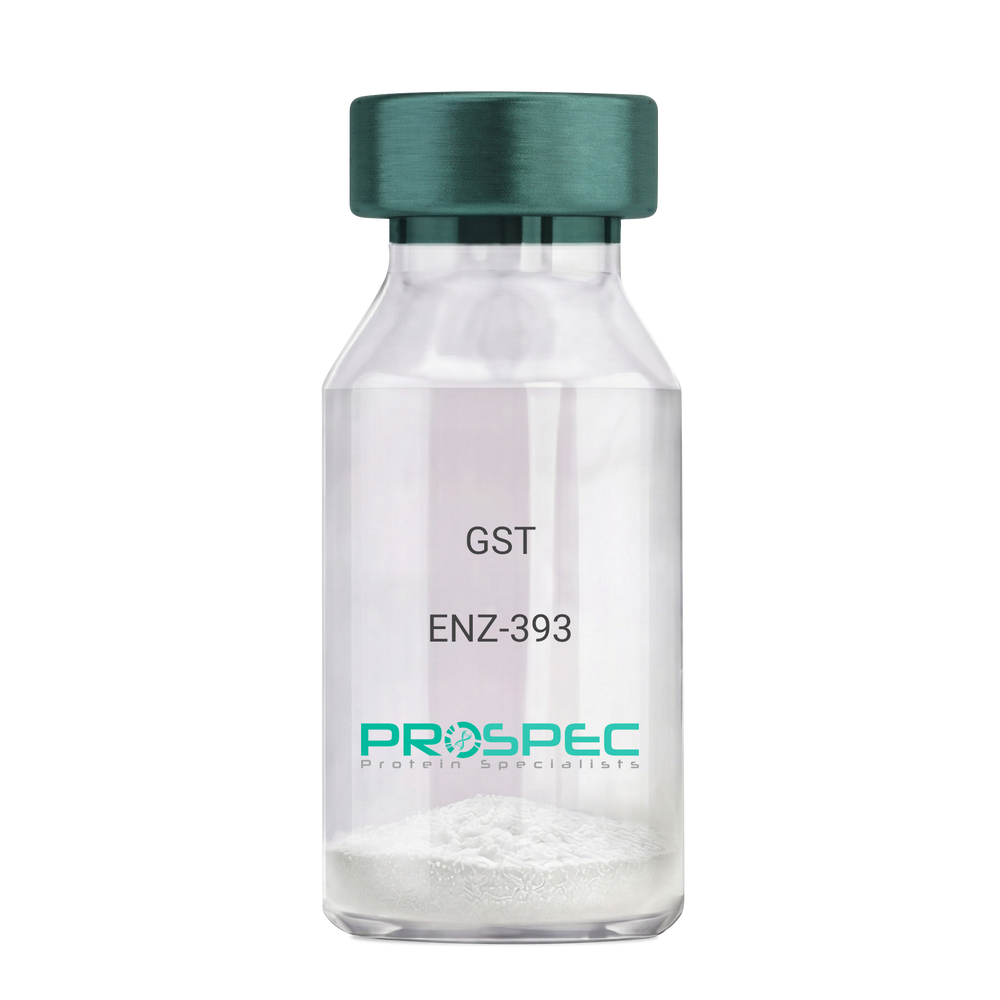
    Name :

    GST

    Description:

    Glutathione S-Transferase Recombinant

    Glutathione S-Transferase, GST, Glutathione S-transferase class-mu 26 kDa isozyme, GST 26, Sj26 antigen, SjGST.

    Product # :

    ENZ-393

    Price :

    Quantity :

    Shipping Method :

    Ice Icon

    Shipped with Ice Packs

    Add To Cart

    More Info

    • description
    • source
    • formulation
    • purity
    • biological activity
    • More Info

    Description

    Recombinant Glutathione S-Transferase full length protein (1-218a.a.) expressed in E.coli, having a molecular mass of 26kDa. GST was isolated from an E. coli strain that carries the coding sequence for Schistosoma japonicum GST under the control of a T7 promoter. The GST is purified by proprietary chromatographic techniques.

    Source

    Escherichia Coli.

    Formulation

    GST supplied in Phosphate Buffered Saline pH 7.4.

    Purity

    Greater than 95% as determined by SDS-PAGE.

    Biological Activity

    Specific activity is >20 units/mg. A unit is defined as the amount of enzyme that conjugate 1.0 u mole of 1-chloro-2,4-dinitrobenzene (CDNB) with reduced glutathione per minute at pH 6.5 at 25°C.

    More Info

    • Introduction

      Antioxidant enzyme Glutathione S- Transferase (GST) is thought to do the primary cellular defense mechanism against reactive oxygen species. GST reduces lipid hydroperoxides through its Se-independent glutathione peroxidase activity. The enzyme also detoxifies lipid peroxidation end products such as 4-hydroxynonenal (4-HNE).
      The soluble GST is a 26 kDa protein which occurs as a dimer in all aerobic organisms. Each monomer has two domains, one that binds GSH and is an /-structure similar to thioredoxin and the other, all helical, that binds the hydrophobic substrate. The GST -fusion protein expression system is a widely used recombinant protein expression system that allows a peptide or a regulatory protein domain to be expressed as a fusion to the C-terminus of Schistosoma japonicum GST. Fusion proteins also possess GST -enzymatic activity and can undergo dimerization similar to in vivo. The fusion protein can be purified via GST -affinity column chromatography. In most cases, the desired peptides or domains are removed from GST by applying a specific protease that recognizes and cleaves the linker between the protein domain and GST. The technique has been widely used to generate different kinds of proteins for crystallization, molecular immunology studies, the production of vaccines and studies involving protein-protein and protein-DNA interactions.

    • Synonyms

      Glutathione S-Transferase, GST, Glutathione S-transferase class-mu 26 kDa isozyme, GST 26, Sj26 antigen, SjGST.

    • Physical Appearance

      Sterile Filtered clear solution.

    • Stability

      Store at 4°C if entire vial will be used within 2-4 weeks. Store, frozen at -20°C for longer periods of time. For long term storage it is recommended to add a carrier protein (0.1% HSA or BSA).Avoid multiple freeze-thaw cycles.

    • Amino Acid Sequence

      MSPILGYWKI KGLVQPTRLL LEYLEEKYEE HLYERDEGDK WRNKKFELGL EFPNLPYYID GDVKLTQSMA IIRYIADKHN MLGGCPKERA EISMLEGAVL DIRYGVSRIA YSKDFETLKV DFLSKLPEML KMFEDRLCHK TYLNGDHVTH PDFMLYDALD VVLYMDPMCL DAFPKLVCFK KRIEAIPQID KYLKSSKYIA WPLQGWQATF GGGDHPPKSD LVPR.

    ProSpec's products are furnished for LABORATORY RESEARCH USE ONLY. The product may not be used as drugs, agricultural or pesticidal products, food additives or household chemicals.

    Glutathione S Transferase
  • View Data Sheet

    Name :

    HCV Core Genotype-3/10

    Description:

    Hepatitis C Virus Core Genotype-3/10 Recombinant

    Product # :

    HCV-201

    Price :

    Quantity :

    Shipping Method :

    Ice Icon

    Shipped with Ice Packs

    Add To Cart

    More Info

    • description
    • source
    • formulation
    • purity
    • More Info

    Description

    The E.coli derived recombinant protein contains the HCV core nucleocapsid immunodominant regions, amino acids 2-119.

    Source

    Escherichia Coli.

    Formulation

    50mM Tris-HCl, pH-8, 60mM NaCl, 10mM glutathione, 0.25% sarkosil & 50% glycerol.

    Purity

    HCV Core Genotype-3/10 protein is >95% pure as determined by 10% PAGE (coomassie staining).

    More Info

    • Introduction

      HCV is a small 50nm, enveloped, single-stranded, positive sense RNAvirus in the family Flaviviridae.
      HCV has a high rate of replication with approximately one trillion particles produced each day in an infected individual. Due to lack of proofreading by the HCV RNA polymerase, the HCV has an exceptionally high mutation rate, a factor that may help it elude the host's immune response. Hepatitis C virus is classified into six genotypes(1-6) with several subtypes within each genotype. The preponderance and distribution of HCV genotypes varies globally. Genotype is clinically important in determining potential response to interferon-based therapy and the required duration of such therapy. Genotypes 1 and 4 are less responsive to interferon-based treatment than are the other genotypes (2, 3, 5 and 6).

    • Stability

      HCV Core Genotype-3/10 although stable at 4°C for 1 week, should be stored below -18°C. Please prevent freeze thaw cycles.

    • Applications

      HCV Core Genotype-3/10 antigen is suitable for ELISA and Western blots, excellent antigen for detection of HCV with minimal specificity problems.

    • Specificity

      Immunoreactive with sera of HCV-infected individuals.

    • Purification Method

      HCV Core Genotype-3/10 protein was purified by proprietary chromatographic technique.

    ProSpec's products are furnished for LABORATORY RESEARCH USE ONLY. The product may not be used as drugs, agricultural or pesticidal products, food additives or household chemicals.

    Hcv Core Genotype 3 10
  • View Data Sheet

    Name :

    M.Pneumoniae P1-C

    Description:

    Mycoplasma Pneumoniae P1-C Recombinant

    Product # :

    PRO-801

    Price :

    Quantity :

    Shipping Method :

    Ice Icon

    Shipped with Ice Packs

    Add To Cart

    More Info

    • description
    • source
    • formulation
    • purity
    • More Info

    Description

    The Recombinant Mycoplasma Pneumoniae C-terminal region (P1C) of the P1 protein was expressed in E. coli containing 362 amino acids. The protein is fused to a 6 His Tag. This peptide can be used as an antigen in the diagnosis of M. pneumoniae infection.

    Source

    E.Coli

    Formulation

    Mycoplasma Pneumoniae P1-C is formulated in 1x PBS pH 7.4.

    Purity

    Greater than 95% as determined by 12% PAGE (Coomassie staining).

    More Info

    • Introduction

      Mycoplasma pneumonia is part of the atypical pneumonia subtype which is caused by the bacteria M. pneumoniae. Mycoplasma pneumonia affects individuals younger than 40. It makes up 15 - 50% of all pneumonia cases in adults and especially in school-aged children. People at great risk for mycoplasma pneumonia comprise of those living or working in busy areas such as schools and homeless shelters, although many people who contract mycoplasma pneumonia have no identifiable risk factor. P1, P30, and P116 of mycoplasma pneumonia membrane proteins have been recognized as adhensive factors, P1 is considered as a main adhension protein of the organism colonization.

    • Physical Appearance

      Streile filtered colorless solution.

    • Stability

      Upon arrival, Store at -20°C.Please prevent freeze-thaw cycles.

    • Applications

      Immunoassay.

    • Purification Method

      The recombinant fusion protein was purified by GSH affinity chromatography technique.

    ProSpec's products are furnished for LABORATORY RESEARCH USE ONLY. The product may not be used as drugs, agricultural or pesticidal products, food additives or household chemicals.

    Mycoplasma Pneumoniae P1 C
  • 1
  • …
  • 13
  • 14
  • 15
  • 16
  • 17
  • …
  • 26
Your browser does not support the video tag.

Products

  • Cytokines
  • Growth Factors
  • Chemokines
  • CD Antigens
  • Neurotrophins
  • Hormones
  • Enzymes
  • Viral Antigens
  • Recombinant Proteins
  • Natural Proteins
  • Monoclonal Antibodies
  • Polyclonal Antibodies
  • Thermal Packaging

About

  • Company
  • Founder
  • Contribution
  • Contact Us
  • My Account

Ordering

  • Ordering Method
  • Ordering - Credit Card
  • Ordering PO
  • Shipping Fees
  • FAQ's

Legal

  • Terms & Conditions
  • Privacy
  • Site Map
  • info@prospecbio.com
  • Facebook
  • Instagram
  • Linkedin Profile

© 2026 ProSpec-Tany TechnoGene Ltd. All Rights Reserved.

  • Choosing a selection results in a full page refresh.
  • Opens in a new window.