Search results
1000 results found for “calbindin”
Name
Description
Product #
Price
Quantity
Shipping Method
- View Data Sheet
Name :
Leptin tA HumanDescription:
Leptin Antagonist Triple Mutant Human Recombinant
Product # :
CYT-352Price :
Quantity :
Shipping Method :
Shipped at Room temp
More Info
- description
- source
- formulation
- purity
- biological activity
- More Info
Description
Leptin Antagonist Triple Mutant Human Recombinant is a single non-glycosilated polypeptide chain containing 146 amino and additional Ala at N-terminus acids and having a molecular weight of 16 kDa, Leptin was mutated, resulting in L39A/D40A/F41A. Leptin Antagonist Triple Mutant Human Recombinant was purified by proprietary chromatographic techniques.
Source
Escherichia coli.
Formulation
The protein was lyophilized from a concentrated (1mg/ml) solution with 0.0045mM NaHCO3.
Purity
Greater than 98.0% as determined by:
(a) Gel filtration analysis.
(b) Analysis by SDS-PAGE.Biological Activity
ProSpec’s Leptin triple antagonist is capable of inhibiting leptin-induced proliferation of BAF/3 cells stably transfected with the long form of human leptin receptor. It also inhibits various leptin effects in several in vitro bioassays.More Info
-
Physical Appearance
White lyophilized (freeze-dried) powder.
-
Stability
Lyophilized Leptin Antagonist Triple Mutant Human Recombinant although stable at room temperature for several weeks, should be stored desiccated below -18°C. Upon reconstitution at > 0.1 mg/ml and up to 2 mg/ml and filter sterilization Leptin mutant can be stored at 4°C or even room temperature for several weeks making it suitable for long term infusion studies using osmotic pumps. At lower concentration addition of a carrier protein (0.1% HSA or BSA) is suggested. Please prevent freeze-thaw cycles.
-
Solubility
It is recommended to reconstitute the lyophilized Leptin Antagonist Triple Mutant Human Recombinant in sterile 0.4% NaHCO3, not less than 100µg/ml, which can then be further diluted to other aqueous solutions.
-
Amino Acid Sequence
The sequence of the first five N-terminal amino acids was determined and was found to be Ala-Val-Pro-Ile-Gln.
-
Protein content
Protein quantitation was carried out by UV spectroscopy at 280 nm using the absorbency value of 0.88 as the extinction coefficient for a 0.1% (1mg/ml) solution at pH 8.0. This value is calculated by the PC GENE computer analysis program of protein sequences (IntelliGenetics).
ProSpec's products are furnished for LABORATORY RESEARCH USE ONLY. The product may not be used as drugs, agricultural or pesticidal products, food additives or household chemicals.
- View Data Sheet
Name :
CAV1 AntibodyDescription:
Caveolin-1, Mouse Anti Human
Caveolin-1, CAV1, CAV, CGL3, BSCL3, VIP21, MSTP085.
Product # :
ANT-027Price :
Quantity :
Shipping Method :
Shipped with Ice Packs
More Info
- formulation
- More Info
Formulation
1mg/ml containing PBS, pH-7.4, & 0.1% Sodium Azide.
More Info
-
Introduction
The scaffolding protein encoded by the CAV1 gene is the key component of the caveolae plasma membranes found in most cell types. Cholesterol is vital for caveolae formation and maintenance, and caveolae structure is highly sensitive to cholesterol depletion or treatment with cholesterol binding drugs. CAV1 can tightly and specifically bind free cholesterol and artificial cholesterol containing vesicles. Caveolin 1 links integrin subunits to the tyrosine kinase FYN, which is an initiating step in coupling integrins to the Ras-ERK pathway and promoting cell cycle progression. CAV1 is a tumor suppressor gene candidate and a negative regulator of the Ras-p42/44 mitogen-activated kinase. Caveolin 1 and caveolin 2 are positioned next to each other on chromosome 7 and express co-localizing proteins which form a stable hetero-oligomeric complex cascade. Mutations in the CAV1 gene are linked to Berardinelli-Seip congenital lipodystrophy.
-
Synonyms
Caveolin-1, CAV1, CAV, CGL3, BSCL3, VIP21, MSTP085.
-
Physical Appearance
Sterile Filtered clear solution.
-
Immunogen
Anti-human CAV1 mAb, is derived from hybridization of mouse FO myeloma cells with spleen cells from BALB/c mice immunized with recombinant human CAV1 amino acids 1-104 purified from E. coli.
-
Ig Subclass
Mouse IgG2b heavy chain and k light chain.
-
Clone
PAT4C1AT.
-
Applications
CAV1 antibody has been tested by ELISA, Western blot to assure specificity and reactivity. Since application varies, however, each investigation should be titrated by the reagent to obtain optimal results. Recommended dilution range for Western blot analysis is 1:250 ~ 1000.
Recommended starting dilution is 1:500. -
Type
Mouse Anti Human Monoclonal.
-
Storage Procedures
For periods up to 1 month store at 4°C, for longer periods of time, store at -20°C. Prevent freeze thaw cycles.
-
Purification Method
CAV1 antibody was purified from mouse ascitic fluids by protein-G affinity chromatography.
ProSpec's products are furnished for LABORATORY RESEARCH USE ONLY. The product may not be used as drugs, agricultural or pesticidal products, food additives or household chemicals.
- View Data Sheet
Name :
ACTN1 HumanDescription:
Actinin Alpha 1 Human Recombinant
ACTN1, Actinin, Alpha 1, Alpha-Actinin Cytoskeletal Isoform, F-Actin Cross-Linking Protein, Non-Muscle Alpha-Actinin-1, BDPLT15, Actinin 1 Smooth Muscle, Alpha-Actinin-1.
Product # :
PRO-2227Price :
Quantity :
Shipping Method :
Shipped with Ice Packs
More Info
- description
- source
- formulation
- purity
- More Info
Description
ACTN1 Human Recombinant produced in E.Coli is a single, non-glycosylated polypeptide chain containing 274 amino acids (1-249 a.a) and having a molecular mass of 31.4kDa. ACTN1 is fused to a 25 amino acid His-tag at N-terminus & purified by proprietary chromatographic techniques.
Source
Escherichia Coli.
Formulation
ACTN1 protein solution (1mg/ml) containing Phosphate Buffered Saline (pH7.4) and 10% glycerol.
Purity
Greater than 95.0% as determined by SDS-PAGE.
More Info
-
Introduction
ACTN1 encodes a nonmuscle, cytoskeletal, alpha actinin isoform and maps to the same site as the structurally similar erythroid beta spectrin gene. Alpha actinins belong to the spectrin gene superfamily which represents a diverse group of cytoskeletal proteins, including the alpha and beta spectrins and dystrophins. Alpha actinin is an actin-binding protein with multiple roles in different cell types. In nonmuscle cells, the cytoskeletal isoform is found along microfilament bundles and adherens-type junctions, where it is involved in binding actin to the membrane. In contrast, skeletal, cardiac, and smooth muscle isoforms are localized to the Z-disc and analogous dense bodies, where they help anchor the myofibrillar actin filaments.
-
Synonyms
ACTN1, Actinin, Alpha 1, Alpha-Actinin Cytoskeletal Isoform, F-Actin Cross-Linking Protein, Non-Muscle Alpha-Actinin-1, BDPLT15, Actinin 1 Smooth Muscle, Alpha-Actinin-1.
-
Physical Appearance
Sterile Filtered colorless solution.
-
Stability
Store at 4°C if entire vial will be used within 2-4 weeks. Store, frozen at -20°C for longer periods of time. For long term storage it is recommended to add a carrier protein (0.1% HSA or BSA).Avoid multiple freeze-thaw cycles.
-
Amino Acid Sequence
MGSSHHHHHH SSGLVPRGSH MGSEFMDHYD SQQTNDYMQP EEDWDRDLLL DPAWEKQQRK TFTAWCNSHL RKAGTQIENI EEDFRDGLKL MLLLEVISGE RLAKPERGKM RVHKISNVNK ALDFIASKGV KLVSIGAEEI VDGNVKMTLG MIWTIILRFA IQDISVEETS AKEGLLLWCQ RKTAPYKNVN IQNFHISWKD GLGFCALIHR HRPELIDYGK LRKDDPLTNL NTAFDVAEKY LDIPKMLDAE DIVGTARPDE KAIMTYVSSF YHAF.
ProSpec's products are furnished for LABORATORY RESEARCH USE ONLY. The product may not be used as drugs, agricultural or pesticidal products, food additives or household chemicals.
- View Data Sheet
Name :
VEGF (121 a.a.) HumanDescription:
Vascular Endothelial Growth Factor (121 a.a.) Human Recombinant
Vascular endothelial growth factor A, VEGF-A, Vascular permeability factor, VPF, VEGF, MGC70609.
Product # :
CYT-343Price :
Quantity :
Shipping Method :
Shipped at Room temp
More Info
- description
- source
- formulation
- purity
- biological activity
- More Info
Description
Vascular Endothelial Growth Factor-121 Human Recombinant produced in E.Coli is a non-glycosylated, polypeptide double chain containing 2x121 amino acids and having a molecular mass of 28.4kDa. VEGF121 circulates more freely than other VEGF forms, which bind more tightly with vascular heparin sulfates.
Source
Escherichia Coli.
Formulation
The protein was lyophilized from a concentrated (1mg/ml) solution with no additives.
Purity
Greater than 98.0% as determined by:
(a) Analysis by RP-HPLC.
(b) Analysis by SDS-PAGE.Biological Activity
VEGF-121 has full biological activity when compared to standards. The activity is determined by the dose-dependent proliferation of HUVECs and is typically 1-6ng/ml corresponding to a specific activity of 166,667-1,000,000U/mg.
More Info
-
Introduction
Vascular endothelial growth factor is an important signaling protein involved in both vasculogenesis and angiogenesis. As its name implies, VEGF activity has been mostly studied on cells of the vascular endothelium, although it does have effects on a number of other cell types (e.g. stimulation monocyte/ macrophagemigration, neurons, cancer cells, kidney epithelial cells ).VEGF mediates increased vascular permeability, induces angiogenesis, vasculogenesis and endothelial cell growth, promotes cell migration, and inhibits apoptosis. In vitro, VEGF has been shown to stimulate endothelial cell mitogenesisand cell migration. VEGF is also a vasodilator and increases microvascular permeability and was originally referred to as vascular permeability factor.
Elevated levels of this protein are linked to POEMS syndrome, also known as Crow-Fukase syndrome. Mutations in this gene have been associated with proliferative and nonproliferative diabetic retinopathy. -
Synonyms
Vascular endothelial growth factor A, VEGF-A, Vascular permeability factor, VPF, VEGF, MGC70609.
-
Physical Appearance
Sterile Filtered White lyophilized (freeze-dried) powder.
-
Stability
Lyophilized Vascular Endothelial Growth Factor 121 although stable at room temperature for 3 weeks, should be stored desiccated below -18°C. Upon reconstitution VEGF-121 should be stored at 4°C between 2-7 days and for future use below -18°C.For long term storage it is recommended to add a carrier protein (0.1% HSA or BSA).Please prevent freeze-thaw cycles.
-
Solubility
It is recommended to reconstitute the lyophilized Vascular Endothelial Growth Factor -121 in sterile 18MΩ-cm H2O not less than 100µg/ml, which can then be further diluted to other aqueous solutions.
-
Amino Acid Sequence
APMAEGGGQN HHEVVKFMDV YQRSYCHPIE TLVDIFQEYP DEIEYIFKPS CVPLMRCGGC CNDEGLECVP TEESNITMQI MRIKPHQGQH IGEMSFLQHN KCECRPKKDR ARQENCDKPR R
ProSpec's products are furnished for LABORATORY RESEARCH USE ONLY. The product may not be used as drugs, agricultural or pesticidal products, food additives or household chemicals.
- View Data Sheet
Name :
WIF1 MouseDescription:
WNT Inhibitory Factor 1 Mouse Recombinant
Wnt inhibitory factor 1, WIF-1, Wif1.
Product # :
PRO-2248Price :
Quantity :
Shipping Method :
Shipped with Ice Packs
More Info
- description
- source
- formulation
- purity
- More Info
Description
WIF1 produced in Sf9 Insect cells is a single, glycosylated polypeptide chain containing 359 amino acids (29-379a.a.) and having a molecular mass of 39.4kDa. (Molecular size on SDS-PAGE will appear at approximately 40-57kDa).WIF1 is expressed with an 8 amino acid His tag at C-Terminus and purified by proprietary chromatographic techniques.
Source
Sf9, Insect cells.
Formulation
WIF1 protein solution (1mg/ml) contains 20mM MES (pH5.5), 1mM DTT, 1mM PMSF and 30% glycerol.
Purity
Greater than 90.0% as determined by SDS-PAGE.
More Info
-
Introduction
WIF1 binds to wnt proteins and inhibits their activities. WIF1 plays a role in mesoderm segmentation. WNT proteins are extracellular signaling molecules that take part in the control of embryonic development & cancer. WIF1 protein contains a WNT inhibitory factor (WIF) domain and 5 epidermal growth factor (EGF)-like domains. WIF1 takes part in mesoderm segmentation. WIF1 protein is found to be present in fish, amphibia and mammals. WIF1 is a recurrent target in human salivary gland oncogenesis. Downregulation of WIF1 takes part in the development and progression of pleomorphic adenomas. WIF1 is a tumor suppressor, specifically in nonfunctioning pituitary tumors.
-
Synonyms
Wnt inhibitory factor 1, WIF-1, Wif1.
-
Physical Appearance
Sterile filtered colorless solution.
-
Stability
Store at 4°C if entire vial will be used within 2-4 weeks.Store, frozen at -20°C for longer periods of time. For long term storage it is recommended to add a carrier protein (0.1% HSA or BSA).Avoid multiple freeze-thaw cycles.
-
Amino Acid Sequence
GQPPEESLYL WIDAHQARVL IGFEEDILIV SEGKMAPFTH DFRKAQQRMP AIPVNIHSMN FTWQAAGQAE YFYEFLSLRS LDKGIMADPT VNVPLLGTVP HKASVVQVGF PCLGKQDGVA AFEVNVIVMN SEGNTILRTP QNAIFFKTCQ QAECPGGCRN GGFCNERRVC ECPDGFYGPH
CEKALCIPRC MNGGLCVTPG FCICPPGFYG VNCDKANCST TCFNGGTCFY PGKCICPPGL EGEQCELSKC PQPCRNGGKC IGKSKCKCPK GYQGDLCSKP VCEPGCGAHG TCHEPNKCQC REGWHGRHCN KRYGASLMHA PRPAGAGLER HTPSLKKAED RRDPPESNYI WVEHHHHHH.
ProSpec's products are furnished for LABORATORY RESEARCH USE ONLY. The product may not be used as drugs, agricultural or pesticidal products, food additives or household chemicals.
- View Data Sheet
Name :
Leptin OvineDescription:
Leptin Ovine Recombinant
OB Protein, Obesity Protein, OBS, Obesity factor.
Product # :
CYT-239Price :
Quantity :
Shipping Method :
Shipped at Room temp
More Info
- description
- source
- formulation
- purity
- biological activity
- More Info
Description
Leptin Ovine Recombinant produced in E.Coli is a single, non-glycosylated, polypeptide chain containing 146 amino acids and having a molecular mass of 16 kDa.The Leptin is purified by proprietary chromatographic techniques.
Source
Escherichia Coli.
Formulation
The protein was lyophilized from a concentrated (1mg/ml) solution with 0.0045mM NaHCO3.
Purity
Greater than 95.0% as determined by:
(a) Analysis by SEC-HPLC.
(b) Analysis by SDS-PAGE.Biological Activity
Biological active as evidenced by inducing proliferation of BAF/3 cells stably transfected with the long form of human leptin receptor.More Info
-
Introduction
A 16-kDa peptide hormone secreted from white adipocytes and implicated in the regulation of food intake and energy balance. Leptin provides the key afferent signal from fat cells in the feedback system that controls body fat stores.
-
Synonyms
OB Protein, Obesity Protein, OBS, Obesity factor.
-
Physical Appearance
Sterile Filtered White lyophilized (freeze-dried) powder.
-
Stability
Lyophilized Leptin although stable at room temperature for 3 weeks, should be stored desiccated below -18°C. Upon reconstitution Leptin should be stored at 4°C between 2-7 days and for future use below -18°C.For long term storage it is recommended to add a carrier protein (0.1% HSA or BSA).Please prevent freeze-thaw cycles.
-
Solubility
It is recommended to reconstitute the lyophilized Leptin in sterile 18MΩ-cm H2O not less than 100µg/ml, which can then be further diluted to other aqueous solutions.
-
Amino Acid Sequence
The sequence of the first five N-terminal amino acids was determined and was found to be Ala-Val-Pro-Ile-Arg.
-
Protein content
Protein quantitation was carried out by two independent methods1. UV spectroscopy at 280 nm using the absorbency value of 0.2 as the extinction coefficient for a 0.1% (1mg/ml) solution. This value is calculated by the PC GENE computer analysis program of protein sequences (IntelliGenetics). 2. Analysis by RP-HPLC,using calibrated solution of Leptin Ovine as a Reference Standard.
ProSpec's products are furnished for LABORATORY RESEARCH USE ONLY. The product may not be used as drugs, agricultural or pesticidal products, food additives or household chemicals.
- View Data Sheet
Name :
Leptin-A TilapiaDescription:
Leptin-A Tilapia Recombinant
Product # :
CYT-1109Price :
Quantity :
Shipping Method :
Shipped at Room temp
More Info
- description
- source
- formulation
- purity
- biological activity
- More Info
Description
Leptin-A Tilapia Recombinant produced in E.Coli is a single, non-glycosylated polypeptide chain containing 161 amino acids and having a molecular mass of 16,491 Dalton. The Leptin-A Tilapia is purified by proprietary chromatographic techniques.
Source
Escherichia Coli.
Formulation
Lyophilized from a concentrated (1mg/ml) solution containing NaHCO3 at 1:2 salt: protein ratio.
Purity
Greater than 95.0% as determined by:
(a) Gel filtration analysis.
(b) Analysis by SDS-PAGE.Biological Activity
Tilapia leptins were found to be biologically active in promoting proliferation of BAF/3 cells stably transfected with the long form of human leptin receptor, but their activity was lower than that of mammalian leptin. Furthermore, the Tilapia leptins were biologically active in promoting STAT‐LUC activation in COS7 cells transfected with Tilapia leptin receptor but not in cells transfected with human leptin receptor. Tilapia Leptin A was more active than Tilapia Leptin B.
More Info
-
Introduction
Leptin is a protein hormone. It is mainly produced in adipose cells that regulate energy homeostasis by restraining hunger. Leptin ties to nuclear receptors in the hypothalamus (arcuate nucleus). Similar to insulin resistance in type II diabetes, in obesity there is a decrease in the sensitivity towards leptin, ending in a failure to identify satiety, even in high levels of energy stores or leptin itself. Full-length cDNA encoding 2 leptin sequences (tLepA and tLepB) and 1 leptin receptor sequence (tLepR) exists in tilapia (Oreochromis niloticus). The full-length cDNA of tLepR is 3423 bp, encoding a protein of 1140 amino acid which contained all functionally important domains conserved among vertebrate leptin receptors. The cDNAs of tLepA and tLepB are 486 bp and 459 bp in length, encoding proteins of 161 aa and 152 aa, respectively. The three-dimensional structures of tLepA and tLepB demonstrates strong conservation of tertiary structure with that of human leptin comprised of 4 helixes.
-
Physical Appearance
Sterile Filtered White lyophilized (freeze-dried) powder.
-
Stability
Lyophilized Leptin-A Tilapia although stable at room temperature for 3 weeks, should be stored desiccated below -18°C. Upon reconstitution Leptin-A Tilapia should be stored at 4°C between 2-7 days and for future use below -18°C. For long term storage it is recommended to add a carrier protein (0.1% HSA or BSA). Please prevent freeze-thaw cycles.
-
Solubility
It is recommended to reconstitute the lyophilized Leptin-A Tilapia in sterile water or 0.4% NaHCO3 adjusted to pH 8-9, not less than 100µg/ml, which can then be further diluted to other aqueous solutions.
-
Amino Acid Sequence
The first six N-terminal amino acids of recombinant Tilapia leptin A are Ala-Pro-Leu-Pro-Val-Glu.
-
Protein content
Protein quantitation was carried out by UV spectroscopy at 280 nm using the absorbency value of 1.33 for 1 mg/ml Leptin-A Tilapia as the extinction coefficient for a 0.1% (1mg/ml) solution at pH 8.0. This value is calculated by the DNAman computer analysis program of protein sequences.
ProSpec's products are furnished for LABORATORY RESEARCH USE ONLY. The product may not be used as drugs, agricultural or pesticidal products, food additives or household chemicals.
- View Data Sheet
Name :
NEFL BovineDescription:
Neurofilament Light Bovine
Neurofilament light polypeptide, NF-L, NEFL, NF68, NFL, 68 kDa neurofilament protein.
Product # :
PRO-2786Price :
Quantity :
Shipping Method :
Shipped at Room temp
More Info
- description
- source
- formulation
- purity
- More Info
Description
NEFL Bovine having a calculated molecular mass of 68 kDa, pI-5.0.
Source
Bovine spinal cord.
Formulation
NEFL was lyophilized from a 1mg/ml solution containing 10mM sodium phosphate buffer pH 7.5, 6M urea, 1mM EDTA, 2mM DTT and 10mM methylammonium chloride.
Purity
Greater than 95.0% as determined by SDS-PAGE.
More Info
-
Synonyms
Neurofilament light polypeptide, NF-L, NEFL, NF68, NFL, 68 kDa neurofilament protein.
-
Physical Appearance
Filtered White lyophilized (freeze-dried) powder.
-
Stability
Store the lyophilized NEFL between 2-8°C, do not freeze. Upon reconstitution NEFL should be stored at -20°C.Please prevent freeze-thaw cycles.
-
Solubility
It is recommended to reconstitute the lyophilized NEFL in sterile 18MΩ-cm H2O not less than 100µg/ml, which can then be further diluted to other aqueous solutions.
-
Background
Neurofilament light chain (NEFL) is a critical component of the neuronal cytoskeleton, primarily found in neurons of the central and peripheral nervous systems. While extensive research has been conducted on NEFL in human and rodent models, the study of NEFL in bovine nervous tissues is an emerging area with potential for advancing our understanding of neuronal biology in larger mammals and its applications in veterinary medicine and neurobiology.
Bovine nervous tissues, such as the brain and spinal cord, are of particular interest due to their relevance in cattle health and the food industry. This research aims to provide a comprehensive exploration of NEFL in bovine nervous tissues, elucidating its functions, structural significance, and potential applications.
The primary objective of this research is to elucidate the role of NEFL in bovine nervous tissues, particularly in maintaining the structural integrity of neurons and axons. In vitro and ex vivo experiments, utilizing bovine neuronal cell cultures and tissue specimens, will be conducted to investigate how NEFL contributes to neuronal morphology, axonal transport, and neuronal resilience. Understanding these mechanisms is fundamental for deciphering the complexities of neuronal biology in bovine species.
The second objective is to assess the relevance of bovine NEFL in veterinary medicine. Studies involving bovine models will be conducted to evaluate the impact of NEFL mutations or variations on neuronal health, disease susceptibility, and neurodegenerative conditions. These investigations may provide valuable insights into potential applications in cattle health and the development of diagnostic tools for neurological disorders.
The third objective is to explore the potential applications of bovine NEFL in neurobiology and biotechnology. Research will investigate the use of bovine NEFL-expressing cells as models for studying neuronal-related diseases and for developing tissue engineering approaches for veterinary medicine and biotechnology.
By delving into the functions and roles of NEFL in bovine nervous tissues, this research aims to expand our knowledge of neuronal biology, its implications for veterinary medicine, and its potential applications in neurobiology and cattle health
ProSpec's products are furnished for LABORATORY RESEARCH USE ONLY. The product may not be used as drugs, agricultural or pesticidal products, food additives or household chemicals.
- View Data Sheet
Name :
SERPINE1 HumanDescription:
Plasminogen Activator Inhibitor-1 Human Recombinant
PAI-1, PAI1, PLANH1, SERPINE1, PAIE, PLASMINOGEN ACTIVATOR INHIBITOR, BETA-MIGRATING ENDOTHELIAL-CELL-DERIVED TYPE.
Product # :
ENZ-357Price :
Quantity :
Shipping Method :
Shipped with Ice Packs
More Info
- description
- source
- formulation
- purity
- biological activity
- More Info
Description
SERPINE1 Human Recombinant fused to an N-terminal His-Tag produced in E.Coli is a single, non-glycosylated polypeptide chain containing 400 amino acids (24-402) and having a molecular mass of 45kDa.SERPINE1 is fused to a 21 amino acid His-Tag at N-terminus and purified by proprietary chromatographic techniques.
Source
Escherichia Coli.
Formulation
50mM NaAc (pH 5.5), 10% glycerol and 0.1M NaCl.
Purity
Greater than 95.0% as determined by SDS-PAGE.
Biological Activity
The IC50 for this effect is less than 3nM, measured by its ability to inhibit uPA cleavage of the substrate Z-GGRAMC.
More Info
-
Introduction
Plasminogen activator inhibitor-1 is the principal inhibitor of tissue plasminogen activator(tPA) and uPA, the activators of plasminogenand hence fibrinolysis(the physiological breakdown of blood clots). It is a serine protease inhibitor(serpin) protein (SERPINE1). The other PAI, plasminogen activator inhibitor-2(PAI-2) is secreted by the placentaand only present in significant amounts during pregnancy. In addition, protease nexinacts as an inhibitor of tPA. SERPINE1, however, is the main inhibitor of the plasminogen activators.
-
Synonyms
PAI-1, PAI1, PLANH1, SERPINE1, PAIE, PLASMINOGEN ACTIVATOR INHIBITOR, BETA-MIGRATING ENDOTHELIAL-CELL-DERIVED TYPE.
-
Physical Appearance
Sterile Filtered clear colorless solution.
-
Stability
Store at 4°C if entire vial will be used within 2-4 weeks. Store, frozen at -20°C for longer periods of time. For long term storage it is recommended to add a carrier protein (0.1% HSA or BSA).Avoid multiple freeze-thaw cycles.
-
Amino Acid Sequence
MGSSHHHHHH SSGLVPRGSH MVHHPPSYVA HLASDFGVRV FQQVAQASKD RNVVFSPYGVASVLAMLQLT TGGETQQQIQ AAMGFKIDDK GMAPALRHLY KELMGPWNKD EISTTDAIFVQRDLKLVQGF MPHFFRLFRS TVKQVDFSEV ERARFIINDW VKTHTKGMIS NLLGKGAVDQLTRLVLVNAL YFNGQWKTPF PDSSTHRRLF HKSDGSTVSV PMMAQTNKFN YTEFTTPDGHYYDILELPYH GDTLSMFIAA PYEKEVPLSA LTNILSAQLI SHWKGNMTRL PRLLVLPKFSLETEVDLRKP LENLGMTDMF RQFQADFTSL SDQEPLHVAQ ALQKVKIEVN ESGTVASSSTAVIVSARMAP EEIIMDRPFL FVVRHNPTGT VLFMGQVMEP.
ProSpec's products are furnished for LABORATORY RESEARCH USE ONLY. The product may not be used as drugs, agricultural or pesticidal products, food additives or household chemicals.
- View Data Sheet
Name :
NFKBIB HumanDescription:
NF-kappa-B Inhibitor Beta Human Recombinant
NF-kappa-B inhibitor beta, NF-kappa-BIB, I-kappa-B-beta, IkB-B, IkB-beta, IkappaBbeta, Thyroid receptor-interacting protein 9, TR-interacting protein 9, TRIP-9, NFKBIB, IKBB, TRIP9.
Product # :
PRO-1046Price :
Quantity :
Shipping Method :
Shipped with Ice Packs
More Info
- description
- source
- formulation
- purity
- More Info
Description
NFKBIB Human Recombinant produced in E.Coli is a single, non-glycosylated polypeptide chain containing 380 amino acids (1-356 a.a) and having a molecular mass of 40.3kDa (Molecular weight on SDS-PAGE will appear higher).NFKBIB is fused to a 24 amino acid His-tag at N-terminus & purified by proprietary chromatographic techniques.
Source
Escherichia Coli.
Formulation
NFKBIB protein solution (1mg/ml) containing 20mM Tris-HCl buffer (pH8.0), 20% glycerol and 1mM DTT.
Purity
Greater than 90.0% as determined by SDS-PAGE.
More Info
-
Introduction
NF-kappa-B inhibitor beta (NFKBIB) is a member of the NF-kappa-B inhibitor family, which inhibit NF-kappa-B by complexing with, and trapping it in the cytoplasm. Phosphorylation of serine residues on these proteins by kinases marks them for destruction via the ubiquitination pathway, thus allowing activation of the NF-kappa-B, which translocates to the nucleus to act as a transcription factor.
-
Synonyms
NF-kappa-B inhibitor beta, NF-kappa-BIB, I-kappa-B-beta, IkB-B, IkB-beta, IkappaBbeta, Thyroid receptor-interacting protein 9, TR-interacting protein 9, TRIP-9, NFKBIB, IKBB, TRIP9.
-
Physical Appearance
Sterile Filtered clear solution.
-
Stability
Store at 4°C if entire vial will be used within 2-4 weeks. Store, frozen at -20°C for longer periods of time. For long term storage it is recommended to add a carrier protein (0.1% HSA or BSA).Avoid multiple freeze-thaw cycles.
-
Amino Acid Sequence
MGSSHHHHHH SSGLVPRGSH MGSHMAGVAC LGKAADADEW CDSGLGSLGP DAAAPGGPGL GAELGPGLSW APLVFGYVTE DGDTALHLAV IHQHEPFLDF LLGFSAGTEY MDLQNDLGQT ALHLAAILGE TSTVEKLYAA GAGLCVAERR GHTALHLACR VGAHACARAL LQPRPRRPRE
APDTYLAQGP DRTPDTNHTP VALYPDSDLE KEEEESEEDW KLQLEAENYE GHTPLHVAVI HKDVEMVRLL RDAGADLDKP EPTCGRSPLH LAVEAQAADV LELLLRAGAN PAARMYGGRT PLGSAMLRPN PILARLLRAH GAPEPEGEDE KSGPCSSSSD SDSGDEGDEY DDIVVHSSRS QTRLPPTPAS KPLPDDPRPV.
ProSpec's products are furnished for LABORATORY RESEARCH USE ONLY. The product may not be used as drugs, agricultural or pesticidal products, food additives or household chemicals.
- View Data Sheet
Name :
LGALS2 MouseDescription:
Galectin-2 Mouse Recombinant
Galectin-2, Gal-2, Lgals2, AI324147, 2200008F12Rik.
Product # :
CYT-019Price :
Quantity :
Shipping Method :
Shipped with Ice Packs
More Info
- description
- source
- formulation
- purity
- More Info
- SDS-PAGE
Description
LGALS2 mouse Recombinant produced E. coli is a single polypeptide chain containing 153 amino acids (1-130) and having a molecular mass of 17.3kDa.LGALS2 is fused to a 23 amino acid His-tag at N-terminus & purified by proprietary chromatographic techniques.
Source
Escherichia Coli.
Formulation
The LGALS2 solution (1mg/ml) contains 20mM Tris-HCl buffer (pH 8.0), 0.1M NaCl, 10% glycerol and 1mM DTT.
Purity
Greater than 95.0% as determined by SDS-PAGE.
SDS-PAGE
More Info
-
Introduction
LGALS2 is a soluble beta-galactoside binding lectin that controls cell-to-cell adhesion and cell-to-extracellular matrix interactions and takes part in tumor progression, pre-mRNA splicing and apoptosis. LGALS2 induces apoptosis in activated T cells and binds to the cytokine lymphotoxin-alpha (LTA) with threat of myocardial infarction.
-
Synonyms
Galectin-2, Gal-2, Lgals2, AI324147, 2200008F12Rik.
-
Physical Appearance
Sterile Filtered colorless solution.
-
Stability
Store at 4°C if entire vial will be used within 2-4 weeks. Store, frozen at -20°C for longer periods of time. For long term storage it is recommended to add a carrier protein (0.1% HSA or BSA).Avoid multiple freeze-thaw cycles.
-
Amino Acid Sequence
MGSSHHHHHH SSGLVPRGSH MGSMSEKFEV KDLNMKPGMS LKIKGKIHND VDRFLINLGQ GKETLNLHFN PRFDESTIVC NTSEGGRWGQ EQRENHMCFS PGSEVKITIT FQDKDFKVTL PDGHQLTFPN RLGHNQLHYL SMGGLQISSF KLE.
-
Background
What is the molecular weight/Mw of LGALS2 MOUSE Protein?
LGALS2 MOUSE Protein has a total Mw of 17.3kDa.
What is the source or expression system of LGALS2 MOUSE Protein?
Escherichia Coli.
What is the Purity of LGALS2 MOUSE Protein?
LGALS2 MOUSE Protein is >95% pure as determined by SDS-PAGE.
What is the Biological Activity of LGALS2 MOUSE Protein?
The biological functionality of LGALS2 MOUSE Protein will be determined in the future.
What is the amino acid sequence of LGALS2 MOUSE Protein?
MGSSHHHHHH SSGLVPRGSH MGSMSEKFEV KDLNMKPGMS LKIKGKIHND VDRFLINLGQ GKETLNLHFN PRFDESTIVC NTSEGGRWGQ EQRENHMCFS PGSEVKITIT FQDKDFKVTL PDGHQLTFPN RLGHNQLHYL SMGGLQISSF KLE.
What applications can LGALS2 MOUSE Protein be used in?
LGALS2 MOUSE Protein can probably be used in western blot, ELISA and Lateral Flow.
What is the endotoxin level for LGALS2 MOUSE Protein?
The endotoxin level is minimal, LGALS2 MOUSE Protein was purified using conventional chromatography techniques.
ProSpec's products are furnished for LABORATORY RESEARCH USE ONLY. The product may not be used as drugs, agricultural or pesticidal products, food additives or household chemicals.
- View Data Sheet
Name :
Visfatin HumanDescription:
Visfatin Human Recombinant
PBEF, Pre-B cell colony-enhancing factor, Nicotinamide phosphoribosyltransferase NAmPRTase, Nampt, MGC117256, DKFZP666B131, 1110035O14Rik.
Product # :
CYT-318Price :
Quantity :
Shipping Method :
Shipped at Room temp
More Info
- description
- source
- formulation
- purity
- biological activity
- More Info
Description
Visfatin Human Recombinant produced in E.Coli is a single, non-glycosylated, polypeptide chain containing 466 amino acids. The total molecular mass is 52.6kDa (calculated). The Visfatin is purified by Flag-affinity chromatography.
Source
Escherichia Coli.
Formulation
Visfatin was lyophilized with no additives.
Purity
Greater than 90.0% as determined by SDS-PAGE.
Biological Activity
The activity is determined by its ability to induce IL-6, IL-1 beta and TNF alpha production from human PBMCs at 100ng/ml.More Info
-
Introduction
Excess adiposity is the most important risk in the development of type 2 diabetes mellitus (T2DM). Adipose tissue produces several proteins (adipocytokines) such as leptin, adiponectin, resistin, tumor necrosis factor-a, and IL-6, that modulate sensitivity and appear to play an important role in the pathogenesis, diabetes, dyslipidemia, inflammation, and atherosclerosis. Visfatin, also known as pre-B cell colony-enhancing factor (PBEF), is a cytokine that is highly expressed in visceral fat and was originally isolated as a secreted factor that synergizes with IL-7 and stem cell factors to promote the growth of B cell precursors. Visfatin homologs have been identified in carp, invertebrate mollusks, and bacteria, as well as in vertebrates, including humans and the mouse. It has been postulated to play a role in innate immunity.
Visfatin exerts mimetic effects that are dose-dependent and quantitatively similar to stimulating muscle and adipocyte glucose transport, and in inhibiting hepatocyte glucose production. Intravenous injection of recombinant visfatin in mice decreased plasma glucose in a dose-dependent fashion. In keeping with its mimetic effects, visfatin was as effective in reducing hyperglycemia in deficient diabetic mice. Visfatin was also found to be bound to and activate receptor, causing receptor phosphorylation and the activation of downstream signaling molecules. However, visfatin did not compete for binding to the receptor, indicating that the two proteins were recognized by different regions of the receptor. Thus, visfatin might play a role in glucose homeostasis and dysregulation in biosynthesis or signal transduction, and might contribute to the pathogenesis of diabetes. -
Synonyms
PBEF, Pre-B cell colony-enhancing factor, Nicotinamide phosphoribosyltransferase NAmPRTase, Nampt, MGC117256, DKFZP666B131, 1110035O14Rik.
-
Physical Appearance
Filtered White lyophilized (freeze-dried) powder.
-
Stability
Lyophilized Visfatin although stable at room temperature for 3 weeks, should be stored desiccated below -18°C. Upon reconstitution Visfatin should be stored at 4°C between 2-7 days and for future use below -18°C.For long term storage it is recommended to add a carrier protein (0.1% HSA or BSA).Please prevent freeze-thaw cycles.
-
Solubility
Centrifuge vial before opening. When reconstituting the product, gently pipet and wash down the sides of the vial to ensure full recovery of the protein into solution. It is recommended to reconstitute the lyophilized product with 20 mM HCl at a concentration of 0.1 mg/mL, which can be further diluted into other aqueous solutions. Wait several minutes for full reconstitution and solubility.
-
Amino Acid Sequence
MPPNTSKVYS YFECREKKTE NSKLRKVKYE ETVFYGLQYI LNKYLKGKVV TKEKIQEAKD VYKEHFQDDV FNEKGWNYIL EKYDGHLPIE IKAVPEGFVI PRGNVLFTVE NTDPECYWLT NWIETILVQS WYPITVATNS REQKKILAKY LLETSGNLDG LEYKLHDFGY RGVSSQETAG IGASAHLVNF KGTDTVAGLA LIKKYYGTKD PVPGYSVPAA EHSTITAWGK DHEKDAFEHI VTQFSSVPVS VVSDSYDIYN ACEKIWGEDL RHLIVSRSTQ APLIIRPDSG NPLDTVLKVL EILGKKFPVT ENSKGYKLLP PYLRVIQGDG VDINTLQEIV EGMKQKMWSI ENIAFGSGGG LLQKLTRDLL NCSFKCSYVV TNGLGINVFK DPVADPNKRS KKGRLSLHRT PAGNFVTLEE GKGDLEEYGQ DLLHTVFKNG KVTKSYSFDE IRKNAQLNIE LEAAHH.
-
Background
About Visfatin Human
Visfatin is a cytokine expressed in visceral fat that was originally isolated as a secreted
element that synergized with stem cell factors and IL-7. One of its main functions is to
enhance the development of B cell precursors.The cytokine is also known as the “Pre-B Cell Colony-Enhancing Factor (PBEF).” It has been
identified in vertebrates, including mice and humans, and it’s being studied due to its link
to inflammatory conditions, beta cell function, and cardiovascular disease.
What’s the Function of Visfatin Human Recombinant?Visfatin human recombinant is produced in E. Coli. It’s a single, non-glycosylated,
polypeptide chain that contains 466 amino acids, it’s purified by FLAG-affinity
chromatography, and it contains a total molecular mass of 52.6 kDa.
What Are the Main Applications of Visfatin Human Recombinant?The cytokine is being researched because of its involvement in glucose homeostasis,
dysregulation in biosynthesis and signal transduction, and the pathogenesis of diabetes.
Visfatin human recombinant is tailored exclusively for laboratory research, ensuring
experts can get further answers regarding the cytokine’s involvement in different
processes, including pathogenesis, diabetes, inflammation, dyslipidemia, and
atherosclerosis.Findings can also help during the identification of high-risk people for cardiovascular
disease and diabetes.
ProSpec's products are furnished for LABORATORY RESEARCH USE ONLY. The product may not be used as drugs, agricultural or pesticidal products, food additives or household chemicals.
- View Data Sheet
Name :
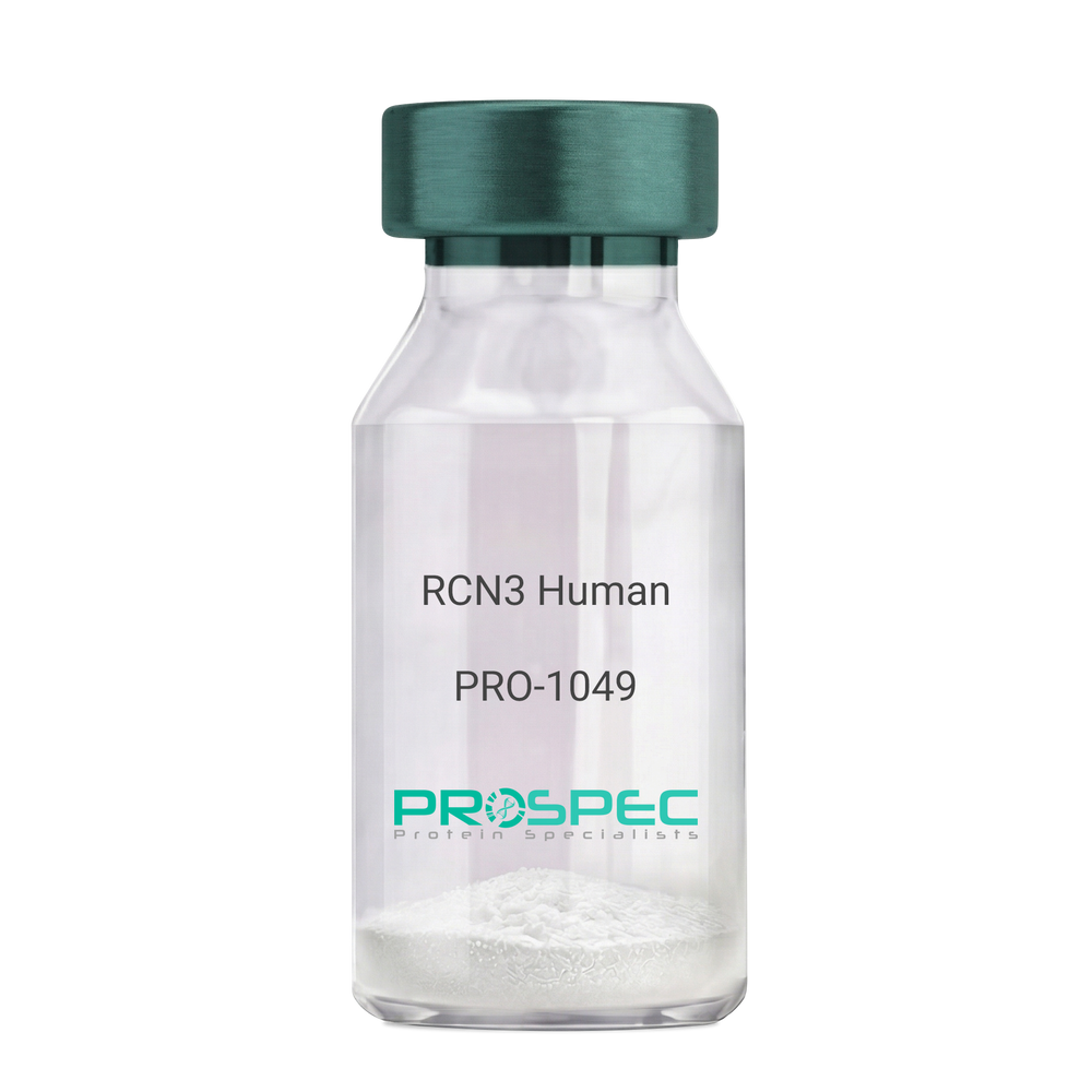
RCN3 HumanDescription:
Reticulocalbin 3 Human Recombinant
Reticulocalbin-3, EF-hand calcium-binding protein RLP49, RCN3, RLP49.
Product # :
PRO-1049Price :
Quantity :
Shipping Method :
Shipped with Ice Packs
More Info
- description
- source
- formulation
- purity
- More Info
Description
RCN3 Human Recombinant produced in E.Coli is a single, non-glycosylated polypeptide chain containing 333 amino acids (21-328 a.a) and having a molecular mass of 37.9kDa.RCN3 is fused to a 25 amino acid His-tag at N-terminus & purified by proprietary chromatographic techniques.
Source
Escherichia Coli.
Formulation
RCN3 protein solution (1mg/ml) containing 20mM Tris-HCl buffer (pH 8.0), 1mM DTT, 10% glycerol and 0.1M NaCl.
Purity
Greater than 95.0% as determined by SDS-PAGE.
More Info
-
Introduction
Reticulocalbin 3 (RCN3) belongs to the CREC (cab45/reticulocalbin/ ERC45/calumenin) family. RCN3 contains 5 Arg-Xaa-Xaa-Arg motifs, which function as target sequences of SPCs (subtilisin-like proprotein convertases), which is a family of serine endoproteases that proteolytically activate proproteins. The synthesis of PACE4 is induced by association and coexpression with RCN3.
-
Synonyms
Reticulocalbin-3, EF-hand calcium-binding protein RLP49, RCN3, RLP49.
-
Physical Appearance
Sterile Filtered colorless solution.
-
Stability
Store at 4°C if entire vial will be used within 2-4 weeks. Store, frozen at -20°C for longer periods of time. For long term storage it is recommended to add a carrier protein (0.1% HSA or BSA).Avoid multiple freeze-thaw cycles.
-
Amino Acid Sequence
MGSSHHHHHH SSGLVPRGSH MGSHMKPSPD AGPHGQGRVH QAAPLSDAPH DDAHGNFQYD HEAFLGREVA KEFDQLTPEE SQARLGRIVD RMDRAGDGDG WVSLAELRAW IAHTQQRHIR DSVSAAWDTY DTDRDGRVGW EELRNATYGH YAPGEEFHDV EDAETYKKML ARDERRFRVA DQDGDSMATR EELTAFLHPE EFPHMRDIVI AETLEDLDRN KDGYVQVEEY IADLYSAEPG EEEPAWVQTE RQQFRDFRDL NKDGHLDGSE VGHWVLPPAQ DQPLVEANHL LHESDTDKDG RLSKAEILGN WNMFVGSQAT NYGEDLTRHH DEL.
ProSpec's products are furnished for LABORATORY RESEARCH USE ONLY. The product may not be used as drugs, agricultural or pesticidal products, food additives or household chemicals.
- View Data Sheet
Name :
Transferrin HumanDescription:
Transferrin Human Recombinant
Serotransferrin, Transferrin, Siderophilin, Beta-1-metal-binding globulin, TF, PRO1557, PRO2086, DKFZp781D0156, HTF.
Product # :
PRO-747Price :
Quantity :
Shipping Method :
Shipped at Room temp
More Info
- description
- source
- formulation
- purity
- biological activity
- More Info
Description
Recombinant Human Transferrin produced in Plant is a non-glycosylated, polypeptide chain containing 679 amino acids and having a molecular mass of 76 kDa. The Recombinant Human Transferrin is purified by proprietary chromatographic techniques.
Source
Oryza sativa (rice).
Formulation
The protein (1mg/ml) was lyophilized with no additives.
Purity
Purity as determined by SDS-PAGE is 97%.
Biological Activity
One mg of Recombinant Human Transferrin will bind to approximately 2 micrograms of Fe.
More Info
-
Introduction
Transferrin is the iron-transport protein of vertebrate serum and donates iron to cells through interaction with a specific membrane receptor, CD71. Transferrin appears to be indispensable for most cells growing in tissue culture.
It is referred to frequently as a growth factor because, in analogy to other growth factor-receptor interactions, proliferating cells express high numbers of transferrin receptors, and the binding of transferrin to their receptors is needed for cells to initiate and maintain their DNA synthesis. Apart from its role as an iron transport protein transferrin acts as a cytokine and has functions that may not be related to its iron-carrying capacity. -
Synonyms
Serotransferrin, Transferrin, Siderophilin, Beta-1-metal-binding globulin, TF, PRO1557, PRO2086, DKFZp781D0156, HTF.
-
Physical Appearance
Sterile Filtered lyophilized (freeze-dried) powder.
-
Stability
Lyophilized Transferrin although stable at room temperature for 3 weeks, should be stored desiccated below -18°C. Upon reconstitution Transferrin Human Recombinant should be stored at 4°C between 2-7 days and for future use below -18°C.For long term storage it is recommended to add a carrier protein (0.1% HSA or BSA).Please prevent freeze-thaw cycles.
-
Solubility
Stock solutions can be prepared by dissolving gently into PBS for several minutes. Recommended stock concentrations are 5mg/ml to 20 mg/ml in PBS, though others can be used as well. Please try to avoid the formation of bubbles when dissolving the protein. Sterile filter through 0.2µm filter.
ProSpec's products are furnished for LABORATORY RESEARCH USE ONLY. The product may not be used as drugs, agricultural or pesticidal products, food additives or household chemicals.
- View Data Sheet
Name :
ThymalinDescription:
Thymulin
Product # :
HOR-047Price :
Quantity :
Shipping Method :
Shipped at Room temp
More Info
- description
- formulation
- purity
- More Info
Description
Thymulin Synthetic is a single, non-glycosylated polypeptide chain containing 9 amino acids, having a molecular mass of 858 Dalton and a Molecular formula of C33H54N12O15.
Formulation
The protein was lyophilized with no additives.
Purity
Greater than 97.0% as determined by analysis by RP-HPLC.
More Info
-
Physical Appearance
Sterile Filtered White lyophilized (freeze-dried) powder.
-
Stability
Lyophilized Thymulin although stable at room temperature for 3 weeks, should be stored desiccated below -18°C. Upon reconstitution Thymulin should be stored at 4°C between 2-7 days and for future use below -18°C. For long term storage it is recommended to add a carrier protein (0.1% HSA or BSA). Please prevent freeze-thaw cycles.
-
Solubility
It is recommended to reconstitute the lyophilized Thymulin in sterile 18MΩ-cm H2O not less than 100 µg/ml, which can then be further diluted to other aqueous solutions.
-
Amino Acid Sequence
Pyr-Ala-Lys-Ser-Gln-Gly-Gly-Ser-Asn-OH.
-
Background
Thymulin, a nonapeptide hormone, is produced primarily by the thymus gland and has been recognized for its pivotal role in the immune system. It plays a significant role in the maturation and differentiation of T lymphocytes, which are crucial for immune function. Beyond its immune-related functions, research has increasingly unveiled the diverse physiological roles of thymulin. This study aims to comprehensively investigate thymulin, shedding light on its immunological functions and exploring its potential applications in various aspects of health and medicine.
The primary objective of this research is to elucidate the mechanisms underlying thymulin's role in immune regulation. In vitro and in vivo experiments will be conducted to explore thymulin's interactions with immune cells, its impact on T cell development and function, and its potential modulation of immune responses. Understanding these mechanisms is fundamental for harnessing thymulin's immunomodulatory properties.
The second objective is to assess the clinical relevance of thymulin in immune-related disorders. Clinical trials and studies involving individuals with autoimmune diseases, immunodeficiencies, and age-related immune decline will be conducted to evaluate the potential therapeutic applications of thymulin. These investigations may offer insights into the use of thymulin as an immunomodulatory agent in various clinical settings.
The third objective is to explore the broader implications of thymulin in health and medicine. Research will investigate its potential roles in areas beyond immunology, such as neuroprotection, wound healing, and tissue regeneration. Understanding the multifaceted properties of thymulin may open new avenues for therapeutic interventions in various medical specialties.
By delving into the diverse functions of thymulin, this research aims to expand our knowledge of its physiological roles and clinical applications. The findings may have implications for the development of innovative approaches in immunology and healthcare, ultimately benefiting patients affected by immune-related disorders and other medical conditions.
What is the molecular weight/Mw of THYMALIN Protein?
THYMALIN Protein has a total Mw of 0.85kDa.
What is the Purity of THYMALIN Protein?
THYMALIN Protein is >97% pure as determined by SDS-PAGE.
What is the Biological Activity of THYMALIN Protein?
The biological functionality of THYMALIN Protein will be determined in the future.
What is the amino acid sequence of THYMALIN Protein?
Pyr-Ala-Lys-Ser-Gln-Gly-Gly-Ser-Asn-OH.
What applications can THYMALIN Protein be used in?
THYMALIN Protein can probably be used in western blot, ELISA and Lateral Flow.
What is the endotoxin level for THYMALIN Protein?
The endotoxin level is minimal, THYMALIN Protein was purified using conventional chromatography techniques.
ProSpec's products are furnished for LABORATORY RESEARCH USE ONLY. The product may not be used as drugs, agricultural or pesticidal products, food additives or household chemicals.
- View Data Sheet
Name :
G CSF Human, PEGDescription:
Granulocyte-Colony Stimulating Factor Pegylated Human Recombinant
CSF-3, MGI-1G, GM-CSF beta, Pluripoietin, Filgrastim, Lenograstim, G-CSF, MGC45931, GCSF.
Product # :
CYT-018Price :
Quantity :
Shipping Method :
Shipped with Ice Packs
More Info
- description
- source
- formulation
- purity
- biological activity
- More Info
Description
Granulocyte Colony Stimulating Factor Human Recombinant produced in E.coli is a single, non-glycosylated, polypeptide chain containing 175 amino acids and having a molecular mass of 18.8kDa. The Pegylated G-CSF is produced by attaching a 20kDa methoxypolyethylene glycol propionaldehyde (mPEG-ALD) to the N-terminal amino acid of G-CSF giving a total molecular mass of 38.8kDa. G-CSF is purified by proprietary chromatographic techniques.
Source
Escherichia Coli.
Formulation
G-CSF is supplied in solution (0.69mg/ml) containing 10mM Acetate Buffer (pH 4.0), and 0.004% Polysorbate 80.
Purity
Greater than 95.0% as determined by SEC-HPLC.
Biological Activity
The ED50, calculated by the dose-dependent proliferation of murine NFS-60 indicator cells is less than 0.1 ng/ml, corresponding to a Specific Activity of 10,000,000IU/mg.
More Info
-
Introduction
GCSF is a cytokine that controls the production, differentiation, and function of granulocytes. The active protein is found extracellularly. Three transcript variants encoding three different isoforms have been found for this gene. Granulocyte/macrophage colony-stimulating factors are cytokines that act in hematopoiesis by controlling the production, differentiation, and function of 2 related white cell populations of the blood, the granulocytes and the monocytes-macrophages. This csf induces granulocytes.
-
Synonyms
CSF-3, MGI-1G, GM-CSF beta, Pluripoietin, Filgrastim, Lenograstim, G-CSF, MGC45931, GCSF.
-
Physical Appearance
Colorless, clear and transparent solution.
-
Stability
G-CSF PEG should be stored refrigerated at 2° to 8°C. Vials should be kept in theirpackaging to protect from light until the time of use. Shaking and freezing should be avoided.
-
Background
What is the molecular weight/Mw of G CSF HUMAN, PEG Protein?
G CSF HUMAN, PEG Protein has a total Mw of 18.8kDa.
What is the source or expression system of G CSF HUMAN, PEG Protein?
Escherichia Coli.
What is the Purity of G CSF HUMAN, PEG Protein?
G CSF HUMAN, PEG Protein is >95% pure as determined by SDS-PAGE.
What is the Biological Activity of G CSF HUMAN, PEG Protein?
The ED50, calculated by the dose-dependent proliferation of murine NFS-60 indicator cells is less than 0.1 ng/ml, corresponding to a Specific Activity of 10,000,000IU/mg.
What is the amino acid sequence of G CSF HUMAN, PEG Protein?
G CSF HUMAN, PEG Protein is composed from 175 amino acids.
What applications can G CSF HUMAN, PEG Protein be used in?
G CSF HUMAN, PEG Protein can probably be used in western blot, ELISA and Lateral Flow.
What is the endotoxin level for G CSF HUMAN, PEG Protein?
The endotoxin level is minimal, G CSF HUMAN, PEG Protein was purified using conventional chromatography techniques.
ProSpec's products are furnished for LABORATORY RESEARCH USE ONLY. The product may not be used as drugs, agricultural or pesticidal products, food additives or household chemicals.
- View Data Sheet
Name :
Adipsin Human, Sf9Description:
Complement Factor D Human Recombinant, Sf9
Complement Factor D (Adipsin), Properdin Factor D, D Component Of Complement (Adipsin), C3 Convertase Activator, EC 3.4.21.46, ADIPSIN, PFD, AND, DF, Complement Factor D Preproprotein, Complement Factor D, EC 3.4.21, Complement factor D.
Product # :
PRO-2213Price :
Quantity :
Shipping Method :
Shipped with Ice Packs
More Info
- description
- source
- formulation
- purity
- More Info
Description
Adipsin produced in Sf9 Baculovirus cells is a single, glycosylated polypeptide chain containing 241 amino acids (21-253a.a.) and having a molecular mass of 26.01kDa. (Molecular size on SDS-PAGE will appear at approximately 28-40kDa). Adipsin is expressed with an 8 amino acid His tag at C-Terminus and purified by proprietary chromatographic techniques.
Source
Sf9, Baculovirus cells.
Formulation
Adipsin protein solution (1mg/ml) contains phosphate buffered saline (pH7.4).
Purity
Greater than 95.0% as determined by SDS-PAGE.
More Info
-
Introduction
Complement Factor D (Adipsin), which belongs to the trypsin family of peptidases, is involved in the alternative complement pathway of the complement system where it cleaves factor B. In the alternative complement pathway, Adipsin is best known for its role in humoral suppression of infectious agents. In addition, Adipsin is a serine protease which is secreted by adipocytes into the bloodstream. Ultimately, Adipsin has a high level of expression in fat, proposing a role for adipose tissue in immune system biology.
-
Synonyms
Complement Factor D (Adipsin), Properdin Factor D, D Component Of Complement (Adipsin), C3 Convertase Activator, EC 3.4.21.46, ADIPSIN, PFD, AND, DF, Complement Factor D Preproprotein, Complement Factor D, EC 3.4.21, Complement factor D.
-
Physical Appearance
Sterile Filtered colorless solution.
-
Stability
Store at 4°C if entire vial will be used within 2-4 weeks. Store, frozen at -20°C for longer periods of time. For long term storage it is recommended to add a carrier protein (0.1% HSA or BSA).Avoid multiple freeze-thaw cycles.
-
Amino Acid Sequence
PPRGRILGGR EAEAHARPYM ASVQLNGAHL CGGVLVAEQW VLSAAHCLED AADGKVQVLL GAHSLSQPEP SKRLYDVLRA VPHPDSQPDT IDHDLLLLQL SEKATLGPAV RPLPWQRVDR DVAPGTLCDV AGWGIVNHAG RRPDSLQHVL LPVLDRATCN RRTHHDGAIT ERLMCAESNR RDSCKGDSGG PLVCGGVLEG VVTSGSRVCG NRKKPGIYTR VASYAAWIDS VLAVEHHHHH H.
ProSpec's products are furnished for LABORATORY RESEARCH USE ONLY. The product may not be used as drugs, agricultural or pesticidal products, food additives or household chemicals.
- View Data Sheet
Name :
PI3 Human, Sf9Description:
Peptidase Inhibitor 3 Human Recombinant, Sf9
Elafin, ESI, SKALP, WAP3, WFDC14.
Product # :
PRO-2653Price :
Quantity :
Shipping Method :
Shipped with Ice Packs
More Info
- description
- source
- formulation
- purity
- More Info
Description
PI3 Human Recombinant produced in Sf9 Baculovirus cells is a single, glycosylated polypeptide chain containing 101 amino acids (23-117a.a) and having a molecular mass of 10.7kDa.PI3 is fused to an 6 amino acid His-tag at C-terminus & purified by proprietary chromatographic techniques.
Source
Sf9, Baculovirus cells.
Formulation
The PI3 solution (0.2mg/ml) contains Phosphate-Buffered Saline (pH 7.4) and 10% glycerol.
Purity
Greater than 85.0% as determined by SDS-PAGE.
More Info
-
Introduction
Peptidase Inhibitor 3, also referred to PI3, is neutrophil and pancreatic elastase-specific inhibitor of skin. The protein may prevent elastase mediated tissue proteolysis. PI3 has shown inhibition of alpha-4-beta-2/CHRNA2-CHRNB2 nicotinic acetylcholine receptor, a weak inhibition on Kv11.1/KCNH2/ERG1 and on the transient receptor potential cation channel subfamily V member 1.
-
Synonyms
Elafin, ESI, SKALP, WAP3, WFDC14.
-
Physical Appearance
Sterile Filtered colorless solution.
-
Stability
Store at 4°C if entire vial will be used within 2-4 weeks. Store, frozen at -20°C for longer periods of time. For long term storage it is recommended to add a carrier protein (0.1% HSA or BSA).Avoid multiple freeze-thaw cycles.
-
Amino Acid Sequence
AVTGVPVKGQ DTVKGRVPFN GQDPVKGQVS VKGQDKVKAQ EPVKGPVSTK PGSCPIILIR CAMLNPPNRC LKDTDCPGIK KCCEGSCGMA CFVPQHHHHH H
ProSpec's products are furnished for LABORATORY RESEARCH USE ONLY. The product may not be used as drugs, agricultural or pesticidal products, food additives or household chemicals.
- View Data Sheet
Name :
Follistatin Human, HisDescription:
Follistatin Human Recombinant, His Tag
FST, FS, Activin-binding protein.
Product # :
CYT-029Price :
Quantity :
Shipping Method :
Shipped at Room temp
More Info
- description
- source
- formulation
- More Info
Description
FST His Protein is 36.0 kDa protein containing 325 amino acid residues of the FST His and the 10 aa N-Terminal His-tag.
Source
E. coli.
Formulation
FST His Tag was filtered (0.4µm) and lyophilized from 0.5mg/ml supplied in 20mM TRIS and 20mM NaCl, pH 7.5.
More Info
-
Introduction
Follistatin is a single-chain gonadal protein that specifically inhibits follicle-stimulating hormone release. The single FST gene encodes two isoforms, FST317 and FST344 containing 317 and 344 amino acids respectively, resulting from alternative splicing of the precursor mRNA. In a study in which 37 candidate genes were tested for linkage and association with polycystic ovary syndrome (PCOS) or hyperandrogenemia in 150 families, evidence was found for linkage between PCOS and follistatin. Follistatin binds directly to activin and functions as an activin antagonist. specific inhibitor of the biosynthesis and secretion of pituitary follicle stimulating hormone (fsh).
-
Synonyms
FST, FS, Activin-binding protein.
-
Stability
Store lyophilized FST His at -20°C. Aliquot the product after reconstitution to avoid repeated freezing/thawing cycles. Reconstituted FST His can be stored at 4°C for a limited period of time; it does not show any change after two weeks at 4°C.
-
Solubility
It is recommended to add deionized water to prepare a working stock solution of approximately 0.5mg/ml and let the lyophilized pellet dissolve completely. Product is not sterile! Please filter the product by an appropriate sterile filter before using it on cell culture.
-
Amino Acid Sequence
MKHHHHHHAS GNCWLRQAKN GRCQVLYKTE LSKEECCSTG RLSTSWTEED VNDNTLFKWM IFNGGAPNCI PCKETCENVD CGPGKKCRMN KKNKPRCVCA PDCSNITWKG PVCGLDGKTY RNECALLKAR CKEQPELEVQ YQGRCKKTCR DVFCPGSSTC VVDQTNNAYC VTCNRICPEP ASSEQYLCGN DGVTYSSACH LRKATCLLGR SIGLAYEGKC IKAKSCEDIQ CTGGKKCLWD FKVGRGRCSL CDELCPDSKS DEPVCASDNA TYASECAMKE AACSSGVLLE VKHSGSCNSI SEDTEEEEED EDQDYSFPIS SILEW.
-
Background
What is the molecular weight/Mw of FOLLISTATIN HUMAN, HIS Protein?
FOLLISTATIN HUMAN, HIS Protein has a total Mw of 36kDa.
What is the source or expression system of FOLLISTATIN HUMAN, HIS Protein?
Escherichia Coli.
What is the Biological Activity of FOLLISTATIN HUMAN, HIS Protein?
The biological functionality of FOLLISTATIN HUMAN, HIS Protein will be determined in the future.
What is the amino acid sequence of FOLLISTATIN HUMAN, HIS Protein?
MKHHHHHHAS GNCWLRQAKN GRCQVLYKTE LSKEECCSTG RLSTSWTEED VNDNTLFKWM IFNGGAPNCI PCKETCENVD CGPGKKCRMN KKNKPRCVCA PDCSNITWKG PVCGLDGKTY RNECALLKAR CKEQPELEVQ YQGRCKKTCR DVFCPGSSTC VVDQTNNAYC VTCNRICPEP ASSEQYLCGN DGVTYSSACH LRKATCLLGR SIGLAYEGKC IKAKSCEDIQ CTGGKKCLWD FKVGRGRCSL CDELCPDSKS DEPVCASDNA TYASECAMKE AACSSGVLLE VKHSGSCNSI SEDTEEEEED EDQDYSFPIS SILEW.
What applications can FOLLISTATIN HUMAN, HIS Protein be used in?
FOLLISTATIN HUMAN, HIS Protein can probably be used in western blot, ELISA and Lateral Flow.
What is the endotoxin level for FOLLISTATIN HUMAN, HIS Protein?
The endotoxin level is minimal, FOLLISTATIN HUMAN, HIS Protein was purified using conventional chromatography techniques.
ProSpec's products are furnished for LABORATORY RESEARCH USE ONLY. The product may not be used as drugs, agricultural or pesticidal products, food additives or household chemicals.
- View Data Sheet
Name :
KEL MouseDescription:
Kell Metallo-Endopeptidase Mouse Recombinant
kell blood group antigen, kell blood group glycoprotein, Kell blood group, Kell blood group glycoprotein homolog, KEL, Kell, CD238 antigen, CD238, ECE3, Kell blood group-metalloendopeptidase, Kell blood group-metalloendopeptidase.
Product # :
ENZ-1158Price :
Quantity :
Shipping Method :
Shipped with Ice Packs
More Info
- description
- source
- formulation
- purity
- More Info
Description
KEL Mouse produced in Sf9 Baculovirus cells is a single, glycosylated polypeptide chain containing 674 amino acids (49-713 aa) and having a molecular mass of 76.3kDa.KEL is fused to a 9 amino acid His tag at C-terminus and purified by proprietary chromatographic techniques.
Source
Sf9, Baculovirus cells.
Formulation
The KEL solution (0.25mg/ml) contains 10% Glycerol and Phosphate-Buffered Saline (pH 7.4).
Purity
Greater than 90.0% as determined by SDS-PAGE.
More Info
-
Introduction
Kell blood group glycoprotein homolog or KEL, is an enzyme, part of the zinc endopeptidase of the neprilysin (NEP) group of proteins. KEL has a crucial part in the production of the potent bioactive ET-3, which also includes enzymes that are endothelin convertingenzymes (PEX, XCE, DINE &various NEP-like proteins). KEL uses a single disulfide bond to XK, a gated membranal transporter. The Kell antigen system that includes two proteins, is very crucial among blood group systems.
-
Synonyms
kell blood group antigen, kell blood group glycoprotein, Kell blood group, Kell blood group glycoprotein homolog, KEL, Kell, CD238 antigen, CD238, ECE3, Kell blood group-metalloendopeptidase, Kell blood group-metalloendopeptidase.
-
Physical Appearance
Sterile Filtered colorless solution.
-
Stability
Store at 4°C if entire vial will be used within 2-4 weeks. Store, frozen at -20°C for longer periods of time. For long term storage it is recommended to add a carrier protein (0.1% HSA or BSA).Avoid multiple freeze-thaw cycles.
-
Amino Acid Sequence
ADPIFRNCGP CPCETPVCME LLDHYLASGN RSVAPCTDFF SFACEKANGT SDSFQALTEE NKSRLWRLLE APGSWHLGSG EEKAFQFYNS CMDTDAIEAS GSGPLIQIIE ELGGWNITGN WTSLDFNQNL RLLMSQYGHF PFFRAYLRPH PAPPHTPIIQ IDQPEFDILL QQEQEQKVYA QILREYVTYL NRLGTLLGSN PQEAQQHASW SIVFTSRLFQ FLRPQQQQQA QDKLFHVVTI DELQEMAPAI DWLSCLQAIF TPMSLNSSQT LVVHDLDYLR NMSQLVEEGL LNHRESIQSY MILGLVDTLS PALDTKFQEA RRELIQELRK LKERPPLPAY PRWMKCVEQT GAFFEPTLAA LFVREAFGPS IQSAAMELFA EIKDAVIIRL KKLSWISEET QKEALNKLAQ LQVEMGAPKR AVKPDIATQE YNDIQLGPSF LQSFLSCVRS LRARNVQSFL QPFPYHRWQK SPWEVNAYYS ISDHMVVFPA GLLQPPFFHP GYPRAVNFGA AGSIMAHELL HIFYQLLLPG GCPACDTHVL QEALLCLERH YAAFPLPSIS SFNGSHTLLE NAADIGGVAI AFQAYSKRIV EHTGELTLPN LDLSPYQLFF RSYAQVMCRG LSSQDPQDPH SPPSLRVHGP LSNTPDFAKH FHCPRGTLLN PSARCKLWHH HHHH.
ProSpec's products are furnished for LABORATORY RESEARCH USE ONLY. The product may not be used as drugs, agricultural or pesticidal products, food additives or household chemicals.
- View Data Sheet
Name :
CDH11 HumanDescription:
Cadherin 11 Human Recombinant
CAD11, CDHOB, OB, OSF-4, Cadherin 11, Osteoblast cadherin, OB-cadherin.
Product # :
PRO-1368Price :
Quantity :
Shipping Method :
Shipped with Ice Packs
More Info
- description
- source
- formulation
- purity
- More Info
Description
CDH11 Human Recombinant produced in E.Coli is a single, non-glycosylated polypeptide chain containing 601 amino acids (54-617 a.a) and having a molecular mass of 66.2kDa. CDH11 is fused to a 37 amino acid His-tag at N-terminus & purified by proprietary chromatographic techniques.
Source
E.coli.
Formulation
CDH11 protein solution (1mg/ml) containing 20mM Tris-HCl buffer (pH8.0), 10% glycerol and 0.1M NaCl.
Purity
Greater than 85% as determined by SDS-PAGE.
More Info
-
Introduction
Cadherin 11 (CDH11) belongs to the cadherin superfamily (integral membrane proteins) which mediate calcium-dependent cell-cell adhesion. CDH11 is defined based on lacking a HAV cell adhesion recognition sequence specific to type I cadherins. CDH11 protein’s expression in osteoblastic cell lines, and its upregulation during differentiation, suggests a specific function in bone development and maintenance. CDH11 contributes to the sorting of heterogeneous cell types.
-
Synonyms
CAD11, CDHOB, OB, OSF-4, Cadherin 11, Osteoblast cadherin, OB-cadherin.
-
Physical Appearance
Sterile Filtered colorless solution.
-
Stability
Store at 4°C if entire vial will be used within 2-4 weeks. Store, frozen at -20°C for longer periods of time. For long term storage it is recommended to add a carrier protein (0.1% HSA or BSA).Avoid multiple freeze-thaw cycles.
-
Amino Acid Sequence
MRGSHHHHHH GMASMTGGQQ MGRDLYDDDD KDRWGSMGWV WNQFFVIEEY TGPDPVLVGR LHSDIDSGDG NIKYILSGEG AGTIFVIDDK SGNIHATKTL DREERAQYTL MAQAVDRDTN RPLEPPSEFI VKVQDINDNP PEFLHETYHA NVPERSNVGT SVIQVTASDA DDPTYGNSAK LVYSILEGQP YFSVEAQTGI IRTALPNMDR EAKEEYHVVI QAKDMGGHMG GLSGTTKVMI TLTDVNDNPP KFPQSVYQMS VSEAAVPGEE VGRVKAKDPD IGENGLVTYN IVDGDGMESF EITTDYETQE GVIKLKKPVD FETKRAYSLK VEAANVHIDP KFISNGPFKD TVTVKIAVED ADEPPMFLAP SYIHEVQENA AAGTVVGRVH AKDPDAANSP IRYSIDRHTD LDRFFTINPE DGFIKTTKPL DREETAWLNI TVFAAEIHNR HQEAKVPVAI RVLDVNDNAP KFAAPYEGFI CESDQTKPLS NQPIVTISAD DKDDTANGPR FIFSLPPEII HNPNFTVRDN RDNTAGVYAR RGGFSRQKQD LYLLPIVISD GGIPPMSSTN TLTIKVCGCD VNGALLSCNA EAYILNAGLS T.
ProSpec's products are furnished for LABORATORY RESEARCH USE ONLY. The product may not be used as drugs, agricultural or pesticidal products, food additives or household chemicals.
- View Data Sheet
Name :
CDH2 HumanDescription:
Cadherin 2 Human Recombinant
Cadherin 2, Cadherin 2, Type 1, N-Cadherin (Neuronal), Neural Cadherin, N-Cadherin, CDw325, NCAD, CDHN, Calcium-Dependent Adhesion Protein, Neuronal, CD325 Antigen, N-Cadherin 1, Cadherin-2, CD325.
Product # :
PRO-2455Price :
Quantity :
Shipping Method :
Shipped with Ice Packs
More Info
- description
- source
- formulation
- purity
- More Info
Description
CDH2 Human Recombinant produced in Sf9 Baculovirus cells is a single, glycosylated polypeptide chain containing 574 amino acids (160-724a.a.) and having a molecular mass of 62.9kDa (Molecular size on SDS-PAGE will appear at approximately 70-100kDa).CDH2 is expressed with a 6 amino acids His tag at C-Terminus and purified by proprietary chromatographic techniques.
Source
Sf9, Baculovirus cells.
Formulation
CDH2 protein solution (0.5mg/ml) contains Phosphate Buffered Saline (pH 7.4) and 20% glycerol.
Purity
Greater than 90.0% as determined by SDS-PAGE.
More Info
-
Introduction
Cadherin-2 isoform 1, also known as CDH2 is a transmembrane, homophilic glycoprotein which belongs to the calcium-dependent cell adhesion molecule family. CDH2 takes part in neurons and later on was discovered to participate in cardiac muscle and in cancer metastasis as well. The CDH2 loss promotes tumorigenesis through releasing membrane-bound β-catenin, and so stimulating Wnt signaling. Furthermore, CDH2 appears to be implicated in tumor development, however this discovery is limited in adrenocortical tumors-ACTs.
-
Synonyms
Cadherin 2, Cadherin 2, Type 1, N-Cadherin (Neuronal), Neural Cadherin, N-Cadherin, CDw325, NCAD, CDHN, Calcium-Dependent Adhesion Protein, Neuronal, CD325 Antigen, N-Cadherin 1, Cadherin-2, CD325.
-
Physical Appearance
Sterile Filtered colorless solution.
-
Stability
Store at 4°C if entire vial will be used within 2-4 weeks. Store, frozen at -20°C for longer periods of time. For long term storage it is recommended to add a carrier protein (0.1% HSA or BSA).Avoid multiple freeze-thaw cycles.
-
Amino Acid Sequence
ADPDWVIPPI NLPENSRGPF PQELVRIRSD RDKNLSLRYS VTGPGADQPP TGIFIINPIS GQLSVTKPLD REQIARFHLR AHAVDINGNQ VENPIDIVIN VIDMNDNRPE FLHQVWNGTV PEGSKPGTYV MTVTAIDADD PNALNGMLRY RIVSQAPSTP SPNMFTINNE TGDIITVAAG LDREKVQQYT LIIQATDMEG NPTYGLSNTA TAVITVTDVN DNPPEFTAMT FYGEVPENRV DIIVANLTVT DKDQPHTPAW NAVYRISGGD PTGRFAIQTD PNSNDGLVTV VKPIDFETNR MFVLTVAAEN QVPLAKGIQH PPQSTATVSV TVIDVNENPY FAPNPKIIRQ EEGLHAGTML TTFTAQDPDR YMQQNIRYTK LSDPANWLKI DPVNGQITTI AVLDRESPNV KNNIYNATFL ASDNGIPPMS GTGTLQIYLL DINDNAPQVL PQEAETCETP DPNSINITAL DYDIDPNAGP FAFDLPLSPV TIKRNWTITR LNGDFAQLNL KIKFLEAGIY EVPIIITDSG NPPKSNISIL RVKVCQCDSN GDCTDVDRIV GAGLGTGAHH HHHH.
ProSpec's products are furnished for LABORATORY RESEARCH USE ONLY. The product may not be used as drugs, agricultural or pesticidal products, food additives or household chemicals.
- View Data Sheet
Name :
LGALS8 HumanDescription:
Galectin-8 Human Recombinant
Galectin-8, Gal-8, Po66 carbohydrate-binding protein, Po66-CBP, Prostate carcinoma tumor antigen 1, PCTA-1, LGALS8.
Product # :
CYT-017Price :
Quantity :
Shipping Method :
Shipped at Room temp
More Info
- description
- source
- formulation
- purity
- biological activity
- More Info
Description
Galectin-8 Human Recombinant produced in E.Coli is a single, non-glycosylated, Polypeptide chain containing 317 amino acids and having a molecular mass of 35.8kDa.The LGALS8 is purified by proprietary chromatographic techniques.
Source
Escherichia Coli.
Formulation
LGALS8 was lyophilized from a concentrated (1mg/ml) solution in 20mM PBS, pH 7.4.
Purity
Greater than 95.0% as determined by:
(a) Analysis by RP-HPLC.
(b) Analysis by SDS-PAGE.Biological Activity
The ED50 of Galectin-8 as determined by its ability to agglutinate human red blood cells is 0.8~4 µg/ml.More Info
-
Introduction
Galectins are a family of animal lectins with an affinity for beta-galactosides. This family has at least 14 identified members. Galectins share similarities in the CRD (the carbohydrate recognition domain). Galectins are synthesized as cytosolic proteins. Though localized principally in the cytoplasm and lacking a classical signal peptide, galectins can also be stimulated to secretion by non-classical pathways or alternatively targeted to the nucleus. Galectins are involved in modulating cell-cell and cell-matrix interactions. Galectin-8 is a tandem-repeat-type member of the galectin family, consisting of 2 CRDs attached by a linker peptide. Galectin-8 is greatly expressed in lung carcinomas, a number of forms of prostate carcinomas, in addition to other tumor cells. Galectin-8 attaches to a subset of cell surface integrins to modulate ECM-integrin interactions. Once immobilized, Galectin-8 promotes cell adhesion by ligation and clustering of cell surface integrin receptors. On the other hand, as a soluble ligand, Galectin-8 can inhibit cell adhesion.
-
Synonyms
Galectin-8, Gal-8, Po66 carbohydrate-binding protein, Po66-CBP, Prostate carcinoma tumor antigen 1, PCTA-1, LGALS8.
-
Physical Appearance
Sterile Filtered White lyophilized (freeze-dried) powder.
-
Stability
Lyophilized LGALS8 although stable at room temperature for 3 weeks, should be stored desiccated below -18°C. Upon reconstitution Galectin-8 should be stored at 4°C between 2-7 days and for future use below -18°C.Please prevent freeze-thaw cycles.
-
Solubility
It is recommended to reconstitute the lyophilized Galectin-8 in sterile 18M-cm H2O not less than 100µg/ml, which can then be further diluted to other aqueous solutions.
-
Amino Acid Sequence
The sequence of the first five N-terminal amino acids was determined and was found to be Met-Met-Leu-Ser-Leu.
-
Background
What is the molecular weight/Mw of LGALS8 HUMAN Protein?
LGALS8 HUMAN Protein has a total Mw of 35.8kDa.
What is the source or expression system of LGALS8 HUMAN Protein?
Escherichia Coli.
What is the Purity of LGALS8 HUMAN Protein?
LGALS8 HUMAN Protein is >95% pure as determined by SDS-PAGE.
What is the Biological Activity of LGALS8 HUMAN Protein?
The ED50 of Galectin-8 as determined by its ability to agglutinate human red blood cells is 0.8~4 µg/ml.
What is the amino acid sequence of LGALS8 HUMAN Protein?
LGALS8 HUMAN Protein is composed from 317 amino acids.
What applications can LGALS8 HUMAN Protein be used in?
LGALS8 HUMAN Protein can probably be used in western blot, ELISA and Lateral Flow.
What is the endotoxin level for LGALS8 HUMAN Protein?
The endotoxin level is minimal, LGALS8 HUMAN Protein was purified using conventional chromatography techniques.
ProSpec's products are furnished for LABORATORY RESEARCH USE ONLY. The product may not be used as drugs, agricultural or pesticidal products, food additives or household chemicals.
- View Data Sheet
Name :
PTH (7-84) N15 HumanDescription:
Parathyroid Hormone (7-84) N15 Labeled Human Recombinant
Parathyrin, PTH, Parathormone.
Product # :
HOR-013Price :
Quantity :
Shipping Method :
Shipped at Room temp
More Info
- description
- source
- formulation
- purity
- More Info
Description
PTH (7-84) N15 Human Recombinant produced in E.coli is a single, non-glycosylated polypeptide chain containing 78 amino acids and having a molecular mass of 8900 Dalton labeled by the stable isotope N15.The PTH (7-84) N15 is purified by proprietary chromatographic techniques.
Source
Escherichia Coli.
Formulation
PTH (7-84) N15 protein was lyophilized from a 0.2µm filtered concentrated solution in 1xPBS, pH 7.4.
Purity
Greater than 97.0% as determined by:
(a) Analysis by RP-HPLC.
(b) Analysis by SDS-PAGE.More Info
-
Introduction
Parathyroid hormone (PTH), or parathormone, is secreted by the parathyroid glands as a polypeptide containing 84 amino acids. It acts to increase the concentration of calcium in the blood, whereas calcitonin (a hormone produced by the parafollicular cells of the thyroid gland) acts to decrease calcium concentration. PTH acts to increase the concentration of calcium in the blood by acting upon parathyroid hormone receptor in three parts of the body: In the bones- It enhances the release of calcium from the large reservoir contained in the bones. Bone resorption is the normal destruction of bone by osteoclasts, which are indirectly stimulated by PTH. Stimulation is indirect since osteoclasts do not have a receptor for PTH; rather, PTH binds to osteoblasts, the cells responsible for creating bone. Binding stimulates osteoblasts to increase their expression of RANKL, which can bind to osteoclast precursors containing RANK, a receptor for RANKL. The binding of RANKL to RANK stimulates these precursors to fuse, forming new osteoclasts which ultimately enhances the resorption of bone. In the kidney- It enhances active reabsorption of calcium from distal tubules and the thick ascending limb. In the intestine- It enhances the absorption of calcium in the intestine by increasing the production of vitamin D and upregulating the enzyme responsible for 1-alpha hydroxylation of 25-hydroxy vitamin D, converting vitamin D to its active form (1,25-dihydroxy vitamin D) which effects the actual absorption of calcium (as Ca2+ ions) by the intestine via calbindin.
-
Synonyms
Parathyrin, PTH, Parathormone.
-
Physical Appearance
Sterile Filtered White lyophilized (freeze-dried) powder.
-
Stability
Lyophilized PTH (7-84) N15 although stable at room temperature for 3 weeks, should be stored desiccated below -18°C. Upon reconstitution PTH (7-84) N15 should be stored at 4°C between 2-7 days and for future use below -18°C. For long term storage it is recommended to add a carrier protein (0.1% HSA or BSA). Please prevent freeze-thaw cycles.
-
Solubility
It is recommended to reconstitute the lyophilized PTH (7-84) N15 in sterile water not less than 100µg/ml, which can then be further diluted to other aqueous solutions.
-
Amino Acid Sequence
LMHNLGKHLN SMERVEWLRK KLQDVHNFVA LGAPLAPRDA GSQRPRKKED NVLVESHEKS LGEADKADVN VLTKAKSQ
ProSpec's products are furnished for LABORATORY RESEARCH USE ONLY. The product may not be used as drugs, agricultural or pesticidal products, food additives or household chemicals.