Search results
1000 results found for “batf”
Name
Description
Product #
Price
Quantity
Shipping Method
- View Data Sheet
Name :
TNFRSF10B Human, Sf9Description:
TNF Ligand Receptor Superfamily Member 10B Human Recombinant, Sf9
TNFRSF10B, CD262, DR5, KILLER, KILLER/DR5, TRAIL-R2, TRAILR2, TRICK2, TRICK2A, TRICK2B, TRICKB, ZTNFR9, Death receptor 5, TNF-related apoptosis-inducing ligand receptor 2.
Product # :
CYT-1055Price :
Quantity :
Shipping Method :
Shipped with Ice Packs
More Info
- description
- source
- formulation
- purity
- biological activity
- More Info
Description
TNFRSF10B produced in Sf9 Baculovirus cells is a single, glycosylated polypeptide chain containing 394 amino acids (56-210a.a.) and having a molecular mass of 43.9kDa. (Molecular size on SDS-PAGE will appear at approximately 40-57kDa).TNFRSF10B is expressed with a 239 amino acid hIgG-His tag at C-Terminus and purified by proprietary chromatographic techniques.
Source
Sf9, Insect cells.
Formulation
TNFRSF10B protein solution (1mg/ml) contains Phosphate Buffered Saline (pH 7.4) and 10% glycerol.
Purity
Greater than 90% as determined by SDS-PAGE.
Biological Activity
Measured by its ability to inhibit cytotoxicity assay using Jurkat human T lymphocyte. The ED50 for this effect ≤ to 5ng/ml with TRAIL
.
More Info
-
Introduction
TRAIL Receptor-1 (DR4) and TRAIL Receptor-2(DR5) are members of the TNFR superfamily of transmembrane proteins and contain a cytoplasmic "death domain", which is capable of activating the cell's apoptotic machinery. These receptors are activated by binding to either membrane anchored or soluble TRAIL/Apo2L.
-
Synonyms
TNFRSF10B, CD262, DR5, KILLER, KILLER/DR5, TRAIL-R2, TRAILR2, TRICK2, TRICK2A, TRICK2B, TRICKB, ZTNFR9, Death receptor 5, TNF-related apoptosis-inducing ligand receptor 2.
-
Physical Appearance
Sterile Filtered colorless solution.
-
Stability
Store at 4°C if entire vial will be used within 2-4 weeks. Store, frozen at -20°C for longer periods of time. For long term storage it is recommended to add a carrier protein (0.1% HSA or BSA).Avoid multiple freeze-thaw cycles.
-
Amino Acid Sequence
ITQQDLAPQQ RAAPQQKRSS PSEGLCPPGH HISEDGRDCI SCKYGQDYST HWNDLLFCLR CTRCDSGEVE LSPCTTTRNT VCQCEEGTFR EEDSPEMCRK CRTGCPRGMV KVGDCTPWSD IECVHKESGT KHSGEVPAVE ETVTSSPGTP ASPCSLEPKS CDKTHTCPPC PAPELLGGPS
VFLFPPKPKD TLMISRTPEV TCVVVDVSHE DPEVKFNWYV DGVEVHNAKT KPREEQYNST YRVVSVLTVL HQDWLNGKEY KCKVSNKALP APIEKTISKA KGQPREPQVY TLPPSRDELT KNQVSLTCLV KGFYPSDIAV EWESNGQPEN NYKTTPPVLD SDGSFFLYSK LTVDKSRWQQ
GNVFSCSVMH EALHNHYTQK SLSLSPGKHH HHHH.
ProSpec's products are furnished for LABORATORY RESEARCH USE ONLY. The product may not be used as drugs, agricultural or pesticidal products, food additives or household chemicals.
- View Data Sheet
Name :
B.Microti p32Description:
Babesia Microti p32 Recombinant
Product # :
PRO-2269Price :
Quantity :
Shipping Method :
Shipped with Ice Packs
More Info
- description
- source
- formulation
- purity
- More Info
Description
Recombinant Babesia Microti p32 produced in SF9 is a glycosylated, polypeptide chain having a calculated molecular mass of 35,808 Dalton. B.Microti p32 is expressed with a 10xHis tag at N-terminus and purified by proprietary chromatographic techniques.
Source
Sf9 insect cells.
Formulation
B.Microti p32 is supplied in 20mM HEPES buffer pH-7.6, 250mM NaCl and 20% glycerol.
Purity
Greater than 80.0% as determined by SDS-PAGE.
More Info
-
Introduction
Babesiosis is a disease caused by apicomplexan parasites of the Babesia genus. The Babesia microti life cycle involves 2 hosts, which include a rodent, mainly the white-footed mouse (Peromyscus leucopus) and a tick in the Ixodes genus. During a blood meal, a Babesia-infected tick introduces sporozoites into the mouse host. Sporozoites pass into the erythrocytes and undergo asexual reproduction (budding). In the blood, some parasites differentiate into male and female gametes, though these cannot be distinguished by light microscopy. The definitive host is the tick. Once ingested by an proper tick, gametes unite and undergo a sporogonic cycle resulting in sporozoites. Transovarial transmission (aka vertical or hereditary transmission) has been detected for "large" Babesia species but not for the "small" Babesia, such as B. microti. Humans enter the cycle when bitten by the infected ticks. Thus during a blood meal, a Babesia-infected tick introduces sporozoites into the human host. Sporozoites then enter erythrocytes and undergo asexual replication (budding). Multiplication of the blood-stage parasites is responsible for the clinical manifestations of the disease. Humans typically are dead-end hosts. However, human-to-human transmission is well acknowledged to occur via contaminated blood transfusions.
-
Physical Appearance
Sterile Filtered clear solution.
-
Stability
Store at 4°C if entire vial will be used within 2-4 weeks. Store, frozen at -20°C for longer periods of time. Avoid multiple freeze-thaw cycles.
ProSpec's products are furnished for LABORATORY RESEARCH USE ONLY. The product may not be used as drugs, agricultural or pesticidal products, food additives or household chemicals.
- View Data Sheet
Name :
VEGF Mouse (121 a.a.), YeastDescription:
Vascular Endothelial Growth Factor (121 a.a.) Mouse Recombinant, Yeast
Vascular endothelial growth factor A, VEGF-A, Vascular permeability factor, VPF, VEGF, MGC70609.
Product # :
CYT-1125Price :
Quantity :
Shipping Method :
Shipped at Room temp
More Info
- description
- source
- formulation
- purity
- biological activity
- More Info
Description
Vascular Endothelial Growth Factor (121 a.a.) Mouse Recombinant produced in yeast is a disulfide-linked homodimer consisting of 2x121 amino acid polypeptide chains, having a molecular mass of approximately 20.7kDa each.VEGF (121 a.a.) is purified by proprietary chromatographic techniques.
Source
Saccharomyces cerevisiae
Formulation
Lyophilized from a 0.2μm filtered concentrated solution in PBS, pH 7.4.
Purity
Greater than 95.0% as determined by:
(a) Analysis by RP-HPLC.
(b) Analysis by SDS-PAGE.Biological Activity
The activity was measured in a cell proliferation assay using HUVEC human umbilical vein endothelial cells and was found to be 1-4 ng/ml.
More Info
-
Introduction
Vascular endothelial growth factor is an important signaling protein involved in both angiogenesis and vasculogenesis.VEGF activity has been mostly studied on cells of the vascular endothelium, although it does have effects on a number of other cell types (e.g. stimulation monocyte/macrophage migration, neurons, cancer cells, kidney epithelial cells ).VEGF mediates increased vascular permeability, induces angiogenesis, vasculogenesis and endothelial cell growth, promotes cell migration, and inhibits apoptosis. In vitro, VEGF has been shown to stimulate endothelial cell mitogenesis and cell migration. VEGF is also a vasodilator and increases microvascular permeability and was originally referred to as vascular permeability factor.
Elevated levels of VEGF is linked to POEMS syndrome, also known as Crow-Fukase syndrome. Mutations in VEGF have been associated with proliferative and nonproliferative diabetic retinopathy. -
Synonyms
Vascular endothelial growth factor A, VEGF-A, Vascular permeability factor, VPF, VEGF, MGC70609.
-
Physical Appearance
Sterile Filtered White lyophilized (freeze-dried) powder.
-
Stability
Lyophilized VEGF (121 a.a.) although stable at room temperature for 3 weeks, should be stored desiccated below -18°C. Upon reconstitution Vascular Endothelial Growth Factor (121 a.a.) should be stored at 4°C between 2-7 days and for future use below -18°C.Please prevent freeze-thaw cycles.
-
Solubility
It is recommended to reconstitute the lyophilized Vascular Endothelial Growth Factor (121 a.a.) in sterile 18MΩ-cm H2O not less than 100µg/ml, which can then be further diluted to other aqueous solutions.
-
Amino Acid Sequence
MAPTTEGEQK SHEVIKFMDV YQRSYCRPIE TLVDIFQEYP DEIEYIFKPS CVPLMRCAGC CNDEALECVP TSESNITMQI MRIKPHQSQH IGEMSFLQHS RCECRPKKDR TKPEKCDKPR R.
ProSpec's products are furnished for LABORATORY RESEARCH USE ONLY. The product may not be used as drugs, agricultural or pesticidal products, food additives or household chemicals.
- View Data Sheet
Name :
VEGF Rat, YeastDescription:
Vascular Endothelial Growth Factor Rat Recombinant, Yeast
Vascular endothelial growth factor A, VEGF-A, Vascular permeability factor, VPF, VEGF, MGC70609.
Product # :
CYT-1126Price :
Quantity :
Shipping Method :
Shipped at Room temp
More Info
- description
- source
- formulation
- purity
- biological activity
- More Info
Description
Vascular Endothelial Growth Factor Rat Recombinant produced in yeast is a disulfide-linked homodimer consisting of 2x165 amino acid polypeptide chains, having a molecular mass of approximately 25.7kDa each.VEGF is purified by proprietary chromatographic techniques.
Source
Saccharomyces cerevisiae
Formulation
Lyophilized from a 0.2μm filtered concentrated solution in PBS, pH 7.4.
Purity
Greater than 90.0% as determined by SDS-PAGE.
Biological Activity
The ED50 was measured in a cell proliferation assay using HUVEC human umbilical vein endothelial cells and was found to be 0.75‑3.75 ng/ml.
More Info
-
Introduction
Vascular endothelial growth factor is an important signaling protein involved in both angiogenesis and vasculogenesis.VEGF activity has been mostly studied on cells of the vascular endothelium, although it does have effects on a number of other cell types (e.g. stimulation monocyte/macrophage migration, neurons, cancer cells, kidney epithelial cells ).VEGF mediates increased vascular permeability, induces angiogenesis, vasculogenesis and endothelial cell growth, promotes cell migration, and inhibits apoptosis. In vitro, VEGF has been shown to stimulate endothelial cell mitogenesis and cell migration. VEGF is also a vasodilator and increases microvascular permeability and was originally referred to as vascular permeability factor.
Elevated levels of VEGF is linked to POEMS syndrome, also known as Crow-Fukase syndrome. Mutations in VEGF have been associated with proliferative and nonproliferative diabetic retinopathy. -
Synonyms
Vascular endothelial growth factor A, VEGF-A, Vascular permeability factor, VPF, VEGF, MGC70609.
-
Physical Appearance
Sterile Filtered White lyophilized (freeze-dried) powder.
-
Stability
Lyophilized VEGF although stable at room temperature for 3 weeks, should be stored desiccated below -18°C. Upon reconstitution Vascular Endothelial Growth Factor should be stored at 4°C between 2-7 days and for future use below -18°C.Please prevent freeze-thaw cycles.
-
Solubility
It is recommended to reconstitute the lyophilized Vascular Endothelial Growth Factor in sterile 18MΩ-cm H2O not less than 100µg/ml, which can then be further diluted to other aqueous solutions.
-
Amino Acid Sequence
MAPTTEGEQK AHEVVKFMDV YQRSYCRPIE TLVDIFQEYP DEIEYIFKPS CVPLMRCAGC CNDEALECVP TSESNVTMQI MRIKPHQSQH IGEMSFLQHS RCECRPKKDR TKPEKHCEPC SERRKHLFVQ DPQTCKCSCK NTDSRCKARQ LELNERTCRC DKPRR.
ProSpec's products are furnished for LABORATORY RESEARCH USE ONLY. The product may not be used as drugs, agricultural or pesticidal products, food additives or household chemicals.
- View Data Sheet
Name :
CFB Human, NativeDescription:
Complement Factor B Human
CFB, C3/C5 convertase, Glycine-rich beta glycoprotein, GBG, PBF2, Properdin factor B, BF, BFD, AHUS4, ARMD14, CFAB, CFBD, FB, FBI12, GBG, H2-Bf.
Product # :
PRO-2698Price :
Quantity :
Shipping Method :
Shipped with Ice Packs
More Info
- description
- source
- formulation
- purity
- More Info
Description
Human Complement Factor B produced in Human plasma is glycosylated polypeptide chain having a total molecular mass of 93kDa.
Source
Human Plasma.
Formulation
CFB protein solution contains 10mM Sodium phosphate and 145mM NaCl, pH 7.2.
Purity
Greater than 95.0% as determined by SDS-PAGE.
More Info
-
Introduction
Complement Factor B, also known as CFB, encodes complement factor B which is a component of the alternative pathway of complement activation. Factor B circulates in the blood as a single chain polypeptide. Once the alternative pathway is activated it is cleaved by complement factor D yielding the noncatalytic chain Ba and the catalytic subunit Bb. The active subunit Bb is a serine protease which connects with C3b to form the alternative pathway C3 convertase. Also, Bb is involved in the proliferation of preactivated B lymphocytes, while Ba inhibits their proliferation.
-
Synonyms
CFB, C3/C5 convertase, Glycine-rich beta glycoprotein, GBG, PBF2, Properdin factor B, BF, BFD, AHUS4, ARMD14, CFAB, CFBD, FB, FBI12, GBG, H2-Bf.
-
Physical Appearance
Sterile filtered solution.
-
Stability
CFB Human is stable at 4°C if entire vial will be used within 2-4 weeks.Store, frozen below -20°C for longer periods of time.For long term storage it is recommended to add a carrier protein (0.1% HSA or BSA).Avoid multiple freeze-thaw cycles.
-
Human Virus Test
Plasma from each donor has been tested and found negative for antibody to HIV-1, HIV-2, HCV and HBSAG.
ProSpec's products are furnished for LABORATORY RESEARCH USE ONLY. The product may not be used as drugs, agricultural or pesticidal products, food additives or household chemicals.
- View Data Sheet
Name :
GM-CSF PorcineDescription:
Granulocyte Macrophage-Colony Stimulating Factor Porcine Recombinant
CSF-2, MGI-1GM, GM-CSF, Pluripoietin-alpha, Molgramostin, Sargramostim.
Product # :
CYT-1095Price :
Quantity :
Shipping Method :
Shipped at Room temp
More Info
- description
- source
- formulation
- purity
- biological activity
- More Info
Description
Granulocyte Macrophage-Colony Stimulating Factor Porcine Recombinant produced in E. coli is a non-glycosylated monomer chain containing 128 amino acids and having a molecular mass of 14.5kDa. GMCSF is purified by proprietary chromatographic techniques.
Source
Escherichia Coli.
Formulation
The protein was lyophilized from a sterile (0.2µm) filtered solution containing 10mM sodium phosphate, pH 7.5.
Purity
Greater than 95.0% as determined by SDS-PAGE.
Biological Activity
The ED50, as determined by TF-1 cell proliferation is 4.52ng/ml corresponding to a specific activity which is 2.2 x 10^5 units/mg.
More Info
-
Introduction
GMCSF is a cytokine that controls the production, differentiation, and function of granulocytes and macrophages. The active form of GMCSF is found extracellularly as a homodimer. GMCSF has been localized to a cluster of related genes at chromosome region 5q31, which is known to be associated with interstitial deletions in the 5q- syndrome and acute myelogenous leukemia. Other genes in the cluster include those encoding interleukins 4, 5, and 13.
GM-CSF stimulates the growth and differentiation of hematopoietic precursor cells from various lineages, including granulocytes, macrophages, eosinophils and erythrocytes. -
Synonyms
CSF-2, MGI-1GM, GM-CSF, Pluripoietin-alpha, Molgramostin, Sargramostim.
-
Physical Appearance
Sterile Filtered White lyophilized (freeze-dried) powder.
-
Stability
Lyophilized GMCSF although stable at room temperature for 3 weeks, should be stored desiccated below -18°C. Upon reconstitution GMCSF should be stored at 4°C between 2-7 days and for future use below -18°C. Please prevent freeze-thaw cycles.
-
Solubility
It is recommended to reconstitute the lyophilized GMCSF in sterile 18MΩ-cm H2O not less than 100µg/ml, which can then be further diluted to other aqueous solutions.
-
Amino Acid Sequence
MAPTRPPSPV TRPWQHVDAI KEALSLLNNS NDTAAVMNET VDVVCEMFDP QEPTCVQTRL NLYKQGLRGS LTRLKSPLTL LAKHYEQHCP LTEETSCETQ SITFKSFKDS LNKFLFTIPF DCWGPVKK.
-
Background
What is the molecular weight/Mw of GM-CSF PORCINE Protein?
GM-CSF PORCINE Protein has a total Mw of 14.5kDa.
What is the source or expression system of GM-CSF PORCINE Protein?
Escherichia Coli.
What is the Purity of GM-CSF PORCINE Protein?
GM-CSF PORCINE Protein is >95% pure as determined by SDS-PAGE.
What is the Biological Activity of GM-CSF PORCINE Protein?
The ED50, as determined by TF-1 cell proliferation is 4.52ng/ml corresponding to a specific activity which is 2.2 x 10^5 units/mg.
What is the amino acid sequence of GM-CSF PORCINE Protein?
MAPTRPPSPV TRPWQHVDAI KEALSLLNNS NDTAAVMNET VDVVCEMFDP QEPTCVQTRL NLYKQGLRGS LTRLKSPLTL LAKHYEQHCP LTEETSCETQ SITFKSFKDS LNKFLFTIPF DCWGPVKK.
What applications can GM-CSF PORCINE Protein be used in?
GM-CSF PORCINE Protein can probably be used in western blot, ELISA and Lateral Flow.
What is the endotoxin level for GM-CSF PORCINE Protein?
The endotoxin level is minimal, GM-CSF PORCINE Protein was purified using conventional chromatography techniques.
ProSpec's products are furnished for LABORATORY RESEARCH USE ONLY. The product may not be used as drugs, agricultural or pesticidal products, food additives or household chemicals.
- View Data Sheet
Name :
TBEV NS3Description:
Tick-Borne Encephalitis Virus NS3 Recombinant
Product # :
TBE-286Price :
Quantity :
Shipping Method :
Shipped with Ice Packs
More Info
- description
- source
- formulation
- purity
- More Info
Description
The E.coli derived recombinant protein contains the Tick-borne Encephalitis Virus NS3 protein epitopes.
Source
Escherichia Coli.
Formulation
20mM MES pH 6.5, 8M urea, 200mM NaCl and 0.05% Tween-20.
Purity
Encephalitis protein is >95% pure as determined by 10% PAGE (coomassie staining).
More Info
-
Introduction
TBE is caused by tick-borne encephalitis virus (TBEV), a member of the family Flaviviridae.
A closely related virus in Far Eastern Eurasia, Russian spring-summer encephalitis virus (RSSEV).
The family Flaviviridae includes other tick-borne viruses are closely related to TBEV and RSSEV, such as Omsk hemorrhagic fever virus & Kyasanur Forest virus.
Louping ill virus is also a member of this family. -
Stability
Encephalitis protein although stable at 4°C for 1 week, should be stored below -18°C. Please prevent freeze thaw cycles.
-
Applications
Encephalitis antigen is suitable for ELISA and Western blots, excellent antigen for detection of Tick-borne encephalitis virus with minimal specificity problems.
-
Specificity
Immunoreactive with sera of encephalitis virus infected individuals.
-
Purification Method
Encephalitis protein was purified by proprietary chromatographic technique.
ProSpec's products are furnished for LABORATORY RESEARCH USE ONLY. The product may not be used as drugs, agricultural or pesticidal products, food additives or household chemicals.
- View Data Sheet
Name :
IGF1 PAT6F8AT AntibodyDescription:
Insulin-Like Growth Factor-1 Clone PAT6F8AT, Mouse Anti Human
Somatomedin C, IGF-I, IGFI, IGF1, IGF-IA, Mechano growth factor, MGF.
Product # :
ANT-538Price :
Quantity :
Shipping Method :
Shipped with Ice Packs
More Info
- formulation
- More Info
Formulation
1mg/ml containing PBS, pH-7.4, 10% Glycerol and 0.02% Sodium Azide.
More Info
-
Introduction
The somatomedins, or insulin-like growth factors (IGFs), comprise a family of peptides that play important roles in mammalian growth and development. IGF1 mediates many of the growth-promoting effects of growth hormone (GH; MIM 139250). Early studies showed that growth hormone did not directly stimulate the incorporation of sulfate into cartilage, but rather acted through a serum factor, termed 'sulfation factor,' which later became known as 'somatomedin' (Daughaday et al., 1972). Three main somatomedins have been characterized: somatomedin C (IGF1), somatomedin A (IGF2; MIM 147470), and somatomedin B (MIM 193190) (Rotwein, 1986; Rosenfeld, 2003).
-
Synonyms
Somatomedin C, IGF-I, IGFI, IGF1, IGF-IA, Mechano growth factor, MGF.
-
Physical Appearance
Sterile filtered colorless solution.
-
Immunogen
Anti-human IGF1 mAb, clone PAT6F8AT, is derived from hybridization of mouse F0 myeloma cells with spleen cells from BALB/c mice immunized with a recombinant human IGF1 protein 49-118 amino acids purified from E. coli.
-
Ig Subclass
Mouse IgG2b heavy chain and k light chain.
-
Clone
PAT6F8AT.
-
Applications
HSPA5 antibody has been tested by ELISA, Western blot analysis to assure specificity and reactivity. Since application varies, however, each investigation should be titrated by the reagent to obtain optimal results. Recommended starting dilution is 1:1000.
-
Type
Mouse Anti Human Monoclonal.
-
Storage Procedures
For periods up to 1 month store at 4°C, for longer periods of time, store at -20°C. Prevent freeze thaw cycles.
-
Purification Method
IGF1 antibody was purified from mouse ascitic fluids by protein-A affinity chromatography.
ProSpec's products are furnished for LABORATORY RESEARCH USE ONLY. The product may not be used as drugs, agricultural or pesticidal products, food additives or household chemicals.
- View Data Sheet
Name :
FGF23 HumanDescription:
Fibroblast Growth Factor-23 Human Recombinant
Tumor-derived hypophosphatemia-inducing factor, HYPF, ADHR, HPDR2, PHPTC, FGF23, FGF-23, Fibroblast Growth Factor-23.
Product # :
CYT-020Price :
Quantity :
Shipping Method :
Shipped at Room temp
More Info
- description
- source
- formulation
- purity
- biological activity
- More Info
Description
Fibroblast Growth Factor-23 Human Recombinant produced in E.Coli is a single, non-glycosylated polypeptide chain containing a total of 228 amino acids and having a molecular mass of 22.5kDa. The FGF-23 is and purified by chromatographic techniques.
Source
Escherichia Coli.
Formulation
The FGF-23 protein (0.5mg/ml) was lyophilized from a 0.2µm filtered concentrated solution in PBS, pH 7.4.
Purity
Greater than 95.0% as determined by SDS-PAGE.
Biological Activity
The biological activity of FGF-23 was measured in a cell proliferation assay using NIH/3T3 mouse embryonic fibroblasts. The ED50 for this effect is typically 0.05-0.5µg/ml in the presence of 5µg/ml of Recombinant Mouse Klotho and 10 µg/ml of HPR.
More Info
-
Introduction
FGF-23 is a member of the fibroblast growth factor (FGF) family. FGF family members possess broad mitogenic and cell survival activities and are involved in a variety of biological processes including embryonic development, cell growth, morphogenesis, tissue repair, tumor growth and invasion. FGF-23 inhibits renal tubular phosphate transport. The FGF-23 gene was identified by its mutations associated with autosomal dominant hypophosphatemic rickets (ADHR), an inherited phosphate wasting disorder. Abnormally high level expression of FGF-23 was found in oncogenic hypophosphatemic osteomalacia (OHO), a phenotypically similar disease caused by abnormal phosphate metabolism. FGF-23 mutations have also been shown to cause familial tumoral calcinosis with hyperphosphatemia.
-
Synonyms
Tumor-derived hypophosphatemia-inducing factor, HYPF, ADHR, HPDR2, PHPTC, FGF23, FGF-23, Fibroblast Growth Factor-23.
-
Physical Appearance
Sterile Filtered white lyophilized powder.
-
Stability
Lyophilized FGF-23 although stable at room temperature for 3 weeks, should be stored desiccated below -18°C. Upon reconstitution FGF-23 should be stored at 4°C between 2-7 days and for future use below -18°C.For long term storage it is recommended to add a carrier protein (0.1% HSA or BSA).Please prevent freeze-thaw cycles.
-
Solubility
It is recommended to reconstitute the lyophilized FGF-23 in sterile 18M-cm H2O not less than 100µg/ml, which can then be further diluted to other aqueous solutions.
-
Amino Acid Sequence
MYPNASPLLG SSWGGLIHLY TATARNSYHL QIHKNGHVDG APHQTIYSAL MIRSEDAGFV VITGVMSRRY LCMDFRGNIF GSHYFDPENC RFQHQTLENG YDVYHSPQYH FLVSLGRAKR AFLPGMNPPP YSQFLSRRNE IPLIHFNTPI PRRHTRSAED DSERDPLNVL KPRARMTPAP ASCSQELPSA EDNSPMASDP LGVVRGGRVN THAGGTGPEG CRPFAKFI.
-
Background
Before it was discovered in 2000, there was a hypothesis that a similar type of protein existed that performed many of the functions we see in FGF23. This was originally referred to as phosphatonin. Various effects were described and noted by researchers including inhibition of production and inhibition of secretion of parathyroid hormone. Derived from the bone Fibroblast Growth Factor 23 is a phosphaturic hormone. It increases phosphate excretion when acting on the kidney and also suppresses the biosynthesis of 1,25(OH)2D3.
Mechanism
Despite various research studies into the topic, many of the mechanisms for the regulations of FGF23 production remain a mystery to the scientific community. While we know that mutations in PHEX, ENPP1 and DMP1 result in the increased expression of FGF23, it is unclear why this occurs. This also means that currently, it is not possible to regulate the production of FGF23 either. We also do not know how signals from FGF23 regulate vitamin D metabolism. However, understanding these types of mechanisms could offer information needed to provide better treatment for deranged bone and mineral metabolism.Interactions
Molecular interactions involving FGF-23, vitamin D and klotho do provide the solution needed to regulate phosphate levels within the body. Furthermore, an interaction between Vitamin D and FGF3 can have an impact on renal phosphate balance. As well as this, when in the presence of klotho, FGF3 does actually increase bioactivity and begins to change systemic phosphate homeostasis.Function
Based on research it seems that the main function for FGF23 is the regulation of phosphate concentration in plasma. It seems to be secreted from the osteocytes due to elevated levels . When acting upon the kidneys, the hormone reduces the expression of NPT2. This is a sodium-phosphate cotransporter found in the proximal tube.
As such, it appears as though FGF23 is able to reduce the reabsorption, all the while maxing the excretion of phosphate. It has also been suggested that the hormone is able to suppress 1-alpha-hydroxylase. If this is the case, it can limit its potential to activate vitamin D and thus impair the ability for calcium absorption.Structure
FGF243 is located on the chromosome 12. It is composed of three exons. The crystal structure of FGF23 is completely different from the common conformation typically adopted by paracrine-acting FGFs. Instead, there is a conformation of the HPR region between beta strands 10 and 12. As well as this, there is a cleft between the other HPR-binding region, the beta1-beta12 loop and this one. This comes before a direct interaction between HPR sulfate and FGF23s backbone atoms. Due to this, endocrine function is benefitted and HPR-binding affinity is reduced for the lipangs.
Certain mutations that cause the protein to be completely resistant to proteolytic cleavage does trigger a surge in activity of the protein and the renal phosphate loss typically found in certain human diseases including hypophosphatemic rickets.
Studies have also revealed that FGF23 is overproduced in certain tumors including phosphaturic mesenchymal tumors. Furthermore a reduced level of activity for this protein is believed to lead to higher phosphate levels and familial tumor calcinosis clinical syndrome.What is the molecular weight/Mw of FGF23 HUMAN Protein?
FGF23 HUMAN Protein has a total Mw of 22.5kDa.
What is the source or expression system of FGF23 HUMAN Protein?
Escherichia Coli.
What is the Purity of FGF23 HUMAN Protein?
FGF23 HUMAN Protein is >95X% pure as determined by SDS-PAGE.
What is the Biological Activity of FGF23 HUMAN Protein?
The biological activity of FGF-23 was measured in a cell proliferation assay using NIH/3T3 mouse embryonic fibroblasts. The ED50 for this effect is typically 0.05-0.5µg/ml in the presence of 5µg/ml of Recombinant Mouse Klotho and 10 µg/ml of HPR.
What is the amino acid sequence of FGF23 HUMAN Protein?
MYPNASPLLG SSWGGLIHLY TATARNSYHL QIHKNGHVDG APHQTIYSAL MIRSEDAGFV VITGVMSRRY LCMDFRGNIF GSHYFDPENC RFQHQTLENG YDVYHSPQYH FLVSLGRAKR AFLPGMNPPP YSQFLSRRNE IPLIHFNTPI PRRHTRSAED DSERDPLNVL KPRARMTPAP ASCSQELPSA EDNSPMASDP LGVVRGGRVN THAGGTGPEG CRPFAKFI.
What applications can FGF23 HUMAN Protein be used in?
FGF23 HUMAN Protein can probably be used in western blot, ELISA and Lateral Flow.
What is the endotoxin level for FGF23 HUMAN Protein?
The endotoxin level is minimal, FGF23 HUMAN Protein was purified using conventional chromatography techniques.
ProSpec's products are furnished for LABORATORY RESEARCH USE ONLY. The product may not be used as drugs, agricultural or pesticidal products, food additives or household chemicals.
- View Data Sheet
Name :
BD5 HumanDescription:
Beta Defensin-5 Human Recombinant
Beta-defensin 105, eta-defensin 5, BD-5, DEFB-5, Defensin, beta 105, DEFB105A, BD5, DEFB105, DEFB5, DEFB105B.
Product # :
CYT-804Price :
Quantity :
Shipping Method :
Shipped at Room temp
More Info
- description
- source
- formulation
- purity
- More Info
Description
BD5 Human Recombinant produced in E.Coli is a single, non-glycosylated, polypeptide chain containing 51 amino acids and having a molecular mass of 5.8 kDa. The Beta Defensin-5 is purified by proprietary chromatographic techniques.
Source
Escherichia Coli.
Formulation
Lyophilized from a 0.2µm filtered concentrated solution in PBS, pH 7.4.
Purity
Greater than 95.0% as determined by:(a) Analysis by RP-HPLC.(b) Analysis by SDS-PAGE.
More Info
-
Introduction
Defensins (alpha & beta) are cationic peptides with a wide spectrum of antimicrobial activity which include a significant arm of the innate immune system. There are 6 human beta-defensins, BD-1, BD-2, BD-3, BD-4, BD-5 and BD-6 which are expressed on some leukocytes and at epithelial surfaces. In addition to their direct antimicrobial activities, beta-defensins can act as chemoattractants towards immature dendritic cells and memory T cells. Beta-defensins contain a 6-cysteine motif which forms 3 intra-molecular disulfide bonds.
-
Synonyms
Beta-defensin 105, eta-defensin 5, BD-5, DEFB-5, Defensin, beta 105, DEFB105A, BD5, DEFB105, DEFB5, DEFB105B.
-
Physical Appearance
Sterile Filtered White lyophilized (freeze-dried) powder.
-
Stability
Lyophilized Beta Defensin-5 although stable at room temperature for 3 weeks, should be stored desiccated below -18°C. Upon reconstitution BD5 should be stored at 4°C between 2-7 days and for future use below -18°C. For long term storage it is recommended to add a carrier protein (0.1% HSA or BSA). Please prevent freeze-thaw cycles.
-
Solubility
It is recommended to reconstitute the lyophilized Beta Defensin-5 in sterile 18M-cm H2O not less than 100µg/ml, which can then be further diluted to other aqueous solutions.
-
Amino Acid Sequence
GLDFSQPFPS GEFAVCESCK LGRGKCRKEC LENEKPDGNC RLNFLCCRQR I.
-
Background
Beta Defensin-5 Human Recombinant: Unleashing the Potential of an Emerging Antimicrobial Peptide
Abstract:
Beta Defensin-5 (hBD-5) human recombinant is an intriguing antimicrobial peptide with diverse properties and promising therapeutic applications. This research paper aims to provide a comprehensive analysis of hBD-5, including its characteristics, mode of action, and potential uses. Furthermore, innovative methodologies for the production and optimization of hBD-5 human recombinant are proposed, offering insights into its future implications in the field of infectious disease management.
Introduction:
The emergence of drug-resistant infections demands innovative solutions to combat pathogens. Antimicrobial peptides, such as hBD-5, have attracted attention due to their broad-spectrum activity. This paper explores the unique features of hBD-5 and presents novel approaches for its production and optimization.
Characteristics and Mode of Action:
hBD-5 is a cationic peptide composed of 41 amino acids and possesses a distinctive structure that contributes to its antimicrobial properties. The mechanism of action involves the disruption of microbial membranes and subsequent destruction of pathogens. Additionally, hBD-5 exhibits immunomodulatory effects by stimulating immune cells and modulating the inflammatory response.
Production of hBD-5 Human Recombinant:
Efficient production methodologies are crucial for the therapeutic application of hBD-5 human recombinant. Various expression systems, including bacterial, yeast, and mammalian cell-based platforms, have been investigated. Each system presents unique advantages and challenges, requiring careful selection to achieve high yields and protein quality. Optimization strategies, such as codon optimization, fusion protein tags, and growth conditions, have been implemented to enhance production efficiency. Purification techniques, such as chromatography and ultrafiltration, have been optimized to isolate high-quality hBD-5 recombinant.
Potential Applications:
hBD-5 human recombinant holds great promise for the treatment of drug-resistant pathogens. Its broad-spectrum antimicrobial activity against bacteria, viruses, and fungi positions it as a valuable therapeutic agent for infectious disease management. Moreover, hBD-5 exhibits potential in wound healing and tissue regeneration, as it promotes cell migration and angiogenesis. Exploring its potential as an adjuvant therapy in combination with existing antibiotics is an exciting area for future research.
Conclusion:
hBD-5 human recombinant represents an emerging antimicrobial peptide with diverse therapeutic potential. Optimizing production methodologies and elucidating its mechanisms of action will further enhance its clinical utility. With its broad-spectrum antimicrobial activity and potential implications in wound healing and adjuvant therapy, hBD-5 human recombinant holds promise as an innovative therapeutic tool.
What is the molecular weight/Mw of BD5 Protein?
BD5 Protein has a total Mw of 5.8kDa.
What is the source or expression system of BD5 Protein?
Escherichia Coli.
What is the Purity of BD5 Protein?
BD5 Protein is >95% pure as determined by SDS-PAGE.
What is the Biological Activity of BD5 Protein?
The biological functionality of BD5 Protein will be determined in the future.
What is the amino acid sequence of BD5 Protein?
GLDFSQPFPS GEFAVCESCK LGRGKCRKEC LENEKPDGNC RLNFLCCRQR I.
What applications can BD5 Protein be used in?
BD5 Protein can probably be used in western blot, ELISA and Lateral Flow.
What is the endotoxin level for BD5 Protein?
The endotoxin level is minimal, BD5 Protein was purified using conventional chromatography techniques.
ProSpec's products are furnished for LABORATORY RESEARCH USE ONLY. The product may not be used as drugs, agricultural or pesticidal products, food additives or household chemicals.
- View Data Sheet
Name :
TACI Human, HisDescription:
Tumor Necrosis Factor Receptor 13B Human Recombinant, His Tag
CD267, CVID, CVID2, TACI, TNFRSF14B, Tumor necrosis factor receptor superfamily, member 13B, Tumor necrosis factor receptor superfamily, member 13B, isoform CRA_a, TNFRSF13B.
Product # :
CYT-032Price :
Quantity :
Shipping Method :
Shipped with Ice Packs
More Info
- description
- source
- formulation
- purity
- More Info
Description
TACI Human Recombinant fused with a 23 amino acid His tag at N-terminus produced in E.Coli is a single, non-glycosylated, polypeptide chain containing 188 amino acids (1- 165a.a.) and having a molecular mass of 20.9kDa. The TACI is purified by proprietary chromatographic techniques.
Source
E.coli.
Formulation
The TACI solution (1mg/1ml) contains 20mM Tris-HCl buffer (pH8.0), 10% glycerol and 0.4M Urea.
Purity
Greater than 90% as determined by SDS-PAGE.
More Info
-
Introduction
TNFRSF13B (TACI) is a transmembrane receptor protein found predominantly on the surface of B cells (a significant part of the immune system). TACI was at first discovered owing to its ability to interact with calcium-modulator and cyclophilin ligand (CAML). Later on, it was found that TACI plays a key role in humoral immunity by interacting with two members of the TNF family. Also, TACI controls T cell-independent B cell antibody responses, isotype switching, and B cell homeostasis.
-
Synonyms
CD267, CVID, CVID2, TACI, TNFRSF14B, Tumor necrosis factor receptor superfamily, member 13B, Tumor necrosis factor receptor superfamily, member 13B, isoform CRA_a, TNFRSF13B.
-
Physical Appearance
Sterile Filtered colorless solution.
-
Stability
Store at 4°C if entire vial will be used within 2-4 weeks. Store, frozen at -20°C for longer periods of time. For long term storage it is recommended to add a carrier protein (0.1% HSA or BSA).Avoid multiple freeze-thaw cycles.
-
Amino Acid Sequence
MGSSHHHHHH SSGLVPRGSH MGSMSGLGRS RRGGRSRVDQ EERFPQGLWT GVAMRSCPEE QYWDPLLGTC MSCKTICNHQ SQRTCAAFCR SLSCRKEQGK FYDHLLRDCI SCASICGQHP KQCAYFCENK LRSPVNLPPE LRRQRSGEVE NNSDNSGRYQ GLEHRGSEAS PALPGLKLSA DQVALVYS.
ProSpec's products are furnished for LABORATORY RESEARCH USE ONLY. The product may not be used as drugs, agricultural or pesticidal products, food additives or household chemicals.
- View Data Sheet
Name :
IL17F RatDescription:
Interleukin-17F Rat Recombinant
Cytokine ML-1, IL-17F, Interleukin-17F precursor, IL17F, ML1, ML-1.
Product # :
CYT-643Price :
Quantity :
Shipping Method :
Shipped at Room temp
More Info
- description
- source
- formulation
- purity
- More Info
Description
IL17F Rat Recombinant produced in E.Coli is a homodimeric, non-glycosylated polypeptide chain containing a total of 270 amino acids and having a molecular mass of 30 kDa. The Rat IL-17F is purified by proprietary chromatographic techniques.
Source
Escherichia Coli.
Formulation
IL17F Rat was lyophilized from a concentrated (1mg/ml) solution containing no additives.
Purity
Greater than 97.0% as determined by(a) Analysis by RP-HPLC.
(b) Analysis by SDS-PAGE.More Info
-
Introduction
IL-17F having an accession number of Q96PD4 is a cytokine that shares sequence similarity with IL17. IL-17F is expressed by activated T cells, and has been shown to stimulate the production of several other cytokines, including IL6, IL8, and CSF2/GM-CSF. IL-17F inhibits the angiogenesis of endothelial cells and induce endothelial cells to produce IL2, TGFB1/TGFB, and monocyte chemoattractant protein-1. IL-17F induces stromal cells to produce proinflammatory and hematopoietic cytokines. Intestinal IL17F gene expression is increased in active CD.
IL-17A & IL-17F alleles influence the susceptibility to and pathophysiological features of ulcerative colitis independently. IL-17F and MIF gene polymorphisms are significantly associated with the development of functional dyspepsia.
The initiation of IL-17F/IL-17R signaling pathway requires the receptor ubiquitination by TRAF6. IL-17F induces expression of IFN-gamma-inducible protein 10 (IP-10) by activating Raf1-mitogen-activated protein kinase 1/2-extracellular-regulated kinase 1/2-p90 ribosomal S6 kinase-cyclic AMP response element-binding protein signaling pathway. -
Synonyms
Cytokine ML-1, IL-17F, Interleukin-17F precursor, IL17F, ML1, ML-1.
-
Physical Appearance
Sterile Filtered White lyophilized (freeze-dried) powder.
-
Stability
Lyophilized Rat IL17F although stable at room temperature for 3 weeks, should be stored desiccated below -18°C. Upon reconstitution Rat IL17F should be stored at 4°C between 2-7 days and for future use below -18°C.For long term storage it is recommended to add a carrier protein (0.1% HSA or BSA).Please prevent freeze-thaw cycles.
-
Solubility
It is recommended to reconstitute the lyophilized Rat IL17F in sterile 18MΩ-cm H2O not less than 100µg/ml, which can then be further diluted to other aqueous solutions.
-
Amino Acid Sequence
MARRNPKVGL SALQKAGNCP PLEDNSVRVD IRIFNQNQGI SVPRDFQNRS SSPWDYNITR DPDRFPSEIA EAQCRHSGCI NAQGQEDGSM NSVPIQQEIL VLRREPQGCS NSFRLEKMLI KVGCTCVTPI VHHAA.
ProSpec's products are furnished for LABORATORY RESEARCH USE ONLY. The product may not be used as drugs, agricultural or pesticidal products, food additives or household chemicals.
- View Data Sheet
Name :
FLT1 Human, HisDescription:
Vascular Endothelial Growth Factor receptor-1 Human Recombinant, His Tag
FLT-1, FLT1, Tyrosine-protein kinase receptor FLT, Flt-1, Tyrosine-protein kinase FRT, Fms-like tyrosine kinase 1, VEGFR-1.
Product # :
PKA-354Price :
Quantity :
Shipping Method :
Shipped with Ice Packs
More Info
- description
- source
- formulation
- purity
- More Info
Description
FLT1 Human Recombinant produced in E.Coli is single, a non-glycosylated, Polypeptide chain containing 298 amino acids fragment (31-328) corresponding to the IgG like domains 1-3 from the mature soluble FLT1 protein, having a total molecular mass of 43kDa and fused with a 4.5kDa amino-terminal hexahistidine tag. The FLT1 His is purified by proprietary chromatographic techniques.
Source
Escherichia Coli.
Formulation
FLT1 His-Tag protein is supplied in 1x PBS and 50% glycerol.
Purity
Greater than 95.0% as determined by SDS-PAGE.
More Info
-
Introduction
Endothelial cells express three different vascular endothelial growth factor (VEGF) receptors, belonging to the family of receptor tyrosine kinases (RTKs). They are named VEGFR-1 (Flt-1), VEGFR-2 (KDR/Flk-1), VEGFR-3 (Flt-4). Their expression is almost exclusively restricted to endothelial cells, but VEGFR-1 can also be found on monocytes, dendritic cells and on trophoblast cells. The flt-1 gene was first described in 1990. The receptor contains seven immunoglobulin-like extracellular domains, a single transmembrane region and an intracellular splited tyrosine kinase domain. Compared to VEGFR-2 the Flt-1 receptor has a higher affinity for VEGF but a weaker signaling activity. VEGFR-1 thus leads not to proliferation of endothelial cells, but mediates signals for differentiation. Interestingly a naturally occuring soluble variant of VEGFR-1 (sVEGFR-1) was found in HUVE supernatants in 1996, which is generated by alternative splicing of the flt-1 mRNA. The biological functions of sVEGFR-1 still are not clear, but it seems to be an endogenous regulator of angiogenesis, binding VEGF with the same affinity as the full-length receptor.
-
Synonyms
FLT-1, FLT1, Tyrosine-protein kinase receptor FLT, Flt-1, Tyrosine-protein kinase FRT, Fms-like tyrosine kinase 1, VEGFR-1.
-
Physical Appearance
Sterile Filtered clear solution.
-
Stability
Store at 4°C if entire vial will be used within 2-4 weeks. Store, frozen at -20°C for longer periods of time. Please avoid freeze thaw cycles.
ProSpec's products are furnished for LABORATORY RESEARCH USE ONLY. The product may not be used as drugs, agricultural or pesticidal products, food additives or household chemicals.
- View Data Sheet
Name :
KGF HumanDescription:
Keratinocyte Growth Factor Human Recombinant
HBGF-7, FGF7, FGF-7, KGF.
Product # :
CYT-219Price :
Quantity :
Shipping Method :
Shipped at Room temp
More Info
- description
- source
- formulation
- purity
- biological activity
- More Info
Description
Keratinocyte Growth Factor-1 Human Recombinant produced in E.Coli is a single, non-glycosylated, polypeptide chain containing 164 amino acids and having a molecular mass of 18995 Dalton.The FGF-7 is purified by proprietary chromatographic techniques.
Source
Escherichia Coli.
Formulation
Lyophilized from a 0.2µm filtered solution in 20mM PB, pH 8.0, 1M NaCl.
Purity
Greater than 96.0% as determined by:
(a) Analysis by RP-HPLC.
(b) Analysis by SDS-PAGE.Biological Activity
The biological activity was determined by the dose-dependent stimulation of thymidine uptake by BaF3 cells expressing KGF receptors yielding an ED50 <10ng/ml, corresponding to a Specific Activity of 1.0×10⁵ IU/mg.More Info
-
Introduction
KGF is a member of the fibroblast growth factor (FGF) family. FGF family members possess broad mitogenic and cell survival activities, and are involved in a variety of biological processes, including embryonic development, cell growth, morphogenesis, tissue repair, tumor growth and invasion. FGF7 is a potent epithelial cell-specific growth factor, whose mitogenic activity is predominantly exhibited in keratinocytes but not in fibroblasts and endothelial cells. Studies of mouse and rat homologs of this gene implicated roles in morphogenesis of epithelium, reepithelialization of wounds, hair development and early lung organogenesis.
-
Synonyms
HBGF-7, FGF7, FGF-7, KGF.
-
Physical Appearance
Sterile Filtered White lyophilized (freeze-dried) powder.
-
Stability
Lyophilized Keratinocyte Growth Factor1 although stable at room temperature for 3 weeks, should be stored desiccated below -18°C. Upon reconstitution FGF7 should be stored at 4°C between 2-7 days and for future use below -18°C.For long term storage it is recommended to add a carrier protein (0.1% HSA or BSA).Please prevent freeze-thaw cycles.
-
Solubility
It is recommended to reconstitute the lyophilized Keratinocyte Growth Factor in sterile 18MΩ-cm H2O not less than 100µg/ml, which can then be further diluted to other aqueous solutions.
-
Amino Acid Sequence
MCNDMTPEQM ATNVNCSSPE RHTRSYDYME GGDIRVRRLF CRTQWYLRID KRGKVKGTQE MKNNYNIMEI RTVAVGIVAI KGVESEFYLA MNKEGKLYAK KECNEDCNFK ELILENHYNT YASAKWTHNG GEMFVALNQK GIPVRGKKTK KEQKTAHFLP MAIT.
-
Background
FGF stands for fibroblast growth factor that have proteins encoded inside them. The FGF family possess broad mitogenetic activities and are involved in a lot of processes in the body. Some of the biological processes that they are included in are embryonic development, cell growth, tumor growth and tissue repair. FGF-7 is often also commonly referred to as keratinocyte growth factor or KGF and studies have focused on the link between the protein and certain tumor growth.
Structure
Studies have found that the crystal structure of FGF7 is comparatively similar to that of FGF10. For instance, FGF7 does interact with D2, linker as well as D3 of the receptor. Similar to other FGFs much of interaction to do with D2 is confined to conserved residues as well as the residue Arg 251 in the linker domain and the hydrophobic surface of D2. That said there are notable differences and some models predict that FGF7 actually interacts with three loops in D3.Mechanism
A member of the FGF family, this factor acts completely exclusively through a subset of FGF receptor isoforms. These are mainly expressed by epithelial cells. Indeed, studies suggest that the factor specifically acts on epithelial cells. This in turn causes increased proliferation differentiation and migrations of the aforementioned cells.Interactions
FGF7 and FGF10 can interact with one of the FGF receptors that is expressed by the epithelial cells. As such, it could be the case that this interaction could cause a protective factor for these epithelial tissues. The protein has also been shown to interact with Perlecan. Also referred to as basement membrane specific heparan sulfate proteoglycan core protein, this is encoded by the HSPG2 gene. A large multi domain this cross-links and binds to various extracellular matrix components as well as self surface molecules. Since FGF7 binds specifically with the perlecan protein, research suggests that i should be considered a novel biological ligand for FGF-7. This could mean that interaction has an influence on both tissue remodeling and cancer growth.Function
Studies have shown that FGF-7 expression is completely unregulated during minor or even chronic injury. This has lead researchers to believe that the protein is used to heal or repair epithelial cells. Furthermore, FGF-1 also triggers the formation of apical ectodermal ridge throughout the development of limbs in the body.
Recently, it has seemed to be the case that FGF-7 has been linked to skin injury repair, and has also been found to play some sort of role in breast cancer. As well as this, it is a vital regulator of HPC’s. It is vital because it can induce de novo activation of these HPCs.Further research has shown that FGF6 is a niche signal that is required for the stimulation of adult liver progenitor cells and this can support liver regeneration. This research has lead to the suggestion that FGF7 could be a possibility therapeutic target for those suffering from liver diseases.
Studies like this are just one of the reasons why researchers continue to explore fgf-7, it’s functions and interactions. -
Protein content
Protein quantitation was carried out by two independent methods: 1. UV spectroscopy at 280 nm using the absorbency value of 0.9 as the extinction coefficient for a 0.1% (1mg/ml) solution. This value is calculated by the PC GENE computer analysis program of protein sequences (IntelliGenetics). 2. Analysis by RP-HPLC, using a calibrated solution of KGF as a Reference Standard.
ProSpec's products are furnished for LABORATORY RESEARCH USE ONLY. The product may not be used as drugs, agricultural or pesticidal products, food additives or household chemicals.
- View Data Sheet
Name :
LTBR HumanDescription:
Lymphotoxin Beta Receptor Human Recombinant
Lymphotoxin Beta Receptor (TNFR Superfamily, Member 3), TNFCR,Tumor Necrosis Factor Receptor 2-Related Protein,Tumor Necrosis Factor Receptor Type III, Tumor Necrosis Factor C Receptor,D12S370, TNFRSF3,TNFR3,Tumor Necrosis Factor Receptor Superfamily Member 3,Lymphotoxin-Beta Receptor,Lymphotoxin B Receptor, LT-BETA-R,TNF-R-III, TNFR2-RP, TNF-RIII,TNFR-III, TNFR-RP, CD18, LTBR.
Product # :
CYT-853Price :
Quantity :
Shipping Method :
Shipped with Ice Packs
More Info
- description
- source
- formulation
- purity
- More Info
Description
LTBR Human Recombinant produced in E.Coli is a single, non-glycosylated polypeptide chain containing 224 amino acids (28-227 a.a) and having a molecular mass of 24.6kDa.LTBR is fused to a 24 amino acid His-tag at N-terminus & purified by proprietary chromatographic techniques.
Source
Escherichia Coli.
Formulation
LTBR protein solution (0.5mg/ml) containing PBS buffer (pH7.4) 10% glycerol and 1mM DTT.
Purity
Greater than 90% as determined by SDS-PAGE.
More Info
-
Introduction
Lymphotoxin Beta Receptor, also known as LTBR takes part in signaling during the development of lymphoid and other organs, lipid metabolism, immune response, and programmed cell death. In addition, the activity of this receptor has been associated to carcinogenesis. Alternatively spliced transcript variants encoding multiple isoforms have been observed for LTBR.
-
Synonyms
Lymphotoxin Beta Receptor (TNFR Superfamily, Member 3), TNFCR,Tumor Necrosis Factor Receptor 2-Related Protein,Tumor Necrosis Factor Receptor Type III, Tumor Necrosis Factor C Receptor,D12S370, TNFRSF3,TNFR3,Tumor Necrosis Factor Receptor Superfamily Member 3,Lymphotoxin-Beta Receptor,Lymphotoxin B Receptor, LT-BETA-R,TNF-R-III, TNFR2-RP, TNF-RIII,TNFR-III, TNFR-RP, CD18, LTBR.
-
Physical Appearance
Sterile filtered colorless solution.
-
Stability
Store at 4°C if entire vial will be used within 2-4 weeks. Store, frozen at -20°C for longer periods of time. For long term storage it is recommended to add a carrier protein (0.1% HSA or BSA).Avoid multiple freeze-thaw cycles.
-
Amino Acid Sequence
MGSSHHHHHH SSGLVPRGSH MGSMSQPQAV PPYASENQTC RDQEKEYYEP QHRICCSRCP PGTYVSAKCS RIRDTVCATC AENSYNEHWN YLTICQLCRP CDPVMGLEEI APCTSKRKTQ CRCQPGMFCA AWALECTHCE LLSDCPPGTE AELKDEVGKG NNHCVPCKAG HFQNTSSPSA RCQPHTRCEN QGLVEAAPGT AQSDTTCKNP LEPLPPEMSG TMLM
ProSpec's products are furnished for LABORATORY RESEARCH USE ONLY. The product may not be used as drugs, agricultural or pesticidal products, food additives or household chemicals.
- View Data Sheet
Name :
USF1 HumanDescription:
Upstream Transcription Factor 1 Human Recombinant
Upstream Transcription Factor 1, Class B Basic Helix-Loop-Helix Protein 11, Major Late Transcription Factor 1, bHLHb11, HYPLIP1, FCHL, FCHL1, MLTF, MLTFI, UEF, Upstream Stimulatory Factor 1, BHLHB11, USF, USF1.
Product # :
PRO-1783Price :
Quantity :
Shipping Method :
Shipped with Ice Packs
More Info
- description
- source
- formulation
- purity
- More Info
Description
USF1 Human Recombinant produced in E.Coli is a single, non-glycosylated polypeptide chain containing 333 amino acids (1-310 a.a) and having a molecular mass of 35.9kDa.USF1 is fused to a 23 amino acid His-tag at N-terminus & purified by proprietary chromatographic techniques.
Source
Escherichia Coli.
Formulation
USF1 protein solution (0.25mg/ml) containing 20mM Tris-HCl buffer (pH 8.0), 0.15M NaCl, 20% glycerol and 1mM DTT.
Purity
Greater than 90.0% as determined by SDS-PAGE.
More Info
-
Introduction
Upstream Transcription Factor 1 (USF1) belongs to the basic helix-loop-helix leucine zipper family, and functions as a cellular transcription factor. USF1 activates transcription via pyrimidine-rich initiator (Inr) elements and E-box motifs. The USF1 gene has been linked to familial combined hyperlipidemia (FCHL). USF1 is a transcription factor which binds to a symmetrical DNA sequence (E-boxes) (5'-CACGTG-3') found in a various viral and cellular promoters.
-
Synonyms
Upstream Transcription Factor 1, Class B Basic Helix-Loop-Helix Protein 11, Major Late Transcription Factor 1, bHLHb11, HYPLIP1, FCHL, FCHL1, MLTF, MLTFI, UEF, Upstream Stimulatory Factor 1, BHLHB11, USF, USF1.
-
Physical Appearance
Sterile Filtered colorless solution.
-
Stability
Store at 4°C if entire vial will be used within 2-4 weeks. Store, frozen at -20°C for longer periods of time. For long term storage it is recommended to add a carrier protein (0.1% HSA or BSA).Avoid multiple freeze-thaw cycles.
-
Amino Acid Sequence
MGSSHHHHHH SSGLVPRGSH MGSMKGQQKT AETEEGTVQI QEGAVATGED PTSVAIASIQ SAATFPDPNV KYVFRTENGG QVMYRVIQVS EGQLDGQTEG TGAISGYPAT QSMTQAVIQG AFTSDDAVDT EGTAAETHYT YFPSTAVGDG AGGTTSGSTA AVVTTQGSEA LLGQATPPGT GQFFVMMSPQ EVLQGGSQRS IAPRTHPYSP KSEAPRTTRD EKRRAQHNEV ERRRRDKINN WIVQLSKIIP DCSMESTKSG QSKGGILSKA CDYIQELRQS NHRLSEELQG LDQLQLDNDV LRQQVEDLKN KNLLLRAQLR HHGLEVVIKN DSN.
ProSpec's products are furnished for LABORATORY RESEARCH USE ONLY. The product may not be used as drugs, agricultural or pesticidal products, food additives or household chemicals.
- View Data Sheet
Name :
NT 3 MouseDescription:
Neurotrophin-3 Mouse Recombinant
Neurotrophic factor, Nerve growth factor-2, NGF-2, HDNF, NT-3, Neurotrophin-3, Ntf3, Ntf-3, AI316846, AI835689, Nt3.
Product # :
CYT-688Price :
Quantity :
Shipping Method :
Shipped at Room temp
More Info
- description
- source
- formulation
- purity
- biological activity
- More Info
Description
Neurotrophin-3 Mouse Recombinant produced in E.Coli is a single, non-glycosylated polypeptide chain containing 119 amino acids and having a molecular mass of 13.6kDa. The NT-3 is purified by proprietary chromatographic techniques.
Source
Escherichia Coli.
Formulation
Lyophilized from 0.02% TFA.
Purity
Greater than 97.0% as determined by(a) Analysis by RP-HPLC.
(b) Analysis by SDS-PAGE.Biological Activity
The activity, as determined by the dose-dependent proliferation of BaF3 cells transfected with the TrkB receptor, is typically in the range of 1-10 ng/ml, corresponding to a specific activity of 100,000-1,000,000 units/mg.
More Info
-
Introduction
NT3 a member of the neurotrophin family, that controls survival and differentiation of mammalian neurons. This protein is closely related to both nerve growth factor and brain-derived neurotrophic factor. It may be involved in the maintenance of the adult nervous system, and may affect development of neurons in the embryo when it is expressed in human placenta. NTF3-deficient mice generated by gene targeting display severe movement defects of the limbs. The mature peptide of this protein is identical in all mammals examined including human, pig, rat and mouse.
-
Synonyms
Neurotrophic factor, Nerve growth factor-2, NGF-2, HDNF, NT-3, Neurotrophin-3, Ntf3, Ntf-3, AI316846, AI835689, Nt3.
-
Physical Appearance
Sterile Filtered White lyophilized (freeze-dried) powder.
-
Stability
Lyophilized NGF2 although stable at room temperature for 3 weeks, should be stored desiccated below -18°C. Upon reconstitution NGF-2 should be stored at 4°C between 2-7 days and for future use below -18°C.For long term storage it is recommended to add a carrier protein (0.1% HSA or BSA).Please prevent freeze-thaw cycles.
-
Solubility
It is recommended to reconstitute the lyophilized Neurotrophin-3 in sterile 18MΩ-cm H2O not less than 100µg/ml, which can then be further diluted to other aqueous solutions.
-
Amino Acid Sequence
YAEHKSHRGE YSVCDSESLW VTDKSSAIDI RGHQVTVLGE IKTGNSPVKQ YFYETRCKEA RPVKNGCRGI DDKHWNSQCK TSQTYVRALT SENNKLVGWR WIRIDTSCVC ALSRKIGRT.
ProSpec's products are furnished for LABORATORY RESEARCH USE ONLY. The product may not be used as drugs, agricultural or pesticidal products, food additives or household chemicals.
- View Data Sheet
Name :
Borrelia DbpBDescription:
Borrelia Burgdorferi Decorin Binding Protein B Recombinant
Product # :
BOR-007Price :
Quantity :
Shipping Method :
Shipped with Ice Packs
More Info
- description
- source
- formulation
- purity
- More Info
Description
Recombinant Borrelia Burgdorferi Decorin Binding Protein B produced in E.coli is a non-glycosylated, polypeptide chain having a calculated molecular mass of 19,353 Dalton. Borrelia DbpB is expressed with a -6x His tag at N-terminus and purified by proprietary chromatographic techniques.
Source
Escherichia Coli.
Formulation
Borrelia DbpB (1.11mg/1ml) is supplied in 16mM HEPES buffer pH-8.0, 300mM NaCl and 20% glycerol.
Purity
Greater than 80.0% as determined by SDS-PAGE.
More Info
-
Introduction
Borrelia belongs to a genus of bacteria of the spirochete phylum. Borrelia causes borreliosis, which is a zoonotic, vector-borne disease transmitted mainly by ticks and some by lice, depending on the species. Of the 36 known species of Borrelia, 12 are distinguished to cause Lyme disease or borreliosis and are transmitted by ticks. The main Borrelia species causing Lyme disease are Borrelia burgdorferi, Borrelia afzelii, and Borrelia garinii. The Borrelia genus members have a linear chromosome which is about 900 kbp in length as well as an excess of both linear and circular plasmids in the 5-220 kbp size range. The plasmids are atypical, as compared to most bacterial plasmids, since they contain many paralogous sequences, a large number of pseudogenes and, in some cases, essential genes. Moreover, a number of the plasmids have features suggesting that they are prophages.
-
Physical Appearance
Sterile Filtered clear solution.
-
Stability
Store at 4°C if entire vial will be used within 2-4 weeks. Store, frozen at -20°C for longer periods of time. Avoid multiple freeze-thaw cycles.
-
Applications
Western blot with Lyme positive plasma.
ProSpec's products are furnished for LABORATORY RESEARCH USE ONLY. The product may not be used as drugs, agricultural or pesticidal products, food additives or household chemicals.
- View Data Sheet
Name :
Borrelia Spielmanii DbpADescription:
Borrelia Spielmanii Decorin Binding Protein A Recombinant
Product # :
BOR-026Price :
Quantity :
Shipping Method :
Shipped with Ice Packs
More Info
- description
- source
- formulation
- purity
- More Info
Description
Recombinant Borrelia Spielmanii Decorin Binding Protein A produced in E.coli is a non-glycosylated, polypeptide chain having a calculated molecular mass of 17kDa. Borrelia Spielmanii DbpA is expressed with a -10x His tag at N-terminus and purified by proprietary chromatographic techniques.
Source
Escherichia Coli.
Formulation
Borrelia Spielmanii DbpA is supplied in 20mM HEPES buffer pH-7.6, 250mM NaCl and 20% glycerol.
Purity
Greater than 80.0% as determined by SDS-PAGE.
More Info
-
Introduction
Borrelia is a part of the genus of bacteria of the spirochete phylum. Borrelia causes borreliosis, which is a zoonotic, vector-borne disease transmitted mostly by ticks and some by lice, depending on the species. Of the 36 known species of Borrelia, 12 are distinguished to cause Lyme disease or borreliosis and are transmitted by ticks. The Gram-negative bacterium Borreliella spielmanii is one of the pathogens of the Borreliella burgdorferi sensu lato complex causing Lyme disease. DbpA, also known as p17 or Osp17, is a heterogeneous protein among the human pathogenic B. burgdorferi sensu lato species.
-
Physical Appearance
Sterile Filtered clear solution.
-
Stability
Store at 4°C if entire vial will be used within 2-4 weeks. Store, frozen at -20°C for longer periods of time. Avoid multiple freeze-thaw cycles.
-
Immunological Functions
1. Binds IgG- and IgM-type human antibodies.2. Immunodot test with Lyme disease positive/negative plasma.
-
Applications
Western blot with Lyme positive plasma.
ProSpec's products are furnished for LABORATORY RESEARCH USE ONLY. The product may not be used as drugs, agricultural or pesticidal products, food additives or household chemicals.
- View Data Sheet
Name :
TNFRSF12A Human, sf9Description:
TNF Ligand Receptor Superfamily Member 12A Human Recombinant, Sf9
Tumor necrosis factor receptor superfamily member 12A, FN14, CD266 antigen, TweakR, tweak-receptor, Fibroblast growth factor-inducible immediate-early response protein 14, FGF-inducible 14, type I transmembrane protein Fn14.
Product # :
CYT-1021Price :
Quantity :
Shipping Method :
Shipped with Ice Packs
More Info
- description
- source
- formulation
- purity
- More Info
Description
TNFRSF12A Human Recombinant produced in Sf9 Baculovirus cells is a single, glycosylated polypeptide chain containing 292 amino acids (28-80a.a.) and having a molecular mass of 32.6kDa (Molecular size on SDS-PAGE will appear at approximately 28-40kDa). TNFRSF12A is expressed with a 239 amino acids hIgG- His tag at C-Terminus and purified by proprietary chromatographic techniques.
Source
Sf9, Baculovirus cells.
Formulation
TNFRSF12A protein solution (1mg/ml) contains Phosphate Buffered Saline (pH 7.4), 1mM DTT and 20% glycerol.
Purity
Greater than 90% as determined by SDS-PAGE.
More Info
-
Introduction
The gene for TNFRSF12A was initially recognized as a fibroblast growth factor inducible immediate early response gene Fn14 in mouse NIH 3T3 fibroblasts. Human TNFRSF12A cDNA encodes a 129 amino acid residue type I transmembrane protein with a 27 aa signal peptide, a 53 aa extracellular domain, a 21 aa transmembrane domain and a 28 aa cytoplasmic domain. Human and mouse TNFRSF12A hold 82% aa sequence identity. TNFRSF12 is the tiniest member of the TNF receptor superfamily and has only one cysteine rich region in its extracellular domain. The TNFRSF12A cytoplasmic domain holds one TRAF binding motif which binds TRAFs 1, 2, and 3. TNFRSF12A binds its ligand TWEAK/TNFSF12A with high affinity to initiate a signal transduction cascade which subject to the cell type, causes different cellular responses such as cell death, cell proliferation, and angiogenesis.
-
Synonyms
Tumor necrosis factor receptor superfamily member 12A, FN14, CD266 antigen, TweakR, tweak-receptor, Fibroblast growth factor-inducible immediate-early response protein 14, FGF-inducible 14, type I transmembrane protein Fn14.
-
Physical Appearance
Sterile Filtered colorless solution.
-
Stability
Store at 4°C if entire vial will be used within 2-4 weeks. Store, frozen at -20°C for longer periods of time. For long term storage it is recommended to add a carrier protein (0.1% HSA or BSA).Avoid multiple freeze-thaw cycles.
-
Amino Acid Sequence
EQAPGTAPCS RGSSWSADLD KCMDCASCRA RPHSDFCLGC AAAPPAPFRL LWPLEPKSCD KTHTCPPCPA PELLGGPSVF LFPPKPKDTL MISRTPEVTC VVVDVSHEDP EVKFNWYVDG VEVHNAKTKP REEQYNSTYR VVSVLTVLHQ DWLNGKEYKC KVSNKALPAP IEKTISKAKG QPREPQVYTL PPSRDELTKN QVSLTCLVKG FYPSDIAVEW ESNGQPENNY KTTPPVLDSD GSFFLYSKLT VDKSRWQQGN VFSCSVMHEA LHNHYTQKSL SLSPGKHHHH HH.
ProSpec's products are furnished for LABORATORY RESEARCH USE ONLY. The product may not be used as drugs, agricultural or pesticidal products, food additives or household chemicals.
- View Data Sheet
Name :
TNFRSF8 igG-His HumanDescription:
CD30 Ligand Receptor, IgG-His Tag Human Recombinant
Tumor Necrosis Factor Receptor Superfamily Member 8, Tumor Necrosis Factor Receptor Superfamily, Member 8, Lymphocyte Activation Antigen CD30, CD30L Receptor, Ki-1 Antigen, D1S166E, CD30, Cytokine Receptor CD30, CD30 Antigen, Ki-1.
Product # :
CYT-983Price :
Quantity :
Shipping Method :
Shipped with Ice Packs
More Info
- description
- source
- formulation
- purity
- More Info
Description
TNFRSF8 produced in Sf9 Baculovirus cells is a single, glycosylated polypeptide chain containing 613 amino acids (19-379 a.a.) and having a molecular mass of 66.7kDa (Molecular size on SDS-PAGE will appear at approximately 25-100kDa). TNFRSF8 is expressed with a 252 amino acid hIgG-His-tag at C-Terminus and purified by proprietary chromatographic techniques.
Source
Sf9, Baculovirus cells.
Formulation
TNFRSF8 protein solution (0.5mg/ml) contains Phosphate Buffered Saline (pH 7.4) and 10% glycerol.
Purity
Greater than 85.0% as determined by SDS-PAGE.
More Info
-
Introduction
Tumor necrosis factor receptor superfamily member 8 (TNFRSF8) is a receptor for TNFSF8/CD30L. TNFRSF8 has a role in the regulation of cellular growth and transformation of activated lymphoblasts. In addition, the TNFRSF8 protein regulates gene expression via activation of NF-kappa-B. TNFRSF8 being a regulator of apoptosis, induces cell death or proliferation, depending on the cell type.
-
Synonyms
Tumor Necrosis Factor Receptor Superfamily Member 8, Tumor Necrosis Factor Receptor Superfamily, Member 8, Lymphocyte Activation Antigen CD30, CD30L Receptor, Ki-1 Antigen, D1S166E, CD30, Cytokine Receptor CD30, CD30 Antigen, Ki-1.
-
Physical Appearance
Sterile Filtered colorless solution.
-
Stability
Store at 4°C if entire vial will be used within 2-4 weeks. Store, frozen at -20°C for longer periods of time. For long term storage it is recommended to add a carrier protein (0.1% HSA or BSA). Avoid multiple freeze-thaw cycles.
-
Amino Acid Sequence
ADPFPQDRPF EDTCHGNPSH YYDKAVRRCC YRCPMGLFPT QQCPQRPTDC RKQCEPDYYL DEADRCTACV TCSRDDLVEK TPCAWNSSRV CECRPGMFCS TSAVNSCARC FFHSVCPAGM IVKFPGTAQK NTVCEPASPG VSPACASPEN CKEPSSGTIP QAKPTPVSPA TSSASTMPVR GGTRLAQEAA SKLTRAPDSP SSVGRPSSDP GLSPTQPCPE GSGDCRKQCE PDYYLDEAGR CTACVSCSRD DLVEKTPCAW NSSRTCECRP GMICATSATN SCARCVPYPI CAAETVTKPQ DMAEKDTTFE APPLGTQPDC NPTPENGEAP ASTSPTQSLL VDSQASKTLP IPTSAPVALS STGKAAAFES RACSLEPKSC DKTHTCPPCP APELLGGPSV FLFPPKPKDT LMISRTPEVT CVVVDVSHED PEVKFNWYVD GVEVHNAKTK PREEQYNSTY RVVSVLTVLH QDWLNGKEYK CKVSNKALPA PIEKTISKAK GQPREPQVYT LPPSRDELTK NQVSLTCLVK GFYPSDIAVE WESNGQPENN YKTTPPVLDS DGSFFLYSKL TVDKSRWQQG NVFSCSVMHE ALHNHYTQKS LSLSPGKHHH HHH.
ProSpec's products are furnished for LABORATORY RESEARCH USE ONLY. The product may not be used as drugs, agricultural or pesticidal products, food additives or household chemicals.
- View Data Sheet
Name :
GCSF MonkeyDescription:
Granulocyte Colony Stimulating Factor Recombinant Rhesus Macaque
CSF3, MGI-1G, GM-CSF beta, Pluripoietin, G-CSF, GCSF.
Product # :
CYT-1121Price :
Quantity :
Shipping Method :
Shipped at Room temp
More Info
- description
- source
- formulation
- purity
- biological activity
- More Info
Description
Granulocyte Colony Stimulating Rhesus Macaque Recombinant produced in E.Coli is a non-glycosylated polypeptide chain containing 174 amino acids and having a molecular mass of approximately 18.9kDa.GCSF is purified by proprietary chromatographic techniques.
Source
Escherichia Coli.
Formulation
Lyophilized from a 0.2μm filtered concentrated solution in PBS, pH7.4.
Purity
Greater than 98.0% as determined by:
(a) Analysis by RP-HPLC.
(b) Analysis by SDS-PAGE.Biological Activity
The ED50 as determined by a cell proliferation assay using murine NFS-60 cells is < 0.05 ng/ml, corresponding to a specific activity of > 2.0 × 10⁷ IU/mg.
More Info
-
Introduction
GCSF is a cytokine that controls the production, differentiation, and function of granulocytes. The active protein is found extracellularly. 3 transcript variants encoding 3 different isoforms have been found for the GCSF gene. Granulocyte/macrophage colony-stimulating factors are cytokines that take part in hematopoiesis by controlling the production, differentiation, and function of 2 related white cell populations of the blood, the granulocytes and the monocytes-macrophages. This csf induces granulocytes.
-
Synonyms
CSF3, MGI-1G, GM-CSF beta, Pluripoietin, G-CSF, GCSF.
-
Physical Appearance
Sterile Filtered White lyophilized (freeze-dried) powder.
-
Stability
Lyophilized GCSF although stable at room temperature for 3 weeks, should be stored desiccated below -18°C. Upon reconstitution Granulocyte Colony Stimulating Rhesus Macaque should be stored at 4°C between 2-7 days and for future use below -18°C.Please prevent freeze-thaw cycles.
-
Solubility
It is recommended to reconstitute the lyophilized Granulocyte Colony Stimulating Rhesus Macaque in sterile 18MΩ-cm H2O not less than 100µg/ml, which can then be further diluted to other aqueous solutions.
-
Amino Acid Sequence
TPLGPASSLP QSFLLKCLEQ VRKIQGDGAA LQEKLCATYK LCHPEELVLL RHSLGIPWAP LSSCPSQALQ LTGCLSQLHS SLFLYQGLLQ ALEGISPELS PTLDTLQLDI ADFATTIWQQ MEDLGMAPAL QPTQGAMPAF TSAFQRRAGG VLVASHLQRF LELAYRVLRH LAQS.
-
Background
What is the molecular weight/Mw of GCSF MONKEY Protein?
GCSF MONKEY Protein has a total Mw of 18.9kDa.
What is the source or expression system of GCSF MONKEY Protein?
Escherichia Coli.
What is the Purity of GCSF MONKEY Protein?
GCSF MONKEY Protein is >98% pure as determined by SDS-PAGE.
What is the Biological Activity of GCSF MONKEY Protein?
The ED50 as determined by a cell proliferation assay using murine NFS-60 cells is < 0.05 ng/ml, corresponding to a specific activity of > 2.0 × 10⁷ IU/mg.
What is the amino acid sequence of GCSF MONKEY Protein?
TPLGPASSLP QSFLLKCLEQ VRKIQGDGAA LQEKLCATYK LCHPEELVLL RHSLGIPWAP LSSCPSQALQ LTGCLSQLHS SLFLYQGLLQ ALEGISPELS PTLDTLQLDI ADFATTIWQQ MEDLGMAPAL QPTQGAMPAF TSAFQRRAGG VLVASHLQRF LELAYRVLRH LAQS.
What applications can GCSF MONKEY Protein be used in?
GCSF MONKEY Protein can probably be used in western blot, ELISA and Lateral Flow.
What is the endotoxin level for GCSF MONKEY Protein?
The endotoxin level is minimal, GCSF MONKEY Protein was purified using conventional chromatography techniques.
ProSpec's products are furnished for LABORATORY RESEARCH USE ONLY. The product may not be used as drugs, agricultural or pesticidal products, food additives or household chemicals.
- View Data Sheet
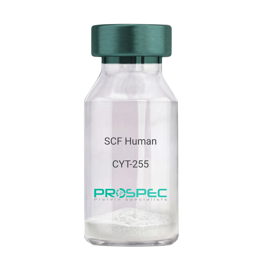
Name :
SCF HumanDescription:
Stem Cell Factor Human Recombinant
Kit ligand Precursor, C-kit ligand, SCF, Mast cell growth factor, MGF, SF, KL-1, Kitl, DKFZp686F2250.
Product # :
CYT-255Price :
Quantity :
Shipping Method :
Shipped at Room temp
More Info
- description
- source
- formulation
- purity
- biological activity
- More Info
Description
Stem Cell Factor Human Recombinant produced in E.Coli is a single, non-glycosylated polypeptide chain containing 165 amino acids and having a molecular mass of 18409 Dalton. The SCF is purified by proprietary chromatographic techniques.
Source
Escherichia Coli.
Formulation
Lyophilized from a concentrated (1mg/ml) solution in water containing 0.02% NaHCO3.
Purity
Greater than 98.0% as determined by:
(a) Analysis by SEC-HPLC.
(b) Analysis by SDS-PAGE.Biological Activity
The ED50 as determined by the dose-dependant stimulation of Human TF-1 cells is < 2 ng/ml, corresponding to a Specific Activity of 500,000 IU/mg.More Info
-
Introduction
Stem cell factor / KIT ligand (SCF) is a cytokine which binds CD117(c-Kit). SCF is also known as "steel factor" or "c-kit ligand". SCF exists in two forms, cell surface bound SCF and soluble (or free) SCF. Soluble SCF is produced by the cleavage of surface bound SCF by metalloproteases. SCF is a growth factor important for the survival, proliferation, and differentiation of hematopoietic stem cells and other hematopoietic progenitor cells. One of its roles is to change the BFU-E (burst-forming unit-erythroid) cells, which are the earliest erythrocyte precursors in the erythrocytic series, into the CFU-E (colony-forming unit-erythroid).
-
Synonyms
Kit ligand Precursor, C-kit ligand, SCF, Mast cell growth factor, MGF, SF, KL-1, Kitl, DKFZp686F2250.
-
Physical Appearance
Sterile Filtered White lyophilized (freeze-dried) powder.
-
Stability
Lyophilized KIT ligand although stable at room temperature for 3 weeks, should be stored desiccated below -18°C. Upon reconstitution SCF should be stored at 4°C between 2-7 days and for future use below -18°C.For long term storage it is recommended to add a carrier protein (0.1% HSA or BSA).Please prevent freeze-thaw cycles.
-
Solubility
It is recommended to reconstitute the lyophilized Stem Cell Factor in sterile 18MΩ-cm H2O not less than 100µg/ml, which can then be further diluted to other aqueous solutions.
-
Amino Acid Sequence
The sequence of the first five N-terminal amino acids was determined and was found to be Met-Glu-Gly-Ile-Cys.
-
Protein content
Protein quantitation was carried out by two independent methods1. UV spectroscopy at 280 nm using the absorbency value of 0.52 as the extinction coefficient for a 0.1% (1mg/ml) solution. This value is calculated by the PC GENE computer analysis program of protein sequences (IntelliGenetics). 2. Analysis by RP-HPLC, using a standard solution of Stem Cell Factor as a Reference Standard.
ProSpec's products are furnished for LABORATORY RESEARCH USE ONLY. The product may not be used as drugs, agricultural or pesticidal products, food additives or household chemicals.
- View Data Sheet
Name :
VEGF Human, YeastDescription:
Vascular Endothelial Growth Factor Human Recombinant, Yeast
Vascular endothelial growth factor A, VEGF-A, Vascular permeability factor, VPF, VEGF, MGC70609.
Product # :
CYT-577Price :
Quantity :
Shipping Method :
Shipped with Ice Packs
More Info
- description
- source
- formulation
- purity
- biological activity
- More Info
Description
Vascular Endothelial Growth Factor Human Recombinant produced in Yeast is a double, glycosylated, polypeptide chain containing 165 amino acids and having a molecular mass of 42 kDa.
Source
Pichia Pastoris.
Formulation
The protein contains 20mM Phosphate- Buffered Saline, pH 7.4.
Purity
Greater than 90.0% as determined by SDS-PAGE.
Biological Activity
Determined by its ability to stimulate 3H-Thymidine incorporation in human umbilical vein endothelial cells, the ED50 for this effect was found to be 2-6ng/ml.More Info
-
Introduction
Vascular endothelial growth factor is an important signaling protein involved in both vasculogenesis and angiogenesis. As its name implies, VEGF activity has been mostly studied on cells of the vascular endothelium, although it does have effects on a number of other cell types (e.g. stimulation monocyte/macrophage migration, neurons, cancer cells, kidney epithelial cells ). VEGF mediates increased vascular permeability, induces angiogenesis, vasculogenesis and endothelial cell growth, promotes cell migration, and inhibits apoptosis. In vitro, VEGF has been shown to stimulate endothelial cell mitogenesis and cell migration. VEGF is also a vasodilator and increases microvascular permeability and was originally referred to as vascular permeability factor.
Elevated levels of this protein is linked to POEMS syndrome, also known as Crow-Fukase syndrome. Mutations in this gene have been associated with proliferative and nonproliferative diabetic retinopathy. -
Synonyms
Vascular endothelial growth factor A, VEGF-A, Vascular permeability factor, VPF, VEGF, MGC70609.
-
Physical Appearance
Sterile Filtered colorless liquid formulation.
-
Stability
Vascular Endothelial Growth Factor Human although stable at 15°C for 2 weeks, should be stored below -18°C. For long term storage it is recommended to add a carrier protein (0.1% HSA or BSA).Please prevent freeze-thaw cycles.
ProSpec's products are furnished for LABORATORY RESEARCH USE ONLY. The product may not be used as drugs, agricultural or pesticidal products, food additives or household chemicals.