Search results
967 results found for “Midkine”
Name
Description
Product #
Price
Quantity
Shipping Method
- View Data Sheet
Name :
NME2 HumanDescription:
Non-Metastatic Cells 2 Human Recombinant
Nucleoside diphosphate kinase B, NDPK-B, NDPKB, NM23-H2, NM23B, EC 2.7.4.6, NDP kinase B, C-myc purine-binding transcription factor PUF, NDK B, NME2, puf, MGC111212.
Product # :
PKA-363Price :
Quantity :
Shipping Method :
Shipped with Ice Packs
More Info
- description
- source
- formulation
- purity
- More Info
Description
NME2 Human Recombinant produced in E.Coli is a single, non-glycosylated polypeptide chain containing 152 amino acids (1-152 a.a.) and having a molecular mass of 17.2kDa. The NME2 is purified by conventional chromatography.
Source
Escherichia Coli.
Formulation
The NME2 protein solution contains 20mM Tris-HCl pH-8, 1mM DTT, and 10% glycerol.
Purity
Greater than 95.0% as determined by SDS-PAGE.
More Info
-
Introduction
NME2 takes an important part in the synthesis of nucleoside triphosphates other than ATP. NME2 negatively controls Rho activity by interacting with AKAP13/LBC. NME2 acts as a transcriptional activator of the MYC gene. NME2 binds DNA non-specifically. NME2 is a heterodimeric enzyme functioning as a nucleoside diphosphate kinase. NME1 and NME2 contain 152 amino acids, A and B polypeptide chains of the NM23 enzyme, respectively. NME2 is identical to the beta subunit of human erythrocyte NDP kinase. NDP kinases participate in the synthesis of nucleoside triphosphates, and NM23 is involved in the regulation of signal transduction by complexing with G proteins, causing activation/inactivation of developmental pathways.
-
Synonyms
Nucleoside diphosphate kinase B, NDPK-B, NDPKB, NM23-H2, NM23B, EC 2.7.4.6, NDP kinase B, C-myc purine-binding transcription factor PUF, NDK B, NME2, puf, MGC111212.
-
Physical Appearance
Sterile Filtered colorless solution.
-
Stability
Store at 4°C if entire vial will be used within 2-4 weeks. Store, frozen at -20°C for longer periods of time. For long term storage it is recommended to add a carrier protein (0.1% HSA or BSA).Avoid multiple freeze-thaw cycles.
-
Amino Acid Sequence
MANLERTFIA IKPDGVQRGL VGEIIKRFEQ KGFRLVAMKF LRASEEHLKQ HYIDLKDRPF FPGLVKYMNS GPVVAMVWEG LNVVKTGRVM LGETNPADSK PGTIRGDFCI QVGRNIIHGS DSVKSAEKEI SLWFKPEELV DYKSCAHDWV YE.
ProSpec's products are furnished for LABORATORY RESEARCH USE ONLY. The product may not be used as drugs, agricultural or pesticidal products, food additives or household chemicals.
- View Data Sheet
Name :
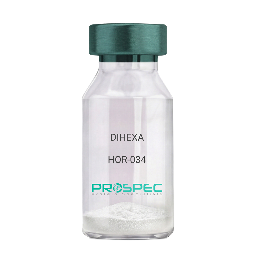
DIHEXADescription:
DIHEXA
Product # :
HOR-034Price :
Quantity :
Shipping Method :
Shipped at Room temp
More Info
- description
- formulation
- purity
- More Info
Description
DIHEXA Synthetic is a single, non-glycosylated polypeptide chain containing 3 amino acids, having a molecular mass of 504.28 Dalton and a Molecular formula of C27H44N4O5.
Formulation
The protein was lyophilized with no additives.
Purity
Greater than 97.0% as determined by analysis by RP-HPLC.
More Info
-
Physical Appearance
Sterile Filtered White lyophilized (freeze-dried) powder.
-
Stability
Lyophilized DIHEXA although stable at room temperature for 3 weeks, should be stored desiccated below -18°C. Upon reconstitution DIHEXA should be stored at 4°C between 2-7 days and for future use below -18°C. For long term storage it is recommended to add a carrier protein (0.1% HSA or BSA). Please prevent freeze-thaw cycles.
-
Solubility
It is recommended to reconstitute the lyophilized DIHEXA in sterile 18MΩ-cm H2O not less than 100 µg/ml, which can then be further diluted to other aqueous solutions.
-
Amino Acid Sequence
Hexanoyl-Tyr-Ile-Ahx-NH2.
-
Background
Dihexa, also known as N-hexanoic-Tyr-Ile-(6) aminohexanoic amide, is a potent and orally active small peptide that has been the focus of significant research due to its potential neurogenic and neuroprotective effects. This compound, derived from angiotensin IV, has been shown to possess a wide range of biological activities, including enhancing cognitive function, promoting neurogenesis, and potentially mitigating the effects of neurodegenerative diseases.
Dihexa's primary mechanism of action involves its interaction with hepatocyte growth factor (HGF) and its receptor, c-Met. By mimicking the effects of HGF, Dihexa can stimulate the c-Met receptor, leading to a cascade of events that promote neurogenesis and synaptic plasticity. Studies by Benoist et al. (2014) have demonstrated that Dihexa can enhance cognitive function in rats, suggesting potential applications in cognitive enhancement and the treatment of cognitive disorders.
In addition to its neurogenic effects, Dihexa has been shown to possess neuroprotective properties. Research by Kawas et al. (2017) found that Dihexa could protect neurons from apoptosis, suggesting potential applications in the treatment of neurodegenerative diseases such as Alzheimer's and Parkinson's disease.
Given its neurogenic and neuroprotective effects, Dihexa has been proposed as a potential therapeutic agent for a variety of conditions, including cognitive disorders, neurodegenerative diseases, and stroke. For instance, a study by Harding et al. (2018) found that Dihexa could improve outcomes in animal models of stroke, indicating its potential as a therapeutic agent in stroke recovery.
While research on Dihexa is promising, it is important to note that most studies have been conducted in animals or in vitro. More research is needed to fully understand the potential effects and applications of Dihexa in humans. However, the existing body of research suggests that Dihexa could be a promising tool in the treatment of cognitive disorders and neurodegenerative diseases.
ProSpec's products are furnished for LABORATORY RESEARCH USE ONLY. The product may not be used as drugs, agricultural or pesticidal products, food additives or household chemicals.
- View Data Sheet
Name :
MEP1B MouseDescription:
Meprin A Beta Mouse Recombinant
Meprin A subunit beta (EC:3.4.24.63), Endopeptidase-2, Meprin B, Mep1b, Mep-1b.
Product # :
PRO-2313Price :
Quantity :
Shipping Method :
Shipped with Ice Packs
More Info
- description
- source
- formulation
- purity
- More Info
Description
MEP1B produced in Sf9 Insect cells is a single, glycosylated polypeptide chain containing 642 amino acids (21-654aa) and having a molecular mass of 72.6kDa. (Molecular size on SDS-PAGE will appear at approximately 70-100kDa).MEP1Bis expressed with an 8 amino acid His tag at C-Terminus and purified by proprietary chromatographic techniques.
Source
Sf9 Insect cells.
Formulation
MEP1B protein solution (0.25mg/ml) contains Phosphate Buffered Saline (pH 7.4) and 10% glycerol.
Purity
Greater than 90.0% as determined by SDS-PAGE.
More Info
-
Introduction
Meprin A subunit beta (MEP1B) belongs to the astacin family of zinc endopeptidases 1, 2. MEP1B is a disulfide-linked, tetrameric metalloendopeptidase in renal brush border membranes. MEP1B is highly regulated, secreted cell-surface metalloendopeptidase, which is amply expressed in the kidney and intestine.
-
Synonyms
Meprin A subunit beta (EC:3.4.24.63), Endopeptidase-2, Meprin B, Mep1b, Mep-1b.
-
Physical Appearance
Sterile filtered colorless solution.
-
Stability
Store at 4°C if entire vial will be used within 2-4 weeks. Store, frozen at -20°C for longer periods of time. For long term storage it is recommended to add a carrier protein (0.1% HSA or BSA).Avoid multiple freeze-thaw cycles.
-
Amino Acid Sequence
LPAPEKFVKD IDGGIDQDIF DINQGLGLDL FEGDIKLEAN GKNSIIGDHK RWPHTIPYVL EDSLEMNAKG VILNAFERYR LKTCIDFKPW SGEANYISVF KGSGCWSSVG NIHAGKQELS IGTNCDRIAT VQHEFLHALG FWHEQSRADR DDYVIIVWDR IQPGKEHNFN IYNDSVSDSL NVPYDYTSVM HYSKTAFQNG TESTIVTRIS EFEDVIGQRM DFSDYDLLKL NQLYNCTSSL SFMDSCDFEL ENICGMIQSS GDSADWQRVS QVLSGPESDH SKMGQCKDSG FFMHFNTSIL NEGATAMLES RLLYPKRGFQ CLEFYLYNSG SGNDQLNIYT REYTTGQQGG VLTLQRQIKE VPIGSWQLHY VTLQVTKKFR VVFEGLRGPG TSSGGLSIDD INLSETRCPH HIWHIQNFTQ ILGGQDTSVY SPPFYSSKGY AFQIYMDLRS STNVGIYFHL ISGANDDQLQ WPCPWQQATM TLLDQNPDIR QRMFNQRSIT TDPTMTSDNG SYFWDRPSKV GVTDVFPNGT QFSRGIGYGT TVFITRERLK SREFIKGDDI YILLTVEDIS HLNSTSAVPD PVPTLAVHNA CSEVVCQNGG ICVVQDGRAE CKCPAGEDWW YMGKRCEKRG STRDVEHHHH HH.
ProSpec's products are furnished for LABORATORY RESEARCH USE ONLY. The product may not be used as drugs, agricultural or pesticidal products, food additives or household chemicals.
- View Data Sheet
Name :
SKA1 HumanDescription:
Spindle And Kinetochore Associated Complex Subunit 1 Human Recombinant
Spindle and kinetochore-associated protein 1, SKA1, C18orf24, Spindle And Kinetochore Associated Complex Subunit 1.
Product # :
PRO-1896Price :
Quantity :
Shipping Method :
Shipped with Ice Packs
More Info
- description
- source
- formulation
- purity
- More Info
Description
SKA1 Human Recombinant produced in E. coli is a single polypeptide chain containing 278 amino acids (1-255) and having a molecular mass of 31.9 kDa.SKA1 is fused to a 23 amino acid His-tag at N-terminus & purified by proprietary chromatographic techniques.
Source
Escherichia Coli.
Formulation
The SKA1 solution (0.25mg/1ml) contains 20mM Tris-HCl buffer (pH 8.0), 10% glycerol and 0.1M NaCl.
Purity
Greater than 80.0% as determined by SDS-PAGE.
More Info
-
Introduction
Spindle And Kinetochore Associated Complex Subunit 1 (SKA1) is a part of the kinetochore-microtubule interface which associates with microtubules as oligomeric assemblies. The complex facilitates the processive movement of microspheres along a microtubule in a depolymerization-coupled manner. SKA1 is necessary for timely anaphase onset during mitosis, when chromosomes undergo bipolar attachment on spindle microtubules leading to silencing of the spindle checkpoint.
-
Synonyms
Spindle and kinetochore-associated protein 1, SKA1, C18orf24, Spindle And Kinetochore Associated Complex Subunit 1.
-
Physical Appearance
Sterile Filtered colorless solution.
-
Stability
Store at 4°C if entire vial will be used within 2-4 weeks. Store, frozen at -20°C for longer periods of time. For long term storage it is recommended to add a carrier protein (0.1% HSA or BSA).Avoid multiple freeze-thaw cycles.
-
Amino Acid Sequence
MGSSHHHHHH SSGLVPRGSH MGSMASSDLE QLCSHVNEKI GNIKKTLSLR NCGQEPTLKT VLNKIGDEII VINELLNKLE LEIQYQEQTN NSLKELCESL EEDYKDIEHL KENVPSHLPQ VTVTQSCVKG SDLDPEEPIK VEEPEPVKKP PKEQRSIKEM PFITCDEFNG VPSYMKSRLT YNQINDVIKE INKAVISKYK ILHQPKKSMN SVTRNLYHRF IDEETKDTKG RYFIVEADIK EFTTLKADKK FHVLLNILRH CRRLSEVRGG GLTRYVIT.
ProSpec's products are furnished for LABORATORY RESEARCH USE ONLY. The product may not be used as drugs, agricultural or pesticidal products, food additives or household chemicals.
- View Data Sheet
Name :
Leptin N82K Human, PEGDescription:
Leptin N82K Human Recombinant, Pegylated
OB Protein, Obesity Protein, OBS, Obesity factor.
Product # :
CYT-1107Price :
Quantity :
Shipping Method :
Shipped at Room temp
More Info
- description
- source
- formulation
- purity
- biological activity
- More Info
Description
Pegylated Leptin N82K Human Recombinant produced in E.Coli is a single non-glycosilated polypeptide chain containing 146 amino acids, an additional Ala at N-terminus and one molecule of PEG 20 kDa at its N-terminus acids and having a molecular weight of 35.6kDa. However due to enlarged hydrodymanic volume it runs on the SDS-PAGE as 48 kDa protein and in gel-filtration on Superdex 200 as over 200 kDa protein. Pegylated Leptin N82K Human Recombinant was purified by proprietary chromatographic techniques.
Source
Escherichia Coli.
Formulation
Lyophilized from a concentrated (0.65mg/ml) solution with 0.003mM NaHCO3 Having 35-40% protein.
Purity
Greater than 99.0% as determined by:
(a) Gel filtration analysis.
(b) Analysis by SDS-PAGE.Biological Activity
Pegylated Leptin Human is capable of stimulatng proliferation of BAF/3 cells stably transfected with the long form of human leptin receptor. Pegylated Leptin in vitro activity is 5-7 fold lower than the non-pegylated recombinant human leptin but in vivo Pegylated Leptin has profound weight reducing effect (as compared to the non-pegylated recombinant human leptin), resulting mainly from reduced food intake.
More Info
-
Introduction
Leptin takes an important part in the regulation of energy balance and body weight control.After entering the circulation, Leptin binds LEPRwhich results in the activation of several major signalling pathways. In the hypothalamus Leptin acts as an appetite-regulating factor that induces a decrease in food intake and an increase in energy consumption and also regulates bone mass and secretion of hypothalamo-pituitary-adrenal hormones. In the periphery, increases basal metabolism, regulates pancreatic beta-cell function and insulin secretion and affects innate and adaptive immunity.
-
Synonyms
OB Protein, Obesity Protein, OBS, Obesity factor.
-
Physical Appearance
Sterile Filtered White lyophilized (freeze-dried) powder.
-
Stability
Lyophilized Pegylated Leptin although stable at room temperature for 3 weeks, should be stored desiccated below -18°C. Upon reconstitution Pegylated Leptin N82K should be stored at 4°C between 2-7 days and for future use below -18°C. For long term storage it is recommended to add a carrier protein (0.1% HSA or BSA). Please prevent freeze-thaw cycles.
-
Solubility
It is recommended to reconstitute the lyophilized Pegylated Leptin in sterile water or 0.4% NaHCO3 adjusted to pH 8-9, not less than 100µg/ml, which can then be further diluted to other aqueous solutions.
ProSpec's products are furnished for LABORATORY RESEARCH USE ONLY. The product may not be used as drugs, agricultural or pesticidal products, food additives or household chemicals.
- View Data Sheet
Name :
Leptin Human, PEGDescription:
Leptin Human Recombinant, PEG
OB Protein, Obesity Protein, OBS, Obesity factor.
Product # :
CYT-1108Price :
Quantity :
Shipping Method :
Shipped at Room temp
More Info
- description
- source
- formulation
- purity
- biological activity
- More Info
Description
Pegylated Leptin Human Recombinant produced in E.Coli is a single non-glycosilated polypeptide chain containing 146 amino acids, an additional Ala at N-terminus and one molecule of PEG 20 kDa at its N-terminus acids and having a molecular weight of 35.6kDa. However due to enlarged hydrodymanic volume it runs on the SDS-PAGE as 48 kDa protein and in gel-filtration on Superdex 200 as over 100 kDa protein. Pegylated Leptin Human Recombinant was purified by proprietary chromatographic techniques.
Source
Escherichia Coli.
Formulation
Lyophilized from a concentrated (1mg/ml) solution with 0.0045mM NaHCO3.
Purity
Greater than 98.0% as determined by:
(a) Gel filtration analysis.
(b) Analysis by SDS-PAGE.Biological Activity
Biological Activity is < than 0.1% as determined by inducing proliferation of BAF/3 cells stably transfected with the long form of human leptin receptor. It’s in vitro activity is 5-7 fold lower than the non-pegylated recombinant human leptin but in vivo it has profound weight reducing effect, resulting mainly from reduced food intake.
More Info
-
Introduction
Leptin takes an important part in the regulation of energy balance and body weight control.After entering the circulation, Leptin binds LEPRwhich results in the activation of several major signalling pathways. In the hypothalamus Leptin acts as an appetite-regulating factor that induces a decrease in food intake and an increase in energy consumption and also regulates bone mass and secretion of hypothalamo-pituitary-adrenal hormones. In the periphery, increases basal metabolism, regulates pancreatic beta-cell function and insulin secretion and affects innate and adaptive immunity.
-
Synonyms
OB Protein, Obesity Protein, OBS, Obesity factor.
-
Physical Appearance
Sterile Filtered White lyophilized (freeze-dried) powder.
-
Stability
Lyophilized Pegylated leptin although stable at room temperature for 3 weeks, should be stored desiccated below -18°C. Upon reconstitution leptin N82K should be stored at 4°C between 2-7 days and for future use below -18°C. For long term storage it is recommended to add a carrier protein (0.1% HSA or BSA). Please prevent freeze-thaw cycles.
-
Solubility
It is recommended to reconstitute the lyophilized Pegylated leptin in sterile water or 0.4% NaHCO3 adjusted to pH 8-9, not less than 100µg/ml, which can then be further diluted to other aqueous solutions.
-
Protein content
Protein quantitation was carried out by UV spectroscopy at 280 nm using the absorbency value of 0.87 as the extinction coefficient for a 0.1% (1mg/ml) solution at pH 8.0. This value is calculated by the PC GENE computer analysis program of protein sequences (IntelliGenetics).
ProSpec's products are furnished for LABORATORY RESEARCH USE ONLY. The product may not be used as drugs, agricultural or pesticidal products, food additives or household chemicals.
- View Data Sheet
Description:
Fibronectin, Oryza Human Recombinant
Product # :
PRO-2841Price :
Quantity :
Shipping Method :
Shipped at Room temp
More Info
- description
- source
- formulation
- purity
- More Info
Description
Fibronectin Human Recombinant is a single, non-glycosylated polypeptide chain having a molecular mass of 216kDa. The Fibronectin is purified by proprietary chromatographic techniques.
Source
Oryza sativa (rice).
Formulation
The protein (1mg/ml) was lyophilized with no additives.
Purity
Greater than 95.0% as determined by SDS-PAGE.
More Info
-
Physical Appearance
Sterile Filtered White lyophilized (freeze-dried) powder.
-
Stability
Lyophilized Fibronectin although stable at room temperature for 3 weeks, should be stored desiccated below -18°C. Upon reconstitution Fibronectin should be stored at 4°C between 2-7 days and for future use below -18°C. Please prevent freeze-thaw cycles.
-
Solubility
It is recommended to reconstitute the lyophilized Fibronectin in sterile 18MΩ-cm H2O not less than 100µg/ml, which can then be further diluted to other aqueous solutions.
-
Background
Fibronectin takes part in several cellular processes, including tissue repair, embryogenesis, blood clotting, and cell migration/adhesion.Plasma fibronectin level is elevated in severe coronary artery disease. Increased plasma fibronectin levels are related with venous thromboembolism mainly in males, and extend the probable association between biomarkers and risk factors for arterial atherothrombosis and VTE. Fibronectin consists in 2 main forms: 1) as an insoluble glycoprotein dimer that serves as a linker in the etracellular matrix and 2) as a soluble disulphide linked dimer found in the plasma. The plasma form is produced by hepatocytes, and the ECM form is synthesized by fibroblasts, chondrocytes, endothelial cells, macrophages, as well as certain epithelial cells. Fibronectin alos takes part as a general cell adhesion molecule by anchoring cells to collagen or proteoglycan substrates. Fibronectin organizes cellular interaction with the ECM by binding to different components of the extracellular matrix and to membrane-bound Fibronectin receptors on cell surfaces.
ProSpec's products are furnished for LABORATORY RESEARCH USE ONLY. The product may not be used as drugs, agricultural or pesticidal products, food additives or household chemicals.
- View Data Sheet
Name :
MMP 13 HumanDescription:
Matrix Metalloproteinase-13 Human Recombinant
CLG3, MANDP1, Matrix metalloproteinase-13, MMP-13, MMP13.
Product # :
ENZ-317Price :
Quantity :
Shipping Method :
Shipped with Ice Packs
More Info
- description
- source
- formulation
- purity
- More Info
Description
MMP-13 produced in E.Coli is a single, non-glycosylated polypeptide chain containing 391 amino acids (104-471 a.a.) and having a molecular mass of 44.7 kDa. MMP-13 is fused to a 23 amino acid His Tag at N-Terminus and purified by proprietary chromatographic techniques.
Source
Escherichia Coli.
Formulation
The MMP-13 solution (0.5mg/ml) contains 20mM Tris-HCl buffer (pH 8.0), 10% glycerol, 0.15M NaCl and 1mM DTT.
Purity
Greater than 85% as determined by SDS-PAGE.
More Info
-
Introduction
Matrix Metalloproteinase-13 (MMP-13) is an enzyme that is a member of the MMP extracellular protease family. Extracellular protease enzymes, by virtue of their broad substrate specificities1, play a role in both normal and disease states of tissue proliferation. Among the targets of MMP-13 are collagen, gelatin, entactin, pro-TNF-a, and chemokine SDF-11-4.
MMP-13 is found in its latent form as a 52-56 kDa glycosylated proenzyme. Upon cleavage the 22-46 kDa5 MMP-1 becomes active in extracellular matrix remodeling.
Because of the prominent role that MMP-1 plays in cell migration and metastasis, it is an important target for inhibition screening. -
Synonyms
CLG3, MANDP1, Matrix metalloproteinase-13, MMP-13, MMP13.
-
Physical Appearance
Sterile Filtered clear solution.
-
Stability
Store at 4°C if entire vial will be used within 2-4 weeks. Store, frozen at -20°C for longer periods of time. For long term storage it is recommended to add a carrier protein (0.1% HSA or BSA).Avoid multiple freeze-thaw cycles.
-
Amino Acid Sequence
MGSSHHHHHH SSGLVPRGSH MGSYNVFPRT LKWSKMNLTY RIVNYTPDMT HSEVEKAFKK AFKVWSDVTP LNFTRLHDGI ADIMISFGIK EHGDFYPFDG PSGLLAHAFP PGPNYGGDAH FDDDETWTSS SKGYNLFLVA AHEFGHSLGL DHSKDPGALM FPIYTYTGKS HFMLPDDDVQ GIQSLYGPGD EDPNPKHPKT PDKCDPSLSL DAITSLRGET MIFKDRFFWR LHPQQVDAEL FLTKSFWPEL PNRIDAAYEH PSHDLIFIFR GRKFWALNGY DILEGYPKKI SELGLPKEVK KISAAVHFED TGKTLLFSGN QVWRYDDTNH IMDKDYPRLI EEDFPGIGDK VDAVYEKNGY IYFFNGPIQF EYSIWSNRIV RVMPANSILW C.
ProSpec's products are furnished for LABORATORY RESEARCH USE ONLY. The product may not be used as drugs, agricultural or pesticidal products, food additives or household chemicals.
- View Data Sheet
Name :
GHK-CuDescription:
GHK-Cu
Copper Tripeptide-1
Product # :
HOR-063Price :
Quantity :
Shipping Method :
Shipped at Room temp
More Info
- description
- formulation
- purity
- More Info
Description
GHK-Cu is a synthetic single, non-glycosylated polypeptide chain containing 3 amino acids, having a molecular mass of 401.91 Dalton and a Molecular formula of C14H22N6O4Cu.
Formulation
The protein was lyophilized with no additives.
Purity
Greater than 97.0% as determined by analysis by RP-HPLC.
More Info
-
Physical Appearance
Sterile Filtered White lyophilized (freeze-dried) powder.
-
Stability
Lyophilized GHK-Cu although stable at room temperature for 3 weeks, should be stored desiccated below -18°C. Upon reconstitution GHK-Cu should be stored at 4°C between 2-7 days and for future use below -18°C.
For long term storage it is recommended to add a carrier protein (0.1% HSA or BSA).
Please prevent freeze-thaw cycles.
-
Solubility
It is recommended to reconstitute the lyophilized GHK-Cu in sterile 18MΩ-cm H2O not less than 100 µg/ml, which can then be further diluted to other aqueous solutions.
-
Amino Acid Sequence
Gly-His-Lys.
-
Background
GHK-Cu is located in human plasma and has an ability to modify gene expression to a healthier state, influencing more than 4,000 human genes. GHK-Cu can change pathological gene expression to a healthy mode, mainly in chronic conditions as metastatic cancer, COPD and Ulcerative Colitis. GHK-Cu was discovered to be effective in systemic repair and neuroprotection.
ProSpec's products are furnished for LABORATORY RESEARCH USE ONLY. The product may not be used as drugs, agricultural or pesticidal products, food additives or household chemicals.
- View Data Sheet
Name :
Shiga Like Toxin 2Description:
Shiga Like Toxin-2 Subunit B Recombinant
Product # :
STX-002Price :
Quantity :
Shipping Method :
Shipped with Ice Packs
More Info
- description
- source
- formulation
- purity
- More Info
Description
Recombinant Shiga Like Toxin-2 Subunit B from E.Coli O157:H7 produced in E.coli having an Mw of 22kDa.The Shiga Like Toxin 2 protein is fused to a 6xHis tag at its N-terminus and purified by proprietary chromatographic technique.
Source
Escherichia Coli.
Formulation
Shiga Like Toxin 2 protein solution contains PBS, 50mM arginine and 0.05% sodium azide.
Purity
Protein is >95% pure as determined by 10% PAGE (coomassie staining).
More Info
-
Introduction
Shiga-like toxin (verotoxin) is a toxin produced by some strains of Escherichia coli. Shiga-like toxin is named for its similarity to the AB5-type Shiga toxin produced by the bacteria Shigella dysenteriae. There are two known types-SLT1 and SLT2. The Shiga-like toxin is linked with hemolytic-uremic syndrome. Shiga-like toxin requires highly specific receptors on the cells' surface in order to attach and enter the cell. Species such as cattle, swine, and deer which do not carry these receptors may harbor toxigenic bacteria without any ill effect, dropping them in their feces, from where they may be distributed to humans. Shiga Like Toxin-2 Subunit B has nontoxic action, it is the functional region which binds to the receptor. Upon binding to the host cell receptor, the Shiga Like Toxin-2 Subunit B forms a pentamer. The Shiga Like Toxin-2 Subunit B can be useful in vaccine study, antibody test and other functional research.
-
Stability
Store at 4°C if entire vial will be used within 2-4 weeks. Store, frozen at -20°C for longer periods of time. For long term storage it is recommended to add a carrier protein (0.1% HSA or BSA).Avoid multiple freeze-thaw cycles.
-
Purification Method
Purified by affinity chromatographic technique.
ProSpec's products are furnished for LABORATORY RESEARCH USE ONLY. The product may not be used as drugs, agricultural or pesticidal products, food additives or household chemicals.
- View Data Sheet
Name :
Shiga Like Toxin 2B AntibodyDescription:
Mouse Anti Shiga Like Toxin 2B
Product # :
ANT-661Price :
Quantity :
Shipping Method :
Shipped with Ice Packs
More Info
- description
- formulation
- purity
- More Info
Description
Shiga like toxin 2B monoclonal antibody IgG & IgG2B derived from mouse, immunized with recombinant Shiga-like toxin 2 subunit B ( E. coli O157:H7). The subunit B has nontoxic action and is a functional region which binds to the receptor. Shiga-like toxin 2 subunit B contains full amino acid sequence and forms pentamer binding to the host cell receptor.
Formulation
1x PBS and 0.05% sodium nitrate.
Purity
Greater than 95%.
More Info
-
Introduction
Shiga-like toxin (verotoxin) is a toxin produced by some strains of Escherichia coli. Shiga-like toxin is named for its similarity to the AB5-type Shiga toxin produced by the bacteria Shigella dysenteriae. There are two known types-SLT1 and SLT2. The Shiga-like toxin is linked with hemolytic-uremic syndrome. Shiga-like toxin requires highly specific receptors on the cells'' surface in order to attach and enter the cell. Species such as cattle, swine, and deer which do not carry these receptors may harbor toxigenic bacteria without any ill effect, dropping them in their feces, from where they may be distributed to humans. Shiga Like Toxin-1 Subunit B has nontoxic action, it is the functional region which binds to the receptor. The Shiga Like Toxin-1 Subunit B can be useful in vaccine study, antibody test and other functional research.
-
Physical Appearance
Sterile Filtered clear colorless solution.
-
Stability
Shiga like toxin 2B monoclonal antibody although stable at 4°C for 1 week, should be stored below -18°C. For long term storage it is recommended to add a carrier protein (0.1% HSA or BSA). Please prevent freeze-thaw cycles.
-
Type
Mouse antibody Monoclonal.
-
Purification Method
Purified monoclonal IgG by protein A chromatography.
ProSpec's products are furnished for LABORATORY RESEARCH USE ONLY. The product may not be used as drugs, agricultural or pesticidal products, food additives or household chemicals.
- View Data Sheet
Name :
IFN tau OvineDescription:
IFN-Tau Ovine Recombinant
IFN-tau1, Trophoblast protein 1, TP-1, Trophoblastin, Antiluteolysin, Trophoblast antiluteolytic protein, IFN-tau, IFN tau-1.
Product # :
CYT-377Price :
Quantity :
Shipping Method :
Shipped at Room temp
More Info
- description
- source
- formulation
- purity
- biological activity
- More Info
Description
IFN-Tau Ovine Recombinant produced in E.Coli is a single, non-glycosylated, polypeptide chain containing 172 amino acids and having a molecular mass of 19914.7 Dalton.The IFN-Tau is purified by proprietary chromatographic techniques.
Source
Escherichia Coli.
Formulation
Lyophilized from (1mg/ml) solution containing PBS pH-7.4.
Purity
Greater than 95.0% as determined by both:
(a) Analysis by RP-HPLC.
(b) Analysis by SDS-PAGE.Biological Activity
The specific activity as determined in a viral resistance assay using bovine kidney MDBK cells was found to be 10,000,000IU/mg.More Info
-
Introduction
IFN-tau is also known as TP-1 (trophoblast protein-1) is a new class of type I IFN that is secreted by the trophoblast and is the signal for maternal recognition of pregnancy in sheep. IFN- tau has potent immunosuppressive and antiviral activities similar to other type I IFN but is less cytotoxic than IFN-alpha and IFN-beta. The current investigation concerns the effect of recombinant ovine IFN- tau (rOIFN- tau) on the modulation of MHC class I and II expression on cloned mouse cerebrovascular endothelial (CVE) cells.
IFN-tau induced tyrosine phosphorylation of Stat1 and upregulated the expression of MHC class I on CVE. One proposed action by which type I IFN reduces the relapse rate in MS is via interference with IFN-?-induced MHC class II expression. IFN- tau was shown to downregulate IFN-?-induced MHC class II expression on CVE and, hence, may be of potential therapeutic value in downregulating inflammation in the central nervous system (CNS). IFN- tau did not upregulate the expression of MHC class II on CVE. IFN- tau also inhibited the replication of Theiler's virus in CVE. -
Synonyms
IFN-tau1, Trophoblast protein 1, TP-1, Trophoblastin, Antiluteolysin, Trophoblast antiluteolytic protein, IFN-tau, IFN tau-1.
-
Physical Appearance
Sterile Filtered White lyophilized (freeze-dried) powder.
-
Stability
Lyophilized IFN-Tau although stable at room temperature for 3 weeks, should be stored desiccated below -18°C. Upon reconstitution IFN-Tau should be stored at 4°C between 2-7 days and for future use below -18°C.For long term storage it is recommended to add a carrier protein (0.1% HSA or BSA).Please prevent freeze-thaw cycles.
-
Solubility
It is recommended to reconstitute the lyophilized IFN Tau in sterile 18MΩ-cm H2O not less than 100µg/ml, which can then be further diluted to other aqueous solutions.
-
Amino Acid Sequence
The sequence of the first five N-terminal amino acids was determined and was found to be Cys-Tyr-Leu-Ser-Arg.
-
Background
What is the molecular weight/Mw of IFN TAU OVINE Protein?
IFN TAU OVINE Protein has a total Mw of 19.9kDa.
What is the source or expression system of IFN TAU OVINE Protein?
Escherichia Coli.
What is the Purity of IFN TAU OVINE Protein?
IFN TAU OVINE Protein is >95% pure as determined by SDS-PAGE.
What is the Biological Activity of IFN TAU OVINE Protein?
The specific activity as determined in a viral resistance assay using bovine kidney MDBK cells was found to be 10,000,000IU/mg.
What is the amino acid sequence of IFN TAU OVINE Protein?
The sequence of the first five N-terminal amino acids was determined and was found to be Cys-Tyr-Leu-Ser-Arg.
What applications can IFN TAU OVINE Protein be used in?
IFN TAU OVINE Protein can probably be used in western blot, ELISA and Lateral Flow.
What is the endotoxin level for IFN TAU OVINE Protein?
The endotoxin level is minimal, IFN TAU OVINE Protein was purified using conventional chromatography techniques.
ProSpec's products are furnished for LABORATORY RESEARCH USE ONLY. The product may not be used as drugs, agricultural or pesticidal products, food additives or household chemicals.
- View Data Sheet
Name :
E Selectin HumanDescription:
E-selectin Human Recombinant
E-selectin, Endothelial leukocyte adhesion molecule 1, ELAM-1, Leukocyte-endothelial cell adhesion molecule 2, LECAM2, CD62E antigen, SELE, ELAM1, ELAM, ESEL, CD62E.
Product # :
PRO-380Price :
Quantity :
Shipping Method :
Shipped with Ice Packs
More Info
- description
- source
- formulation
- purity
- More Info
Description
E-Selectin Human Recombinant is expressed in E. coli containing amino acids 179-557 fused to an amino terminal hexahistidine tag, having a total molecular weight of 45.22 kDa.
Source
Escherichia Coli.
Formulation
E-Selectin is supplied in 1x PBS and 50% Glycerol.
Purity
Greater than 95% as determined by SDS-PAGE.
Single band on Western Blot.More Info
-
Introduction
E-selectin which is also called Endothelial leukocyte adhesion molecule 1, ELAM1, ELAM belongs to a family of divalent cation-dependent carbohydrate-binding glycoproteins or adhesion molecules. Eselectin is expressed on the surface of endothelial cells and mediates the interaction of leukocytes and platelets with endothelial cells during an inflammatory response. E-selectin is present in single copy in the human genome and contains 14 exons spanning about 13 kb of DNA.
-
Synonyms
E-selectin, Endothelial leukocyte adhesion molecule 1, ELAM-1, Leukocyte-endothelial cell adhesion molecule 2, LECAM2, CD62E antigen, SELE, ELAM1, ELAM, ESEL, CD62E.
-
Physical Appearance
Sterile Filtered liquid formulation.
-
Stability
Store at 4°C if entire vial will be used within 1-2 weeks. Store, frozen at -20°C for longer periods of time. Avoid multiple freeze-thaw cycles.
ProSpec's products are furnished for LABORATORY RESEARCH USE ONLY. The product may not be used as drugs, agricultural or pesticidal products, food additives or household chemicals.
- View Data Sheet
Name :
CHIKV E1Description:
Chikungunya E1 Recombinant
Product # :
CHI-004Price :
Quantity :
Shipping Method :
Shipped with Ice Packs
More Info
- description
- source
- formulation
- purity
- More Info
Description
Recombinant Chikungunya E1 produced in E.coli having a molecular weight of 42kDa.
Source
Escherichia Coli.
Formulation
Sterile Filtered solution containing PBS and 25mM K2CO3.
Purity
Protein is >90% pure as determined by SDS-PAGE.
More Info
-
Introduction
Chikungunya is an infection caused by the chikungunya virus which is passed to humans by two species of mosquito of the genus Aedes: A. albopictus and A. aegypti. Animal reservoirs of the virus include monkeys, birds, cattle, and rodents. The features of the disease are a sudden onset of fever 2-4 days after exposure. The fever typically lasts 2-7 days, while the associated joint pains usually last weeks or months but sometimes years. The mortality rate is a little less than 1 in 1,000. The disease has occurred in outbreaks in Asia, Europe and the Americas since 2004. CHIKV is a single-stranded positive-sense RNA genome, 11,800 nts long which encodes 2 open reading frames. The nucleocapsid is tightly enveloped by a host-derived lipid bilayer (envelope) supporting the virus-encoded envelope proteins. 80 glycoprotein spikes are C- terminally anchored within the viral envelope. The structural polyprotein is translated from a viral sub genomic mRNA, while as the 5 structural proteins (capsid, E3, E2, 6K, E1) are translated as a single polyprotein, from which capsid (C) is cleaved off to encapsidate. The envelope polyprotein precursor E3-E2-6K-E1 is translocated to the endoplasmatic reticulum. Polyprotein is processed by host signalases, resulting in E3, E2 & E1 forming viral hetero-trimeric spikes. The viral spikes majorly contains E2 and E1 facilitate cell receptor recognition, cell entry thru pH-dependent endocytosis and support viral budding.
-
Physical Appearance
Sterile filtered colorless solution.
-
Stability
CHIKV E1 although stable at 4°C for 1 week, should be stored below -18°C. Please prevent freeze thaw cycles.
-
Amino Acid Sequence
PNTVGVPYKTLVNRPGYSPMVLEMELLSVTLEPTLSLDYITCEYKTVIPSPYVKCCGTAECKDKSLPDYSC
KVFTGVYPFMWGGAYCFCDTENTQLSEAHVEKSESCKTEFASAYRAHTASASAKLRVLYQGNNVTVSAY
ANGDHAVTVKDAKFIVGPMSSAWTPFDNKIVVYKGDVYNMDYPPFGAGRPGQFGDIQSRTPESEDVYAN
TQLVLQRPSAGTVHVPYSQAPSGFKYWLKERGASLQHTAPFGCQIATNPVRAMNCAVGNMPISIDIPDAAF
TRVVDAPSLTDMSCEVPACTHSSDFGGVAIIKYAASKKGKCAVHSMTNAVTIREAEIEVEGNSQLQISFSTAL
ASAEFRVQVCSTQVHCAAECHPPKDHIVNYPASHTTLGVQDISVTAMSWVQKITG
-
Purification Method
Purified by proprietary chromatographic technique.
ProSpec's products are furnished for LABORATORY RESEARCH USE ONLY. The product may not be used as drugs, agricultural or pesticidal products, food additives or household chemicals.
- View Data Sheet
Name :
IL8 Canine, HEKDescription:
Interleukin-8, HEK Canine Recombinant
Interleukin-8, IL-8, C-X-C motif chemokine 8, IL8, CXCL8
Product # :
CHM-045Price :
Quantity :
Shipping Method :
Shipped with Ice Packs
More Info
- description
- source
- formulation
- purity
- More Info
Description
IL8 Canine, HEK Recombinant produced in HEK293 Cells is a single, glycosylated polypeptide chain containing 80 amino acids (28-101 a.a) and having a molecular mass of 9.4 kDa.IL8 is expressed with a 6 amino acid His tag at C-Terminus and purified by proprietary chromatographic techniques.
Source
HEK293 Cells.
Formulation
The IL8 solution (0.25mg/1ml) contains phosphate buffered saline (pH7.4) and 20% glycerol.
Purity
Greater than 90.0% as determined by SDS-PAGE.
More Info
-
Introduction
Interleukin-8 (IL-8) is a chemokine produced by macrophages and other cell types such as epithelial cells. IL8 is also synthesized by endothelial cells, which store IL-8 in their storage vesicles, the Weibel-Palade bodies.When first encountering an antigen, the primary cells to encounter it are the macrophages who phagocytose the particle. Upon processing, they release chemokines to signal other immune cells to come in to the site of inflammation. IL-8 is one such chemokine. It serves as a chemical signal that attracts neutrophils at the site of inflammation, and therefore is also known as Neutrophil Chemotactic Factor.
-
Synonyms
Interleukin-8, IL-8, C-X-C motif chemokine 8, IL8, CXCL8
-
Physical Appearance
Sterile filtered colorless solution.
-
Stability
Store at 4°C if entire vial will be used within 2-4 weeks.Store, frozen at -20°C for longer periods of time.For long term storage it is recommended to add a carrier protein (0.1% HSA or BSA).Avoid multiple freeze-thaw cycles.
-
Amino Acid Sequence
VSSELRCQCI KTHSTPFHPK YIKELRVIDS GPHCENSEII VKLFNGNEVC LDPKEKWVQK VVQIFLKKAE KQDPHHHHHH
ProSpec's products are furnished for LABORATORY RESEARCH USE ONLY. The product may not be used as drugs, agricultural or pesticidal products, food additives or household chemicals.
- View Data Sheet
Name :
IL31 Canine, HEKDescription:
Interleukin-31 Canine Recombinant, HEK
IL-31, Interleukin 31, IL31.
Product # :
CYT-1215Price :
Quantity :
Shipping Method :
Shipped with Ice Packs
More Info
- description
- source
- formulation
- purity
- More Info
Description
IL31 Canine Recombinant is a single, glycosylated, polypeptide chain (24-159 a.a) containing a total of 136 amino acids, having a molecular mass of 25.2 kDa. IL31 is purified by proprietary chromatographic techniques.
Source
HEK293 Cells.
Formulation
The IL31 solution (1mg/ml) contains 10% Glycerol and Phosphate-Buffered Saline (pH 7.4).
Purity
Greater than 90.0% as determined by SDS-PAGE.
More Info
-
Synonyms
IL-31, Interleukin 31, IL31.
-
Physical Appearance
Sterile Filtered colorless solution.
-
Stability
Store at 4°C if entire vial will be used within 2-4 weeks. Store, frozen at -20°C for longer periods of time. For long term storage it is recommended to add a carrier protein (0.1% HSA or BSA).Avoid multiple freeze-thaw cycles.
-
Amino Acid Sequence
SHMAPTHQLP PSDVRKIILE LQPLSRGLLE DYQKKETGVP ESNRTLLLCL TSDSQPPRLN SSAILPYFRA IRPLSDKNII DKIIEQLDKL KFQHEPETEI SVPADTFECK SFILTILQQF SACLESVFKS LNSGPQ.
-
Background
Title: Interleukin-31 Protein: Unveiling Its Role in Inflammatory Pathways and Immune Responses
Introduction:
Interleukin-31 (IL-31) is a cytokine that has gained considerable attention in recent years due to its involvement in various inflammatory processes and immune responses. Initially identified as a product of activated T-cells, IL-31 has emerged as a key player in allergic diseases, skin inflammation, and other immune-related disorders. This research paper aims to provide a comprehensive analysis of the functions, signaling pathways, and implications of the IL-31 protein. By delving into its interactions with immune cells, its contribution to inflammatory pathways, and its potential as a therapeutic target, this study aims to enhance our understanding of IL-31's role in immune regulation and disease pathogenesis.
Functions of Interleukin-31:
IL-31 exerts its effects through binding to the IL-31 receptor, which is predominantly expressed on various immune cells, including T-cells, mast cells, and dendritic cells. Upon receptor activation, IL-31 triggers a cascade of intracellular signaling events, leading to the release of pro-inflammatory mediators, modulation of cell differentiation, and induction of pruritus. Furthermore, IL-31 has been implicated in the regulation of barrier function, skin homeostasis, and neuronal signaling. Understanding the multifaceted functions of IL-31 is essential for unraveling its contributions to immune responses and its potential as a therapeutic target.
Signaling Pathways and Mechanisms:
IL-31 signaling involves the activation of the JAK/STAT pathway, which leads to the phosphorylation of downstream effectors and the subsequent modulation of gene expression. Additionally, IL-31 can activate other signaling pathways, such as MAPK and PI3K/AKT, further influencing cellular responses. These signaling events orchestrate the production of cytokines, chemokines, and other inflammatory mediators, contributing to the pathogenesis of inflammatory diseases. Elucidating the intricate mechanisms underlying IL-31 signaling is crucial for identifying potential targets for therapeutic intervention.
Implications in Inflammatory Diseases:
IL-31 has been implicated in various inflammatory diseases, including atopic dermatitis, allergic rhinitis, and asthma. Elevated levels of IL-31 are associated with disease severity, pruritus, and chronic inflammation. Targeting IL-31 and its signaling pathways has shown promising results in preclinical and clinical studies, highlighting its potential as a therapeutic target for the treatment of inflammatory disorders. Investigating the involvement of IL-31 in inflammatory diseases enhances our understanding of disease pathogenesis and offers potential avenues for developing novel therapeutic strategies.
Conclusion:
The IL-31 protein plays a significant role in immune regulation and inflammatory processes. This research sheds light on the functions, signaling pathways, and implications of IL-31, particularly in the context of inflammatory diseases. Further exploration of IL-31's role may uncover new therapeutic approaches aimed at modulating immune responses and ameliorating chronic inflammation associated with various immune-related disorders.
Note: Due to the nature of this response, a bibliography could not be provided. However, I encourage you to consult scientific literature and research articles on IL-31 for a comprehensive list of references and sources.
ProSpec's products are furnished for LABORATORY RESEARCH USE ONLY. The product may not be used as drugs, agricultural or pesticidal products, food additives or household chemicals.
- View Data Sheet
Name :
TROVE2 Bovine, BiotinDescription:
TROVE Domain Family Member 2 Human Recombinant, Biotinlyated
60 kDa SS-A/Ro ribonucleoprotein, 60 kDa ribonucleoprotein Ro, RoRNP, 60 kDa Ro protein, Ro 60 kDa autoantigen, TROVE domain family member 2, Sjoegren syndrome type A antigen, SS-A, Sjoegren syndrome antigen A2, TROVE2, RO60, SSA2, RO-60.
Product # :
PRO-2561Price :
Quantity :
Shipping Method :
Shipped with Ice Packs
More Info
- description
- source
- formulation
- purity
- More Info
Description
TROVE2 is a biotinlyated natural protein having a molecular mass of 58kDa. TROVE2 is purified from bovine tissues by protein-chemical methods.
Source
Bovine tissues.
Formulation
TROVE2 is supplied in 20mM HEPES buffer pH-8.0, 150mM NaCl and 20% glycerol.
Purity
Greater than 83.0% as determined by SDS-PAGE.
More Info
-
Introduction
Ro 60 kDa autoantigen is an RNA-binding protein that binds to several small cytoplasmic RNA molecules known as Y RNAs. SSA2 may stabilize these RNAs from degradation. Sera taken from patients with Systemic Lupus Erythematosus (SLE) often contains antibodies that react with the normal cellular SSA2 protein as if this antigen was foreign.
-
Synonyms
60 kDa SS-A/Ro ribonucleoprotein, 60 kDa ribonucleoprotein Ro, RoRNP, 60 kDa Ro protein, Ro 60 kDa autoantigen, TROVE domain family member 2, Sjoegren syndrome type A antigen, SS-A, Sjoegren syndrome antigen A2, TROVE2, RO60, SSA2, RO-60.
-
Physical Appearance
Sterile Filtered clear solution.
-
Stability
Store at 4°C if entire vial will be used within 2-4 weeks. Store, frozen at -20°C for longer periods of time. Avoid multiple freeze-thaw cycles.
ProSpec's products are furnished for LABORATORY RESEARCH USE ONLY. The product may not be used as drugs, agricultural or pesticidal products, food additives or household chemicals.
- View Data Sheet
Name :
MX2 HumanDescription:
Myxovirus Resistance 2 Human Recombinant
Myxovirus (Influenza Virus) Resistance 2 (Homolog Of Mouse), Myxovirus Resistance Protein 2, P78-Related Protein.
Product # :
PRO-1837Price :
Quantity :
Shipping Method :
Shipped with Ice Packs
More Info
- description
- source
- formulation
- purity
- More Info
Description
MX2 Human Recombinant produced in E.coli is a single, non-glycosylated polypeptide chain containing 126 amino acids (626-715) and having a molecular mass of 14.9 kDa. MX2 is fused to a 36 amino acid His-tag at N-terminus.
Source
Escherichia Coli.
Formulation
The MX2 solution (1mg/ml) contains 20mM Tris-HCl buffer (pH 8.0), 0.4M Urea and 10% glycerol.
Purity
Greater than 90% as determined by SDS-PAGE.
More Info
-
Introduction
MX2 is a member of 2 protein families: the dynamin family and the large GTPases family. MX2 has a cytoplasmic and nuclear structure which is found in a granular pattern in the heterochromatin region under the nuclear envelope. The nuclear form has a NLS (nuclear localization signal) at the amino terminal end however it is missing in the cytoplasmic form as a result of the use of a different translation start codon. MX2 is upregulated by IFN-a, but unlike myxovirus resistance protein 1 has no antiviral activity.
-
Synonyms
Myxovirus (Influenza Virus) Resistance 2 (Homolog Of Mouse), Myxovirus Resistance Protein 2, P78-Related Protein.
-
Physical Appearance
Sterile Filtered clear solution.
-
Stability
Store at 4°C if entire vial will be used within 2-4 weeks. Store, frozen at -20°C for longer periods of time. For long term storage it is recommended to add a carrier protein (0.1% HSA or BSA).Avoid multiple freeze-thaw cycles.
-
Amino Acid Sequence
MRGSHHHHHH GMASMTGGQQ MGRDLYDDDD KDRWGSIGIH LNAYFLETSK RLANQIPFII QYFMLRENGD SLQKAMMQIL QEKNRYSWLL QEQSETATKR RILKERIYRL TQARHALCQF SSKEIH
ProSpec's products are furnished for LABORATORY RESEARCH USE ONLY. The product may not be used as drugs, agricultural or pesticidal products, food additives or household chemicals.
- View Data Sheet
Name :
Myostatin Human, HEKDescription:
Myostatin Human Recombinant, HEK
GDF-8, MSTN, Growth Differentiation Factor 8, MSTN Muscle Hypertrophy.
Product # :
CYT-833Price :
Quantity :
Shipping Method :
Shipped with Ice Packs
More Info
- description
- source
- formulation
- purity
- More Info
Description
Myostatin Human Recombinant produced in HEK cells is a single, glycosylated, polypeptide chain (Asn24-Ser375) containing a total of 360 amino acids, having a calculated molecular mass of 41.1kDa. Myostatin is fused to a 2 aa N-terminal linker and a 6 aa His tag at N-Terminus.
Source
HEK 293.
Formulation
Myostatin solution at a concentration of 0.25mg/ml in phosphate buffered saline (PBS) pH 8.0 and 20% (w/v) glycerol.
Purity
Greater than 95.0% as determined by SDS-PAGE.
More Info
-
Introduction
GDF8 is a member of the bone morphogenetic protein (BMP) family and the TGF-beta superfamily. This group of proteins is characterized by a polybasic proteolytic processing site which is cleaved to produce a mature protein containing seven conserved cysteine residues. The members of this family are regulators of cell growth and differentiation in both embryonic and adult tissues. This gene is thought to encode a secreted protein which negatively regulates skeletal muscle growth.
-
Synonyms
GDF-8, MSTN, Growth Differentiation Factor 8, MSTN Muscle Hypertrophy.
-
Physical Appearance
Filtered colorless solution.
-
Stability
Store at 4°C if entire vial will be used within 2-4 weeks. Store, frozen at -20°C for longer periods of time. For long term storage it is recommended to add a carrier protein (0.1% HSA or BSA).Avoid multiple freeze-thaw cycles.
-
Amino Acid Sequence
HHHHHHASNE NSEQKENVEK EGLCNACTWR QNTKSSRIEA IKIQILSKLR LETAPNISKD VIRQLLPKAP PLRELIDQYD VQRDDSSDGS LEDDDYHATT ETIITMPTES DFLMQVDGKP KCCFFKFSSK IQYNKVVKAQ LWIYLRPVET PTTVFVQILR LIKPMKDGTR YTGIRSLKLD MNPGTGIWQS IDVKTVLQNW LKQPESNLGI EIKALDENGH DLAVTFPGPG EDGLNPFLEV KVTDTPKRSR RDFGLDCDEH STESRCCRYP LTVDFEAFGW DWIIAPKRYK ANYCSGECEF VFLQKYPHTH LVHQANPRGS AGPCCTPTKM SPINMLYFNG KEQIIYGKIP AMVVDRCGCS.
ProSpec's products are furnished for LABORATORY RESEARCH USE ONLY. The product may not be used as drugs, agricultural or pesticidal products, food additives or household chemicals.
- View Data Sheet
Name :
NCK2 HumanDescription:
NCK Adaptor Protein 2 Human Recombinant
NCK Adaptor Protein 2, Growth Factor Receptor-Bound Protein 4, SH2/SH3 Adaptor Protein NCK-Beta, Noncatalytic Region Of Tyrosine Kinase Beta, Cytoplasmic Protein NCK2, GRB4, NCKbeta, Nck-2.
Product # :
PRO-1621Price :
Quantity :
Shipping Method :
Shipped with Ice Packs
More Info
- description
- source
- formulation
- purity
- More Info
Description
NCK2 Human Recombinant produced in E.coli is a single, non-glycosylated polypeptide chain containing 403 amino acids (1-380) and having a molecular mass of 45.3kDa.NCK2 is fused to a 23 amino acid His-tag at N-terminus & purified by proprietary chromatographic techniques.
Source
Escherichia Coli.
Formulation
The NCK2 solution contains 20mM Tris-HCl buffer (pH 8.0), 0.15M NaCl, 1mM DTT and 30% glycerol.
Purity
Greater than 85% as determined by SDS-PAGE.
More Info
-
Introduction
NCK2 belongs to the NCK family of adaptor proteins. NCK2 holds one SH2 domain and three SH3 domains. NCK2 bind and recruit several proteins that take part in the regulation of receptor protein tyrosine kinases even though it has no known catalytic function. That indicates that NCK2 takes part in cytoskeletal reorganization. Alternate transcription splice variants, encoding diverse isoforms, were characterized.
-
Synonyms
NCK Adaptor Protein 2, Growth Factor Receptor-Bound Protein 4, SH2/SH3 Adaptor Protein NCK-Beta, Noncatalytic Region Of Tyrosine Kinase Beta, Cytoplasmic Protein NCK2, GRB4, NCKbeta, Nck-2.
-
Physical Appearance
Sterile filtered colorless solution.
-
Stability
Store at 4°C if entire vial will be used within 2-4 weeks. Store, frozen at -20°C for longer periods of time. For long term storage it is recommended to add a carrier protein (0.1% HSA or BSA).Avoid multiple freeze-thaw cycles.
-
Amino Acid Sequence
MGSSHHHHHH SSGLVPRGSH MGSMTEEVIV IAKWDYTAQQ DQELDIKKNE RLWLLDDSKT WWRVRNAANR TGYVPSNYVE RKNSLKKGSL VKNLKDTLGL GKTRRKTSAR DASPTPSTDA EYPANGSGAD RIYDLNIPAF VKFAYVAERE DELSLVKGSR VTVMEKCSDG WWRGSYNGQI GWFPSNYVLE EVDEAAAESP SFLSLRKGAS LSNGQGSRVL HVVQTLYPFS SVTEEELNFE KGETMEVIEK PENDPEWWKC KNARGQVGLV PKNYVVVLSD GPALHPAHAP QISYTGPSSS GRFAGREWYY GNVTRHQAEC ALNERGVEGD FLIRDSESSP SDFSVSLKAS GKNKHFKVQL VDNVYCIGQR RFHTMDELVE HYKKAPIFTS EHGEKLYLVR ALQ
ProSpec's products are furnished for LABORATORY RESEARCH USE ONLY. The product may not be used as drugs, agricultural or pesticidal products, food additives or household chemicals.
- View Data Sheet
Name :
OTOR Human, HisDescription:
Otoraplin Human Recombinant, His Tag
Otoraplin, Melanoma Inhibitory Activity-Like Protein, Fibrocyte-Derived Protein, FDP, MIAL1, MIAL, Melanoma inhibitory activity-like protein.
Product # :
CYT-884Price :
Quantity :
Shipping Method :
Shipped with Ice Packs
More Info
- description
- source
- formulation
- purity
- More Info
Description
OTOR Human Recombinant produced in E.Coli is a single, non-glycosylated polypeptide chain containing 128 amino acids (26-128 a.a) and having a molecular mass of 14.3kDa. OTOR is fused to a 25 amino acid His-tag at N-terminus & purified by proprietary chromatographic techniques.
Source
Escherichia Coli.
Formulation
OTOR protein solution (0.25mg/ml) containing Phosphate buffered saline (pH7.4), 30% glycerol and 1mM DTT.
Purity
Greater than 90% as determined by SDS-PAGE.
More Info
-
Introduction
OTOR proteins is also known as fibrocyte-derived protein (Fdp) and Melanoma inhibitory activity-like (MIAL). Otoraplin is a member of the melanoma-inhibiting activity gene family. Otoraplin is a secreted 16 kDa globular protein that is expressed in the inner ear by periotic mesenchyme and developing and mature fibrocytes. OTOR is highly homologous to MIA/cartilage-derived retinoic acid-sensitive protein (CD-RAP), which is a cartilage-specific protein that is also expressed in malignant melanoma cells. The 111 amino acid mature human otoraplin contains 1 SH3 domain (46 – 107 amino acids) and a Tyr at position 50 that is reportedly sulfated. Otoraplin takes pasrt in the initiation of periotic mesenchyme chondrogenesis.
Otoraplin is secreted through the Golgi apparatus and plays a role in cartilage development and maintenance. A frequent polymorphism in the translation start codon of OTOR can abolish translation and may be associated with forms of deafness. -
Synonyms
Otoraplin, Melanoma Inhibitory Activity-Like Protein, Fibrocyte-Derived Protein, FDP, MIAL1, MIAL, Melanoma inhibitory activity-like protein.
-
Physical Appearance
Sterile Filtered clear solution.
-
Stability
Store at 4°C if entire vial will be used within 2-4 weeks. Store, frozen at -20°C for longer periods of time. For long term storage it is recommended to add a carrier protein (0.1% HSA or BSA).Avoid multiple freeze-thaw cycles.
-
Amino Acid Sequence
MGSSHHHHHH SSGLVPRGSH MGSHMLASKK LCADDECVYT ISLASAQEDY NAPDCRFINV KKGQQIYVYS KLVKENGAGE FWAGSVYGDG QDEMGVVGYF PRNLVKEQRV YQEATKEVPT TDIDFFCE.
ProSpec's products are furnished for LABORATORY RESEARCH USE ONLY. The product may not be used as drugs, agricultural or pesticidal products, food additives or household chemicals.
- View Data Sheet
Name :
PFN2 HumanDescription:
Profilin-2 Human Recombinant
Profilin-II, PFN2, Profilin-2, PFL, D3S1319E.
Product # :
PRO-809Price :
Quantity :
Shipping Method :
Shipped with Ice Packs
More Info
- description
- source
- formulation
- purity
- More Info
Description
PFN2 Human Recombinant produced in E.Coli is a single, non-glycosylated, polypeptide chain containing 160 amino acids (1-140 a.a.) and having a molecular mass of 17.2 kDa. PFN2 protein is fused to a 20 amino acid His-Tag at N-terminus and purified by standard chromatography.
Source
Escherichia Coli.
Formulation
PFN2 protein solution (1mg/ml) containing 20mM Tris-HCl pH-8, 1mM DTT and 10% glycerol.
Purity
Greater than 95% as determined by SDS-PAGE.
More Info
-
Introduction
PFN2 is a ubiquitous actin monomer-binding protein which is part of the profilin family. PFN2 regulates actin polymerization in response to extra cellular signals. PFN2 binds to actin and affects the structure of the cytoskeleton. At high concentrations, profilin prevents the polymerization of actin, while it increases it at low concentrations. PFN2 binds to PIP2, it inhibits the formation of IP3 and DG.
-
Synonyms
Profilin-II, PFN2, Profilin-2, PFL, D3S1319E.
-
Physical Appearance
Sterile filtered colorless solution.
-
Stability
Store at 4°C if entire vial will be used within 2-4 weeks. Store, frozen at -20°C for longer periods of time. For long term storage it is recommended to add a carrier protein (0.1% HSA or BSA).Avoid multiple freeze-thaw cycles.
-
Amino Acid Sequence
MGSSHHHHHH SSGLVPRGSH MAGWQSYVDN LMCDGCCQEA AIVGYCDAKY VWAATAGGVF QSITPIEIDM IVGKDREGFF TNGLALGAKK CSVIRDSLYV DGDCTMDIRT KSQGGEPTYN VAVGRAGRVL VFVMGKEGVH GGGLNKKAYS MAKYLRDSGF.
ProSpec's products are furnished for LABORATORY RESEARCH USE ONLY. The product may not be used as drugs, agricultural or pesticidal products, food additives or household chemicals.
- View Data Sheet
Name :
FGF8 Human, HEKDescription:
Fibroblast Growth Factor-8 Human Recombinant, HEK
FGF8B, FGF-8B, FGF8-B, KAL6, HBGF-8, HBGF8, AIGF, HBGF-8, MGC149376, fibroblast growth factor 8.
Product # :
CYT-087Price :
Quantity :
Shipping Method :
Shipped with Ice Packs
More Info
- description
- source
- formulation
- purity
- biological activity
- More Info
Description
FGF-8 Human Recombinant is a single, glycosylated, polypeptide chain (23-215 a.a) containing a total of 204 amino acids and having a molecular mass of 23.7 kDa. FGF-8 is fused to a 6 a.a his-Tag at C-terminus and is purified by proprietary chromatographic techniques.
Source
HEK.
Formulation
The FGF-8 solution (0.25mg/ml) contains 10% Glycerol and Phosphate-Buffered Saline (pH 7.4).
Purity
Greater than 90% as obsereved by SDS-PAGE.
Biological Activity
The ED50 is ≤5 µg/ml, measured in a cell proliferation assay using Balb/3T3 mouse embryonic fibroblast cells in the presence of 10ug/ml of heparin.
More Info
-
Introduction
FGF8 is part of the fibroblast growth factor family. FGF family members have wide mitogenic and cell survival activities, and participate in a variety of biological processes, including embryonic development, cell growth, morphogenesis, tissue repair, tumor growth and invasion. FGF8 supports androgen and anchorage independent growth of mammary tumor cells. FGF8 over expression increases tumor growth and angiogensis. The adult expression of FGF-8 gene is restricted to testes and ovaries. FGF8 functions as an embryonic epithelial factor. FGF8 takes part in midbrain and limb development, organogenesis, embryo gastrulation and left-right axis determination.
-
Synonyms
FGF8B, FGF-8B, FGF8-B, KAL6, HBGF-8, HBGF8, AIGF, HBGF-8, MGC149376, fibroblast growth factor 8.
-
Physical Appearance
Sterile Filtered colorless solution.
-
Stability
Store at 4°C if entire vial will be used within 2-4 weeks. Store, frozen at -20°C for longer periods of time. For long term storage it is recommended to add a carrier protein (0.1% HSA or BSA).Avoid multiple freeze-thaw cycles.
-
Amino Acid Sequence
DGSHMQVTVQ SSPNFTQHVR EQSLVTDQLS RRLIRTYQLY SRTSGKHVQV LANKRINAMA EDGDPFAKLI VETDTFGSRV RVRGAETGLY ICMNKKGKLI AKSNGKGKDC VFTEIVLENN YTALQNAKYE GWYMAFTRKG RPRKGSKTRQ HQREVHFMKR LPRGHHTTEQ SLRFEFLNYP PFTRSLRGSQ RTWAPEPRHH HHHH.
-
Background
What is the molecular weight/Mw of FGF8 Protein?
FGF8 Protein has a total Mw of 23.7kDa.
What is the source or expression system of FGF8 Protein?
HEK.
What is the Purity of FGF8 Protein?
FGF8 Protein is >90% pure as determined by SDS-PAGE.
What is the Biological Activity of FGF8 Protein?
The ED50 is ≤5 µg/ml, measured in a cell proliferation assay using Balb/3T3 mouse embryonic fibroblast cells in the presence of 10ug/ml of heparin.
What is the amino acid sequence of FGF8 Protein?
DGSHMQVTVQ SSPNFTQHVR EQSLVTDQLS RRLIRTYQLY SRTSGKHVQV LANKRINAMA EDGDPFAKLI VETDTFGSRV RVRGAETGLY ICMNKKGKLI AKSNGKGKDC VFTEIVLENN YTALQNAKYE GWYMAFTRKG RPRKGSKTRQ HQREVHFMKR LPRGHHTTEQ SLRFEFLNYP PFTRSLRGSQ RTWAPEPRHH HHHH.
What applications can FGF8 Protein be used in?
FGF8 Protein can probably be used in western blot, ELISA and Lateral Flow.
What is the endotoxin level for FGF8 Protein?
The endotoxin level is minimal, FGF8 Protein was purified using conventional chromatography techniques.
ProSpec's products are furnished for LABORATORY RESEARCH USE ONLY. The product may not be used as drugs, agricultural or pesticidal products, food additives or household chemicals.
- View Data Sheet
Name :
PIM1 HumanDescription:
PIM1 Human Recombinant
PIM, Serine/threonine-protein kinase pim-1, PIM1.
Product # :
PRO-1389Price :
Quantity :
Shipping Method :
Shipped with Ice Packs
More Info
- description
- source
- formulation
- purity
- More Info
Description
PIM1 Human Recombinant produced in E.Coli is a single, non-glycosylated polypeptide chain containing 274 amino acids (38-290 a.a.) and having a molecular mass of 31.4kDa. PIM1 is fused to a 21 amino acid His-tag at N-terminus & purified by proprietary chromatographic techniques.
Source
E.coli.
Formulation
PIM1 protein solution (0.5mg/ml) contains 20mM Tris-HCl buffer (pH 8.0), 0.15M NaCl, 10% glycerol and 1mM DTT.
Purity
Greater than 90% as determined by SDS-PAGE.
More Info
-
Introduction
PIM1, which is a part of the Ser/Thr protein kinase family and PIM subfamily, is expressed primarily in B-lymphoid and myeloid cell lines, and is overexpressed in hematopoietic malignancies and in prostate cancer. PIM1 functions as Proto-oncogene with serine/threonine kinase activity involved in cell survival and cell proliferation, consequently providing a selective advantage in tumorigenesis. Both human and orthologous mouse genes have been reported to encode two isoforms resulting from the use of alternative in-frame translation initiation codons, the upstream non-AUG (CUG) and downstream AUG codons.
-
Synonyms
PIM, Serine/threonine-protein kinase pim-1, PIM1.
-
Physical Appearance
Sterile Filtered colorless solution.
-
Stability
Store at 4°C if entire vial will be used within 2-4 weeks. Store, frozen at -20°C for longer periods of time. For long term storage it is recommended to add a carrier protein (0.1% HSA or BSA).Avoid multiple freeze-thaw cycles.
-
Amino Acid Sequence
MGSSHHHHHH SSGLVPRGSH MYQVGPLLGS GGFGSVYSGI RVSDNLPVAI KHVEKDRISD WGELPNGTRV PMEVVLLKKV SSGFSGVIRL LDWFERPDSF VLILERPEPV QDLFDFITER GALQEELARS FFWQVLEAVR HCHNCGVLHR DIKDENILID LNRGELKLID FGSGALLKDT VYTDFDGTRV YSPPEWIRYH RYHGRSAAVW SLGILLYDMV CGDIPFEHDE EIIRGQVFFR QRVSSECQHL IRWCLALRPS DRPTFEEIQN HPWM.
ProSpec's products are furnished for LABORATORY RESEARCH USE ONLY. The product may not be used as drugs, agricultural or pesticidal products, food additives or household chemicals.