Search results
1000 results found for “EBV”
Name
Description
Product #
Price
Quantity
Shipping Method
- View Data Sheet
Name :
HIV-1 p24 PairedDescription:
Mouse Anti HIV-1 p24 Paired
Product # :
ANT-772Price :
Quantity :
Shipping Method :
Shipped with Ice Packs
More Info
- description
- formulation
- purity
- More Info
- Citations
Description
HIV-1 p24 Paired antibodies coating and conjugating are used for lateral flow immunoassay and having a Mw of 24kDa. Please note that when ordering for example: 100µg antibody we ship 50µg from each of the antibodies (100µg in total).
Formulation
*HIV-1 p24 Label antibody contains PBS, pH 7.4.
* HIV-1 p24 Capture antibody contains PBS, pH 7.4.
Purity
Greater than 95%.
Citations
More Info
-
Introduction
Human immunodeficiency virus (HIV) is a retrovirus that can lead to a condition in which the immune system begins to fail, leading to opportunistic infections. HIV primarily infects vital cells in the human immune system such as helper T cells (specifically CD4+ T cells), macrophage sand dendritic cells. HIV infection leads to low levels of CD4+ T cells through three main mechanisms: firstly, direct viral killing of infected cells; secondly, increased rates of apoptosis in infected cells; and thirdly, killing of infected CD4+ T cells by CD8 cytotoxic lymphocytes that recognize infected cells. When CD4+ T cell numbers decline below a critical level, cell-mediated immunity is lost, and the body becomes progressively more susceptible to opportunistic infections. HIV was classified as a member of the genus Lentivirus, part of the Retroviridae family. Lentiviruses have many common morphologies and biological properties. Many species are infected by lentiviruses, which are characteristically responsible for long-duration illnesses with a long incubation period. Lentiviruses are transmitted as single-stranded, positive-sense, enveloped RNA viruses. Upon entry of the target cell, the viral RNA genome is converted to double-stranded DNA by a virally encoded reverse transcriptase that is present in the virus particle. This viral DNA is then integrated into the cellular DNA by a virally encoded integrase so that the genome can be transcribed. Once the virus has infected the cell, two pathways are possible: either the virus becomes latent and the infected cell continues to function, or the virus becomes active and replicates, and a large number of virus particles are liberated that can then infect other cells. The p24 protein is detected in patient blood as early as 2 weeks after HIV infection, further reducing the window period necessary to accurately detect the HIV status of the patient.
-
Physical Appearance
2 vials of sterile filtered clear colorless solution.
-
Stability
HIV-1 p24 antibody although stable at 4°C for 1 week, should be stored below -18°C. For long term storage it is recommended to add a carrier protein (0.1% HSA or BSA). Please prevent freeze-thaw cycles.
-
Applications
Lateral flow immunoassay.
-
Type
Mouse Anti Human Monoclonal.
-
Purification Method
Purified by protein A affinity column.
ProSpec's products are furnished for LABORATORY RESEARCH USE ONLY. The product may not be used as drugs, agricultural or pesticidal products, food additives or household chemicals.
- View Data Sheet
Name :
HSV-2 gDDescription:
Herpes Simplex Virus-2 gD Recombinant
Product # :
HSV-222Price :
Quantity :
Shipping Method :
Shipped with Ice Packs
More Info
- description
- formulation
- purity
- More Info
- Citations
Description
The E.Coli derived 39.7 kDa recombinant protein contains the HSV-2 gD immunodominant regions 266-394 amino acids, fused with 26 kDa GST-tag.
Formulation
25mM Tris pH 7.2, 1mM EDTA, and 50% glycerol.
Purity
HSV-2 gD protein is >95% pure as determined by 10% PAGE (coomassie staining).
Citations
More Info
-
Introduction
Entry of HSV into the host cell involves interactions of several viral glycoproteins with cell surface receptors. The virus particle is covered by an envelope which, when bound to specific receptors on the cell surface, will fuse with the cell membrane and create an opening, or pore, through which the virus enters the host cell. The sequential stages of HSV entry are analagous to those of other viruses. At first, complementary receptors on the virus and cell surface bring the two membranes into proximity. In an intermediate state, the two membranes begin to merge, forming a hemifusion state. Finally, a stable entry pore is formed through which the viral envelope contents are introduced to the host cell.
-
Physical Appearance
Sterile Filtered clear solution.
-
Stability
HSV-2 gD Protein although stable at 4°C for 1 week, should be stored below -18°C. Please prevent freeze thaw cycles.
-
Specificity
Immunoreactive with sera of HSV-infected individuals.
-
Purification Method
HSV-2 gD protein was purified by proprietary chromatographic technique.
ProSpec's products are furnished for LABORATORY RESEARCH USE ONLY. The product may not be used as drugs, agricultural or pesticidal products, food additives or household chemicals.
- View Data Sheet
Name :
Influenza-B Paired AntibodyDescription:
Mouse Anti Influenza-B Paired
Product # :
ANT-652Price :
Quantity :
Shipping Method :
Shipped with Ice Packs
More Info
- description
- formulation
- purity
- More Info
- Citations
Description
Influenza-B conjugation antibody and Influenza-B coating antibody are used to develop rapid test for Influenza-B rapid test. Please note that when ordering for example: 100µg antibody we ship 50µg from each of the antibodies (100µg in total).
Formulation
* Influenza-B conjugation antibody in PBS, PH 7.4 .
* Influenza-B coating antibody in PBS, PH 7.4 .Purity
Greater than 90%.
Citations
More Info
-
Introduction
Influenza-B virus is a genus in the virus family Orthomyxoviridae. The only species in this genus is called "Influenza B virus". Influenza B virus only infects humans and seals. This limited host range is apparently in contrast with those caused by the similar Influenza virus A as both mutate by both genetic drift and reassortment. Influenza-B virus evolves slower than A viruses and faster than C viruses. Influenza-B virus mutates at a rate 2-3 times lower than type A. However, influenza B mutates enough that lasting immunity is not possible. The Influenza B virus capsid is enveloped while its virion consists of a matrix protein + envelope + nucleoprotein complex + nucleocapsid, and a polymerase complex. Influenza B is sometimes spherical and sometimes filamentous. Its 500 or so surface projections are made of hemagglutinin and neuraminidase.
The Influenza B virus is 14648 nucleotides long and consists of eight segments of linear negative-sense, single-stranded RNA. The multipartite genome is encapsidated, each segment in a separate nucleocapsid, and the nucleocapsids are surrounded by one envelope. -
Physical Appearance
2 vials of sterile Filtered clear colorless solution.
-
Stability
For periods up to 1 month Influenza-B Paired Antibody should be stored at 4°C, for longer periods of time, store at -20°C. Prevent freeze thaw cycles.
-
Applications
Lateral flow immunoassay.
-
Type
Mouse antibody Monoclonal.
-
Purification Method
Purified monoclonal IgG2b by protein A chromatography.
ProSpec's products are furnished for LABORATORY RESEARCH USE ONLY. The product may not be used as drugs, agricultural or pesticidal products, food additives or household chemicals.
- View Data Sheet
Name :
SARS MERS RBD, ActiveDescription:
SARS MERS Spike Receptor Binding Domain Recombinant, Active
Middle East respiratory syndrome coronavirus, Human betacoronavirus 2c EMC/2012, MERS-CoV, MERS, MERSCoV RBD, MERS RBD, receptor binding domain, RBD, Spike RBD protein, Spike glycoprotein, S glycoprotein, E2, Peplomer protein
Product # :
SARS-060Price :
Quantity :
Shipping Method :
Shipped with Ice Packs
More Info
- description
- source
- formulation
- purity
- biological activity
- More Info
- Citations
Description
SARS MERS RBD Recombinant produced in Sf9 Baculovirus cells is a single, glycosylated polypeptide chain containing 258 amino acids (358-606 aa) and having a molecular mass of 28.2kDa. SARS MERS RBD is fused to a 6 amino acid His-tag at C-terminus & purified by proprietary chromatographic techniques.
Source
Sf9, Baculovirus cells.
Formulation
The SARS MERS RBD solution (0.5mg/ml) contains 10% glycerol and Phosphate-Buffered Saline (pH 7.4).
Purity
Greater than 90.0% as determined by SDS-PAGE.
Biological Activity
Measured by its binding ability in a functional ELISA with Human DPPIV/CD26 (CAT# enz-1187).
Citations
More Info
-
Synonyms
Middle East respiratory syndrome coronavirus, Human betacoronavirus 2c EMC/2012, MERS-CoV, MERS, MERSCoV RBD, MERS RBD, receptor binding domain, RBD, Spike RBD protein, Spike glycoprotein, S glycoprotein, E2, Peplomer protein
-
Physical Appearance
Sterile filtered colorless solution.
-
Stability
Store at 4°C if entire vial will be used within 2-4 weeks.Store, frozen at -20°C for longer periods of time.For long term storage it is recommended to add a carrier protein (0.1% HSA or BSA).Avoid multiple freeze-thaw cycles.
-
Amino Acid Sequence
ADPSGVYSVS SFEAKPSGSV VEQAEGVECD FSPLLSGTPP QVYNFKRLVF TNCNYNLTKL LSLFSVNDFT CSQISPAAIA SNCYSSLILD YFSYPLSMKS DLSVSSAGPI SQFNYKQSFS NPTCLILATV PHNLTTITKP LKYSYINKCS RLLSDDRTEV PQLVNANQYS PCVSIVPSTV WEDGDYYRKQ LSPLEGGGWL VASGSTVAMT EQLQMGFGIT VQYGTDTNSV CPKLEFANDT KIASQLGNCV EYHHHHHH.
-
Background
The severe acute respiratory syndrome coronavirus (SARS-CoV) and Middle East respiratory syndrome coronavirus (MERS-CoV) have posed significant global health threats in recent years. Central to their pathogenesis is the interaction between the viral spike proteins and host cell receptors. This research aims to investigate the receptor binding domain (RBD) of SARS and MERS spike proteins and its implications for viral entry and the development of therapeutic interventions. Understanding the molecular mechanisms underlying viral-host interactions can pave the way for targeted therapeutic strategies against these deadly coronaviruses.
Structure and Function of SARS and MERS Spike RBD:
The spike proteins of SARS-CoV and MERS-CoV are critical for viral entry into host cells. These proteins consist of two subunits: S1, responsible for receptor binding, and S2, involved in membrane fusion. The receptor binding domain (RBD) within the S1 subunit specifically interacts with host cell receptors, enabling viral attachment and entry. The RBDs of SARS and MERS spike proteins exhibit unique structural features and binding affinities for their respective receptors.
Interaction with ACE2 and DPP4 Receptors:
The SARS-CoV spike protein RBD interacts with the angiotensin-converting enzyme 2 (ACE2) receptor, which is abundantly expressed in the respiratory tract. The binding of SARS-CoV RBD to ACE2 facilitates viral entry into host cells. On the other hand, the MERS-CoV spike protein RBD interacts with the dipeptidyl peptidase 4 (DPP4) receptor, predominantly expressed in the lungs and other tissues. The ACE2 and DPP4 receptors play crucial roles in determining the host range and tissue tropism of SARS and MERS coronaviruses.
Implications for Viral Pathogenesis:
The binding of SARS and MERS spike RBDs to their respective receptors triggers conformational changes in the spike protein, leading to membrane fusion and subsequent viral entry. This process is crucial for viral replication and the spread of infection within the host. The specificity and affinity of the RBD-receptor interaction influence viral tropism, tissue damage, and disease severity. Understanding the determinants of RBD-receptor binding can provide insights into viral pathogenesis and potential therapeutic targets.
Development of Therapeutic Interventions:
The RBD of SARS and MERS spike proteins represents a promising target for the development of antiviral therapeutics. Several strategies have been explored, including monoclonal antibodies and small molecule inhibitors, to disrupt the RBD-receptor interaction and inhibit viral entry. These approaches aim to block the binding interface between the spike RBD and the host receptor, thereby preventing viral attachment and entry. Additionally, vaccine development efforts have focused on generating neutralizing antibodies against the RBD to elicit protective immune responses.
Conclusion:
The investigation of the receptor binding domain (RBD) of SARS and MERS spike proteins sheds light on the molecular mechanisms underlying viral entry and pathogenesis. The specific interactions between the spike RBD and host cell receptors play a crucial role in determining viral tropism and tissue damage. Targeting the RBD-receptor interaction holds promise for the development of effective therapeutics against SARS-CoV, MERS-CoV, and potentially other related coronaviruses. Further research and development efforts are needed to exploit the potential of the spike RBD as a therapeutic target and to combat future coronavirus outbreaks.
ProSpec's products are furnished for LABORATORY RESEARCH USE ONLY. The product may not be used as drugs, agricultural or pesticidal products, food additives or household chemicals.
- View Data Sheet
Name :
EFNB1 HumanDescription:
Ephrin-B1 Human Recombinant
ephrin-B1, LERK2, EPLG2, Elk-L, EPH-related receptor tyrosine kinase ligand 2, EFL-3, ELK ligand, CFND, CFNS, Craniofrontonasal Syndrome (craniofrontonasal dysplasia), MGC8782.
Product # :
PRO-918Price :
Quantity :
Shipping Method :
Shipped with Ice Packs
More Info
- description
- source
- formulation
- purity
- More Info
- Citations
Description
EFNB1 Human Recombinant produced in E.coli is a single, non-glycosylated polypeptide chain containing 231 amino acids (28-237) and having a molecular mass of 25.3 kDa.The EFNB1 is fused to a 21 amino acid His-Tag at N-terminus and purified by proprietary chromatographic techniques.
Source
E.coli.
Formulation
The EFNB1 solution (1mg/ml) contains 20mM Tris-HCl buffer (pH 8.0), 0.4M Urea and 5% glycerol.
Purity
Greater than 95% as determined by SDS-PAGE.
Citations
More Info
-
Introduction
EFNB1 is a member of the Eph family. The cell-surface proteins Ephrins split into two groups, ephrin-A and ephrin-B, based on their structure and function and perform as ligands for Eph receptors. The transmembrane EFNB1 proteins have conserved cytoplasmic tyrosine residues that are phosphorylated upon interaction with an EphB receptor. In addition, EFNB1 transduces outside-in signals by C-terminal protein interfaces which influence integrin-mediated cell attachment and migration.
-
Synonyms
ephrin-B1, LERK2, EPLG2, Elk-L, EPH-related receptor tyrosine kinase ligand 2, EFL-3, ELK ligand, CFND, CFNS, Craniofrontonasal Syndrome (craniofrontonasal dysplasia), MGC8782.
-
Physical Appearance
Sterile Filtered colorless solution.
-
Stability
Store at 4°C if entire vial will be used within 2-4 weeks. Store, frozen at -20°C for longer periods of time. For long term storage it is recommended to add a carrier protein (0.1% HSA or BSA).Avoid multiple freeze-thaw cycles.
-
Amino Acid Sequence
MGSSHHHHHH SSGLVPRGSH MLAKNLEPVS WSSLNPKFLS GKGLVIYPKI GDKLDIICPR AEAGRPYEYY KLYLVRPEQA AACSTVLDPN VLVTCNRPEQ EIRFTIKFQE FSPNYMGLEF KKHHDYYITS TSNGSLEGLE NREGGVCRTR TMKIIMKVGQ DPNAVTPEQL TTSRPSKEAD NTVKMATQAP GSRGSLGDSD GKHETVNQEE KSGPGASGGS SGDPDGFFNS K
ProSpec's products are furnished for LABORATORY RESEARCH USE ONLY. The product may not be used as drugs, agricultural or pesticidal products, food additives or household chemicals.
- View Data Sheet
Name :
GCVDescription:
Ganciclovir
Product # :
SYN-001Price :
Quantity :
Shipping Method :
Shipped at Room temp
More Info
- description
- purity
- More Info
- Citations
Description
Ganciclovir is a white to off-white crystalline powder with a molecular formula of C9H13N5O4 and a molecular weight of 255.23.
Purity
Greater than 99.0%.
Citations
More Info
-
Introduction
Ganciclovir (GCV) is a pro-drug nucleoside analog that is activated by phosphorylation. It is useful in the study of gene therapy in cancer research.
Ganciclovir is a synthetic analogue of 2'-deoxy-guanosine. It is initially phosphorylated to a deoxyguanosine triphosphate (dGTP) analogue. This mechanism competitively inhibits the integration of dGTP by viral DNA polymerase, resulting in the termination of elongation of viral DNA. Upon expression of a viral suicide gene encoding thymidine kinase, the non-toxic pro-drug is converted to a phosphorylated active analog and is incorporated into the DNA of replicating eukaryotic cells, causing death of the malignant dividing cell. The cell cycle is irreversibly arrested at the G2-M checkpoint. Gap junction involvement in the ganciclovir bystander effect has been studied. Ganciclovir has been used to study loss of telomeres and to evaluate sensitivity of viruses to antiviral treatments.
Ganciclovir is used in molecular biology for selection against random recombination events when homologous recombination of a gene of interest is required. -
Physical Appearance
Sterile Filtered White lyophilized (freeze-dried) powder.
-
Stability
Lyophilized Ganciclovir although stable at room temperature for 3 weeks, should be stored at 4°C.
-
Solubility
It is recommended to reconstitute the lyophilized Ganciclovir in sterile 18MΩ-cm H2O not less than 100µg/ml, which can then be further diluted to other aqueous solutions.
ProSpec's products are furnished for LABORATORY RESEARCH USE ONLY. The product may not be used as drugs, agricultural or pesticidal products, food additives or household chemicals.
- View Data Sheet
Name :
HCV NS3 47.8kDaDescription:
Hepatitis C Virus NS3 47.8kDa Recombinant
Product # :
HCV-010Price :
Quantity :
Shipping Method :
Shipped at Room temp
More Info
- description
- source
- formulation
- purity
- More Info
- Citations
Description
Recombinant HCV NS3 Genotype 1a produced in E.coli is a non-glycosylated polypeptide chain having a molecular mass of 47.8kDa and fused to a His tag at N-terminus.
Source
Escherichia Coli.
Formulation
Lyophilized from 1mg/ml in 20mM sodium carbonate pH-9.6.
Purity
Greater than 95.0% as determined by SDS-PAGE.
Citations
More Info
-
Introduction
HCV is a small 50nm, enveloped, single-stranded, positive sense RNAvirus in the family Flaviviridae.
HCV has a high rate of replication with approximately one trillion particles produced each day in an infected individual. Due to lack of proofreading by the HCV RNA polymerase, the HCV has an exceptionally high mutation rate, a factor that may help it elude the host's immune response. Hepatitis C virus is classified into six genotypes(1-6) with several subtypes within each genotype. The preponderance and distribution of HCV genotypes varies globally. Genotype is clinically important in determining potential response to interferon-based therapy and the required duration of such therapy. Genotypes 1 and 4 are less responsive to interferon-based treatment than are the other genotypes (2, 3, 5 and 6). -
Physical Appearance
Sterile Filtered White lyophilized (freeze-dried) powder.
-
Stability
HCV NS3 although stable at room temperature for 4 weeks, should be stored below -18°C. Please prevent freeze thaw cycles.
-
Solubility
It is recommended to reconstitute the lyophilized HCV NS3 in sterile 18M-cm H2O not less than 100µg/ml, which can then be further diluted to other aqueous solutions.
ProSpec's products are furnished for LABORATORY RESEARCH USE ONLY. The product may not be used as drugs, agricultural or pesticidal products, food additives or household chemicals.
- View Data Sheet
Name :
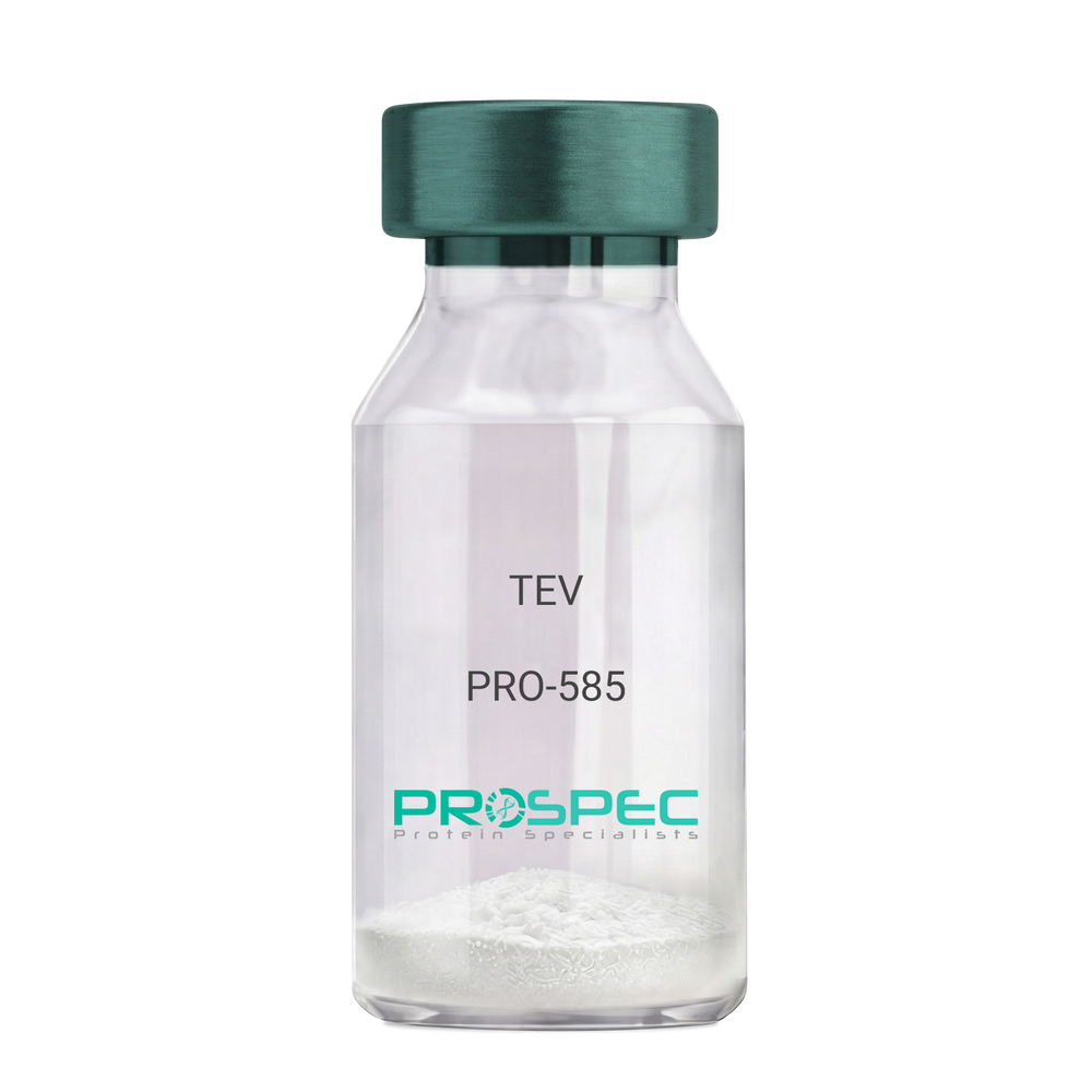
TEVDescription:
Tobacco Etch Virus Protease Recombinant
rTEV, TEV, P1 protease.
Product # :
PRO-585Price :
Quantity :
Shipping Method :
Shipped with Ice Packs
More Info
- description
- source
- formulation
- purity
- More Info
- Citations
Description
Recombinant TEV Protease (rTEV) is a site-specific protease purified from E. coli. The protease can be used for the removal of affinity tags from fusion proteins. The seven-amino-acid recognition site for rTEV is Glu-Asn-Leu-Tyr-Phe-Gln-Gly with cleavage occurring between Gln and Gly. The optimal temperature for cleavage is 30°C; however, the enzyme can be used at temperatures as low as 4°C. The rTEV contains His tag.The rTEV is purified by proprietary chromatographic techniques.
Source
Escherichia Coli.
Formulation
The rTEV contains 25mM Tris, Ph 8.0, 75mM NaCl, 5mM EDTA, 10mM GSH, 50% Glycerol.
Purity
Greater than 90.0% as determined by analysis by SDS-PAGE.
Citations
More Info
-
Introduction
TEV protease is the common name for the 27 kDa catalytic domain of the Nuclear Inclusion a (NIa) protein encoded by the tobacco etch virus (TEV). Because its sequence specificity is far more stringent than that of factor Xa, thrombin, or enterokinase, TEV protease is a very useful reagent for cleaving fusion proteins. TEV protease recognizes a linear epitope of the general form E-Xaa-Xaa-Y -Xaa-Q-(G/S), with cleavage occurring between Q and G or Q and S. The most commonly used sequence is ENLYFQG.
-
Synonyms
rTEV, TEV, P1 protease.
-
Physical Appearance
Sterile liquid formulation.
-
Stability
rTEV although stable at 4°C for 1 week, should be stored below -18°C. Please prevent freeze thaw cycles.
-
Cleavage Conditions
A number of variables can be changed to optimize the cleavage of any specific protein. The amount of rTEV, the temperature of the incubation, and the time needed for cleavage may be examined. If the protein of interest is heat-labile, then 4°C incubations are recommended. Reactions at 4°C will require longer incubation times and/or more rTEV.
-
Unit Definition
One unit is defined as the amount of enzyme needed to cleave 3 ug of fusion protein in 1 hour to 85 % completion at 30°C in a buffer containing 50 mM Tris-HCl, pH 8.0, 0.5 mM EDTA, and 1 mM DTT.
ProSpec's products are furnished for LABORATORY RESEARCH USE ONLY. The product may not be used as drugs, agricultural or pesticidal products, food additives or household chemicals.
- View Data Sheet
Name :
Lassa CapsidDescription:
Lassa Capsid Recombinant
Product # :
LSV-001Price :
Quantity :
Shipping Method :
Shipped with Ice Packs
More Info
- description
- source
- formulation
- purity
- More Info
- Citations
Description
The E.Coli derived Recombinant Lassa Nucleoprotein containing 420 amino acids of Lassa virus nucleoprotein. The Lassa Capsid protein is fused to a 6xHis tag at C-terminus and purified by proprietary chromatographic technique.
Source
Escherichia Coli.
Formulation
Lassa Capsid protein solution contains 25mM K2CO3 and PBS.
Purity
Lassa Capsid is >90% pure as determined by 10% PAGE (coomassie staining).
Citations
More Info
-
Introduction
Lassa virus, which is a member of the Arenaviridae virus family, causes acute viral hemorrhagic fever. It was first reported in 1969 in the town of Lassa, in Borno State, Nigeria. Natal multimammate mouse is the primary reservoir of Lassa virus, found in sub-Saharan Africa. The virus is transmitted by contact with the feces or urine of animals, contributing to its high rate of incidence. Lassa fever occurs generally in West Africa. Lassa fever results in 300,000 to 500,000 cases annually and causes about 5,000 deaths each year. Outbreaks of this disease have been observed in Nigeria, Liberia, Sierra Leone, Guinea, and the Central African Republic. Protein structure: Lassa virus genome is comprised of two single-stranded RNA molecules defined as small (S) and large (L). Two genes on the S segment encode the nucleoprotein (NP) and two envelope glycoproteins (GP1 and GP2); while, the L segment encodes the viral polymerase (L protein) and RING finger Z matrix protein. GP1 subunit serves a putative role in receptor binding, while the structure of GP2 subunit is consistent with viral transmembrane fusion proteins. NP is a virion protein which binds and protects the viral RNA.
-
Physical Appearance
Sterile Filtered solution.
-
Stability
Lassa Capsid protein although stable at 4°C for 1 week, should be stored below -18°C. Please prevent freeze thaw cycles.
-
Applications
Immunoassay.
ProSpec's products are furnished for LABORATORY RESEARCH USE ONLY. The product may not be used as drugs, agricultural or pesticidal products, food additives or household chemicals.
- View Data Sheet
Name :
CHIKV CapsidDescription:
Chikungunya Capsid Recombinant
Product # :
CHI-005Price :
Quantity :
Shipping Method :
Shipped with Ice Packs
More Info
- description
- source
- formulation
- purity
- More Info
- Citations
Description
Recombinant Chikungunya Full length capsid protein containing 220 amino acids produced in E.coli having a molecular weight of 25kDa.
Source
Escherichia Coli.
Formulation
Sterile Filtered solution containing 1x PBS, 2M urea and 25mM K2CO3.
Purity
Protein is >90% pure as determined by SDS-PAGE.
Citations
More Info
-
Introduction
Chikungunya is an infection caused by the chikungunya virus which is passed to humans by two species of mosquito of the genus Aedes: A. albopictus and A. aegypti. Animal reservoirs of the virus include monkeys, birds, cattle, and rodents. The features of the disease are a sudden onset of fever 2-4 days after exposure. The fever typically lasts 2-7 days, while the associated joint pains usually last weeks or months but sometimes years. The mortality rate is a little less than 1 in 1,000. The disease has occurred in outbreaks in Asia, Europe and the Americas since 2004. CHIKV is a single-stranded positive-sense RNA genome, 11,800 nts long which encodes 2 open reading frames. The nucleocapsid is tightly enveloped by a host-derived lipid bilayer (envelope) supporting the virus-encoded envelope proteins. 80 glycoprotein spikes are C- terminally anchored within the viral envelope. The structural polyprotein is translated from a viral sub genomic mRNA, while as the 5 structural proteins (capsid, E3, E2, 6K, E1) are translated as a single polyprotein, from which capsid (C) is cleaved off to encapsidate. The envelope polyprotein precursor E3-E2-6K-E1 is translocated to the endoplasmatic reticulum. Polyprotein is processed by host signalases, resulting in E3, E2 & E1 forming viral hetero-trimeric spikes. The viral spikes majorly contains E2 and E1 facilitate cell receptor recognition, cell entry thru pH-dependent endocytosis and support viral budding.
-
Physical Appearance
Sterile filtered colorless solution.
-
Stability
CHIKV Capsid although stable at 4°C for 1 week, should be stored below -18°C. Please prevent freeze thaw cycles.
-
Purification Method
Purified by proprietary chromatographic technique.
ProSpec's products are furnished for LABORATORY RESEARCH USE ONLY. The product may not be used as drugs, agricultural or pesticidal products, food additives or household chemicals.
- View Data Sheet
Name :
HIV-1 p24 CoreDescription:
HIV-1 p24 Core Recombinant
Product # :
HIV-103Price :
Quantity :
Shipping Method :
Shipped with Ice Packs
More Info
- description
- source
- formulation
- purity
- More Info
- Citations
Description
HIV-1 p24 Core is a 24kDa non-glycosylated polypeptide chain, containing the full length sequence of the HIV-1 p24.
Source
Escherichia Coli.
Formulation
50mM Tris, pH 8.5 and 8M urea.
Purity
Greater than 95.0% as determined by HPLC analysis and SDS-PAGE.
Citations
More Info
-
Introduction
Human immunodeficiency virus (HIV) is a retrovirusthat can lead to a condition in which the immune systembegins to fail, leading to opportunistic infections. HIV primarily infects vital cells in the humanimmune systemsuch as helper T cells(specifically CD4+ T cells), macrophagesand dendritic cells. HIV infection leads to low levels of CD4+ T cells through three main mechanisms: firstly, direct viral killing of infected cells; secondly, increased rates of apoptosisin infected cells; and thirdly, killing of infected CD4+ T cells by CD8 cytotoxic lymphocytesthat recognize infected cells. When CD4+ T cell numbers decline below a critical level, cell-mediated immunityis lost, and the body becomes progressively more susceptible to opportunistic infections. HIV was classified as a member of the genus Lentivirus, part of the family of Retroviridae. Lentiviruses have many common morphologies and biological properties. Many species are infected by lentiviruses, which are characteristically responsible for long-duration illnesses with a long incubation period. Lentiviruses are transmitted as single-stranded, positive-sense, enveloped RNA viruses. Upon entry of the target cell, the viral RNA genomeis converted to double-stranded DNAby a virally encoded reverse transcriptasethat is present in the virus particle. This viral DNA is then integrated into the cellular DNA by a virally encoded integraseso that the genome can be transcribed. Once the virus has infected the cell, two pathways are possible: either the virus becomes latentand the infected cell continues to function, or the virus becomes active and replicates, and a large number of virus particles are liberated that can then infect other cells.
-
Physical Appearance
Sterile filtered colorless clear solution.
-
Stability
HIV-1 p24 core although stable at 4°C for 1 week, should be stored below -18°C. Please prevent freeze thaw cycles.
-
Specificity
Immunoreactive with all sera of HIV-1 infected individuals.
ProSpec's products are furnished for LABORATORY RESEARCH USE ONLY. The product may not be used as drugs, agricultural or pesticidal products, food additives or household chemicals.
- View Data Sheet
Name :
Zika NS1 Paired AntibodyDescription:
Mouse Anti Zika NS1 Paired
Product # :
ANT-008Price :
Quantity :
Shipping Method :
Shipped with Ice Packs
More Info
- description
- formulation
- purity
- More Info
- Citations
Description
Zika NS1 conjugation antibody and Zika NS1 capture antibody are used to develop rapid test for Zika NS1 rapid test. Please note that when ordering for example: 100µg antibody we ship 50µg from each of the antibodies (100µg in total).
Formulation
* Zika NS1 gold conjugation antibody in PBS, 10mg BSA/ml & 0.1% Sodium Nitrate.
* Zika NS1 capture antibody in PBS, 10mg BSA/ml & 0.1% Sodium Nitrate.Purity
Greater than 95%.
Citations
More Info
-
Introduction
Zika virus (ZIKV) belongs to the family Flaviviridae and the genus Flavivirus, it is transmitted by daytime-active Aedes mosquitoes, such as A. aegypti and A. albopictus. The Zika virus is related to the dengue, yellow fever, Japanese encephalitis, and West Nile viruses. Much like the other flaviviruses, Zika virus is enveloped and icosahedral and has a nonsegmented, single-stranded, positive-sense RNA genome. Zika fever is an infection, which often causes no symptoms or only mild ones, like a mild form of dengue fever, and it is treated by rest. As of February 2016, there has been mounting evidence that Zika fever in pregnant women can cause abnormal brain development in their fetuses by mother-to-child transmission, which may result in miscarriage or microcephaly, however it is not yet known whether Zika virus causes microcephaly. Furthermore, a connection has been established with neurologic conditions in infected adults, including Guillain–Barre syndrome.
-
Physical Appearance
2 vials of sterile filtered clear colorless solution.
-
Stability
Zika antibody although stable at 4°C for 1 week, should be stored below -18°C. For long term storage it is recommended to add a carrier protein (0.1% HSA or BSA). Please prevent freeze-thaw cycles.
-
Applications
Lateral flow immunoassay.
-
Type
Mouse antibody Monoclonal.
-
Purification Method
Purified monoclonal IgG by protein A chromatography.
ProSpec's products are furnished for LABORATORY RESEARCH USE ONLY. The product may not be used as drugs, agricultural or pesticidal products, food additives or household chemicals.
- View Data Sheet
Name :
EDA2R Human, Sf9Description:
Ectodysplasin A2 Receptor Human Recombinant, Sf9
Tumor necrosis factor receptor superfamily member 27, X-linked ectodysplasin-A2 receptor, EDA-A2 receptor, Ectodysplasin A2 Receptor, X-Linked Ectodysplasin-A2 Receptor, EDA-A2 Receptor, TNFRSF27, XEDAR, Tumor Necrosis Factor Receptor Superfamily Member XEDAR, Tumor Necrosis Factor Receptor Superfamily Member 27, Ectodysplasin A2 Isoform Receptor, EDA-A2R, EDAA2R.
Product # :
PRO-2399Price :
Quantity :
Shipping Method :
Shipped with Ice Packs
More Info
- description
- source
- formulation
- purity
- More Info
- Citations
Description
EDA2R Human Recombinant produced in Sf9 Baculovirus cells is a single, glycosylated polypeptide chain containing 380 amino acids (1-138a.a.) and having a molecular mass of 42.5kDa (Molecular size on SDS-PAGE will appear at approximately 40-57kDa). EDA2R is expressed with a 242 amino acid hIgG-His tag at C-Terminus and purified by proprietary chromatographic techniques.
Source
Sf9, Baculovirus cells.
Formulation
EDA2R protein solution (0.5mg/ml) contains Phosphate Buffered Saline (pH 7.4), 20% glycerol and 1mM DTT.
Purity
Greater than 90.0% as determined by SDS-PAGE.
Citations
More Info
-
Introduction
EDA2R (ectodysplasin A2 receptor) mediates the activation of the NF-kappa-B and JNK pathways. Activation seems to be mediated through binding to TRAF3 and TRAF6. In addition, Mutations in EDA give rise to a clinical syndrome characterized by loss of hair, sweat glands, and teeth. EDA2R specifically binds to EDA-A2 isoform. This protein is a type III transmembrane protein of the TNFR (tumor necrosis factor receptor) superfamily, and contains 3 cysteine-rich repeats and a single transmembrane domain however it lacks an N-terminal signal peptide. Alternatively spliced transcript variants have been found for this gene. Among the diseases associated with EDA2R are ectodermal dysplasia 1, hypohidrotic, x-linked, and hypohidrotic ectodermal dysplasia.
-
Synonyms
Tumor necrosis factor receptor superfamily member 27, X-linked ectodysplasin-A2 receptor, EDA-A2 receptor, Ectodysplasin A2 Receptor, X-Linked Ectodysplasin-A2 Receptor, EDA-A2 Receptor, TNFRSF27, XEDAR, Tumor Necrosis Factor Receptor Superfamily Member XEDAR, Tumor Necrosis Factor Receptor Superfamily Member 27, Ectodysplasin A2 Isoform Receptor, EDA-A2R, EDAA2R.
-
Physical Appearance
Sterile Filtered clear solution.
-
Stability
Store at 4°C if entire vial will be used within 2-4 weeks. Store, frozen at -20°C for longer periods of time.For long term storage it is recommended to add a carrier protein (0.1% HSA or BSA).Please avoid freeze thaw cycles.
-
Amino Acid Sequence
ADPMDCQENE YWDQWGRCVT CQRCGPGQEL SKDCGYGEGG DAYCTACPPR RYKSSWGHHR CQSCITCAVI NRVQKVNCTA TSNAVCGDCL PRFYRKTRIG GLQDQECIPC TKQTPTSEVQ CAFQLSLVEA DAPTVPPQEA TLEPKSCDKT HTCPPCPAPE LLGGPSVFLF PPKPKDTLMI SRTPEVTCVV VDVSHEDPEV KFNWYVDGVE VHNAKTKPRE EQYNSTYRVV SVLTVLHQDW LNGKEYKCKV SNKALPAPIE KTISKAKGQP REPQVYTLPP SRDELTKNQV SLTCLVKGFY PSDIAVEWES NGQPENNYKT TPPVLDSDGS FFLYSKLTVD KSRWQQGNVF SCSVMHEALH NHYTQKSLSL SPGKHHHHHH.
ProSpec's products are furnished for LABORATORY RESEARCH USE ONLY. The product may not be used as drugs, agricultural or pesticidal products, food additives or household chemicals.
- View Data Sheet
Name :
EIF5A2 HumanDescription:
Eukaryotic Translation Initiation Factor 5A2 Human Recombinant
eIF-5A-2, EIF-5A2, eIF5AII, Eukaryotic translation initiation factor 5A-2, Eukaryotic initiation factor 5A isoform 2, EIF5A2.
Product # :
PRO-846Price :
Quantity :
Shipping Method :
Shipped with Ice Packs
More Info
- description
- source
- formulation
- purity
- More Info
- Citations
Description
EIF5A2 Recombinant E.coli produced in E.Coli is a single, non-glycosylated polypeptide chain containing 173 amino acids (1-153 a.a.) and having a molecular mass of 18.9 kDa. The EIF5A2 is fused to a 20 amino acid His-Tag at N-terminus and purified by proprietary chromatographic techniques.
Source
Escherichia Coli.
Formulation
EIF5A2 Human solution containing 20mM Tris-HCl pH-8 & 10% glycerol.
Purity
Greater than 85.0% as determined by SDS-PAGE.
Citations
More Info
-
Introduction
EIF5A2 is part of the eukaryotic initiation factor 5A subfamily, is an vital protein strongly linked to cellular polyamine homeostasis. EIF5A2 promotes the formation of the first peptide bond during the initial stage of protein synthesis. EIF5A2 is the single eukaryotic protein to have a hypusine residue, which is a post-translational modification of a lysine by the addition of a butylamino group.
-
Synonyms
eIF-5A-2, EIF-5A2, eIF5AII, Eukaryotic translation initiation factor 5A-2, Eukaryotic initiation factor 5A isoform 2, EIF5A2.
-
Physical Appearance
Sterile filtered colorless solution.
-
Stability
Store at 4°C if entire vial will be used within 2-4 weeks. Store, frozen at -20°C for longer periods of time. For long term storage it is recommended to add a carrier protein (0.1% HSA or BSA).Avoid multiple freeze-thaw cycles.
-
Amino Acid Sequence
MGSSHHHHHH SSGLVPRGSH MADEIDFTTG DAGASSTYPM QCSALRKNGF VVLKGRPCKI VEMSTSKTGK HGHAKVHLVG IDIFTGKKYE DICPSTHNMD VPNIKRNDYQ LICIQDGYLS LLTETGEVRE DLKLPEGELG KEIEGKYNAG EDVQVSVMCA MSEEYAVAIK PCK.
ProSpec's products are furnished for LABORATORY RESEARCH USE ONLY. The product may not be used as drugs, agricultural or pesticidal products, food additives or household chemicals.
- View Data Sheet
Name :
ERCC1 HumanDescription:
Excision Repair Cross-Complementing 1 Human Recombinant
Excision Repair Cross-complementing rodent repair deficiency complementation group 1 (includes overlapping antisense sequence), RAD10, COFS4, UV20, DNA excision repair protein ERCC-1.
Product # :
PRO-1106Price :
Quantity :
Shipping Method :
Shipped with Ice Packs
More Info
- description
- source
- formulation
- purity
- More Info
- Citations
Description
ERCC1 Human Recombinant produced in E. coli is a single polypeptide chain containing 297 amino acids (1-273) and having a molecular mass of 32.5kDa.ERCC1 is fused to a 24 amino acid His-tag at N-terminus & purified by proprietary chromatographic techniques.
Source
E.coli.
Formulation
The ERCC1 solution (0.5mg/1ml) contains 20mM Tris-HCl buffer (pH 8.0), 100mM NaCl, 1mM DTT and 20% glycerol.
Purity
Greater than 90% as determined by SDS-PAGE.
Citations
More Info
-
Introduction
ERCC1 polypeptide is essential for nucleotide excision repair (NER) of damaged DNA and is homologous to Saccharomyces cerevisiae RAD10, which takes part in repair and mitotic intra-chromosomal recombination. NER mechanism includes dual incisions on both sides of the damage catalyzed by two nucleases. In mammalian cells XPG cleaves 3' of the DNA lesion while the ERCC1-XPF complex makes the 5' incision.
-
Synonyms
Excision Repair Cross-complementing rodent repair deficiency complementation group 1 (includes overlapping antisense sequence), RAD10, COFS4, UV20, DNA excision repair protein ERCC-1.
-
Physical Appearance
Sterile Filtered colorless solution.
-
Stability
Store at 4°C if entire vial will be used within 2-4 weeks. Store, frozen at -20°C for longer periods of time. For long term storage it is recommended to add a carrier protein (0.1% HSA or BSA).Avoid multiple freeze-thaw cycles.
-
Amino Acid Sequence
MGSSHHHHHH SSGLVPRGSH MGSHMDPGKD KEGVPQPSGP PARKKFVIPL DEDEVPPGVA KPLFRSTQSL PTVDTSAQAA PQTYAEYAIS QPLEGAGATC PTGSEPLAGE TPNQALKPGA KSNSIIVSPR QRGNPVLKFV RNVPWEFGDV IPDYVLGQST CALFLSLRYH NLHPDYIHGR LQSLGKNFAL RVLLVQVDVK DPQQALKELA KMCILADCTL ILAWSPEEAG RYLETYKAYE QKPADLLMEK LEQDFVSRSL EQLIAASRED LALCPGLGPQ KARRLFDVLH EPFLKVP
ProSpec's products are furnished for LABORATORY RESEARCH USE ONLY. The product may not be used as drugs, agricultural or pesticidal products, food additives or household chemicals.
- View Data Sheet
Name :
HEL antibodyDescription:
Hen Egg Lysozyme, Mouse antibody
Product # :
ANT-149Price :
Quantity :
Shipping Method :
Shipped at Room temp
More Info
- formulation
- More Info
- Citations
Formulation
1mg/ml in PBS (after reconstitution).
Citations
More Info
-
Solubility
Reconstitute with H20 to get a 1 mg/ml concentration. Mix gently, wash the sides of the vial and wait 30-60 seconds before use.
-
Immunogen
Purified HEL.
-
Ig Subclass
Mouse IgG2b.
-
Clone
NYRHEL.
-
Titer
By direct ELISA, 1:5,000 dilution will yield 0.2 O.D using alkaline phosphatase conjugated rabbit anti-mouse Ig (Jackson Laboratories).
-
Shipping Conditions
Antibody is shipped lyophilized at ambient temperature.
-
Type
Mouse Antibody Monoclonal.
-
Storage Procedures
In lyophilized form, for long periods, store at 4°C in a dry environment. After reconstitution, if not intended for use within a month, aliquot and store at -20°C.
-
Purification Method
Ion exchange column.
ProSpec's products are furnished for LABORATORY RESEARCH USE ONLY. The product may not be used as drugs, agricultural or pesticidal products, food additives or household chemicals.
- View Data Sheet
Name :
CRKL HumanDescription:
V-crk Sarcoma Virus CT10 Oncogene Homolog (Avian)-Like Human Recombinant
Crk-like protein, CRKL.
Product # :
PRO-063Price :
Quantity :
Shipping Method :
Shipped with Ice Packs
More Info
- description
- source
- formulation
- purity
- More Info
- Citations
Description
CRKL Human Recombinant fused with a 20 amino acid His tag at N-terminus produced in E.Coli is a single, non-glycosylated, polypeptide chain containing 323 amino acids (1-303 a.a.) and having a molecular mass of 35.9kDa (Molecular size on SDS-PAGE will appear higher). The CRKL is purified by proprietary chromatographic techniques.
Source
Escherichia Coli.
Formulation
The CRKL solution (1 mg/ml) 20mM Tris-HCl buffer (pH 8.0), 10% glycerol, 2mM DTT and 0.1M NaCl.
Purity
Greater than 90.0% as determined by SDS-PAGE.
Citations
More Info
-
Introduction
Crk-like protein (CRKL) is a protein kinase containing SH2 and SH3 (src homology) domains which activates the RAS and JUN kinase signaling pathways and transforms fibroblasts in a RAS-dependent manner. CRKL is a substitute of the BCR-ABL tyrosine kinase, it also has a role in fibroblast transformation by BCR-ABL, and has oncogenic potential.
-
Synonyms
Crk-like protein, CRKL.
-
Physical Appearance
Sterile Filtered colorless solution.
-
Stability
Store at 4°C if entire vial will be used within 2-4 weeks. Store, frozen at -20°C for longer periods of time. For long term storage it is recommended to add a carrier protein (0.1% HSA or BSA).Avoid multiple freeze-thaw cycles.
-
Amino Acid Sequence
MGSSHHHHHH SSGLVPRGSH MSSARFDSSD RSAWYMGPVS RQEAQTRLQG QRHGMFLVRDSSTCPGDYVL SVSENSRVSH YIINSLPNRR FKIGDQEFDH LPALLEFYKI HYLDTTTLIE PAPRYPSPPM GSVSAPNLPT AEDNLEYVRT LYDFPGNDAE DLPFKKGEIL VIIEKPEEQW WSARNKDGRV GMIPVPYVEK LVRSSPHGKH GNRNSNSYGI PEPAHAYAQP QTTTPLPAVS GSPGAAITPL PSTQNGPVFA KAIQKRVPCA YDKTALALEV GDIVKVTRMN INGQWEGEVN GRKGLFPFTH VKIFDPQNPD ENE.
ProSpec's products are furnished for LABORATORY RESEARCH USE ONLY. The product may not be used as drugs, agricultural or pesticidal products, food additives or household chemicals.
- View Data Sheet
Name :
BNIP1 HumanDescription:
BCL2/Adenovirus E1B 19kDa Interacting Protein 1 Human Recombinant
BCL2/Adenovirus E1B 19kDa Interacting Protein 1, NIP1, BCL2/Adenovirus E1B 19 KDa Protein-Interacting Protein 1, Transformation-Related Gene 8 Protein, TRG-8, BCL2/Adenovirus E1B 19kD-Interacting Protein 1, Vesicle Transport Protein SEC20, SEC20L, SEC20, Vesicle transport protein SEC20.
Product # :
PRO-2150Price :
Quantity :
Shipping Method :
Shipped with Ice Packs
More Info
- description
- source
- formulation
- purity
- More Info
- Citations
Description
BNIP1 Human Recombinant produced in E.Coli is a single, non-glycosylated polypeptide chain containing 222 amino acids (1-199 a.a) and having a molecular mass of 25.2kDa. BNIP1 is fused to a 23 amino acid His-tag at N-terminus & purified by proprietary chromatographic techniques.
Source
Escherichia Coli.
Formulation
BNIP1 protein solution (1.0 mg/ml) containing 20mM Tris-HCl (pH8.0) and 10% glycerol.
Purity
Greater than 95% as determined by SDS-PAGE.
Citations
More Info
-
Introduction
BNIP1 belongs to the of the BCL2/adenovirus E1B 19 kd-interacting protein (BNIP) family. BNIP1 interacts with the E1B 19 kDa protein, which defends cells from virally-induced cell death. In addition, BNIP1 interacts with E1B 19 kDa-like sequences of BCL2 which is an additional apoptotic protector. Adding up, BNIP1 is implicated in vesicle transport into the endoplasmic reticulum. Alternative splicing results of BNIP1 in four protein products with identical N- and C-termini have been found.
-
Synonyms
BCL2/Adenovirus E1B 19kDa Interacting Protein 1, NIP1, BCL2/Adenovirus E1B 19 KDa Protein-Interacting Protein 1, Transformation-Related Gene 8 Protein, TRG-8, BCL2/Adenovirus E1B 19kD-Interacting Protein 1, Vesicle Transport Protein SEC20, SEC20L, SEC20, Vesicle transport protein SEC20.
-
Physical Appearance
Sterile filtered colorless solution.
-
Stability
Store at 4°C if entire vial will be used within 2-4 weeks. Store, frozen at -20°C for longer periods of time. For long term storage it is recommended to add a carrier protein (0.1% HSA or BSA).Avoid multiple freeze-thaw cycles.
-
Amino Acid Sequence
MGSSHHHHHH SSGLVPRGSH MGSMAAPQDV HVRICNQEIV KFDLEVKALI QDIRDCSGPL SALTELNTKV KEKFQQLRHR IQDLEQLAKE QDKESEKQLL LQEVENHKKQ MLSNQASWRK ANLTCKIAID NLEKAELLQG GDLLRQRKTT KESLAQTSST ITESLMGISR MMAQQVQQSE EAMQSLVTSS RTILDANEEF KSMSGTIQLG RKLITKYNRR EL.
ProSpec's products are furnished for LABORATORY RESEARCH USE ONLY. The product may not be used as drugs, agricultural or pesticidal products, food additives or household chemicals.
- View Data Sheet
Name :
HBsAg adw2Description:
Hepatitis B Surface Antigen, adw2 Recombinant
Product # :
HBS-885Price :
Quantity :
Shipping Method :
Shipped with Ice Packs
More Info
- description
- source
- formulation
- purity
- More Info
- Citations
Description
HbsAg subtype adw2 produced in E.Coli, is a single, non-glycosylated, polypeptide chain containing 226 amino acids and having a molecular weight of approximately 23.0kDa. HBsAg adw2 migrates on SDS-PAGE as a 23kDa monomer band with minor amounts of dimer (46kDa) and trimer (69kDa) forms.The HBsAg is fused to a 6 His tag on C-terminus and purified by proprietary chromatographic techniques.
Source
Escherichia Coli.
Formulation
Sterile filtered solution containing 50mM potassium phosphate.
Purity
Greater than 90.0% as determined by SDS-PAGE.
Citations
More Info
-
Introduction
HBsAg is the surface antigenof the Hepatitis-B-Virus (HBV). The capsidof a virus has different surface proteins from the rest of the virus. The antigen is a protein that binds specifically on one of these surface proteins. It is commonly referred to as the Australian Antigen.
-
Physical Appearance
Sterile filtered colorless solution.
-
Stability
Store at 4°C if entire vial will be used within 2-4 weeks. Store, frozen at -20°C for longer periods of time.For long term storage it is recommended to add a carrier protein (0.1% HSA or BSA). Avoid multiple freeze-thaw cycles.
-
Amino Acid Sequence
MENITSGFLGPLLVLQAGFFLLTRILTIPQSLDSWWTSLNFLGGSPVCLGQNSQSPTSNH
SPTSCPPICPGYRWMCLRRFIIFLFILLLCLIFLLVLLDYQGMLPVCPLIPGSTTTSTGP
CKTCTTPAQGNSMFPSCCCTKPTDGNCTCIPIPSSWAFAKYLWEWASVRFSWLSLLVPFV
QWFVGLSPTVWLSAIWMMWYWGPSLYSIVSPFIPLLPIFFCLWVYI
ProSpec's products are furnished for LABORATORY RESEARCH USE ONLY. The product may not be used as drugs, agricultural or pesticidal products, food additives or household chemicals.
- View Data Sheet
Name :
LY6G AntibodyDescription:
LY6G, Rat Anti Mouse Antibody
Lymphocyte antigen 6G, Ly-6G, Ly-6G.1, Ly6g, Gr1, Gr-1.
Product # :
ANT-158Price :
Quantity :
Shipping Method :
Shipped at Room temp
More Info
- formulation
- More Info
- Citations
Formulation
1mg/ml in PBS (after reconstitution).
Citations
More Info
-
Introduction
Myeloid differentiation antigen Gr1 (Ly6G) is a GPI-anchored protein, which is briefly expressed on monocytes in the bone marrow. The level of the Ly6G expression in the bone marrow completely correlates with granulocyte differentiation and maturation. Ly6G ligation on murine neutrophils inhibits neutrophil recruitment, thus providing the first evidence of a function for the Ly6G molecule. Ly6G is seen primarily on neutrophils, also in a subgroup of eosinophils, differentiating pre-monocytes and plasmacytoid dendritic cells.
-
Synonyms
Lymphocyte antigen 6G, Ly-6G, Ly-6G.1, Ly6g, Gr1, Gr-1.
-
Solubility
Reconstitute with H20. Mix gently, wash the sides of the vial and wait 30-60 seconds before use.
-
Immunogen
Ly-6G-transfected cell line.
-
Ig Subclass
Rat IgG2a.
-
Clone
YRmLy-6G.
-
Applications
Flow cytometry, immunohistochemistry, cell depletion.
This antibody recognizes only Ly-6G which is expressed on most myeloid cells in the bone marrow and all peripheral granulocytes. For flow cytometry, use 10 µl/106 cells. Exact titer for cell depletion in vivo (cytotoxicity) should be determined by the investigator. -
Shipping Conditions
Antibody is shipped lyophilized at ambient temperature.
-
Available Conjugates
This antibody is also available conjugated to biotin and FITC.
-
Type
Rat Anti Mouse Monoclonal.
-
Storage Procedures
In lyophilized form, for long periods, store at 4°C in a dry environment. After reconstitution, if not intended for use within a month, aliquot and store at -20°C.
-
Purification Method
Protein A column.
ProSpec's products are furnished for LABORATORY RESEARCH USE ONLY. The product may not be used as drugs, agricultural or pesticidal products, food additives or household chemicals.
- View Data Sheet
Name :
HBV Pre-SDescription:
Hepatitis B Virus Pre-S Recombinant
Product # :
HBV-231Price :
Quantity :
Shipping Method :
Shipped with Ice Packs
More Info
- description
- source
- formulation
- purity
- More Info
- Citations
Description
Recombinant Hepatitis B Virus Pre-S produced in E.coli is a full length Pre-S of HBV subtype adw2, containing 174 amino acids, having a molecular weight of about 25kDa including a vector sequence and linked with a 6xHis tag at C- terminus.
Source
Escherichia Coli.
Formulation
Sterile Filtered solution containing PBS, and 25mM K2CO3.
Purity
Protein is >95% pure as determined by 12% PAGE (coomassie staining).
Citations
More Info
-
Introduction
Hepatitis B virus surface gene encodes 3 proteins: small (HBs), middle (MHBs) and large (LHBs) surface protein. These 3 proteins are translated from distinct start codons, however share a common reading frame and stop codon. HBs contains 226 amino acids, it is a major viral envelope component. MHBs has extra 55 amino acids (Pre-S2) located to the N-terminus of HBs, LHBs has an additional 119 amino acids (Pre-S1). Pre-S is a region is comprised of Pre-S1 and S2, Pre-S1 has a vital role in receptor recognition, while Pre-S2 plays a part in translocation of virus into host cell. Clinically, HBV pre-S is linked with HBsAg and HBV DNA copies, which indicates active replication of virus, reduction of pre-S is typically associated with decreased HBV DNA copies, pointing to an improved predictor of treatment. In addition, Pre-S is a potential vaccine under development to enhance currently used HBsAg vaccine.
-
Physical Appearance
Sterile Filtered solution.
-
Stability
HBV Pre-S although stable at 4°C for 1 week, should be stored below -18°C. Please prevent freeze thaw cycles.
-
Applications
Immunoassay.
-
Purification Method
Purified by proprietary chromatographic technique.
ProSpec's products are furnished for LABORATORY RESEARCH USE ONLY. The product may not be used as drugs, agricultural or pesticidal products, food additives or household chemicals.
- View Data Sheet
Name :
T.pallidum p47 (Partial)Description:
Treponema pallidum p47 (Partial) Recombinant
Product # :
TRP-249Price :
Quantity :
Shipping Method :
Shipped with Ice Packs
More Info
- description
- source
- formulation
- purity
- More Info
- Citations
Description
The E.Coli derived recombinant protein is fused at the C- terminus with a 6xHis Tag and contains the T.Pallidum p47 immunodominant regions.
Source
Escherichia Coli.
Formulation
70mM Tris-HCl pH8.0, 50mM NaCl, 50% Glycerol and 1.5M urea.
Purity
Treponema Pallidum protein is >95% pure as determined by 10% PAGE (coomassie staining).
Citations
More Info
-
Introduction
Treponema pallidum is a gram-negative spirochaete bacterium and is considered to be metabolically crippled. There are at least four known subspecies: T. pallidum pallidum, T. pallidum pertenue, T. pallidum carateum and T. pallidum endemicum. The helical structure of T. pallidum pallidum allows it to move in a corkscrew motion through viscous mediums such as mucus. Treponema pallidum subsp. pallidum has one of the smallest bacterial genomes at 1.14 million base pairs (Mb) and has limited metabolic capabilities, reflecting its adaptation through genome reduction to the rich environment of mammalian tissue.
-
Stability
Treponema Pallidum although stable at 4°C for 1 week, should be stored below -18°C. Please prevent freeze thaw cycles.
-
Applications
Treponema Pallidum protein is suitable for ELISA and Western blots, excellent antigen for detection of T.Pallidum with minimal specificity problems.
-
Specificity
Immunoreactive with sera of T.Pallidum infected individuals.
-
Purification Method
Treponema Pallidum protein was purified by proprietary chromatographic technique.
ProSpec's products are furnished for LABORATORY RESEARCH USE ONLY. The product may not be used as drugs, agricultural or pesticidal products, food additives or household chemicals.
- View Data Sheet
Name :
HB-EGF MouseDescription:
HB-EGF Mouse Recombinant
DTR, HEGFL, diphtheria toxin receptor, DTSF.
Product # :
CYT-068Price :
Quantity :
Shipping Method :
Shipped at Room temp
More Info
- description
- source
- formulation
- purity
- biological activity
- More Info
- sds-page
- Citations
Description
HB-EGF Mouse Recombinant produced in E.Coli is a single, non-glycosylated, polypeptide chain containing 86 amino acids (63-148 a.a.) and having a molecular mass of 9.8 kDa.The HB-EGF is purified by proprietary chromatographic techniques
Source
Escherichia Coli.
Formulation
The protein was filtered (0.2µm) and lyophilized from a concentrated solution containing 10mM PB and 500mM NaCl, pH7.4.
Purity
Greater than 97.0% as determined by:
(a) Analysis by RP-HPLC.
(b) Analysis by SDS-PAGE.Biological Activity
The ED50 was determined by a cell proliferation assay using balb/c 3T3 cells is < 1.0 ng/ml, corresponding to a specific activity of > 1.0×10⁶ units/mg.sds-page
Citations
More Info
-
Introduction
HB-EGF is an EGF related growth factor which signals via the EGF receptor, and stimulates the proliferation of SMC (smooth muscle cells), fibroblasts, epithelial cells and keratinocytes. HB-EGF is expressed in various cell types and tissues, including vascular endothelial cells and SMC, macrophages, skeletal muscle, keratinocytes and particular tumor cells. HB-EGF’s ability to explicitly bind sulfate proteoglycans is dissimilar from other EGF-like molecules, and might be related to the enhanced mitogenic activity, relative to EGF, that HB-EGF exerts on smooth muscle cells.
-
Synonyms
DTR, HEGFL, diphtheria toxin receptor, DTSF.
-
Physical Appearance
Sterile Filtered White lyophilized (freeze-dried) powder.
-
Stability
Lyophilized Mouse HB-EGF Recombinant although stable at room temperature for 3 weeks, should be stored desiccated below -18°C. Upon reconstitution HB-EGF should be stored at 4°C between 2-7 days and for future use below -18°C.For long term storage it is recommended to add a carrier protein (0.1% HSA or BSA).Please prevent freeze-thaw cycles.
-
Solubility
It is recommended to reconstitute the lyophilized Mouse HB-EGF in sterile 18M-cm H2O not less than 100 µg/ml, which can then be further diluted to other aqueous solutions.
-
Amino Acid Sequence
DLEGTDLNLF KVAFSSKPQG LATPSKERNG KKKKKGKGLG KKRDPCLRKY KDYCIHGECR YLQEFRTPSC KCLPGYHGHR CHGLTL.
-
Background
What is the molecular weight/Mw of HB-EGF Protein?
HB-EGF Protein has a total Mw of 9.8kDa.
What is the source or expression system of HB-EGF Protein?
Escherichia Coli.
What is the Purity of HB-EGF Protein?
HB-EGF Protein is >97% pure as determined by SDS-PAGE.
What is the Biological Activity of HB-EGF Protein?
The ED50 was determined by a cell proliferation assay using balb/c 3T3 cells is < 1.0 ng/ml, corresponding to a specific activity of > 1.0×10⁶ units/mg.
What is the amino acid sequence of HB-EGF Protein?
DLEGTDLNLF KVAFSSKPQG LATPSKERNG KKKKKGKGLG KKRDPCLRKY KDYCIHGECR YLQEFRTPSC KCLPGYHGHR CHGLTL.
What applications can HB-EGF Protein be used in?
HB-EGF Protein can probably be used in western blot, ELISA and Lateral Flow.
What is the endotoxin level for HB-EGF Protein?
The endotoxin level is minimal, HB-EGF Protein was purified using conventional chromatography techniques.
ProSpec's products are furnished for LABORATORY RESEARCH USE ONLY. The product may not be used as drugs, agricultural or pesticidal products, food additives or household chemicals.
- View Data Sheet
Name :
HBsAg preS2Description:
Hepatitis B Surface Antigen, preS2 Recombinant
Product # :
HBS-874Price :
Quantity :
Shipping Method :
Shipped at Room temp
More Info
- description
- source
- formulation
- purity
- More Info
- Citations
Description
The Recombinant Hepatitis B Surface Antigen preS2 is approximately 5.7 kDa, a single non-glycosylated polypeptide chain containing 55 amino acids. Purified by proprietary chromatographic technique.
Source
E.coli.
Formulation
HBsAg protein was lyophilized from 0.2μm filtered (1mg/ml) solution in 20mM PB, pH 7.4 and 50mM NaCl.
Purity
Greater than 95.0% as determined by:
(a) Analysis by RP-HPLC.
(b) Analysis by SDS-PAGE.Citations
More Info
-
Introduction
Hepatitis B virus (HBV) is a human pathogen, causing serious liver disease. The HBV surface protein antigens (HBsAg) are comprised of three carboxyl co terminal HBs proteins termed large (LHBs), middle (MHBs) and small (SHBs, also called major) protein. LHBs and MHBs also share the highly hydrophobic, repetitive, membrane spanning S domain. In addition, MHBs has a 55 amino acid region called preS2.
-
Stability
This lyophilized preparation is stable at 2-8°C, but should be kept at -20°C for long term storage, preferably desiccated. Upon reconstitution, the preparation is stable for up to one week at 2-8°C. For maximal stability, apportion the reconstituted preparation into working aliquots and store at -20°C to -70°C. Avoid repeated freeze/thaw cycles.
-
Solubility
We recommend that this vial be briefly centrifuged prior to opening to bring the contents to the bottom. Reconstitute in sterile distilled water or aqueous buffer containing 0.1% BSA to a concentration of 0.1-1.0 mg/mL. Stock solutions should be apportioned into working aliquots and stored at <-20°C. Further dilutions should be made in appropriate buffered solutions.
-
Amino Acid Sequence
MQWNSTTFHQALLDPKVRGLYFPAGGSSSGTVNPVPTTASP ISSIFSRTGDPAPN.
ProSpec's products are furnished for LABORATORY RESEARCH USE ONLY. The product may not be used as drugs, agricultural or pesticidal products, food additives or household chemicals.