Search results
1000 results found for “Anti Coagulation Factors”
Name
Description
Product #
Price
Quantity
Shipping Method
- View Data Sheet
Name :
SULT2A1 HumanDescription:
Sulfotransferase Family, Cytosolic, 2A, Member 1 Human Recombinant
Sulfotransferase family cytosolic 2A dehydroepiandrosterone (DHEA)-preferring member 1, DHEA-ST, STD, HST, ST2, ST2A1, ST2A3, Alcohol/hydroxysteroid sulfotransferase, bile-salt sulfotranasferase 2A1, EC 2.8.2.14.
Product # :
ENZ-141Price :
Quantity :
Shipping Method :
Shipped with Ice Packs
More Info
- description
- source
- formulation
- purity
- More Info
Description
SULT2A1 Human Recombinant produced in E.Coli is a single, non-glycosylated polypeptide chain containing 305 amino acids (1-285a.a) and having a molecular mass of 35.9kDa.SULT2A1 is fused to a 20 amino acid His-tag at N-terminus & purified by proprietary chromatographic techniques.
Source
Escherichia Coli.
Formulation
SULT2A1 protein solution (1mg/1ml) containing 20mM Tris-HCl buffer (pH8.0), 20% glycerol, 0.1M NaCl and 1mM DTT.
Purity
Greater than 95.0% as determined by SDS-PAGE.
More Info
-
Introduction
SULT2A1 is a member fo the sulfotransferase family. SULT2A1 is primarily expressed in liver and adrenal tissues, and to a lesser extent in kidney. SULT2A1 catalyzes the 3'-phosphoadenosine 5'-phosphosulfate-dependent sulfation of an extensive selection of steroids in human liver and adrenal tissues, and in addition is responsible for a larg number of the sulfation of bile acids in human liver.
-
Synonyms
Sulfotransferase family cytosolic 2A dehydroepiandrosterone (DHEA)-preferring member 1, DHEA-ST, STD, HST, ST2, ST2A1, ST2A3, Alcohol/hydroxysteroid sulfotransferase, bile-salt sulfotranasferase 2A1, EC 2.8.2.14.
-
Physical Appearance
Sterile Filtered clear solution.
-
Stability
Store at 4°C if entire vial will be used within 2-4 weeks. Store, frozen at -20°C for longer periods of time. For long term storage it is recommended to add a carrier protein (0.1% HSA or BSA).Avoid multiple freeze-thaw cycles.
-
Amino Acid Sequence
MGSSHHHHHH SSGLVPRGSH MSDDFLWFEG IAFPTMGFRS ETLRKVRDEF VIRDEDVIIL TYPKSGTNWL AEILCLMHSK GDAKWIQSVP IWERSPWVES EIGYTALSET ESPRLFSSHL PIQLFPKSFF SSKAKVIYLM RNPRDVLVSG YFFWKNMKFI KKPKSWEEYF EWFCQGTVLY GSWFDHIHGW MPMREEKNFL LLSYEELKQD TGRTIEKICQ FLGKTLEPEE LNLILKNSSF QSMKENKMSN YSLLSVDYVV DKAQLLRKGV SGDWKNHFTV AQAEDFDKLF QEKMADLPRE LFPWE
ProSpec's products are furnished for LABORATORY RESEARCH USE ONLY. The product may not be used as drugs, agricultural or pesticidal products, food additives or household chemicals.
- View Data Sheet
Name :
M CSF RatDescription:
Macrophage-Colony Stimulating Factor Rat Recombinant
Macrophage colony-stimulating factor 1, CSF-1, MCSF, Csf1, Csfm.
Product # :
CYT-856Price :
Quantity :
Shipping Method :
Shipped at Room temp
More Info
- description
- source
- formulation
- purity
- biological activity
- More Info
Description
Macrophage Colony Stimulating Factor Rat Recombinant produced in E.coli is a non-glycosylated homodimer, containing 2 x 155 amino acids and having a total molecular mass of 36.2 kDa.MCSF is purified by proprietary chromatographic techniques.
Source
Escherichia Coli.
Formulation
Lyophilized from a sterile filtered aqueous solution containing 10mM Na3PO4, pH 7.5.
Purity
Greater than 95.0% as determined by SDS-PAGE.
Biological Activity
The activity as determined by dose-dependent induction of M-NFS-60 cell proliferation is 1.65 ng/ml. This corresponds to an expected specific activity of 6.1x10⁵ units/mg.
More Info
-
Introduction
Granulocyte/Macrophage Colony-Stimulating Factors are cytokines that act in hematopoiesis by controlling the production, differentiation, and function of 2 related white cell populations of the blood, the granulocytes and the monocytes-macrophages. MCSF induces cells of the monocyte/macrophage lineage. MCSF plays a role in immunological defenses, bone metabolism, lipoproteins clearance, fertility and pregnancy.
-
Synonyms
Macrophage colony-stimulating factor 1, CSF-1, MCSF, Csf1, Csfm.
-
Physical Appearance
Sterile Filtered White lyophilized (freeze-dried) powder.
-
Stability
Lyophilized MCSF although stable at room temperature for 3 weeks, should be stored desiccated below -18°C. Upon reconstitution MCSF should be stored at 4°C between 2-7 days and for future use below -18°C.For long term storage it is recommended to add a carrier protein (0.1% HSA or BSA).Please prevent freeze-thaw cycles.
-
Solubility
It is recommended to reconstitute the lyophilized MCSF in sterile 18M-cm H2O not less than 100µg/ml, which can then be further diluted to other aqueous solutions.
-
Amino Acid Sequence
MEVSEHCSHM IGNGHLQILQ QLIDSQMETA CLIEYKFVDQ EQLDDPVCYL KKAFVLVQVI IEETMRFKDN TPNANATERL QELSMKLNSC FIKDYKEQNE ACVQTYKESP LRLLEKIKNF FNETKNFLEK DWNIFSKNCN DSLAKCSSRD VVTKP.
ProSpec's products are furnished for LABORATORY RESEARCH USE ONLY. The product may not be used as drugs, agricultural or pesticidal products, food additives or household chemicals.
- View Data Sheet
Name :
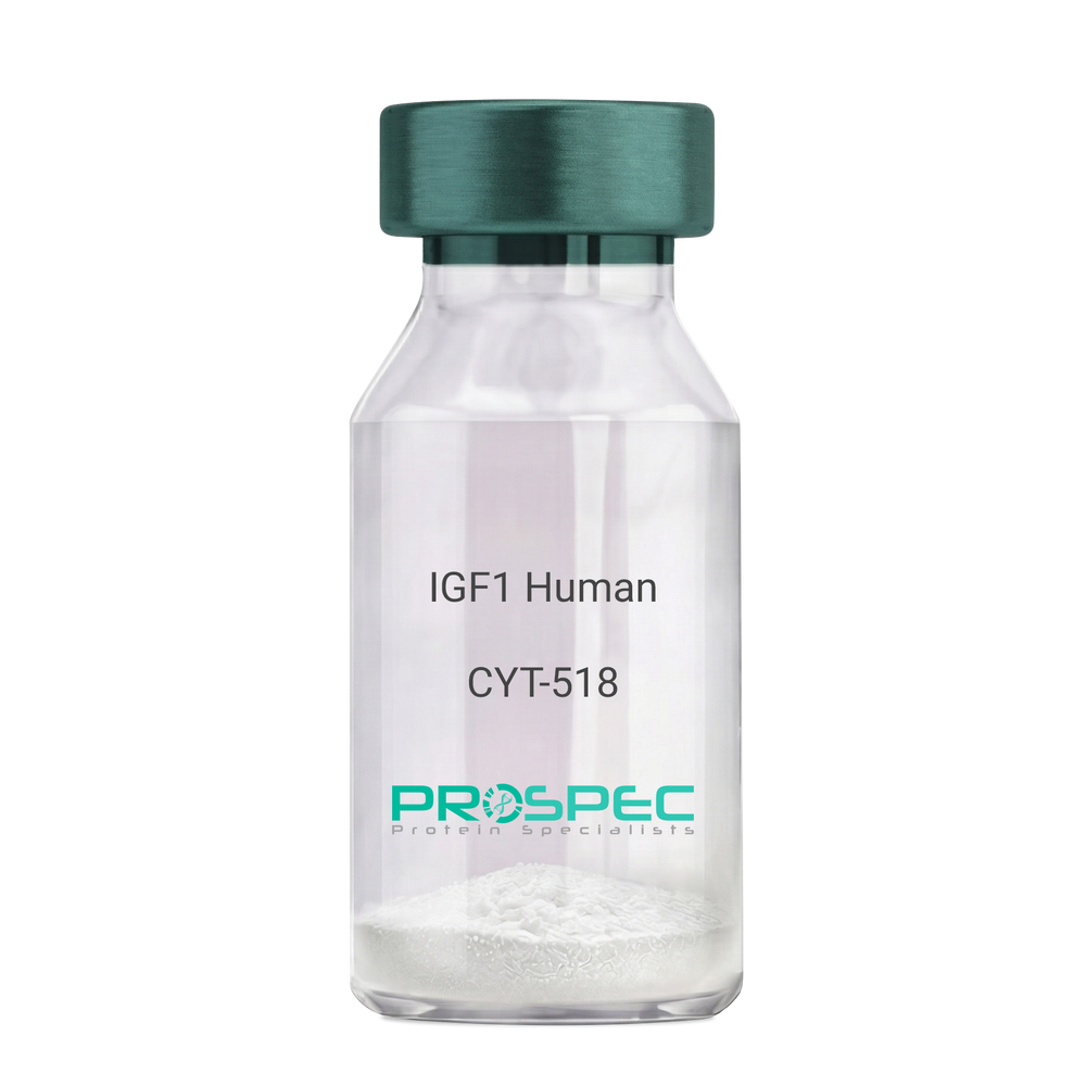
IGF1 HumanDescription:
IGF-1 Human Recombinant
Somatomedin C, IGF-I, IGFI, IGF1, IGF-IA MGF.
Product # :
CYT-216Price :
Quantity :
Shipping Method :
Shipped at Room temp
More Info
- description
- source
- formulation
- purity
- biological activity
- More Info
Description
IGF1 Human Recombinant produced in E.Coli is a single, non-glycosylated, polypeptide chain containing 70 amino acids and having a molecular mass of 7.6kDa.IGF-1 is purified by proprietary chromatographic techniques.
Source
Escherichia Coli.
Formulation
The protein was lyophilized from a concentrated solution in PBS, pH 7.0.
Purity
Greater than 97.0% as determined by:
(a) Analysis by RP-HPLC.
(b) Analysis by SDS-PAGE.Biological Activity
The biological activity was determined by the cell proliferation assay using serum free human MCF-7 cells in <2ng/ml, corresponding to a Specific Activity of >5.0 x 10⁵IU/mg.
More Info
-
Introduction
The somatomedins, IGFs, comprise a family of peptides that play important roles in mammalian growth and development. IGF1 mediates many of the growth-promoting effects of GH. Early studies showed that GH did not directly stimulate the incorporation of sulfate into cartilage, but rather acted through a serum factor, termed 'sulfation factor,' which later became known as 'somatomedin. Three main somatomedins have been characterized: somatomedin C (IGF1), somatomedin A (IGF2; MIM 147470), and somatomedin B.
-
Synonyms
Somatomedin C, IGF-I, IGFI, IGF1, IGF-IA MGF.
-
Physical Appearance
Sterile Filtered White lyophilized (freeze-dried) powder.
-
Stability
Lyophilized IGF1 although stable at room temperature for 3 weeks, should be stored desiccated below -18°C. Upon reconstitution IGF1 should be stored at 4°C between 2-7 days and for future use below -18°C.For long term storage it is recommended to add a carrier protein (0.1% HSA or BSA).Please prevent freeze-thaw cycles.
-
Solubility
It is recommended to reconstitute the lyophilized IGF-1 in sterile 18MΩ-cm H2O not less than 100µg/ml, which can then be further diluted to other aqueous solutions.
-
Amino Acid Sequence
GPETLCGAEL VDALQFVCGD RGFYFNKPTG YGSSSRRAPQ TGIVDECCFR SCDLRRLEMY CAPLKPAKSA.
-
Background
IGF stands for INS like growth factors, which are proteins that have a high similarity to INS. They are part of a complicated process that uses cells to communication with the physiologic environment around them. IGF’s complex system is often called the ‘axis’. This consists of:
● Two cell-surface receptors (IGF1R and IGF2R)
● Two ligands (IGF-1 and IGF-2)
● A family of six high-affinity IGF-binding proteins.
● Proteases.The Effect IGF Has On The Body
Many unique tissue types express the IGF-1 receptor, and the effects can vary. It induces the survival of neurons, may catalyse skeletal muscle hypertrophy by inducing protein synthesis, and by blocking muscle atrophy. It works as a protector for cartilage cells, and may work to be an anabolic factor for the bones. When used in high concentrations, it can activate the INS receptor, and can even complement the effects that INS has on the body.Diseases And IGF
Diseases and IGF are closely related, and a number of them can be affected. The INS IGF axis is thought to have an effect on aging, with an increased life span shown in fruit flies when used in studies.
It is also important to note the crucial role that IGF plays in cancer and diabetes - IGF- 1 has been shown to stimulate growth in both prostate and breast cancer cells. The degree of risk that IGF-1 poses is up for debate, and many scientists are not in agreement. IGF has also shown to have the ability to decrease blood glucose levels, although not quite as effective as INS.How Was IGF Discovered?
Investigators began studying the effects of biological substances on cells and tissues outside the body when IGFs were discovered. The name is self explanatory in the fact that IGF performs INS actions in some tissues, but is less potent than INS at decreasing blood sugar. Its fundamental action is to stimulate growth, whether that be within the epidermal growth factor or the nerve growth factor.What’s The Difference Between IGF-1 And IGF-2?
The two types of IGF are IGF-1 and IGF-2. Although the names are similar, the specific actions that they take are different - they bind and activate completely difference receptors. The major actor in them both is the effect that they have on cell growth. Most of the actions of the pituitary GH are mediated by IGFs, but predominantly IGF-1. GH works to stimulate many tissues within the body, especially the liver which then secretes IGF-1. This then causes hypertrophy, or in layman's terms, an increase in cell size, as well as hyperplasia which is an increase in the number of cells.
The IGF-1 concentration will increase during childhood and hit peak during puberty, but will then decrease afterwards, as does the hormone secretion itself. It has been proven that children and adults with a deficiency of the GH have low serum IGF-1 concentrations when put in comparison with others in the same age range. Patients who have conditions like acromegaly have been shown to have increased serum IGF-1 concentrations. The production of IGF-2 is less dependent on the secretion of GH than IGF-1, and is much less important for stimulating linear growth.
ProSpec's products are furnished for LABORATORY RESEARCH USE ONLY. The product may not be used as drugs, agricultural or pesticidal products, food additives or household chemicals.
- View Data Sheet
Name :
ADI1 AntibodyDescription:
Acireductone Dioxygenase 1, Mouse Anti Human
APL1, ARD, FLJ10913, HMFT1638, MTCBP-1, SIPL, 1,2-dihydroxy-3-keto-5-methylthiopentene dioxygenase, Acireductone dioxygenase, Fe-ARD, Membrane-type 1 matrix metalloproteinase cytoplasmic tail-binding protein 1, Submergence-induced protein-like factor.
Product # :
ANT-466Price :
Quantity :
Shipping Method :
Shipped with Ice Packs
More Info
- formulation
- More Info
Formulation
1mg/ml containing PBS, pH-7.4, 10% Glycerol and 0.02% Sodium Azide.
More Info
-
Introduction
Acireductone dioxygenase 1 (ADI1) is a part of the acireductone dioxygenase family of metal-binding enzymes, which are involved in methionine salvage. ADI1 regulates mRNA processing in the nucleus, and carries out different functions depending on its localization. Related pseudogenes have been defined on chromosomes 8 and 20. ADI1 down-regulates cell migration arbitrated by MMP14.
-
Synonyms
APL1, ARD, FLJ10913, HMFT1638, MTCBP-1, SIPL, 1,2-dihydroxy-3-keto-5-methylthiopentene dioxygenase, Acireductone dioxygenase, Fe-ARD, Membrane-type 1 matrix metalloproteinase cytoplasmic tail-binding protein 1, Submergence-induced protein-like factor.
-
Physical Appearance
Sterile filtered colorless solution.
-
Immunogen
Anti-human ADI1 mAb, clone PAT27E8A, is derived from hybridization of mouse F0 myeloma cells with spleen cells from BALB/c mice immunized with a recombinant human ADI1 protein 1-179 amino acids purified from E. coli.
-
Ig Subclass
Mouse IgG1 heavy chain and Kappa light chain.
-
Clone
PAT27E8A.
-
Applications
The antibody has been tested by ELISA, Western blot analysis to assure specificity and reactivity. Since application varies, however, each investigation should be titrated by the reagent to obtain optimal results. Recommended dilution range for Western blot analysis is 1:500 ~ 1:5000. Recommended starting dilution is 1:1000.
-
Type
Mouse Anti Human Monoclonal.
-
Storage Procedures
For periods up to 1 month store at 4°C, for longer periods of time, store at -20°C. Prevent freeze thaw cycles.
ProSpec's products are furnished for LABORATORY RESEARCH USE ONLY. The product may not be used as drugs, agricultural or pesticidal products, food additives or household chemicals.
- View Data Sheet
Name :
GH Antagonist ChickenDescription:
Growth Hormone Antagonist Chicken Recombinant
GH1, GH, GHN, GH-N, hGH-N, Pituitary growth hormone, Growth hormone 1, Somatotropin.
Product # :
CYT-538Price :
Quantity :
Shipping Method :
Shipped at Room temp
More Info
- description
- source
- formulation
- purity
- biological activity
- More Info
Description
Somatotropin Chicken Antagonist Recombinant mutein G119R produced in E.Coli is a single, non-glycosylated polypeptide chain containing 191 amino acids with an additional Ala at the N-terminus and having a molecular mass of 22.3 kDa. The Chicken Growth-Hormone Antagonist Recombinant is purified by proprietary chromatographic techniques.
Source
Escherichia Coli.
Formulation
The protein was lyophilized from a concentrated (1mg/ml) solution with 0.03% NaHCO3 pH-8.
Purity
Greater than 98.0% as determined by:
(a) Analysis by SEC-HPLC.
(b) Analysis by SDS-PAGE.Biological Activity
Recombinant Chicken Growth Hormone G119R mutant did not bind to ovine GHR-ECD and was devoid of any biological activity in FDC-P1 3B9 cells.
However, in binding experiments that were carried out using chicken liver membranes, both ovine GH and chicken GH showed similar IC50 values in competition with 125I-ovine GH, while the IC50 of G119R mutein was 10-fold higher.
These results emphasize the importance of species specificity and indicate the possibility of antagonistic activity of chGH G119R in homologous system.More Info
-
Introduction
GH is a member of the somatotropin/prolactin family of hormones which play an important role in growth control. The gene, along with four other related genes, is located at the growth hormone locus on chromosome 17 where they are interspersed in the same transcriptional orientation; an arrangement which is thought to have evolved by a series of gene duplications. The five genes share a remarkably high degree of sequence identity. Alternative splicing generates additional isoforms of each of the five growth hormones, leading to further diversity and potential for specialization. This particular family member is expressed in the pituitary but not in placental tissue as is the case for the other four genes in the growth hormone locus. Mutations in or deletions of the gene lead to growth hormone deficiency and short stature.
-
Synonyms
GH1, GH, GHN, GH-N, hGH-N, Pituitary growth hormone, Growth hormone 1, Somatotropin.
-
Physical Appearance
Sterile Filtered White lyophilized (freeze-dried) powder.
-
Stability
Lyophilized Growth-Hormone Chicken antagonist although stable at room temperature for at least two weeks, should be stored desiccated below -18°C. Upon reconstitution and filter sterilization GH can be stored at 4°C, pH 9 for up to 4 weeks. For long term storage and more diluted solutions it is recommended to add a carrier protein (0.1% HSA or BSA). Please prevent freeze-thaw cycles.
-
Solubility
It is recommended to reconstitute the lyophilized Growth-Hormone Chicken antagonist in 0.4% NaHCO3 or water adjusted to pH 8-9, not less than 100µg/ml, which can then be further diluted to other aqueous solutions, preferably in a presence of a carrier protein such as BSA or similar.
-
Amino Acid Sequence
The sequence of the first five N-terminal amino acids was determined and was found to be Ala-Thr-Phe-Pro-Ala.
-
Protein content
Protein quantitation was carried out by: UV spectroscopy at 280 nm using the absorbency value of 0.74 as the extinction coefficient for a 0.1% (1mg/ml) solution. This value is calculated by the PC GENE computer analysis program of protein sequences (IntelliGenetics).
ProSpec's products are furnished for LABORATORY RESEARCH USE ONLY. The product may not be used as drugs, agricultural or pesticidal products, food additives or household chemicals.
- View Data Sheet
Name :
TDP2 HumanDescription:
Tyrosyl-DNA Phosphodiesterase 2 Human Recombinant
AD022, dJ30M3.3, EAP2, EAPII, RP1-30M3.3, TTRAP, 5'-tyrosyl-DNA phosphodiesterase,hTDP2, ETS1-associated protein 2, ETS1-associated protein II.
Product # :
ENZ-698Price :
Quantity :
Shipping Method :
Shipped with Ice Packs
More Info
- description
- source
- formulation
- purity
- More Info
Description
TDP2 Human Recombinant produced in E.coli is a single, non-glycosylated polypeptide chain containing 385 amino acids (1-362) and having a molecular mass of 43.3kDa. TDP2 is fused to a 23 amino acid His-tag at N-terminus & purified by proprietary chromatographic techniques.
Source
Escherichia Coli.
Formulation
The TDP2 solution (0.5mg/ml) contains 20mM Tris-HCl buffer, (pH 8.0), 0.15M NaCl, 10% glycerol and 1mM DTT.
Purity
Greater than 85.0% as determined by SDS-PAGE.
More Info
-
Introduction
Tyrosyl-DNA Phosphodiesterase 2 (TDP2), belongs to the CCR4/nocturin family of divalent cation-dependent phosphodiesterases.TDP2 associates with CD40, tumor necrosis factor (TNF) receptor-75 and TNF receptor associated factors (TRAFs), and inhibits nuclear factor-kappa-B activation. TDP2 is characterized by similar sequence and structure as APE1 endonuclease, which participates in DNA repair and the activation of transcription factors.
-
Synonyms
AD022, dJ30M3.3, EAP2, EAPII, RP1-30M3.3, TTRAP, 5'-tyrosyl-DNA phosphodiesterase,hTDP2, ETS1-associated protein 2, ETS1-associated protein II.
-
Physical Appearance
Sterile filtered colorless solution.
-
Stability
Store at 4°C if entire vial will be used within 2-4 weeks. Store, frozen at -20°C for longer periods of time. For long term storage it is recommended to add a carrier protein (0.1% HSA or BSA). Avoid multiple freeze-thaw cycles.
-
Amino Acid Sequence
MGSSHHHHHH SSGLVPRGSH MGSMELGSCL EGGREAAEEE GEPEVKKRRL LCVEFASVAS CDAAVAQCFL AENDWEMERA LNSYFEPPVE ESALERRPET ISEPKTYVDL TNEETTDSTT SKISPSEDTQ QENGSMFSLI TWNIDGLDLN NLSERARGVC SYLALYSPDV IFLQEVIPPY YSYLKKRSSN YEIITGHEEG YFTAIMLKKS RVKLKSQEII PFPSTKMMRN LLCVHVNVSG NELCLMTSHL ESTRGHAAER MNQLKMVLKK MQEAPESATV IFAGDTNLRD REVTRCGGLP NNIVDVWEFL GKPKHCQYTW DTQMNSNLGI TAACKLRFDR IFFRAAAEEG HIIPRSLDLL GLEKLDCGRF PSDHWGLLCN LDIIL.
ProSpec's products are furnished for LABORATORY RESEARCH USE ONLY. The product may not be used as drugs, agricultural or pesticidal products, food additives or household chemicals.
- View Data Sheet
Name :
Anthrax LF AntibodyDescription:
Anthrax LF Polyclonal Antibody
Product # :
ANT-181Price :
Quantity :
Shipping Method :
Shipped with Ice Packs
More Info
- formulation
- More Info
Formulation
100µgs in 200µl PBS containing 0.2% gelatin and 0.05% sodium azide.
More Info
-
Introduction
Anthrax PA (Protective Antigen) and Anthrax LF (Lethal Factor), along with Anthrax EF (Edema Factor), are toxins produced by Bacillus anthracis. All three of the proteins must be present for toxicity to occur. It begins with the binding of PA to its cell-surface receptor, ATR (Anthrax Toxin Receptor), at which point PA is cleaved and the resulting 63 kD carboxyl-terminal fragment binds to LF and EF. LF is a protease that is known to cleave members of the mitogen-activated protein kinase kinase (MAPKK) family(2).
-
Stability
Store at 4C, stable for 6 months. For long-term storage, store at -20C.
-
Immunogen
The antibody was developed by immunizing rabbits with synthetic peptides corresponding to amino acids 47-65 of the Anthrax LF protein (~90 kD).
-
Applications
This polyclonal antibody can be used for detection of Anthrax LF protein in western blot.
Not tested in other applications. -
Isotype
Rabbit Ig.
-
Type
Polyclonal Antibody.
ProSpec's products are furnished for LABORATORY RESEARCH USE ONLY. The product may not be used as drugs, agricultural or pesticidal products, food additives or household chemicals.
- View Data Sheet
Name :
Ecotin E.ColiDescription:
Ecotin E.Coli Recombinant
E. coli serine protease inhibitor.
Product # :
ENZ-058Price :
Quantity :
Shipping Method :
Shipped with Ice Packs
More Info
- description
- source
- formulation
- purity
- More Info
Description
Ecotin produced in E.Coli is a single, non-glycosylated polypeptide chain containing 163 amino acids (21-162a.a.) and having a molecular mass of 18.3kDa.Ecotin is fused to a 20 amino acid His-tag at N-terminus & purified by proprietary chromatographic techniques.
Source
Escherichia Coli.
Formulation
The Ecotin protein solution (1mg/ml) is formulated in 20mM Tris-HCl buffer (pH8.0), 1mM DTT, 50mM NaCl and 10% glycerol.
Purity
Greater than 95% as determined by SDS-PAGE.
More Info
-
Introduction
Ecotin inhibits pancreatic serine proteases. Ecotin protein inhibits chymotrypsin, trypsin, elastases, factor X, kallikrein as well as a variety of other proteases. The power of inhibition is not linked to a specific protease specificity.
-
Synonyms
E. coli serine protease inhibitor.
-
Physical Appearance
Sterile Filtered clear solution.
-
Stability
Store at 4°C if entire vial will be used within 2-4 weeks. Store, frozen at -20°C for longer periods of time.Please avoid freeze thaw cycles.
-
Amino Acid Sequence
MGSSHHHHHH SSGLVPRGSH MAESVQPLEK IAPYPQAEKG MKRQVIQLTP QEDESTLKVE LLIGQTLEVD CNLHRLGGKL ENKTLEGWGY DYYVFDKVSS PVSTMMACPD GKKEKKFVTA YLGDAGMLRY NSKLPIVVYT PDNVDVKYRV WKAEEKIDNA VVR
ProSpec's products are furnished for LABORATORY RESEARCH USE ONLY. The product may not be used as drugs, agricultural or pesticidal products, food additives or household chemicals.
- View Data Sheet
Name :
IGFBP7 HumanDescription:
Insulin-Like Growth Factor Binding Protein-7 Human Recombinant
Insulin-like growth factor-binding protein 7, IBP-7, IGF-binding protein 7, IGFBP-7, IGFBP-rP1, MAC25 protein, PGI2-stimulating factor, Prostacyclin-stimulating factor, Tumor-derived adhesion factor, TAF, IGFBP7, MAC25, PSF, AGM, FSTL2, RAMSVPS, IGFBP-7v, IGFBPRP1.
Product # :
CYT-788Price :
Quantity :
Shipping Method :
Shipped at Room temp
More Info
- description
- source
- formulation
- purity
- More Info
Description
Recombinant Human IGFBP7 produced in E.coli cells is a non-glycosylated, homodimeric protein containing 2x256 amino acid chains and having a molecular mass of 26.4kDa. The IGFBP-7 is purified by proprietary chromatographic techniques.
Source
Escherichia Coli.
Formulation
The IGFBP7 was lyophilized from a 0.2µm filtered concentrated solution in 20mM Tris-HCl, pH 8.5 and 150mM NaCl.
Purity
Greater than 97.0% as determined by:
(a) Analysis by RP-HPLC.
(b) Analysis by SDS-PAGE.More Info
-
Introduction
Insulin-like Growth Factor-Binding Protein 7 (IGFBP7) is a member of the IGFBP family. IGFBP family members are all cysteine rich proteins with conserved cysteine and have an IGFBP domain, a Kazal-like domain and an Ig-like C2-type domain. IGFBP7 is expressed in a broad range of normal human tissues and it mostly shows reduced expression in cancer cell lines of prostate, breast, colon, and lung origin. IGFBP7 has a role in skeletal myogenesis by binding to IGF in a manner which inhibits IGF induced differentiation of skeletal myoblasts, without disturbing IGF induced proliferation. Moreover, IGFBP7 suppresses growth and colony formation of prostate and breast cancer cell lines via an IGF independent mechanism, which triggers a delay in the G1 phase of the cell cycle, and increased apoptosis.
-
Synonyms
Insulin-like growth factor-binding protein 7, IBP-7, IGF-binding protein 7, IGFBP-7, IGFBP-rP1, MAC25 protein, PGI2-stimulating factor, Prostacyclin-stimulating factor, Tumor-derived adhesion factor, TAF, IGFBP7, MAC25, PSF, AGM, FSTL2, RAMSVPS, IGFBP-7v, IGFBPRP1.
-
Physical Appearance
Sterile Filtered White lyophilized (freeze-dried) powder.
-
Stability
Lyophilized IGFBP7 although stable at room temperature for 3 weeks, should be stored desiccated below -18°C. Upon reconstitution IGFBP-7 should be stored at 4°C between 2-7 days and for future use below -18°C. For long term storage it is recommended to add a carrier protein (0.1% HSA or BSA).Please prevent freeze-thaw cycles.
-
Solubility
It is recommended to reconstitute the lyophilized IGFBP-7 in sterile 20mM AcOH (acetic Acid) not less than 100µg/ml, which can then be further diluted to other aqueous solutions.
-
Amino Acid Sequence
SSSDTCGPCE PASCPPLPPL GCLLGETRDA CGCCPMCARG EGEPCGGGGA GRGYCAPGME CVKSRKRRKG KAGAAAGGPG VSGVCVCKSR YPVCGSDGTT YPSGCQLRAA SQRAESRGEK AITQVSKGTC EQGPSIVTPP KDIWNVTGAQ VYLSCEVIGI PTPVLIWNKV KRGHYGVQRT ELLPGDRDNL AIQTRGGPEK HEVTGWVLVS PLSKEDAGEY ECHASNSQGQ ASASAKITVV DALHEIPVKK GEGAEL.
ProSpec's products are furnished for LABORATORY RESEARCH USE ONLY. The product may not be used as drugs, agricultural or pesticidal products, food additives or household chemicals.
- View Data Sheet
Name :
BDNF HumanDescription:
Brain-Derived Neurotrophic Factor Human Recombinant
Brain-Derived Neurotrophic Factor, BDNF, MGC34632.
Product # :
CYT-207Price :
Quantity :
Shipping Method :
Shipped at Room temp
More Info
- description
- source
- formulation
- purity
- biological activity
- More Info
- Activity
Description
BDNF Human Recombinant produced in E.Coli is a homodimer, non-glycosylated, polypeptide chain containing 2 x 119 amino acids (and an N-terminal Met) and having a total molecular mass of 28kDa. BDNF Human Recombinant is purified by proprietary chromatographic techniques.
Source
Escherichia Coli.
Formulation
The protein was lyophilized with 20mM PB and 400mM NaCl, pH 7.2.
Purity
BDNF is greater than 950% as determined SDS-PAGE.
Biological Activity
The activity was determined using Immobilized Human TrkB-His tag protein 2ug/ml (100 μl/well) for its binding to NHS-Biotin BDNF. The ED50 of was found to be ≤20ng/mLActivity
More Info
-
Introduction
BDNF promotes the survival of neuronal populations that are all located either in the central nervous system or directly connected to it. BDNF is a major regulator of synaptic transmission and plasticity at adult synapses in many regions of the cns. The versatility of BDNF is emphasized by its contribution to a range of adaptive neuronal responses including long-term potentiation (ltp), long-term depression (ltd), certain forms of short-term synaptic plasticity, as well as homeostatic regulation of intrinsic neuronal excitability.
-
Synonyms
Brain-Derived Neurotrophic Factor, BDNF, MGC34632.
-
Physical Appearance
Sterile Filtered White lyophilized (freeze-dried) powder.
-
Stability
Lyophilized BDNF although stable at room temperature for 3 weeks, should be stored desiccated below -18°C. Upon reconstitution BDNF should be stored at 4°C between 2-7 days and for future use below -18°C.For long term storage it is recommended to add a carrier protein (0.1% HSA or BSA).Please prevent freeze-thaw cycles.
-
Solubility
It is recommended to reconstitute the lyophilized BDNF in sterile 18M-cm H2O not less than 100µg/ml, which can then be further diluted to other aqueous solutions.
-
Amino Acid Sequence
HSDPARRGE LSVCDSISEW VTAADKKTAV DMSGGTVTVL EKVPVSKGQL KQYFYETKCN PMGYTKEGCR GIDKRHWNSQ CRTTQSYVRA LTMDSKKRIG WRFIRIDTSC VCTLTIKRGR.
-
Background
Final Thoughts
Although more research is needed on the safety and effectiveness of BDNF human recombinant, trials suggest that this laboratory-produced protein may be effective in managing and treating several neurological and psychiatric disorders. It's important for experts to stay up to date on the latest developments and research to learn more about potential risks and benefits.
What is the molecular weight/Mw of BDNF Protein?
BDNF Protein has a total Mw of 27kDa.
What is the source or expression system of BDNF Protein?
Escherichia Coli.
What is the Purity of BDNF Protein?
BDNF Protein is >97% pure as determined by SDS-PAGE.
What is the Biological Activity of BDNF Protein?
The ED50, as determined by the dose-dependent induction of C6 cells proliferation, is 1.3-2µg/ml.
What is the amino acid sequence of BDNF Protein?
MHSDPARRGE LSVCDSISEW VTAADKKTAV DMSGGTVTVL EKVPVSKGQL KQYFYETKCN PMGYTKEGCR GIDKRHWNSQ CRTTQSYVRA LTMDSKKRIG WRFIRIDTSC VCTLTIKRGR.
What applications can BDNF Protein be used in?
BDNF Protein can probably be used in western blot, ELISA and Lateral Flow.
What is the endotoxin level for BDNF Protein?
The endotoxin level is minimal, BDNF Protein was purified using conventional chromatography techniques.
-
Protein content
BDNF quantitation was carried out by two independent methods:1. UV spectroscopy at 280 nm using the absorbency value of 1.6 as the extinction coefficient for a 0.1% (1mg/ml) solution. This value is calculated by the PC GENE computer analysis program of protein sequences (IntelliGenetics). 2. Analysis by RP-HPLC, using a standard solution of Brain-derived Neurotrophic Factor as a Reference Standard.
ProSpec's products are furnished for LABORATORY RESEARCH USE ONLY. The product may not be used as drugs, agricultural or pesticidal products, food additives or household chemicals.
- View Data Sheet
Name :
TBCB AntibodyDescription:
Tubulin Folding Cofactor B, Mouse Anti Human
Tubulin folding cofactor B, Cytoskeleton-associated protein CKAPI, cytoskeleton associated protein 1, CKAP1, CG22, Tubulin-specific chaperone B.
Product # :
ANT-526Price :
Quantity :
Shipping Method :
Shipped with Ice Packs
More Info
- formulation
- More Info
Formulation
1mg/ml containing PBS, pH-7.4, 10% Glycerol and 0.02% Sodium Azide.
More Info
-
Introduction
TBCB is a member of the Microtubules family. Biosynthesis of functional microtubules include the involvement of a number of chaperones, termed Tubulin folding cofactors A (TBCA), B (TBCB), C (TBCC), D (TBCD) and E (TBCE), which operate on folding intermediates downstream of the cytosolic chaperon, known as TCP. The 244 amino acid cytoplasmic protein TBCB has one CAP-Gly domain and is universally expressed. TBCB takes part in the regulation of tubulin heterodimer dissociation and operates as a negative regulator of axonal growth.
-
Synonyms
Tubulin folding cofactor B, Cytoskeleton-associated protein CKAPI, cytoskeleton associated protein 1, CKAP1, CG22, Tubulin-specific chaperone B.
-
Physical Appearance
Sterile filtered colorless solution.
-
Immunogen
Anti-human TBCB mAb, clone PAT1F6AT, is derived from hybridization of mouse F0 myeloma cells with spleen cells from BALB/c mice immunized with a recombinant human TBCB protein 1-244 amino acids purified from E. coli.
-
Ig Subclass
Mouse IgG2b heavy chain and k light chain.
-
Clone
PAT1F6AT.
-
Applications
The antibody has been tested by ELISA, Western blot analysis to assure specificity and reactivity. Since application varies, however, each investigation should be titrated by the reagent to obtain optimal results. Recommended starting dilution is 1:1500.
-
Type
Mouse Anti Human Monoclonal.
-
Storage Procedures
For periods up to 1 month store at 4°C, for longer periods of time, store at -20°C. Prevent freeze thaw cycles.
-
Purification Method
TBCB antibody was purified from mouse ascitic fluids by protein-A affinity chromatography.
ProSpec's products are furnished for LABORATORY RESEARCH USE ONLY. The product may not be used as drugs, agricultural or pesticidal products, food additives or household chemicals.
- View Data Sheet
Name :
TMEFF2 AntibodyDescription:
Tomoregulin-2, Mouse Anti Human
TMEFF2, Tomoregulin-2, Transmembrane protein with EGF-like and two follistatin-like domains, Hyperplastic polyposis protein 1, HPP1, TENB2, TPEF, TR, CT120.2.
Product # :
ANT-428Price :
Quantity :
Shipping Method :
Shipped with Ice Packs
More Info
- formulation
- More Info
Formulation
1mg/ml containing PBS, pH-7.4, & 0.1% Sodium Azide.
More Info
-
Introduction
TMEFF2 is a gene encoding a plasma membrane protein with two follistatin-like domains and one epidermal growth factor–like domain. TMEFF2 has limited normal tissue distribution and is highly overexpressed in prostate cancer. The shedded form of TMEFF2 up-regulates cancer cell proliferation, most likely by promoting ERK1/2 phosphorylation. TMEFF2 is down-regulated in tumor cell lines in response to a high level of methylation in the 5'' region.
-
Synonyms
TMEFF2, Tomoregulin-2, Transmembrane protein with EGF-like and two follistatin-like domains, Hyperplastic polyposis protein 1, HPP1, TENB2, TPEF, TR, CT120.2.
-
Physical Appearance
Sterile filtered colorless solution.
-
Immunogen
Anti-human TMEFF2 mAb, is derived from hybridization of mouse F0 myeloma cells with spleen cells from BALB/c mice immunized with recombinant human TMEFF2 amino acids 41-150 purified from E. coli.
-
Ig Subclass
Mouse IgG2b heavy chain and κ light chain.
-
Clone
PJ4B6AT.
-
Applications
TMEFF2 antibody has been tested by ELISA, Western blot and immunohistochemistry analysis to assure specificity and reactivity. Since application varies, however, each investigation should be titrated by the reagent to obtain optimal results. Recommended dilution range for Western blot is 1:1,000~1:2,000 and immunohistochemistry analysis is 1:50~100. Recommended starting dilution for Western blot is 1:1,000 and Immunohistochemistry is 1:50.
-
Type
Mouse Anti Human Monoclonal.
-
Storage Procedures
For periods up to 1 month store at 4°C, for longer periods of time, store at -20°C. Prevent freeze thaw cycles.
-
Purification Method
TMEFF2 antibody was purified from mouse ascitic fluids by protein-G affinity chromatography.
ProSpec's products are furnished for LABORATORY RESEARCH USE ONLY. The product may not be used as drugs, agricultural or pesticidal products, food additives or household chemicals.
- View Data Sheet
Name :
SULT2B1 HumanDescription:
Sulfotransferase Family, Cytosolic, 2B, Member 1 Human Recombinant
SULT2B1, HSST2, EC 2.8.2.2, Sulfotransferase 2B1, Hydroxysteroid sulfotransferase 2, ST2B1, Sulfotransferase family cytosolic 2B member 1.
Product # :
ENZ-512Price :
Quantity :
Shipping Method :
Shipped with Ice Packs
More Info
- description
- source
- formulation
- purity
- More Info
Description
SULT2B1 Human Recombinant produced in E.Coli is a single, non-glycosylated, polypeptide chain containing 365 amino acids (1-365 a.a.) and having a molecular mass of 41.3 kDa. SULT2B1 protein is purified by standard chromatography.
Source
Escherichia Coli.
Formulation
SULT2B1 Human solution containing 20mM Tris-HCl pH-7.5, & 10% glycerol.
Purity
Greater than 85% as determined by SDS-PAGE.
More Info
-
Introduction
SULT2B1 catalyzes the sulfate conjugation of numerous hormones, neurotransmitters, drugs and xenobiotic compounds. Sulfonation enhances the water solubility of molecules, and therefore their renal excretion, however it can also result in bioactivation to form active metabolites. SULT2B1b is localized in the cytosol and nuclei of human cells. SULT2B1b is selective for the sulfation of 3beta-hydroxysteroids such as dehydroepiandrosterone and pregnenolone, and participates in cholesterol sulfation in human skin.
-
Synonyms
SULT2B1, HSST2, EC 2.8.2.2, Sulfotransferase 2B1, Hydroxysteroid sulfotransferase 2, ST2B1, Sulfotransferase family cytosolic 2B member 1.
-
Physical Appearance
Sterile filtered colorless solution.
-
Stability
Store at 4°C if entire vial will be used within 2-4 weeks. Store, frozen at -20°C for longer periods of time. For long term storage it is recommended to add a carrier protein (0.1% HSA or BSA). Avoid multiple freeze-thaw cycles.
-
Amino Acid Sequence
MDGPAEPQIP GLWDTYEDDI SEISQKLPGE YFRYKGVPFP VGLYSLESIS LAENTQDVRD DDIFIITYPK SGTTWMIEII CLILKEGDPS WIRSVPIWER APWCETIVGA FSLPDQYSPR LMSSHLPIQI FTKAFFSSKA KVIYMGRNPR DVVVSLYHYS KIAGQLKDPG TPDQFLRDFL KGEVQFGSWF DHIKGWLRMK GKDNFLFITY EELQQDLQGS VERICGFLGR PLGKEALGSV VAHSTFSAMK ANTMSNYTLL PPSLLDHRRG AFLRKGVCGD WKNHFTVAQS EAFDRAYRKQ MRGMPTFPWD EDPEEDGSPD PEPSPEPEPK PSLEPNTSLE REPRPNSSPS PSPGQASETP HPRPS.
ProSpec's products are furnished for LABORATORY RESEARCH USE ONLY. The product may not be used as drugs, agricultural or pesticidal products, food additives or household chemicals.
- View Data Sheet
Name :
C1QTNF6 HumanDescription:
Complement C1q Tumor Necrosis Factor-Related Protein 6 Human Recombinant
CTRP6, C1QTNF6, ZACRP6, Complement C1q tumor necrosis factor-related protein 6.
Product # :
PRO-655Price :
Quantity :
Shipping Method :
Shipped with Ice Packs
More Info
- description
- source
- formulation
- purity
- More Info
Description
C1QTNF6 Human Recombinant produced in E.Coli is a single, non-glycosylated, Polypeptide chain containing 242 amino acids and having a molecular mass of 27 kDa. The protein contains an extra His tag at N-terminus. The C1QTNF6 amino acid sequence is identical to UniProtKB/Swiss-Prot entry Q9BXI9 amino acids 29–259.The C1QTNF6 is purified by proprietary chromatographic techniques.
Source
Escherichia Coli.
Formulation
0.23mg/ml in 30mM Acetate Buffer pH-4.
Purity
Greater than 95% as determined by SDS PAGE.
More Info
-
Introduction
C1QTNF6 is a collagen type-IV specific for basement membranes. C1QTNF6 is vital for embryonic development.
-
Synonyms
CTRP6, C1QTNF6, ZACRP6, Complement C1q tumor necrosis factor-related protein 6.
-
Physical Appearance
Sterile Filtered clear solution.
-
Stability
Store at 4°C if entire vial will be used within 2-4 weeks. Store, frozen at -20°C for longer periods of time. Avoid multiple freeze-thaw cycles.
-
Amino Acid Sequence
MKHHHHHHAS TFDRAVASGC QRCCDSEDPL DPAHVSSASS SGRPHALPEI RPYINITILK GDKGDPGPMG LPGYMGREGP QGEPGPQGSK GDKGEMGSPG APCQKRFFAF SVGRKTALHS GEDFQTLLFE RVFVNLDGCF DMATGQFAAP LRGIYFFSLN VHSWNYKETY VHIMHNQKEA VILYAQPSER SIMQSQSVML DLAYGDRVWV RLFKRQRENA IYSNDFDTYI TFSGHLIKAEDD.
ProSpec's products are furnished for LABORATORY RESEARCH USE ONLY. The product may not be used as drugs, agricultural or pesticidal products, food additives or household chemicals.
- View Data Sheet
Name :
NCF4 HumanDescription:
Neutrophil Cytosolic Factor 4 Human Recombinant
Neutrophil cytosol factor 4, NCF-4, Neutrophil NADPH oxidase factor 4, SH3 and PX domain-containing protein 4, p40-phox, p40phox, NCF4, SH3PXD4, NCF, SH3PXD4.
Product # :
PRO-1258Price :
Quantity :
Shipping Method :
Shipped with Ice Packs
More Info
- description
- source
- formulation
- purity
- More Info
Description
NCF4 Human Recombinant produced in E.Coli is a single, non-glycosylated polypeptide chain containing 359 amino acids (1-339 a.a) and having a molecular mass of 41.1kDa.NCF4 is fused to a 20 amino acid His-tag at N-terminus & purified by proprietary chromatographic techniques.
Source
Escherichia Coli.
Formulation
NCF4 protein solution (1mg/ml) containing 20mM Tris-HCl buffer (pH 8.0), 0.1M NaCl, 10% glycerol and 1mM DTT.
Purity
Greater than 90.0% as determined by SDS-PAGE.
More Info
-
Introduction
Neutrophil Cytosolic Factor 4 (NCF4) is a cytosolic regulatory factor of the superoxide-producing phagocyte NADPH-oxidase, which is a multicomponent enzyme system imperative for host defense. The NCF4 protein is preferentially expressed in cells of myeloid lineage. NCF4 interacts mainly with neutrophil cytosolic factor 2 (NCF2/p67-phox) to create a complex with neutrophil cytosolic factor (NCF1/p47-phox), which further interacts with the small G protein RAC1 and translocates to the membrane upon cell stimulation. This complex subsequently activates flavocytochrome b, the membrane-integratedcatalytic core of the enzyme system. The PX domain of the NCF4 protein can bind phospholipid products of the PI(3) kinase, suggesting its part in PI(3) kinase-mediated signaling events. The phosphorylation of the NCF4 protein negatively regulates the enzyme activity.
-
Synonyms
Neutrophil cytosol factor 4, NCF-4, Neutrophil NADPH oxidase factor 4, SH3 and PX domain-containing protein 4, p40-phox, p40phox, NCF4, SH3PXD4, NCF, SH3PXD4.
-
Physical Appearance
Sterile Filtered colorless solution.
-
Stability
Store at 4°C if entire vial will be used within 2-4 weeks. Store, frozen at -20°C for longer periods of time. For long term storage it is recommended to add a carrier protein (0.1% HSA or BSA).Avoid multiple freeze-thaw cycles.
-
Amino Acid Sequence
MGSSHHHHHH SSGLVPRGSH MAVAQQLRAE SDFEQLPDDV AISANIADIE EKRGFTSHFV FVIEVKTKGG SKYLIYRRYR QFHALQSKLE ERFGPDSKSS ALACTLPTLP AKVYVGVKQE IAEMRIPALN AYMKSLLSLP VWVLMDEDVR IFFYQSPYDS EQVPQALRRL RPRTRKVKSV SPQGNSVDRM AAPRAEALFD FTGNSKLELN FKAGDVIFLL SRINKDWLEG TVRGATGIFP LSFVKILKDF PEEDDPTNWL RCYYYEDTIS TIKDIAVEED LSSTPLLKDL LELTRREFQR EDIALNYRDA EGDLVRLLSD EDVALMVRQA RGLPSQKRLF PWKLHITQKD NYRVYNTMP.
ProSpec's products are furnished for LABORATORY RESEARCH USE ONLY. The product may not be used as drugs, agricultural or pesticidal products, food additives or household chemicals.
- View Data Sheet
Name :
ARF6 HumanDescription:
ADP-Ribosylation Factor 6 Human Recombinant
ADP-ribosylation factor 6, ARF6, DKFZp564M0264.
Product # :
PRO-032Price :
Quantity :
Shipping Method :
Shipped with Ice Packs
More Info
- description
- source
- formulation
- purity
- More Info
Description
ARF6 produced in E.Coli is a single, non-glycosylated polypeptide chain containing 195 amino acids (1-175a.a.) and having a molecular mass of 22.2kDa.ARF6 is fused to a 20 amino acid His-tag at N-terminus & purified by proprietary chromatographic techniques.
Source
Escherichia Coli.
Formulation
The ARF6 protein solution (1mg/1ml) is formulated in 20mM Tris-HCl buffer (pH 8.0), 2mM DTT, 0.2mM PMSF and 20% glycerol.
Purity
Greater than 95% as determined by SDS-PAGE.
More Info
-
Introduction
ARF6 is a part of the ADP ribosylation factor family of GTP-binding proteins. ARF6 is restricted to the plasma membrane, and controls vesicular trafficking, remodeling of membrane lipids, and signaling pathways which lead to actin remodeling. Furthermore, ARF6 is a key player in conservation of organelle integrity, assembly of coat proteins and activation of phospholipase D.
-
Synonyms
ADP-ribosylation factor 6, ARF6, DKFZp564M0264.
-
Physical Appearance
Sterile Filtered clear solution.
-
Stability
Store at 4°C if entire vial will be used within 2-4 weeks. Store, frozen at -20°C for longer periods of time.Please avoid freeze thaw cycles.
-
Amino Acid Sequence
MGSSHHHHHH SSGLVPRGSH MGKVLSKIFG NKEMRILMLG LDAAGKTTIL YKLKLGQSVT TIPTVGFNVE TVTYKNVKFN VWDVGGQDKI RPLWRHYYTG TQGLIFVVDC ADRDRIDEAR QELHRIINDR EMRDAIILIF ANKQDLPDAM KPHEIQEKLG LTRIRDRNWY VQPSCATSGD GLYEGLTWLT SNYKS
ProSpec's products are furnished for LABORATORY RESEARCH USE ONLY. The product may not be used as drugs, agricultural or pesticidal products, food additives or household chemicals.
- View Data Sheet
Name :
ABHD10 HumanDescription:
Abhydrolase Domain Containing 10 Human Recombinant
Abhydrolase domain containing 10 mitochondrial2, FLJ11342, EC 3.4.-.-.
Product # :
ENZ-612Price :
Quantity :
Shipping Method :
Shipped with Ice Packs
More Info
- description
- source
- formulation
- purity
- More Info
Description
ABHD10 Human Recombinant produced in E. coli is a single polypeptide chain containing 279 amino acids (53-306) and having a molecular mass of 30.9kDa.ABHD10 is fused to a 25 amino acid His-tag at N-terminus & purified by proprietary chromatographic techniques.
Source
E.coli.
Formulation
The ABHD10 solution (1mg/1ml) contains 20mM Tris-HCl buffer (pH 8.0), 1mM DTT, 100mM NaCl and 20% glycerol.
Purity
Greater than 85% as determined by SDS-PAGE.
More Info
-
Introduction
Abhydrolase domain-containing protein 10 (ABHD10) is a member of the AB hydrolase superfamily.
-
Synonyms
Abhydrolase domain containing 10 mitochondrial2, FLJ11342, EC 3.4.-.-.
-
Physical Appearance
Sterile Filtered colorless solution.
-
Stability
Store at 4°C if entire vial will be used within 2-4 weeks. Store, frozen at -20°C for longer periods of time. For long term storage it is recommended to add a carrier protein (0.1% HSA or BSA).Avoid multiple freeze-thaw cycles.
-
Amino Acid Sequence
MGSSHHHHHH SSGLVPRGSH MGSHMTSLSF LNRPDLPNLA YKKLKGKSPG IIFIPGYLSY MNGTKALAIE EFCKSLGHAC IRFDYSGVGS SDGNSEESTL GKWRKDVLSI IDDLADGPQI LVGSSLGGWL MLHAAIARPE KVVALIGVAT AADTLVTKFN QLPVELKKEV EMKGVWSMPS KYSEEGVYNV QYSFIKEAEH HCLLHSPIPV NCPIRLLHGM KDDIVPWHTS MQVADRVLST DVDVILRKHS DHRMREKADI QLLVYTIDDL IDKLSTIVN
ProSpec's products are furnished for LABORATORY RESEARCH USE ONLY. The product may not be used as drugs, agricultural or pesticidal products, food additives or household chemicals.
- View Data Sheet
Name :
TNFSF14 HumanDescription:
LIGHT Human Recombinant
Tumor necrosis factor ligand superfamily member 14, CD258, Tnfsf14, Light
Product # :
CYT-1202Price :
Quantity :
Shipping Method :
Shipped with Ice Packs
More Info
- description
- source
- formulation
- purity
- More Info
Description
Recombinant Human LIGHT (74-240 aa) having a Mw of 23kDa was purified from E. coli.The Recombinant Human LIGHT is purified by proprietary chromatographic technique.
Source
Escherichia Coli.
Formulation
TNFSF14 solution contains PBS & 25mM K2CO3.
Purity
Protein is >95% pure as determined by 10% PAGE (coomassie staining).
More Info
-
Introduction
TNFRSF14, a member of the TNF receptor superfamily, is a type I transmembrane protein. TNFRSF14 is expressed in peripheral blood T cells, B cells, monocytes and in various tissues enriched in lymphoid cells. TNFRSF14 operates as a co-stimulatory factor for the activation of lymphoid cells and as a deterrent to infection by herpesvirus. Additionally, TNFRSF14 encourages the proliferation of T cells, and triggers apoptosis of various tumor cells.
-
Synonyms
Tumor necrosis factor ligand superfamily member 14, CD258, Tnfsf14, Light
-
Physical Appearance
Sterile Filtered clear solution.
-
Stability
Store at 4°C if entire vial will be used within 2-4 weeks.Store, frozen at -20°C for longer periods of time.For long term storage it is recommended to add a carrier protein (0.1% HSA or BSA).Avoid multiple freeze-thaw cycles.
-
Amino Acid Sequence
DGPAGSWEQL IQERRSHEVN PAAHLTGANS SLTGSGGPLL WETQLGLAFL RGLSYHDGAL VVTKAGYYYI YSKVQLGGVG CPLGLASTIT HGLYKRTPRY PEELELLVSQ QSPCGRATSS SRVWWDSSFL GGVVHLEAGE KVVVRVLDER LVRLRDGTRS YFGAFMV
ProSpec's products are furnished for LABORATORY RESEARCH USE ONLY. The product may not be used as drugs, agricultural or pesticidal products, food additives or household chemicals.
- View Data Sheet
Name :
CTF1 MouseDescription:
Cardiotrophin-1 Mouse Recombinant
CTF1, CT1, CT-1, Cardiophin 1.
Product # :
CYT-151Price :
Quantity :
Shipping Method :
Shipped at Room temp
More Info
- description
- source
- formulation
- purity
- biological activity
- More Info
Description
CTF1 Mouse Recombinant produced in E.Coli is a single, non-glycosylated, polypeptide chain containing 202 amino acids and having a molecular mass of 21.3kDa.The CTF1 Mouse is purified by proprietary chromatographic techniques.
Source
Escherichia Coli.
Formulation
Lyophilized from a 0.2µm filtered concentrated solution in PBS, pH 7.4.
Purity
Greater than 98.0% as determined by SDS-PAGE.
Biological Activity
The ED50 as determined by the dose-dependent proliferation of TF-1 cells was < 1.0ng/ml, corresponding to a specific activity of > 1,000,000units/mg.More Info
-
Introduction
Cardiotrophin 1 (CT-1) is a 201 amino acid member of the interleukin-6 superfamily. It was identified by its ability to induce hypertrophic response in cardiac myocytes. CT-1 mRNA levels were found both in cardiac myocytes and in cardiac nonmyocytes. CT 1 was also detected in abundance in normal adult human lung and was expressed in both fetal and adult airway smooth muscle cells. CT 1 activates gp130 dependent signaling and stimulates the Janus kinase/signal transducers and activators of transcription (JAK/STAT) pathway to transduce hypertrophic and cytoprotective signals in cardiac myocytes.
CT 1 has also a neurotrophic function. CTF1 deficiency causes increased motoneuron cell death in spinal cord and brainstem nuclei of mice during a period between embryonic day 14 and the first postnatal week. Moreover, CT-1 is a hepatocyte survival factor that efficiently reduces hepatocellular damage in animal models of acute liver injury. Cardiotrophin 1 expression is augmented after hypoxic stimulation and it can protect cardiac cells when added either prior to simulated ischaemia or at the time of reoxygenation following simulated ischaemia. Cardiotrophin 1 can induce expression of the protective heat shock proteins (hsps) in cardiac cells.
Cardiotrophin-1 increased ventricular expression of ANP, brain natriuretic peptide (BNP) and angiotensinogen mRNA.
Cardiophin 1 levels were significantly elevated in patients with heart failure, patients with dilatative cardiomyopathy, moderate/severe mitral regurgitation, stable and unstable angina and after acute myocardial infarction. -
Synonyms
CTF1, CT1, CT-1, Cardiophin 1.
-
Physical Appearance
Sterile Filtered White lyophilized (freeze-dried) powder.
-
Stability
Lyophilized CTF1 although stable at room temperature for 3 weeks, should be stored desiccated below -18°C. Upon reconstitution CTF1 should be stored at 4°C between 2-7 days and for future use below -18°C.Please prevent freeze-thaw cycles.
-
Solubility
It is recommended to reconstitute the lyophilized CTF1 in sterile 4mM HCl to a concentration of 0.1-0.5 mg/ml. Stock solutions should be apportioned into working aliquots and stored at <-200C. Further dilutions should be made in appropriate buffered solutions.
-
Amino Acid Sequence
SQREGSLEDH QTDSSISFLP HLEAKIRQTH NLARLLTKYA EQLLEEYVQQ QGEPFGLPGF SPPRLPLAGL SGPAPSHAGL PVSERLRQDA AALSVLPALL DAVRRRQAEL NPRAPRLLRS LEDAARQVRA LGAAVETVLA ALGAAARGPG PEPVTVATLF TANSTAGIFS AKVLGFHVCG LYGEWVSRTE GDLGQLVPGG VA
-
Background
What is the molecular weight/Mw of CTF1 Protein?
CTF1 Protein has a total Mw of 21.3kDa.
What is the source or expression system of CTF1 Protein?
Escherichia Coli.
What is the Purity of CTF1 Protein?
CTF1 Protein is >98% pure as determined by SDS-PAGE.
What is the Biological Activity of CTF1 Protein?
The ED50 as determined by the dose-dependent proliferation of TF-1 cells was < 1.0ng/ml, corresponding to a specific activity of > 1,000,000units/mg.
What is the amino acid sequence of CTF1 Protein?
SQREGSLEDH QTDSSISFLP HLEAKIRQTH NLARLLTKYA EQLLEEYVQQ QGEPFGLPGF SPPRLPLAGL SGPAPSHAGL PVSERLRQDA AALSVLPALL DAVRRRQAEL NPRAPRLLRS LEDAARQVRA LGAAVETVLA ALGAAARGPG PEPVTVATLF TANSTAGIFS AKVLGFHVCG LYGEWVSRTE GDLGQLVPGG VA
What applications can CTF1 Protein be used in?
CTF1 Protein can probably be used in western blot, ELISA and Lateral Flow.
What is the endotoxin level for CTF1 Protein?
The endotoxin level is minimal, CTF1 Protein was purified using conventional chromatography techniques.
ProSpec's products are furnished for LABORATORY RESEARCH USE ONLY. The product may not be used as drugs, agricultural or pesticidal products, food additives or household chemicals.
- View Data Sheet
Name :
MIG BovineDescription:
MIG (CXCL9) Bovine Recombinant
Small inducible cytokine B9, CXCL9, Gamma INF-induced monokine, MIG, chemokine (C-X-C motif) ligand 9, CMK, Humig, SCYB9, crg-10, monokine induced by gamma-INF.
Product # :
CHM-041Price :
Quantity :
Shipping Method :
Shipped at Room temp
More Info
- description
- source
- formulation
- purity
- biological activity
- More Info
Description
MIG (CXCL9) BovineRecombinant produced in E.Coli is a non-glycosylated polypeptide chain containing 104 amino acids and having a molecular mass of approximately 18.0kDa.MIG is purified by proprietary chromatographic techniques.
Source
Escherichia Coli.
Formulation
Lyophilized from a 0.2μm filtered concentrated solution in 20 mM PB and 500mM NaCl, pH 7.0.
Purity
Greater than 96.0% as determined by:
(a) Analysis by RP-HPLC.
(b) Analysis by SDS-PAGE.Biological Activity
The biological activity determined by a chemotaxis bioassay using human lymphocytes is 0.1-1.0 ng/ml.
More Info
-
Introduction
Chemokine (C-X-C motif) ligand 9 (CXCL9) is a small cytokine belongs to the CXC chemokine family that is also known as Monokine induced by gamma INF (MIG). CXCL9 is closely related to two other CXC chemokines called CXCL10 and CXCL11, whose genes are located near the gene for CXCL9 on human chromosome 4. CXCL9, CXCL10 and CXCL11 all elicit their chemotactic functions by interacting with the chemokine receptor CXCR3.
-
Synonyms
Small inducible cytokine B9, CXCL9, Gamma INF-induced monokine, MIG, chemokine (C-X-C motif) ligand 9, CMK, Humig, SCYB9, crg-10, monokine induced by gamma-INF.
-
Physical Appearance
Sterile Filtered White lyophilized (freeze-dried) powder.
-
Stability
Lyophilized MIG although stable at room temperature for 3 weeks, should be stored desiccated below -18°C. Upon reconstitution MIG (CXCL9) should be stored at 4°C between 2-7 days and for future use below -18°C.Please prevent freeze-thaw cycles.
-
Solubility
It is recommended to reconstitute the lyophilized MIG (CXCL9) in sterile 18MΩ-cm H2O not less than 100µg/ml, which can then be further diluted to other aqueous solutions.
-
Amino Acid Sequence
VPAIRNGRCS CINTSQGMIH PKSLKDLKQF APSPSCEKTE IIATMKNGNE ACLNPDLPEV KELIKEWEKQ VNQKKKQRKG KKYKKTKKVP KVKRSQRPSQ KKTT.
ProSpec's products are furnished for LABORATORY RESEARCH USE ONLY. The product may not be used as drugs, agricultural or pesticidal products, food additives or household chemicals.
- View Data Sheet
Name :
TNFR HumanDescription:
Tumor Necrosis Factor Receptor Human Recombinant
Tumor necrosis factor receptor superfamily member 1A, Tumor necrosis factor receptor 1, Tumor necrosis factor receptor type I, TNF-R1, TNF-RI, TNFR-I, p60, p55, CD120a, TNFRSF1A, TNFAR, TNFR1, FPF, TBP1, TNF-R, p55-R, TNFR55, TNFR60, TNF-R-I, TNF-R55, MGC19588.
Product # :
CYT-707Price :
Quantity :
Shipping Method :
Shipped at Room temp
More Info
- description
- source
- formulation
- purity
- More Info
Description
TNFR Human Recombinant produced in E.Coli is a single, non-glycosylated, polypeptide chain containing 162 amino acids and having a total molecular mass of 18.2 kDa. TNFR Human Recombinant is purified by proprietary chromatographic techniques.
Source
Escherichia Coli.
Formulation
The TNFR protein was lyophilized from 10mM sodium phosphate buffer pH-7.5.
Purity
Greater than 97.0% as determined by:
(a) Analysis by RP-HPLC.
(b) Analysis by SDS-PAGE.More Info
-
Introduction
TNFR1 belongs to the TNF-receptor superfamily. TNFR1 is a receptor for TNFSF2/TNF-alpha and homotrimeric TNFSF1/lymphotoxin-alpha.
There are 2 types of soluble TNF receptors: sTNFR-I and sTNFR-II, which act to neutralize the biological activities of TNF alpha and TNF beta. The levels of these soluble receptors seem to increase as a result of shedding of the extracellular domains of the membrane bound receptors. TNF-a, TNFR1 and TNFR2 have roles in cellular differentiation. TNFR1 and TNFR2 function in cell type-specific renal injury.
TNFR1 is capable of signaling both cell survival and apoptosis. TNFR1-induced apoptosis requires 2 sequential signaling complexes. TNFR1 is capable of activating NF-kappaB, mediate apoptosis, and function as a regulator of inflammation. Oxidative stress promotes TNFR1 and TNFR2 self-interaction, ligand-independent and enhanced ligand-dependent TNF signaling. TNFR1 contributes to the induction of non-cytocidal TNF effects including anti-viral state and activation of the acid sphingomyelinase. Human TNFR1 has a major region which controls cell surface expression. High levels of soluble TNF receptors are found in the amniotic fluid of pregnant women.
Germline mutations of the extracellular domains of TNFR1 are linked to the autosomal dominant periodic fever syndrome. The impaired receptor clearance is believed to be a mechanism of the disease. Familial hibernian fever (FHF) is caused by defects in TNFRSF1A gene. -
Synonyms
Tumor necrosis factor receptor superfamily member 1A, Tumor necrosis factor receptor 1, Tumor necrosis factor receptor type I, TNF-R1, TNF-RI, TNFR-I, p60, p55, CD120a, TNFRSF1A, TNFAR, TNFR1, FPF, TBP1, TNF-R, p55-R, TNFR55, TNFR60, TNF-R-I, TNF-R55, MGC19588.
-
Physical Appearance
Sterile Filtered White lyophilized (freeze-dried) powder.
-
Stability
Lyophilized TNFR although stable at room temperature for 3 weeks, should be stored desiccated below -18C. Upon reconstitution TNFR should be stored at 4C between 2-7 days and for future use below -18C. For long term storage it is recommended to add a carrier protein (0.1% HSA or BSA). Please prevent freeze-thaw cycles.
-
Solubility
It is recommended to reconstitute the lyophilized TNFR in sterile 18M-cm H2O not less than 100µg/ml, which can then be further diluted to other aqueous solutions.
-
Amino Acid Sequence
MDSVCPQGKY IHPQNNSICC TKCHKGTYLY NDCPGPGQDT DCRECESSGSF TASENHLRHC LSCSKCRKEM GQVEKSSCTV DRDTVCGCRK NQYRHYWSEN LFQCFNCSLC LNGTVHLSCQ EKQNTVCTCH AGFFLRENEC VSCSNCKKSL ECTKLCLPQI EN.
ProSpec's products are furnished for LABORATORY RESEARCH USE ONLY. The product may not be used as drugs, agricultural or pesticidal products, food additives or household chemicals.
- View Data Sheet
Name :
NFKBIB HumanDescription:
NF-kappa-B Inhibitor Beta Human Recombinant
NF-kappa-B inhibitor beta, NF-kappa-BIB, I-kappa-B-beta, IkB-B, IkB-beta, IkappaBbeta, Thyroid receptor-interacting protein 9, TR-interacting protein 9, TRIP-9, NFKBIB, IKBB, TRIP9.
Product # :
PRO-1046Price :
Quantity :
Shipping Method :
Shipped with Ice Packs
More Info
- description
- source
- formulation
- purity
- More Info
Description
NFKBIB Human Recombinant produced in E.Coli is a single, non-glycosylated polypeptide chain containing 380 amino acids (1-356 a.a) and having a molecular mass of 40.3kDa (Molecular weight on SDS-PAGE will appear higher).NFKBIB is fused to a 24 amino acid His-tag at N-terminus & purified by proprietary chromatographic techniques.
Source
Escherichia Coli.
Formulation
NFKBIB protein solution (1mg/ml) containing 20mM Tris-HCl buffer (pH8.0), 20% glycerol and 1mM DTT.
Purity
Greater than 90.0% as determined by SDS-PAGE.
More Info
-
Introduction
NF-kappa-B inhibitor beta (NFKBIB) is a member of the NF-kappa-B inhibitor family, which inhibit NF-kappa-B by complexing with, and trapping it in the cytoplasm. Phosphorylation of serine residues on these proteins by kinases marks them for destruction via the ubiquitination pathway, thus allowing activation of the NF-kappa-B, which translocates to the nucleus to act as a transcription factor.
-
Synonyms
NF-kappa-B inhibitor beta, NF-kappa-BIB, I-kappa-B-beta, IkB-B, IkB-beta, IkappaBbeta, Thyroid receptor-interacting protein 9, TR-interacting protein 9, TRIP-9, NFKBIB, IKBB, TRIP9.
-
Physical Appearance
Sterile Filtered clear solution.
-
Stability
Store at 4°C if entire vial will be used within 2-4 weeks. Store, frozen at -20°C for longer periods of time. For long term storage it is recommended to add a carrier protein (0.1% HSA or BSA).Avoid multiple freeze-thaw cycles.
-
Amino Acid Sequence
MGSSHHHHHH SSGLVPRGSH MGSHMAGVAC LGKAADADEW CDSGLGSLGP DAAAPGGPGL GAELGPGLSW APLVFGYVTE DGDTALHLAV IHQHEPFLDF LLGFSAGTEY MDLQNDLGQT ALHLAAILGE TSTVEKLYAA GAGLCVAERR GHTALHLACR VGAHACARAL LQPRPRRPRE
APDTYLAQGP DRTPDTNHTP VALYPDSDLE KEEEESEEDW KLQLEAENYE GHTPLHVAVI HKDVEMVRLL RDAGADLDKP EPTCGRSPLH LAVEAQAADV LELLLRAGAN PAARMYGGRT PLGSAMLRPN PILARLLRAH GAPEPEGEDE KSGPCSSSSD SDSGDEGDEY DDIVVHSSRS QTRLPPTPAS KPLPDDPRPV.
ProSpec's products are furnished for LABORATORY RESEARCH USE ONLY. The product may not be used as drugs, agricultural or pesticidal products, food additives or household chemicals.
- View Data Sheet
Name :
ACOT8 HumanDescription:
Acyl-CoA Thioesterase 8 Human Recombinant
Acyl-coenzyme A thioesterase 8, hACTE-III, HNAACTE, the, PTE-1, PTE-2, PTE1, PTE2, Acyl-CoA thioesterase 8, Choloyl-coenzyme A thioesterase, HIV-Nef-associated acyl-CoA thioesterase, PTE-2, Peroxisomal acyl-coenzyme A thioester hydrolase 1, Peroxisomal long-chain acyl-CoA thioesterase 1 Thioesterase II, ACTEIII, hACTEIII, the, ACOT8.
Product # :
ENZ-712Price :
Quantity :
Shipping Method :
Shipped with Ice Packs
More Info
- description
- source
- formulation
- purity
- More Info
Description
ACOT8 Human Recombinant produced in E.coli is a single, non-glycosylated polypeptide chain containing 342 amino acids (1-319) and having a molecular mass of 38.3kDa. ACOT8 is fused to a 23 amino acid His-tag at N-terminus & purified by proprietary chromatographic techniques.
Source
Escherichia Coli.
Formulation
The ACOT8 solution (0.5mg/ml) contains 20mM Tris-HCl buffer (pH 8.0), 0.2M NaCl, 40% glycerol and 2mM DTT.
Purity
Greater than 95.0% as determined by SDS-PAGE.
More Info
-
Introduction
Acyl-CoA Thioesterase 8 (ACOT8) is a group of enzymes which catalyze the hydrolysis of acyl-CoAs to the free fatty acid and coenzyme A (CoASH), granting the potential to regulate intracellular levels of acyl-CoAs, free fatty acids and CoASH. ACOT8 mediate Nef-induced down-regulation of CD4. ACOT8 contends with BAAT (Bile acid CoA: amino acid N-acyltransferase) for bile acid-CoA substrate (such as chenodeoxycholoyl-CoA). ACOT8 prefers medium-length fatty acyl-CoAs.
-
Synonyms
Acyl-coenzyme A thioesterase 8, hACTE-III, HNAACTE, the, PTE-1, PTE-2, PTE1, PTE2, Acyl-CoA thioesterase 8, Choloyl-coenzyme A thioesterase, HIV-Nef-associated acyl-CoA thioesterase, PTE-2, Peroxisomal acyl-coenzyme A thioester hydrolase 1, Peroxisomal long-chain acyl-CoA thioesterase 1 Thioesterase II, ACTEIII, hACTEIII, the, ACOT8.
-
Physical Appearance
Sterile Filtered colorless solution.
-
Stability
Store at 4°C if entire vial will be used within 2-4 weeks. Store, frozen at -20°C for longer periods of time. For long term storage it is recommended to add a carrier protein (0.1% HSA or BSA).Avoid multiple freeze-thaw cycles.
-
Amino Acid Sequence
MGSSHHHHHH SSGLVPRGSH MGSMSSPQAP EDGQGCGDRG DPPGDLRSVL VTTVLNLEPL DEDLFRGRHY WVPAKRLFGG QIVGQALVAA AKSVSEDVHV HSLHCYFVRA GDPKLPVLYQ VERTRTGSSF SVRSVKAVQH GKPIFICQAS FQQAQPSPMQ HQFSMPTVPP PEELLDCETL IDQYLRDPNL QKRYPLALNR IAAQEVPIEI KPVNPSPLSQ LQRMEPKQMF WVRARGYIGE GDMKMHCCVA AYISDYAFLG TALLPHQWQH KVHFMVSLDH SMWFHAPFRA DHWMLYECES PWAGGSRGLV HGRLWRQDGV LAVTCAQEGV IRVKPQVSES KL.
ProSpec's products are furnished for LABORATORY RESEARCH USE ONLY. The product may not be used as drugs, agricultural or pesticidal products, food additives or household chemicals.
- View Data Sheet
Name :
HBsAgA AntibodyDescription:
Hepatitis B surface Antigen A, Polyclonal Goat Antibody
Product # :
ANT-163Price :
Quantity :
Shipping Method :
Shipped with Ice Packs
More Info
- description
- formulation
- More Info
Description
Affinity-purified Goat antibodies against the Native Ad/AyHBsAg antigens.1mg/ml in PBS, was passed through a 0.2um Filter into a 50ml sterile conic Tube.
Formulation
(1ml) PBS.
More Info
-
Introduction
Hepatitis B is one of a few known non-retroviralviruses which employ reverse transcriptionas a part of its replication process. (HIV, a completely unrelated virus, also uses reverse transcription, but it is a retrovirus.) HBV invades the cell by binding to surface receptor and become internalized. The viral core particles then migrate to the hepatocyte nucleus and the partially double-stranded, relaxed circular genomes (RC-DNA) are repaired to form a covalently closed circular DNA (cccDNA), which is the template for viral genomic and sub-genomic RNAs by cellular RNA polymerase II. Of these, the pregenomic RNA (pgRNA is selectively packaged into progeny capsids and is then reverse-transcribed into new RC-DNA. The core can either bud into the endoplasmic reticulum to be enveloped or exported from the cell or recycled back into the genome for conversion to cccDNA.
-
Physical Appearance
Sterile Filtered solution.
-
Host
Rabbit.
-
Specificity
Immunoreactive with all HBsAg A antigens. Direct ELISA shows signal above cut-off at antibody levels of 100ng/test and above.
-
Type
Polyclonal Goat Antibody.
-
Storage Procedures
Store at -20°C.
ProSpec's products are furnished for LABORATORY RESEARCH USE ONLY. The product may not be used as drugs, agricultural or pesticidal products, food additives or household chemicals.