Search results
1000 results found for “omentin”
Name
Description
Product #
Price
Quantity
Shipping Method
- View Data Sheet
Description:
Mouse Anti SARS Nucleocapsid Biotinylated
Product # :
ANT-764Price :
Quantity :
Shipping Method :
Shipped with Ice Packs
More Info
- description
- formulation
- purity
- More Info
Description
SARS Nucleoprotein Biotinylated antibody clone PT3851 is specific for the nucleocapsid SARS-CoV protein and recognizes the nucleoprotein of SARS-CoV-2 by ELISA.
Formulation
1mg/ml in DPBS, 1% BSA and 0.1% Proclin 950.
Purity
Greater than 90% as determined by SDS-PAGE gel.
More Info
-
Introduction
SARS Coronavirus is an enveloped virus containing three outer structural proteins, namely the membrane (M), envelope (E), and spike (S) proteins. Spike (S)-glycoprotein of the virus interacts with a cellular receptor and mediates membrane fusion to allow viral entry into susceptible target cells. Accordingly, S-protein plays an important role in virus infection cycle and is the primary target of neutralizing antibodies.It has recently been shown that SARS is caused by a human coronavirus. Human coronaviruses are the major cause of upper respiratory tract illness in humans, such as the common cold. Coronaviruses are positive-stranded RNA viruses, featuring the largest viral RNA genomes known to date (27-31 kb). The first step in coronavirus infection is binding of the viral spike protein, a 139-kDa protein, to certain receptors on host cells. The spike protein is the main surface antigen of the coronavirus. The most prominent protein in the culture supernatants infected with SARS virus is a 46 kDa nucleocapsid protein. This suggests that the nucleocapsid protein is a major immunogen that may be useful for early diagnostics.
-
Physical Appearance
Sterile Filtered liquid formulation.
-
Stability
Store at 4°C, stable for 2 weeks. For long-term storage, store at -20°C.
-
Immunogen
Recombinant protein fragment 1-49 a.a. of the SARS nucleoprotein.
-
Clone
PT3851
-
Applications
ELISA.
-
Isotype
Mouse IgG2b.
-
Type
Mouse antibody Monoclonal.
-
Purification Method
Protein A affinity purified.
ProSpec's products are furnished for LABORATORY RESEARCH USE ONLY. The product may not be used as drugs, agricultural or pesticidal products, food additives or household chemicals.
- View Data Sheet
Name :
CD40 Antibody, BiotinDescription:
CD40, Mouse Anti-Human, Biotin
p50, Bp50, CDW40, MGC9013, TNFRSF5, CD40.
Product # :
ANT-264Price :
Quantity :
Shipping Method :
Shipped at Room temp
More Info
- formulation
- More Info
Formulation
1mg/ml in PBS (after reconstitution).
More Info
-
Introduction
CD40 is part of the TNF-receptor superfamily. CD40 is a necessary mediator for vast range of immune and inflammatory responses together with T cell-dependent immunoglobulin class switching, memory B cell growth, and germinal center formation. The transcription factor AKNA regulates the expression of CD40 and its ligand, which is vital for homotypic cell interactions. Adaptor protein TNFR2 interacts with CD40 and serves as a mediator of the signal transduction. The interaction of CD40 and its ligand is crucial for amyloid-beta-induced microglial activation, and thus is thought to be an early event in Alzheimer disease pathogenesis. CD40 is the receptor for TNFSF5/CD40L.
-
Synonyms
p50, Bp50, CDW40, MGC9013, TNFRSF5, CD40.
-
Solubility
Reconstitute with of H20. Mix gently, wash the sides of the vial and wait 30-60 seconds before use.
-
Immunogen
Activated Human B cells.
-
Ig Subclass
Mouse IgG1.
-
Clone
hCD40.
-
Applications
Staining antibody. For staining, use 10µl/1,000,000 cells.
-
Available Conjugates
This antibody is also available unconjugated and conjugated to FITC. For staining with biotin or FITC-conjugated antibody use 5-10µl/106 cells.
-
Type
Mouse Anti Human Monoclonal.
-
Storage Procedures
Lyophilized: store at 4°C. After reconstitution, if not intended for use within a month, aliquot and store at -20°C.
-
Purification Method
Protein-A.
ProSpec's products are furnished for LABORATORY RESEARCH USE ONLY. The product may not be used as drugs, agricultural or pesticidal products, food additives or household chemicals.
- View Data Sheet
Name :
Y.Enterocolitica (O:9) YopDDescription:
Yersinia Enterocolitica (O:9) YopD Recombinant
Product # :
PRO-2276Price :
Quantity :
Shipping Method :
Shipped with Ice Packs
More Info
- description
- source
- formulation
- purity
- More Info
Description
Recombinant Yersinia Enterocolitica (O:9) YopD produced in SF9 is a glycosylated, polypeptide chain having a calculated molecular mass of 34,603 Dalton. Y.Enterocolitica (O:9) YopD is expressed with a 10xHis tag at N-terminus and purified by proprietary chromatographic techniques.
Source
Escherichia Coli.
Formulation
Y.Enterocolitica (O:9) YopD is supplied in 20mM HEPES buffer pH-7.6, 250mM NaCl and 20% glycerol.
Purity
Greater than 80.0% as determined by SDS-PAGE.
More Info
-
Introduction
Yersinia enterocolitica is a Gram-negative bacillus-shaped bacterium, which is a member of the Enterobacteriaceae family. Y.Enterocolitica is motile at temperatures between 22-29°C, however becomes non-motile at normal human body temperature. Y. Enterocolitica infection causes the yersiniosis disease, which is an animal-borne disease occurring in humans, as well as in a various groups of animals such as cattle, deer, pigs, and birds. Yersinia enterocolitica is a heterogeneous group of strains, which are conventionally classified by bio-typing into six bio-groups on the basis of phenotypic characteristics, and by serotyping into more than 57 “O” serogroups, on the basis of their O (lipopolysaccharide or LPS) surface antigen. Five of the six biogroups (1B and 2–5) are considered as pathogens. Nevertheless, only a few of these serogroups have been linked with disease in either humans or animals. Strains which belong to serogroups O:3 (biogroup 4), O:5,27 (biogroups 2 and 3), O:8 (biogroup 1B), and O:9 (biogroup 2) are most frequently isolated worldwide from human samples. Still, the main Y. enterocolitica serogroup in many European countries is serogroup O:3 followed by O:9, whereas the serogroup O:8 is mostly detected in the United States.
-
Physical Appearance
Sterile Filtered clear solution.
-
Stability
Store at 4°C if entire vial will be used within 2-4 weeks. Store, frozen at -20°C for longer periods of time. Avoid multiple freeze-thaw cycles.
-
Immunological Functions
1. Binds IgG- and IgM- and IgA-type human antibodies.2. Immunodot test with positive/negative sera panels.
ProSpec's products are furnished for LABORATORY RESEARCH USE ONLY. The product may not be used as drugs, agricultural or pesticidal products, food additives or household chemicals.
- View Data Sheet
Name :
CD31 Biotin AntibodyDescription:
CD31-Biotinylated, Mouse Anti Human
Platelet endothelial cell adhesion molecule, PECAM-1, EndoCAM, GPIIA', CD31 antigen, PECAM1, CD31.
Product # :
ANT-053Price :
Quantity :
Shipping Method :
Shipped at Room temp
More Info
- formulation
- More Info
Formulation
1mg/ml in PBS (after reconstitution).
More Info
-
Introduction
CD31 is a glycoprotein expressed on endothelial cells, platelets, macrophages and Kupffer cells, granulocytes, T / NK cells, lymphocytes, megakaryocytes, fibroblasts, osteoclasts, neutrophils. In humans, the gene encoding CD31 is found on chromosome 17.
CD31 is a cluster of differentiation molecule. It is also called PECAM-1 for platelet endothelial cell adhesion molecule. It plays a key role in removing aged neutrophils from the body. Both the neutrophil and the macrophage express CD31 on their membranes, and during the testing process, these CD31 molecules bind the two cells together. If the neutrophil is healthy, it will send a signal to the macrophage, and the CD31 molecules will detach.
CD31 is also expressed in certain tumors, including epithelioid hemangioendothelioma, epithelioid sarcoma-like hemangioendothelioma, other vascular tumors, histiocytic malignancies, and plasmacytomas. It is rarely found in some sarcomas and carcinomas. -
Synonyms
Platelet endothelial cell adhesion molecule, PECAM-1, EndoCAM, GPIIA', CD31 antigen, PECAM1, CD31.
-
Solubility
Reconstitute with H20. Mix gently, wash the sides of the vial and wait 30-60 seconds before use.
-
Immunogen
Enriched human platelets
-
Ig Subclass
mouse IgG1
-
Clone
hCD31
-
Applications
Flow cytometry, immunohistochemistry. For staining, use 10µl/106 cells.
-
Note
CD31 is expressed in high levels on endothelial cells and in lower levels on monocytes, granulocytes and platelets.
-
Shipping Conditions
Antibody is shipped lyophilized at ambient temperature.
-
Available Conjugates
This antibody is also available conjugated to FITC and biotin. For flow cytometry use 5-10µl/106 cells.
-
Type
Mouse Anti Human Monoclonal.
-
Storage Procedures
In lyophilized form, for long periods, store at 4°C in a dry environment. After reconstitution, if not intended for use within a month, aliquot and store at -20°C.
-
Purification Method
Protein A
ProSpec's products are furnished for LABORATORY RESEARCH USE ONLY. The product may not be used as drugs, agricultural or pesticidal products, food additives or household chemicals.
- View Data Sheet
Name :
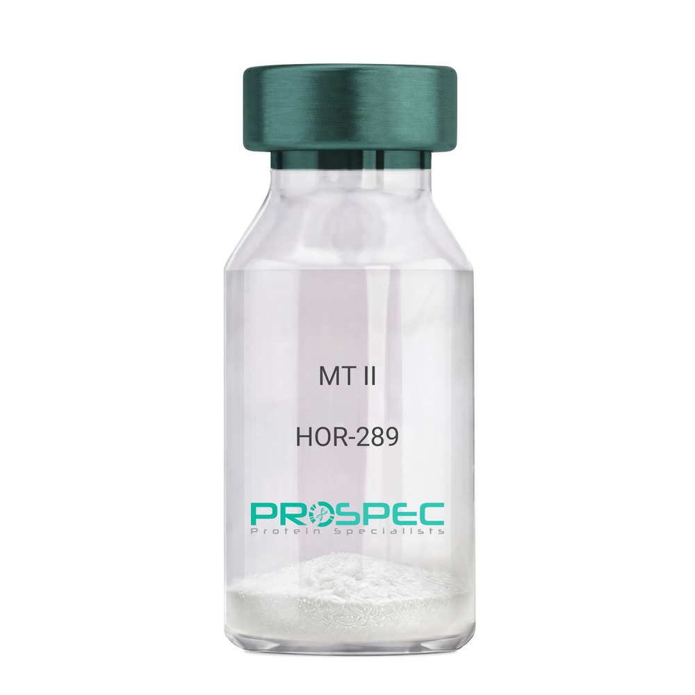
MT IIDescription:
Melanotan-II
Melanotan-II, MT-II, Melanotan.
Product # :
HOR-289Price :
Quantity :
Shipping Method :
Shipped at Room temp
More Info
- description
- formulation
- purity
- More Info
Description
The molecular formula of Melanotan-II is C50H69N15O9. It has the amino acid sequence of Ac-Nle-cyclo [Asp-His-D-Phe-Arg-Trp-Lys]-NH2 and a molecular weight of 1024.2 Dalton. The CAS Number is 121062-08-6.
Formulation
The protein (1mg/ml) was lyophilized with no additives.
Purity
Greater than 99% as determined by by RP-HPLC.
More Info
-
Introduction
Melanotan and Melanotan-II are both analogs of the peptide hormone alpha melanocyte stimulating hormone (a-MSH) that tend to induce skin tanning. Unlike Melanotan, Melanotan-II has the additional effect of increasing libido. Melanotan II has aphrodisiac properties.
-
Synonyms
Melanotan-II, MT-II, Melanotan.
-
Physical Appearance
Sterile Filtered White lyophilized (freeze-dried) powder.
-
Stability
Lyophilized Melanotan-II although stable at room temperature for 3 weeks, should be stored desiccated below -18°C. Upon reconstitution MT-II should be stored at 4°C between 2-7 days and for future use below -18°C.For long term storage it is recommended to add a carrier protein (0.1% HSA or BSA).Please prevent freeze-thaw cycles.
-
Solubility
It is recommended to reconstitute the lyophilized Melanotan-II in sterile 18MΩ-cm H2O not less than 100 µg/ml, which can then be further diluted to other aqueous solutions.
ProSpec's products are furnished for LABORATORY RESEARCH USE ONLY. The product may not be used as drugs, agricultural or pesticidal products, food additives or household chemicals.
- View Data Sheet
Name :
CSTB AntibodyDescription:
Cystatin B, Mouse Anti Human
Cystatin-B, Stefin-B, Liver thiol proteinase inhibitor, CPI-B, CSTB, CST6, EPM1, PME, STFB.
Product # :
ANT-339Price :
Quantity :
Shipping Method :
Shipped with Ice Packs
More Info
- formulation
- More Info
Formulation
1mg/ml containing PBS, pH-7.4, & 0.1% Sodium Azide.
More Info
-
Introduction
Type 1 cystatins are also called stefins which function as intracellular thiol protease inhibitors. Cystatin-B protein is able to form a dimer stabilized by noncovalent forces, inhibiting papain and cathepsins l, h and b. CSTB protein protects proteases leakage from lysosomes. Mutations in Stefin-B gene cause primary defects in patients with progressive myoclonic epilepsy (EPM1), a degenerative disease of the central nervous system. CSTB is overexpressed & elevated in the serum of HCC patients. Cystatin-B in vivo has a polymeric structure which is sensitive to the redox environment. Cystatin-B inhibits bone resorption by down-regulating intracellular cathepsin K activity despite increased osteoclast survival. Protein and mRNA levels of stefin B are significantly lower in atypical benign meningiomas. Stefins-A & Stefin-B which belong to the type-1 Cystatins, are up-regulated in lung tumours and thus able to counteract harmful tumour-associated proteolytic activity. Human stefin-A & Stef
-
Synonyms
Cystatin-B, Stefin-B, Liver thiol proteinase inhibitor, CPI-B, CSTB, CST6, EPM1, PME, STFB.
-
Immunogen
Anti-human CSTB mAb is derived from hybridization of mouse F0 myeloma cells with spleen cells from BALB/c mice immunized with recombinant human CSTB amino acids 1-98 purified from E. coli.
-
Ig Subclass
Mouse IgG2b heavy chain and κ light chain.
-
Clone
P2F1AT.
-
Applications
CSTB antibody has been tested by ELISA and Western blot analysis to assure specificity and reactivity. Since application varies, however, each investigation should be titrated by the reagent to obtain optimal results. Recommended dilution range for Western blot analysis is 1:1,000~ 2,000. Recommended starting dilution is 1:1,000.
-
Type
Mouse Anti Human Monoclonal.
-
Storage Procedures
For periods up to 1 month store at 4°C, for longer periods of time, store at -20°C. Prevent freeze thaw cycles.
-
Purification Method
CSTB antibody was purified from mouse ascitic fluids by protein-G affinity chromatography.
ProSpec's products are furnished for LABORATORY RESEARCH USE ONLY. The product may not be used as drugs, agricultural or pesticidal products, food additives or household chemicals.
- View Data Sheet
Name :
HBsAg adwDescription:
Hepatitis B Surface Antigen, adw Recombinant
Product # :
HBS-872Price :
Quantity :
Shipping Method :
Shipped with Ice Packs
More Info
- description
- source
- formulation
- purity
- More Info
Description
HbsAg adw produced Pichia Pastoris, having a molecular weight of approximately 24.0 kDa as shown on SDS-PAGE.
Source
Pichia Pastoris.
Formulation
Sterile Filtered solution containing 20mM Phosphate Buffer, 154mM sodium chloride, pH 7.1.
Purity
Greater than 95.0% as determined by SDS-PAGE.
More Info
-
Introduction
HBsAg is the surface antigenof the Hepatitis-B-Virus (HBV). The capsidof a virus has different surface proteins from the rest of the virus. The antigen is a protein that binds specifically on one of these surface proteins. It is commonly referred to as the Australian Antigen.
-
Physical Appearance
Sterile Filtered pale solution.
-
Stability
HBsAg Should be stored at 4°C.DO NOT FREEZE.
ProSpec's products are furnished for LABORATORY RESEARCH USE ONLY. The product may not be used as drugs, agricultural or pesticidal products, food additives or household chemicals.
- View Data Sheet
Name :
CD4 biotin AntibodyDescription:
CD4-Biotinylated, Mouse Anti-Human
gp55, HLA-2, L3 / T4, Ly-4, T cell antigen T4/LEU3, T4, sCD4, CD4mut.
Product # :
ANT-167Price :
Quantity :
Shipping Method :
Shipped at Room temp
More Info
- formulation
- More Info
Formulation
1mg/ml in PBS (after reconstitution).
More Info
-
Introduction
CD4 is a cell-surface glycoprotein found on the mature helper T cells and immature thymocytes, as well as on monocytes and macrophages. (Some cytotoxic T cells have CD4 protein as well.) Normally, about 65% of T cells in the blood are CD4+ (have CD4 protein protruding from their membrane). A mature T cell with either have CD4 or CD8, but not both. During one stage of development T cells develop CD4 and CD8 receptors, but they eventually are differentiated in the thymus and become more specialized.
-
Synonyms
gp55, HLA-2, L3 / T4, Ly-4, T cell antigen T4/LEU3, T4, sCD4, CD4mut.
-
Solubility
Reconstitute with of H20. Mix gently, wash the sides of the vial and wait 30-60 seconds before use.
-
Immunogen
Purified human PBL CD4+ T cells.
-
Ig Subclass
Mouse IgG2b.
-
Clone
hCD4.
-
Applications
Blocking and staining antibody. Blocks blinding of HIV to CD4. For staining, use 10 ml/1,000,000 cells. Titer for blocking T cell activation should be determined by the investigator.
-
Available Conjugates
This antibody is also available conjugated to FITC.
-
Type
Mouse Anti Human Monoclonal.
-
Storage Procedures
Lyophilized: store at 4oC. After reconstitution, if not intended for use within a month, aliquot and store at -20oC.
-
Purification Method
Ion exchange column.
ProSpec's products are furnished for LABORATORY RESEARCH USE ONLY. The product may not be used as drugs, agricultural or pesticidal products, food additives or household chemicals.
- View Data Sheet
Name :
CX3CL1 RatDescription:
Fractalkine Rat Recombinant (CX3CL1)
Fractalkine, CX3CL1, Neurotactin, CX3C membrane-anchored chemokine, Small inducible cytokine D1, NTN, NTT, CXC3, CXC3C, SCYD1, ABCD-3, C3Xkine.
Product # :
CHM-005Price :
Quantity :
Shipping Method :
Shipped at Room temp
More Info
- description
- source
- formulation
- purity
- biological activity
- More Info
Description
Fractalkine Rat Recombinant produced in E.Coli is a single, non-glycosylated, polypeptide chain containing 76 amino acids and having a molecular mass of 8.7kDa.The Fractalkine is purified by proprietary chromatographic techniques.
Source
Escherichia Coli.
Formulation
Lyophilized from a 0.2µm filtered concentrated solution in PBS, pH 7.4.
Purity
Greater than 97.0% as determined by SDS-PAGE.
Biological Activity
Determined by its ability to chemoattract human monocytes using a concentration range of 5.0-10.0 ng/ml, corresponding to a specific activity of 100,000-200,000units/mg.More Info
-
Introduction
Fractalkine soluble form is chemotactic for t-cells and monocytes, but not for neutrophils. Fractalkine membrane-bound form promotes adhesion of those leukocytes to endothelial cells. Fractalkine regulates leukocyte adhesion and migration processes at the endothelium and binds to CX3CR1. Natural Human Fractalkine is produced as a long protein (373-amino acid) with an extended mucin-like stalk and a chemokine domain on top. The mucin-like stalk permits it to bind to the cell surface. Fractalkine gene is located on human chromosome 16 along with some CC chemokines known as CCL17 and CCL22.
-
Synonyms
Fractalkine, CX3CL1, Neurotactin, CX3C membrane-anchored chemokine, Small inducible cytokine D1, NTN, NTT, CXC3, CXC3C, SCYD1, ABCD-3, C3Xkine.
-
Physical Appearance
Sterile Filtered White lyophilized (freeze-dried) powder.
-
Stability
Lyophilized FractalkineRat although stable at room temperature for 3 weeks, should be stored desiccated below -18°C. Upon reconstitution Fractalkine should be stored at 4°C between 2-7 days and for future use below -18°C.Please prevent freeze-thaw cycles.
-
Solubility
It is recommended to reconstitute the lyophilized Fractalkine in sterile 18MΩ-cm H2O not less than 100µg/ml, which can then be further diluted to other aqueous solutions.
-
Amino Acid Sequence
QHLGMTKCNI TCHKMTSPIP VTLLIHYQLN QESCGKRAII LETRQHRHFC ADPKEKWVQD AMKHLDHQTA ALTRNG
-
Background
What is the molecular weight/Mw of CX3CL1 RAT Protein?
CX3CL1 RAT Protein has a total Mw of 8.7kDa.
What is the source or expression system of CX3CL1 RAT Protein?
Escherichia Coli.
What is the Purity of CX3CL1 RAT Protein?
CX3CL1 RAT Protein is >97% pure as determined by SDS-PAGE.
What is the Biological Activity of CX3CL1 RAT Protein?
Determined by its ability to chemoattract human monocytes using a concentration range of 5.0-10.0 ng/ml, corresponding to a specific activity of 100,000-200,000units/mg.
What is the amino acid sequence of CX3CL1 RAT Protein?
QHLGMTKCNI TCHKMTSPIP VTLLIHYQLN QESCGKRAII LETRQHRHFC ADPKEKWVQD AMKHLDHQTA ALTRNG
What applications can CX3CL1 RAT Protein be used in?
CX3CL1 RAT Protein can probably be used in western blot, ELISA and Lateral Flow.
What is the endotoxin level for CX3CL1 RAT Protein?
The endotoxin level is minimal, CX3CL1 RAT Protein was purified using conventional chromatography techniques.
ProSpec's products are furnished for LABORATORY RESEARCH USE ONLY. The product may not be used as drugs, agricultural or pesticidal products, food additives or household chemicals.
- View Data Sheet
Name :
KRT18 HumanDescription:
Cytokeratin 18 Human Recombinant
Keratin type I cytoskeletal 18, Cytokeratin-18, CK-18, Keratin-18, K18, KRT18,CYK18,Cell proliferation-inducing gene 46 protein.
Product # :
PRO-349Price :
Quantity :
Shipping Method :
Shipped at Room temp
More Info
- description
- source
- formulation
- purity
- More Info
Description
Cytokeratin 18 Human Recombinant produced in E.Coli is a single, non-glycosylated polypeptide chain having a calculated molecular mass of 48,201 Dalton, showing a 45kDa band on SDS-page, pI-5.7.The KRT18 is purified by proprietary chromatographic techniques.
Source
Escherichia Coli.
Formulation
The protein (1mg/ml) was lyophilized from a sterile solution containing 30mM Tris-HCl pH-8, 9.5M urea, 2mM EDTA, 2mM DTT and 10mM methylammonium chloride.
Purity
Greater than 95.0% as determined by analysis by SDS-PAGE.
More Info
-
Introduction
KRT18 encodes the type I intermediate filament chain keratin 18. Keratin 18, together with its filament partner keratin 8, are perhaps the most commonly found members of the intermediate filament gene family. They are expressed in single layer epithelial tissues of the body. Mutations in this gene have been linked to cryptogenic cirrhosis. Two transcript variants encoding the same protein have been found for this gene.
-
Synonyms
Keratin type I cytoskeletal 18, Cytokeratin-18, CK-18, Keratin-18, K18, KRT18,CYK18,Cell proliferation-inducing gene 46 protein.
-
Physical Appearance
Sterile Filtered White lyophilized (freeze-dried) powder.
-
Stability
Lyophilized CK-18 although stable at room temperature for 3 weeks, should be stored between 2-8°C. Upon reconstitution CK-18 should be stored at 4°C between 2-7 days and for future use below -18°C. For long term storage it is recommended to add a carrier protein (0.1% HSA or BSA). Please prevent freeze-thaw cycles.
-
Solubility
It is recommended to reconstitute the lyophilized 1mg CK-18 in sterile 18MΩ-cm H2O not less than 700µl, which can then be further diluted to other aqueous solutions.
-
Reconstitution to filaments
Performed by mixing equimolar amounts of cytokeratins of type I and type II at concentrations of approx. 0.5 mg/ml, both dissolved in 9.5M urea buffer (see above). Protofilaments and filament complexes are obtained by dialyzing the resulting polypeptide solution stepwise to a concentration of 4M urea and then to low salt condition (50mM NaCl, 2mM dithiothreitol, 10mM Tris-HCI, pH 7.4). For immunization purposes, the solution can be further dialyzed against PBS (phosphate buffered saline, e.g. Dulbecco's PBS).
ProSpec's products are furnished for LABORATORY RESEARCH USE ONLY. The product may not be used as drugs, agricultural or pesticidal products, food additives or household chemicals.
- View Data Sheet
Name :
Lymphotactin HumanDescription:
Lymphotactin Human Recombinant (XCL1)
XCL1, Cytokine SCM-1, ATAC, Lymphotaxin, SCM-1-alpha, Small inducible cytokine C1, XC chemokine ligand 1, LTN, LPTN, SCM1, SCM-1, SCYC1, SCM-1a.
Product # :
CHM-314Price :
Quantity :
Shipping Method :
Shipped at Room temp
More Info
- description
- source
- formulation
- purity
- biological activity
- More Info
Description
Lymphotactin Human Recombinant produced in E.Coli is a single, non-glycosylated, polypeptide chain containing 92 amino acids and having a molecular mass of 10007 Dalton. The Lymphotactin is purified by proprietary chromatographic techniques.
Source
Escherichia Coli.
Formulation
The XCL1 was lyophilized from a concentrated (1mg/ml) solution in water containing no additives.
Purity
Greater than 99.0% as determined by:
(a) Analysis by RP-HPLC.
(b) Analysis by SDS-PAGE.Biological Activity
The Biological activity is calculated by its ability to chemoattract human T cells at 10-100ng/ml corresponding to a Specific Activity of 10,000-100,000IU/mg.More Info
-
Introduction
Chemokine (C motif) ligand (XCL1) is a small cytokine belonging to the XC chemokine family that is also known as lymphotactin. It is found in high levels in spleen, thymus, intestine and peripheral blood leukocytes, and at lower levels in lung, prostate gland and ovary. Cellular sources for XCL1 include activated thymic and peripheral blood CD8+ T cells. This chemokine attracts T cells. In humans, XCL1 is closely related to another chemokine called XCL2, whose geneis found at the same locus on chromosome 1. XCL1 induces it chemotactic function by binding to a chemokine receptor called XCR1.
-
Synonyms
XCL1, Cytokine SCM-1, ATAC, Lymphotaxin, SCM-1-alpha, Small inducible cytokine C1, XC chemokine ligand 1, LTN, LPTN, SCM1, SCM-1, SCYC1, SCM-1a.
-
Physical Appearance
Sterile Filtered White lyophilized (freeze-dried) powder.
-
Stability
Lyophilized Lymphotactin although stable at room temperature for 3 weeks, should be stored desiccated below -18°C. Upon reconstitution XCL1 should be stored at 4°C between 2-7 days and for future use below -18°C. For long term storage it is recommended to add a carrier protein (0.1% HSA or BSA). Please avoid freeze-thaw cycles.
-
Solubility
It is recommended to reconstitute the lyophilized Lymphotactin in sterile 18MΩ-cm H2O not less than 100µg/ml, which can then be further diluted to other aqueous solutions.
-
Amino Acid Sequence
The sequence of the first five N-terminal amino acids was determined and was found to be Gln-Ser-Glu-Val-Ser.
ProSpec's products are furnished for LABORATORY RESEARCH USE ONLY. The product may not be used as drugs, agricultural or pesticidal products, food additives or household chemicals.
- View Data Sheet
Name :
HCV NS3, BiotinDescription:
Hepatitis C Virus NS3, Biotin Recombinant
Product # :
HCV-244Price :
Quantity :
Shipping Method :
Shipped with Ice Packs
More Info
- description
- formulation
- purity
- More Info
Description
The E.coli derived recombinant Biotin Labeled protein contains the HCV NS3 immunodominant regions, a.a. 1450-1643, 22 kDa. The Biotin labeled protein is fused with a 6xHis-Tag at N-terminus.
Formulation
(1mg/ml) 1.5M urea, 20mM Tris-HCl pH 8.0 and 10mM β-mercaptoethanol.
Purity
HCV NS3 Biotin labeled protein is >95% pure as determined by 10% PAGE (coomassie staining).
More Info
-
Introduction
HCV is a small 50nm, enveloped, single-stranded, positive sense RNAvirus in the family Flaviviridae.
HCV has a high rate of replication with approximately one trillion particles produced each day in an infected individual. Due to lack of proofreading by the HCV RNA polymerase, the HCV has an exceptionally high mutation rate, a factor that may help it elude the host's immune response. Hepatitis C virus is classified into six genotypes(1-6) with several subtypes within each genotype. The preponderance and distribution of HCV genotypes varies globally. Genotype is clinically important in determining potential response to interferon-based therapy and the required duration of such therapy. Genotypes 1 and 4 are less responsive to interferon-based treatment than are the other genotypes (2, 3, 5 and 6). -
Stability
HCV NS3 Biotin although stable at 4°C for 1 week, should be stored below -18°C. Please prevent freeze thaw cycles.
-
Applications
HCV NS3 Biotin labeled antigen is suitable for ELISA and Western blots, excellent antigen for detection of HCV with minimal specificity problems.
-
Specificity
Reactivity with human HCV positive serum undetermined.
-
Purification Method
HCV NS3 Biotin labeled protein was purified by proprietary chromatographic technique.
ProSpec's products are furnished for LABORATORY RESEARCH USE ONLY. The product may not be used as drugs, agricultural or pesticidal products, food additives or household chemicals.
- View Data Sheet
Name :
Activin B HumanDescription:
Activin-B Human Recombinant
Inhibin beta B (activin AB beta polypeptide), Inhibin, beta-2, Activin beta-B chain, MGC157939.
Product # :
CYT-058Price :
Quantity :
Shipping Method :
Shipped at Room temp
More Info
- description
- source
- formulation
- purity
- More Info
Description
Activin B human Recombinant produced in Nicotiana benthamiana plant is a beta-B single chain (aa 293-406) containing 123 amino acids (molecular formula C615H910N178O177S12). Activin B is fused to a 10-His-tag at the N-terminal having the total molecular mass of 14kDa and purified by standard chromatographic techniques.
Source
Nicotiana benthamiana plant
Formulation
Lyophilized from 1mg/ml solution in 0.05M Tris-HCl buffer pH 7.4.
Purity
Greater than 97.0% as determined by Analysis by SDS-PAGE.
More Info
-
Introduction
Inhibins are dimeric peptide hormones produced by female ovarian granulose cells and male Sertoli cells as well as a variety of other tissues. Inhibins have two isoforms, A and B, with the same alpha subunit but different beta subunits. Inhibin A is a dimer of alpha and beta A subunits, inhibin B is a dimer of alpha and beta B subunits.
Inhibins are thought to inhibit the production of follicle-stimulating hormone (FSH) by the pituitary gland. In addition, Inhibins are also thought to play a role in the control of gametogenesis, and embryonic and fetal development. -
Synonyms
Inhibin beta B (activin AB beta polypeptide), Inhibin, beta-2, Activin beta-B chain, MGC157939.
-
Physical Appearance
Sterile Filtered White lyophilized (freeze-dried) powder.
-
Stability
Lyophilized Activin B although stable at room temperature for 3 weeks, should be stored desiccated below -18°C. Upon reconstitution Activin B should be stored at 4°C between 2-7 days and for future use below -18°C. For long term storage it is recommended to add a carrier protein (0.1% HSA or BSA). Please prevent freeze-thaw cycles.
-
Solubility
It is recommended to reconstitute the lyophilized Activin B in sterile 18M-cm H2O not less than 100µg/ml, which can then be further diluted to other aqueous solutions.
-
Amino Acid Sequence
HHHHHHHHHH GLECDGRTNL CCRQQFFIDF RLIGWNDWII APTGYYGNYC EGSCPAYLAG VPGSASSFHT AVVNQYRMRG LNPGTVNSCC IPTKLSTMSM LYFDDEYNIV KRDVPNMIVE ECG
-
Background
What is the molecular weight / Mw of Activin B Protein?
Activin A Protein has a total Mw of 14 kDa.
What is the source or expression system of Activin B Protein?
Nicotinia
What is the Purity of Activin B Protein?
Activin B Protein is >97% pure as determined by SDS-PAGE.
What is the Biological Activity of Activin B Protein?
The biological functionality of Activin-B Protein will be determined in the future.
What is the endotoxin level for Activin B Protein?
The endotoxin level is minimal, ACTIVIN B Protein was purified using conventional chromatography techniques.
What is the amino acid sequence of ACTIVIN B Protein?
HHHHHHHHHH GLECDGRTNL CCRQQFFIDF RLIGWNDWII APTGYYGNYC EGSCPAYLAG VPGSASSFHT AVVNQYRMRG LNPGTVNSCC IPTKLSTMSM LYFDDEYNIV KRDVPNMIVE ECG
What applications can ACTIVIN B Protein be used in?
ACTIVIN A Protein can probably be used in western blot, ELISA and Lateral Flow.
ProSpec's products are furnished for LABORATORY RESEARCH USE ONLY. The product may not be used as drugs, agricultural or pesticidal products, food additives or household chemicals.
- View Data Sheet
Name :
Calumenin HumanDescription:
Calumenin Human Recombinant
CALU, Crocalbin, IEF SSP 9302, FLJ90608, Calumenin.
Product # :
PRO-2465Price :
Quantity :
Shipping Method :
Shipped with Ice Packs
More Info
- description
- source
- formulation
- purity
- biological activity
- More Info
Description
Calumenin Human Recombinant produced in E. coli is a single polypeptide chain containing 297 amino acids (20-315) and having a molecular mass of 35kDa. Calumenin is purified by proprietary chromatographic techniques.
Source
Escherichia Coli.
Formulation
The Calumenin solution (0.5mg/1ml) contains phosphate buffered saline (pH7.4) and 10% glycerol.
Purity
Greater than 90.0% as determined by SDS-PAGE.
Biological Activity
The IC50 value is < 2.0nM. The inhibitory function of Cystatin 5 on protease activity of papain was measured by a fluorometric assay using Z-FR-AMC at pH 7.5 at 25C.More Info
-
Introduction
Calumenin is a calcium-binding protein located in the endoplasmic reticulum (ER) and sarcoplasmic reticulum (SR) of mammalian tissues which plays a role in ER functions as protein folding and sorting. Calumenin belongs to a family of multiple EF-hand proteins (CERC) that include reticulocalbin, ERC-55, and Cab45 and the product of this gene. Calumenin binds 7 calcium ions having low affinity and takes part in such ER functions as protein folding and sorting.
-
Synonyms
CALU, Crocalbin, IEF SSP 9302, FLJ90608, Calumenin.
-
Physical Appearance
Sterile Filtered clear solution.
-
Stability
Store at 4°C if entire vial will be used within 2-4 weeks. Store, frozen at -20°C for longer periods of time. For long term storage it is recommended to add a carrier protein (0.1% HSA or BSA).Avoid multiple freeze-thaw cycles.
-
Amino Acid Sequence
MKPTEKKDRV HHEPQLSDKV HNDAQSFDYD HDAFLGAEEA KTFDQLTPEE SKERLGKIVS KIDGDKDGFV TVDELKDWIK FAQKRWIYED VERQWKGHDL NEDGLVSWEE YKNATYGYVL DDPDPDDGFN YKQMMVRDER RFKMADKDGD LIATKEEFTA FLHPEEYDYM KDIVVQETME DIDKNADGFI DLEEYIGDMY SHDGNTDEPE WVKTEREQFV EFRDKNRDGK MDKEETKDWI LPSDYDHAEA EARHLVYESD QNKDGKLTKE EIVDKYDLFV GSQATDFGEA LVRHDEF.
ProSpec's products are furnished for LABORATORY RESEARCH USE ONLY. The product may not be used as drugs, agricultural or pesticidal products, food additives or household chemicals.
- View Data Sheet
Name :
AtosibanDescription:
Atosiban
Product # :
HOR-239Price :
Quantity :
Shipping Method :
Shipped at Room temp
More Info
- description
- formulation
- purity
- More Info
- HPLC, MS
Description
Atosiban also called ADH (Anti-Diuretic Hormone) has a molecular formula of C43H67N11O12S2, 3-Mercaptopropionyl-D-Tyr(ET)-Ile-Thr-Asn-Cys-Pro-Orn-Gly-NH2 having a Mw of 994.2 Dalton.
Formulation
The Atosiban peptide was lyophilized with no additives.
Purity
Greater than 98.0% as determined by analysis by RP-HPLC.
HPLC, MS
More Info
-
Introduction
Atosiban is the first oxytocin antagonist to be specifically developed for the treatment of preterm labor. Atosiban has a specific mode of action, inhibiting oxytocin-induced uterine contractions by blocking oxytocin receptors in the uterus. Extensive clinical investigations have shown Atosiban to be at least as effective as current tocolytic agents. In addition, due to its novel and specific mode of action, Atosiban has a markedly improved maternal side effects profile compared with conventional therapies.
-
Physical Appearance
Sterile Filtered White lyophilized (freeze-dried) powder.
-
Stability
Lyophilized Atosiban although stable at room temperature for 3 weeks, should be stored desiccated below -18°C. Upon reconstitution Atosiban should be stored at 4°C between 2-7 days and for future use below -18°C. For long term storage it is recommended to add a carrier protein (0.1% HSA or BSA).Please prevent freeze-thaw cycles.
-
Solubility
It is recommended to reconstitute the lyophilized Atosiban in sterile 18MΩ-cm H2O not less than 100µg/ml, which can then be further diluted to other aqueous solutions.
ProSpec's products are furnished for LABORATORY RESEARCH USE ONLY. The product may not be used as drugs, agricultural or pesticidal products, food additives or household chemicals.
- View Data Sheet
Name :
IFIH1 HumanDescription:
Interferon Induced With Helicase C Domain 1 Human Recombinant
Interferon-induced helicase C domain-containing protein 1, Clinically amyopathic dermatomyositis autoantigen 140 kDa, CADM-140 autoantigen, Helicase with 2 CARD domains, Helicard, Interferon-induced with helicase C domain protein 1, Melanoma differentiation-associated protein 5, MDA-5, Murabutide down-regulated protein, RIG-I-like receptor 2, RLR-2, RNA helicase-DEAD box protein 116, IFIH1, MDA5, RH116, Hlcd, IDDM19.
Product # :
PRO-1505Price :
Quantity :
Shipping Method :
Shipped with Ice Packs
More Info
- description
- source
- formulation
- purity
- More Info
Description
IFIH1 Human Recombinant produced in SF9 is a glycosylated, polypeptide chain having a calculated molecular mass of 152,000 Dalton. IFIH1 is expressed with a -10xHis tag at N-terminus and purified by proprietary chromatographic techniques.
Source
Sf9 Insect Cells.
Formulation
IFIH1 is supplied in 20mM HEPES buffer pH-7.9, 550mM NaCl and 6M Urea.
Purity
Greater than 93.0% as determined by SDS-PAGE.
More Info
-
Introduction
IFIH1 is a DEAD box protein which is upregulated in response to treatment with beta-interferon and a protein kinase C-activating compound, mezerein. Irreversible reprogramming of melanomas can be attained by therapy with both these agents; treatment with either agent alone only achieves reversible differentiation. DEAD box proteins are implicated in several cellular processes involving alteration of RNA secondary structure such as translation initiation, nuclear and mitochondrial splicing, and ribosome and spliceosome assembly.
-
Synonyms
Interferon-induced helicase C domain-containing protein 1, Clinically amyopathic dermatomyositis autoantigen 140 kDa, CADM-140 autoantigen, Helicase with 2 CARD domains, Helicard, Interferon-induced with helicase C domain protein 1, Melanoma differentiation-associated protein 5, MDA-5, Murabutide down-regulated protein, RIG-I-like receptor 2, RLR-2, RNA helicase-DEAD box protein 116, IFIH1, MDA5, RH116, Hlcd, IDDM19.
-
Physical Appearance
Sterile Filtered clear solution.
-
Stability
Store at 4°C if entire vial will be used within 2-4 weeks. Store, frozen at -20°C for longer periods of time. Avoid multiple freeze-thaw cycles.
ProSpec's products are furnished for LABORATORY RESEARCH USE ONLY. The product may not be used as drugs, agricultural or pesticidal products, food additives or household chemicals.
- View Data Sheet
Name :
DCTN6 HumanDescription:
Dynactin 6 Human Recombinant
Dynactin 6, WS3, Dynactin Subunit P27, Protein WS-3, P27, Novel RGD-Containing Protein, Dynactin Subunit 6, WS-3, Dynactin subunit 6.
Product # :
PRO-2094Price :
Quantity :
Shipping Method :
Shipped with Ice Packs
More Info
- description
- source
- formulation
- purity
- More Info
Description
DCTN6 Human Recombinant produced in E.Coli is a single, non-glycosylated polypeptide chain containing 213 amino acids (1-190 a.a) and having a molecular mass of 23.1kDa. DCTN6 is fused to a 23 amino acid His-tag at N-terminus & purified by proprietary chromatographic techniques.
Source
Escherichia Coli.
Formulation
DCTN6 protein solution (1mg/ml) containing Phosphate Buffered Saline (pH7.4) and 10% glycerol.
Purity
Greater than 85.0% as determined by SDS-PAGE.
More Info
-
Introduction
Dynactin 6, also known as DCTN6 is a member of the dynactin subunits 5/6 family. DCTN6 includes an RGD (Arg-Gly-Asp) motif in the N-terminal region, which confers adhesive properties to macromolecular proteins such as fibronectin. DCTN6 has a high degree of sequence resemblance with the mouse homolog, which has been found to participate in mitochondrial biogenesis. Moreover, the precise biological function of DCTN6 is unknown.
-
Synonyms
Dynactin 6, WS3, Dynactin Subunit P27, Protein WS-3, P27, Novel RGD-Containing Protein, Dynactin Subunit 6, WS-3, Dynactin subunit 6.
-
Physical Appearance
Sterile Filtered colorless solution.
-
Stability
Store at 4°C if entire vial will be used within 2-4 weeks. Store, frozen at -20°C for longer periods of time. For long term storage it is recommended to add a carrier protein (0.1% HSA or BSA).Avoid multiple freeze-thaw cycles.
-
Amino Acid Sequence
MGSSHHHHHH SSGLVPRGSH MGSMAEKTQK SVKIAPGAVV CVESEIRGDV TIGPRTVIHP KARIIAEAGP IVIGEGNLIE EQALIINAYP DNITPDTEDP EPKPMIIGTN NVFEVGCYSQ AMKMGDNNVI ESKAYVGRNV ILTSGCIIGA CCNLNTFEVI PENTVIYGAD CLRRVQTERP QPQTLQLDFL MKILPNYHHL KKTMKGSSTP VKN.
ProSpec's products are furnished for LABORATORY RESEARCH USE ONLY. The product may not be used as drugs, agricultural or pesticidal products, food additives or household chemicals.
- View Data Sheet
Name :
C17ORF49 HumanDescription:
Chromosome 17 Open Reading Frame 49 Human Recombinant
Chromosome 17 open reading frame 49, BPTF-associated protein of 18 kDa, chromatin complexes subunit BAP18, MLL1/MLL complex subunit C17orf49, MGC49942, BAP18.
Product # :
PRO-1192Price :
Quantity :
Shipping Method :
Shipped with Ice Packs
More Info
- description
- source
- formulation
- purity
- More Info
Description
C17ORF49 Human Recombinant produced in E. coli is a single polypeptide chain containing 195 amino acids (1-172) and having a molecular mass of 20.3 kDa.C17ORF49 is fused to a 23 amino acid His-tag at N-terminus & purified by proprietary chromatographic techniques.
Source
E.coli.
Formulation
The C17ORF49 solution (1mg/1ml) contains 20mM Tris-HCl buffer (pH 8.0), 0.1M NaCl, 1mM DTT and 10% glycerol.
Purity
Greater than 90% as determined by SDS-PAGE.
More Info
-
Introduction
C17ORF49 is a component of chromatin complexes such as the MLL1/MLL and NURF complexes.
-
Synonyms
Chromosome 17 open reading frame 49, BPTF-associated protein of 18 kDa, chromatin complexes subunit BAP18, MLL1/MLL complex subunit C17orf49, MGC49942, BAP18.
-
Physical Appearance
Sterile Filtered colorless solution.
-
Stability
Store at 4°C if entire vial will be used within 2-4 weeks. Store, frozen at -20°C for longer periods of time. For long term storage it is recommended to add a carrier protein (0.1% HSA or BSA).Avoid multiple freeze-thaw cycles.
-
Amino Acid Sequence
MGSSHHHHHH SSGLVPRGSH MGSMTSASTK VGEIFSAAGA AFTKLGELTM QLHPVADSSP AGAKWTETEI EMLRAAVKRF GDDLNHISCV IKERTVAQIK ATVKRKVYED SGIPLPAESP KKGPKKVASG VLSPPPAAPP PSSSSVPEAG GPPIKKQKAD VTLSALNDSD ANSDVVDIEG LGETPPAKKL NFDQA
ProSpec's products are furnished for LABORATORY RESEARCH USE ONLY. The product may not be used as drugs, agricultural or pesticidal products, food additives or household chemicals.
- View Data Sheet
Name :
POFUT1 HumanDescription:
Protein O-Fucosyltransferase 1 Human Recombinant
FUT12, O-Fuc-T, O-FucT-1, O-FUT, GDP-fucose protein O-fucosyltransferase 1, Peptide-O-fucosyltraferase 1,KIAA0180.
Product # :
ENZ-679Price :
Quantity :
Shipping Method :
Shipped with Ice Packs
More Info
- description
- source
- formulation
- purity
- More Info
Description
POFUT1 Human Recombinant produced in E. coli is a single polypeptide chain containing 385 amino acids (27-388) and having a molecular mass of 43.7 kDa. POFUT1 is fused to a 23 amino acid His-tag at N-terminus & purified by proprietary chromatographic techniques.
Source
Escherichia Coli.
Formulation
The POFUT1 solution (1mg/1ml) contains 20mM Tris-HCl buffer (pH 8.0), 2M Urea and 10% glycerol.
Purity
Greater than 85.0% as determined by SDS-PAGE.
More Info
-
Introduction
GDP-fucose protein O-fucosyltransferase 1 (POUFUT1), belongs to the glycosyltransferase O-Fuc family. POUFUT1 encodes a member of the glycosyltransferase O-Fuc family and Expressed mainly in pancreas, kidney, lung, heart, brain, liver, placenta and skeletal muscle.POUFUT1 adds O-fucose through an O-glycosidic linkage to Preserve serine or threonine residues in the epidermal growth factor-like repeats of a number of cell surface and emitted proteins. POUFUT1 is involved in ligand-induced receptor signaling. Alternative splicing of this gene results in 2 transcript variants encoding different isoforms.POFUT1 participates in Notch signaling, as Notch ligands can use as POFUT1 substrates.
-
Synonyms
FUT12, O-Fuc-T, O-FucT-1, O-FUT, GDP-fucose protein O-fucosyltransferase 1, Peptide-O-fucosyltraferase 1,KIAA0180.
-
Physical Appearance
Sterile Filtered colorless solution.
-
Stability
Store at 4°C if entire vial will be used within 2-4 weeks. Store, frozen at -20°C for longer periods of time. For long term storage it is recommended to add a carrier protein (0.1% HSA or BSA).Avoid multiple freeze-thaw cycles.
-
Amino Acid Sequence
MGSSHHHHHH SSGLVPRGSH MGSGSWDPAG YLLYCPCMGR FGNQADHFLG SLAFAKLLNR TLAVPPWIEY QHHKPPFTNL HVSYQKYFKL EPLQAYHRVI SLEDFMEKLA PTHWPPEKRV AYCFEVAAQR SPDKKTCPMK EGNPFGPFWD QFHVSFNKSE LFTGISFSAS YREQWSQRFS PKEHPVLALP GAPAQFPVLE EHRPLQKYMV WSDEMVKTGE AQIHAHLVRP YVGIHLRIGS DWKNACAMLK DGTAGSHFMA SPQCVGYSRS TAAPLTMTMC LPDLKEIQRA VKLWVRSLDA QSVYVATDSE SYVPELQQLF KGKVKVVSLK PEVAQVDLYI LGQADHFIGN CVSSFTAFVK RERDLQGRPS SFFGMDRPPK LRDEF.
ProSpec's products are furnished for LABORATORY RESEARCH USE ONLY. The product may not be used as drugs, agricultural or pesticidal products, food additives or household chemicals.
- View Data Sheet
Name :
VTN HumanDescription:
Vitronectin Human Recombinant
Vitronectin precursor, V75, VN, VNT, Vitronectin, VTN, S-protein, Serum-spreading factor, Vitronectin V65 subunit, Vitronectin V10 subunit, Somatomedin-B.
Product # :
PRO-2008Price :
Quantity :
Shipping Method :
Shipped with Ice Packs
More Info
- description
- source
- formulation
- purity
- More Info
Description
VTN Human Recombinant produced in E.Coli is a single, non-glycosylated polypeptide chain containing 482 amino acids (20-478 a.a) and having a molecular mass of 54.7kDa. VTN is fused to a 23 amino acid His-tag at N-terminus & purified by proprietary chromatographic techniques.
Source
Escherichia Coli.
Formulation
VTN protein solution (0.5mg/ml) containing 20mM Tris-HCl buffer (pH 8.0), 10% glycerol and 0.4M Urea.
Purity
Greater than 90.0% as determined by SDS-PAGE.
More Info
-
Introduction
Vitronectin (VTN) which is a part of the pexin family is a cell adhesion and spreading factor found in serum and tissues. VTN interacts with glycosaminoglycans and proteoglycans. VTN inhibits the membrane-damaging effect of the terminal cytolytic complement pathway and binds to numerous serpin serine protease inhibitors. Scientists have been noticed an over expression of VTN, integrins and plasminogen in migrating cells during wound healing.
-
Synonyms
Vitronectin precursor, V75, VN, VNT, Vitronectin, VTN, S-protein, Serum-spreading factor, Vitronectin V65 subunit, Vitronectin V10 subunit, Somatomedin-B.
-
Physical Appearance
Sterile Filtered colorless solution.
-
Stability
Store at 4°C if entire vial will be used within 2-4 weeks. Store, frozen at -20°C for longer periods of time. For long term storage it is recommended to add a carrier protein (0.1% HSA or BSA).Avoid multiple freeze-thaw cycles.
-
Amino Acid Sequence
MGSSHHHHHH SSGLVPRGSH MGSDQESCKG RCTEGFNVDK KCQCDELCSY YQSCCTDYTA ECKPQVTRGD VFTMPEDEYT VYDDGEEKNN ATVHEQVGGP SLTSDLQAQS KGNPEQTPVL KPEEEAPAPE VGASKPEGID SRPETLHPGR PQPPAEEELC SGKPFDAFTD LKNGSLFAFR GQYCYELDEK AVRPGYPKLI RDVWGIEGPI DAAFTRINCQ GKTYLFKGSQ YWRFEDGVLD PDYPRNISDG FDGIPDNVDA ALALPAHSYS GRERVYFFKG KQYWEYQFQH QPSQEECEGS SLSAVFEHFA MMQRDSWEDI FELLFWGRTS AGTRQPQFIS RDWHGVPGQV DAAMAGRIYI SGMAPRPSLA KKQRFRHRNR KGYRSQRGHS RGRNQNSRRP SRATWLSLFS SEESNLGANN YDDYRMDWLV PATCEPIQSV FFFSGDKYYR VNLRTRRVDT VDPPYPRSIA QYWLGCPAPG HL.
ProSpec's products are furnished for LABORATORY RESEARCH USE ONLY. The product may not be used as drugs, agricultural or pesticidal products, food additives or household chemicals.
- View Data Sheet
Name :
FBLIM1 HumanDescription:
Filamin Binding LIM Protein 1 Human Recombinant
Filamin binding LIM protein 1, CAL, FBLP-1, FBLP1, RP11-169K16.5, Migfilin, Mitogen-inducible 2-interacting protein, MIG2-interacting protein.
Product # :
PRO-1473Price :
Quantity :
Shipping Method :
Shipped with Ice Packs
More Info
- description
- source
- formulation
- purity
- More Info
Description
FBLIM1 Human Recombinant produced in E.coli is a single, non-glycosylated polypeptide chain containing 396 amino acids (1-373) and having a molecular mass of 43.1 kDa. FBLIM1 is fused to a 23 amino acid His-tag at N-terminus & purified by proprietary chromatographic techniques.
Source
Escherichia Coli.
Formulation
The FBLIM1 solution (0.25mg/ml) contains 20mM Tris-HCl buffer (pH 8.0), 0.15M NaCl, 10% glycerol and 1mM DTT.
Purity
Greater than 85.0% as determined by SDS-PAGE.
More Info
-
Introduction
Filamin binding LIM protein 1 (FBLIM1) plays a role as an anchoring site for cell-ECM adhesion proteins and filamin-containing actin filaments. FBLIM1 is involved in cell shape spreading and motility. FBLIM1 participates in the regulation of filamin-mediated cross-linking and stabilization of actin filaments. FBLIM1 promotes stimulation of integrins and regulates integrin-mediated cell-cell adhesion.
-
Synonyms
Filamin binding LIM protein 1, CAL, FBLP-1, FBLP1, RP11-169K16.5, Migfilin, Mitogen-inducible 2-interacting protein, MIG2-interacting protein.
-
Physical Appearance
Sterile Filtered colorless solution.
-
Stability
Store at 4°C if entire vial will be used within 2-4 weeks. Store, frozen at -20°C for longer periods of time. For long term storage it is recommended to add a carrier protein (0.1% HSA or BSA).Avoid multiple freeze-thaw cycles.
-
Amino Acid Sequence
MGSSHHHHHH SSGLVPRGSH MGSMASKPEK RVASSVFITL APPRRDVAVA EEVRQAVCEA RRGRPWEAPA PMKTPEAGLA GRPSPWTTPG RAAATVPAAP MQLFNGGCPP PPPVLDGEDV LPDLDLLPPP PPPPPVLLPS EEEAPAPMGA SLIADLEQLH LSPPPPPPQA PAEGPSVQPG PLRPMEEELP PPPAEPVEKG ASTDICAFCH KTVSPRELAV EAMKRQYHAQ CFTCRTCRRQ LAGQSFYQKD GRPLCEPCYQ DTLERCGKCG EVVRDHIIRA LGQAFHPSCF TCVTCARCIG DESFALGSQN EVYCLDDFYR KFAPVCSICE NPIIPRDGKD AFKIECMGRN FHENCYRCED CRILLSVEPT DQGCYPLNNH LFCKPCHVKR SAAGCC.
ProSpec's products are furnished for LABORATORY RESEARCH USE ONLY. The product may not be used as drugs, agricultural or pesticidal products, food additives or household chemicals.
- View Data Sheet
Name :
LA/SS-B Human, BiotinDescription:
LA / SS-B Human Recombinant, Biotinylated
Lupus La protein, Sjoegren syndrome type B antigen, SS-B, La ribonucleoprotein, La autoantigen, SSB, La, LARP3, LA/SS-B, La(SS-B).
Product # :
PRO-2562Price :
Quantity :
Shipping Method :
Shipped with Ice Packs
More Info
- description
- source
- formulation
- purity
- More Info
Description
LA/SS-B Human Recombinant produced in SF9 is a single, glycosylated, polypeptide chain having a calculated molecular mass of 48 kDa. The LA/SS-B is expressed with a -6x His tag at N-terminus and purified by proprietary chromatographic techniques.
Source
Sf9 insect cells.
Formulation
The protein solution contains 20mM HEPES, pH 7.5, 400mM NaCl, 20% Glycerol.
Purity
Greater than 80.0% as determined by SDS-PAGE.
More Info
-
Introduction
The La protein is a 47 kDa polypeptide that frequently acts as an autoantigen in systemic lupus erythematosus and Sjogren's syndrome patients. La is involved in various aspects of RNA metabolism, including binding and protecting 3-prime UUU(OH) elements of newly RNA polymerase III - transcribed RNA, processing 5-prime and 3-prime ends of pre-tRNA precursors, acting as an RNA chaperone, and binding viral RNAs linked to hepatitis C virus. It occurs in both the nucleus and the cytoplasm, where it assumes different roles. In the nucleus, La protein facilitates the production of tRNAs, acting as an RNA polymerase III (RNAP III) transcription factor by attaching to the U-rich 3'UTR of nascent transcripts, aiding in their folding and maturation. In the cytoplasm, La protein facilitates the translation of specific mRNAs, acting as a translation factor. As an RNA binding protein (RBP), La protein associates with subsets of mRNAs which contain a 5'-terminal oligopyrimidine (5'TOP) motif known to direct protein synthesis. The binding of La protein to particular classes of RNA molecules regulates their downstream processing, guards them from endonuclease digestion, and organizes their export from the nucleus. La/SS-B appears to be readily disposed to proteolysis, which results in many smaller (42kD, 320, and 270) nevertheless still immunoreactive polypeptides. La/SS-B antigen is strongly conserved across species. Anti-La/SS-B autoantibodies were originally found as precipitating autoantibodies in sera of Sjogren's Syndrome patients and referred to as SjT. Anti-La/SS-B precipitins are most frequently found in Sjogren's Syndrome, Systemic Lupus Erythematosus (SLE) and Subacute Cutaneous Lupus. Also, there seems to be a correlation between anti-La/SS-B and the absence of nephritis in SLE patients.
-
Synonyms
Lupus La protein, Sjoegren syndrome type B antigen, SS-B, La ribonucleoprotein, La autoantigen, SSB, La, LARP3, LA/SS-B, La(SS-B).
-
Physical Appearance
Sterile Filtered clear solution.
-
Stability
Store at 4°C if entire vial will be used within 2-4 weeks. Store, frozen at -20°C for longer periods of time. Avoid multiple freeze-thaw cycles.
-
Immunological Functions
1. Binds IgG type human auto antibodies.2. Functional Streptavidin based ELISA test (analysis of positive/negative samples.)
ProSpec's products are furnished for LABORATORY RESEARCH USE ONLY. The product may not be used as drugs, agricultural or pesticidal products, food additives or household chemicals.
- View Data Sheet
Name :
KRT18 AntibodyDescription:
Cytokeratin 18, Mouse Anti Human
Keratin type I cytoskeletal 18, Cytokeratin-18, CK-18, Keratin-18, K18, KRT18,CYK18,Cell proliferation-inducing gene 46 protein.
Product # :
ANT-025Price :
Quantity :
Shipping Method :
Shipped with Ice Packs
More Info
- formulation
- More Info
Formulation
1mg/ml containing PBS, pH-7.4, & 0.1% Sodium Azide.
More Info
-
Introduction
KRT18 encodes the type I intermediate filament chain keratin 18. Keratin 18, together with its filament partner keratin 8, are perhaps the most commonly found members of the intermediate filament gene family. They are expressed in single layer epithelial tissues of the body. Mutations in this gene have been linked to cryptogenic cirrhosis. Two transcript variants encoding the same protein have been found for this gene.
-
Synonyms
Keratin type I cytoskeletal 18, Cytokeratin-18, CK-18, Keratin-18, K18, KRT18,CYK18,Cell proliferation-inducing gene 46 protein.
-
Physical Appearance
Sterile filtered colorless solution.
-
Immunogen
Anti-human KRT18 mAb, is derived from hybridization of mouse FO myeloma cells with spleen cells from BALB/c mice immunized with recombinant human KRT18 amino acids 79-430 purified from E. coli.
-
Ig Subclass
Mouse IgG2b heavy chain and k light chain.
-
Clone
PAT2G3AT.
-
Applications
KRT18 antibody has been tested by ELISA, Western blot and Immunofluorescence analysis to assure specificity and reactivity. Since application varies, however, each investigation should be titrated by the reagent to obtain optimal results. Recommended dilution range for Western blot analysis and Immunofluorescence is 1:250 ~ 1000.
Recommended starting dilution is 1:500. -
Type
Mouse Anti Human Monoclonal.
-
Storage Procedures
For periods up to 1 month store at 4°C, for longer periods of time, store at -20°C. Prevent freeze thaw cycles.
-
Purification Method
KRT18 antibody was purified from mouse ascitic fluids by protein-G affinity chromatography.
ProSpec's products are furnished for LABORATORY RESEARCH USE ONLY. The product may not be used as drugs, agricultural or pesticidal products, food additives or household chemicals.
- View Data Sheet
Name :
Cystatin-C ProteinDescription:
Cystatin-C Human Recombinant
Cystatin-C, Cystatin-3, Neuroendocrine basic polypeptide, Gamma-trace, Post-gamma-globulin, CST3, MGC117328.
Product # :
PRO-2601Price :
Quantity :
Shipping Method :
Shipped with Ice Packs
More Info
- description
- source
- formulation
- purity
- More Info
Description
Cystatin-C Human Recombinant produced in E.Coli is a single, non- glycosylated polypeptide chain containing 120 amino acids and having a molecular mass of 13.3kDa. Cystatin-C is purified by proprietary chromatographic techniques.
Source
Escherichia Coli.
Formulation
Cystatin-C is supplied as a 0.2 μm filtered solution containing 20mM Tris-HCl, 50 % glycerol, pH 8.0 and 300mM NaCl.
Purity
Greater than 98.0% as determined by:
(a) Analysis by RP-HPLC.
(b) Analysis by SDS-PAGE.More Info
-
Introduction
Cystatins are a superfamily of cysteine proteinase inhibitors found in both plants and animals. They comprise a group of proteinase inhibitors, widely distributed in tissues and body fluids, and form tight complexes with cysteine proteases such as cathepsin B, H, L and S. Cystatin C, a secreted molecule of this family, is of interest from biochemical, medicine and evolutionary points of view. Cystatin C is increased in patients with malignant diseases, and is related to the insufficiency of renal function and appears to be a better marker than creatinine. On the other hand, low levels of cystatin C involve cause the breakdown of the elastic laminae and subsequently, the atherosclerosis and abdominal aortic aneurysm.
-
Synonyms
Cystatin-C, Cystatin-3, Neuroendocrine basic polypeptide, Gamma-trace, Post-gamma-globulin, CST3, MGC117328.
-
Physical Appearance
Sterile Filtered colorless solution.
-
Stability
Store at 4°C if entire vial will be used within 2-4 weeks. Store, frozen at -20°C for longer periods of time. For long term storage it is recommended to add a carrier protein (0.1% HSA or BSA).Avoid multiple freeze-thaw cycles.
-
Amino Acid Sequence
SSPGKPPRLV GGPMDASVEE EGVRRALDFA VGEYNKASND MYHSRALQVV RARKQIVAGV NYFLDVELGR TTCTKTQPNL DNCPFHDQPH LKRKAFCSFQ IYAVPWQGTM TLSKSTCQDA.
ProSpec's products are furnished for LABORATORY RESEARCH USE ONLY. The product may not be used as drugs, agricultural or pesticidal products, food additives or household chemicals.