Search results
302 results found for “family with sequence similarity”
Name
Description
Product #
Price
Quantity
Shipping Method
- View Data Sheet
Name :
TNFRSF10B HumanDescription:
TNF Ligand Receptor Superfamily Member 10B Human Recombinant
Tumor necrosis factor receptor superfamily member 10B, Death receptor 5, TNF-related apoptosis-inducing ligand receptor 2, TRAIL receptor 2, TRAIL-R2, CD262, TNFRSF10B, DR5, KILLER, TRAILR2, TRICK2, ZTNFR9, TRICKB, TRICK2A, TRICK2B, KILLER/DR5.
Product # :
CYT-069Price :
Quantity :
Shipping Method :
Shipped at Room temp
More Info
- description
- source
- formulation
- purity
- biological activity
- More Info
Description
TNFRSF10B Human Recombinant produced in E.Coli is a single, non-glycosylated, polypeptide chain containing 132 amino acids and having a molecular mass of 14.8kDa.The TNFRSF10B is purified by proprietary chromatographic techniques.
Source
Escherichia Coli.
Formulation
Lyophilized from a 0.2µm filtered concentrated solution in PBS, pH7.4.
Purity
Greater than 95.0% as determined by SDS-PAGE.
Biological Activity
The TNFRSF10B reduced the production of LPS-induced TNF by its ability to neutralize endogenous TRAIL in fresh human PBMC. In this assay, endogenous TRAIL is induced during a 24 hour exposure to LPS (10ng/mL) but in the presence of TNFRSF10B, TRAIL-induced TNF is suppressed.More Info
-
Introduction
TRAIL Receptor-1 (DR4) and TRAIL Receptor-2(DR5) are members of the TNFR superfamily of transmembrane proteins and contain a cytoplasmic "death domain", which is capable of activating the cell's apoptotic machinery. These receptors are activated by binding to either membrane anchored or soluble TRAIL/Apo2L.
-
Synonyms
Tumor necrosis factor receptor superfamily member 10B, Death receptor 5, TNF-related apoptosis-inducing ligand receptor 2, TRAIL receptor 2, TRAIL-R2, CD262, TNFRSF10B, DR5, KILLER, TRAILR2, TRICK2, ZTNFR9, TRICKB, TRICK2A, TRICK2B, KILLER/DR5.
-
Physical Appearance
Sterile Filtered White lyophilized (freeze-dried) powder.
-
Stability
Lyophilized TNFRSF10B although stable at room temperature for 3 weeks, should be stored desiccated below -18°C. Upon reconstitution TNFRSF10B should be stored at 4°C between 2-7 days and for future use below -18°C. For long term storage it is recommended to add a carrier protein (0.1% HSA or BSA).Please prevent freeze-thaw cycles.
-
Solubility
It is recommended to reconstitute the lyophilized TNFRSF10B in sterile 18M-cm H2O not less than 100µg/ml, which can then be further diluted to other aqueous solutions.
-
Amino Acid Sequence
ESALITQQD LAPQQRVAPQ QKRSSPSEGL CPPGHHISED GRDCISCKYG QDYSTHWNDL LFCLRCTRCD SGEVELSPCT TTRNTVCQCE EGTFREEDSP EMCRKCRTGC PRGMVKVGDC TPWSDIECVH KES.
ProSpec's products are furnished for LABORATORY RESEARCH USE ONLY. The product may not be used as drugs, agricultural or pesticidal products, food additives or household chemicals.
- View Data Sheet
Name :
CAMP HumanDescription:
Cathelicidin Antimicrobial Peptide Human Recombinant
CAP-18, CAP18, CRAMP, FALL-39, FALL39, HSD26, LL37, 18 kDa cationic antimicrobial protein, FALL-39 peptide antibiotic, Cathelicidin antimicrobial peptide.
Product # :
PRO-1405Price :
Quantity :
Shipping Method :
Shipped with Ice Packs
More Info
- description
- source
- formulation
- purity
- More Info
Description
CAMP Human Recombinant produced in E.Coli is a single, non-glycosylated polypeptide chain containing 163 amino acids (34-173 a.a.) and having a molecular mass of 18.4kDa.CAMP is fused to a 23 amino acid His-tag at N-terminus.
Source
Escherichia Coli.
Formulation
CAMP protein solution (0.5mg/ml) contains 20mM Tris-HCl buffer (pH 8.0), 0.4M UREA and 10% glycerol.
Purity
Greater than 85.0% as determined by SDS-PAGE.
More Info
-
Introduction
CAMP belongs to the antimicrobial peptide family, contains highly conserved N-terminal signal peptide, a cathelin domain and a structurally variable cationic antimicrobial peptide that produced by extracellular proteolysis from the C-terminus. CAMP has numerous functions besides the antimicrobial activity such as: cell chemotaxis, immune mediator induction and inflammatory response regulation.
-
Synonyms
CAP-18, CAP18, CRAMP, FALL-39, FALL39, HSD26, LL37, 18 kDa cationic antimicrobial protein, FALL-39 peptide antibiotic, Cathelicidin antimicrobial peptide.
-
Physical Appearance
Sterile Filtered clear solution.
-
Stability
Store at 4°C if entire vial will be used within 2-4 weeks. Store, frozen at -20°C for longer periods of time. For long term storage it is recommended to add a carrier protein (0.1% HSA or BSA).Avoid multiple freeze-thaw cycles.
-
Amino Acid Sequence
MGSSHHHHHH SSGLVPRGSH MGSQVLSYKE AVLRAIDGIN QRSSDANLYR LLDLDPRPTM DGDPDTPKPV SFTVKETVCP RTTQQSPEDC DFKKDGLVKR CMGTVTLNQA RGSFDISCDK DNKRFALLGD FFRKSKEKIG KEFKRIVQRI KDFLRNLVPR TES.
ProSpec's products are furnished for LABORATORY RESEARCH USE ONLY. The product may not be used as drugs, agricultural or pesticidal products, food additives or household chemicals.
- View Data Sheet
Name :
PLA2G12 HumanDescription:
Secreted Phospholipase A2-XII Human Recombinant
Group XIIA secretory phospholipase A2, EC 3.1.1.4, Phosphatidylcholine 2-acylhydrolase GXII, GXII sPLA2, PLA2G12, sPLA2-XII, PLA2G12A.
Product # :
ENZ-330Price :
Quantity :
Shipping Method :
Shipped at Room temp
More Info
- description
- source
- formulation
- purity
- More Info
Description
Secreted Phospholipase A2-XII Human Recombiannt was produced with N-terminal His-Tag. PLA2G12 His-Tagged Fusion Protein is 20.6 kDa containing 167 amino acid residues of the human secreted phospholipase A2-XII and 16 additional amino acid residues – His-Tag (underlined).
Source
Escherichia Coli.
Formulation
Filtered (0.4µm) and lyophilized from 0.5 mg/ml in 0.01M Tris buffer pH 8.6.
Purity
Greater than 95% as determined by SDS PAGE.
More Info
-
Introduction
Phospholipase A2 (PLA2) catalyzes the hydrolysis of the sn-2 position of membrane glycerophospholipids to liberate arachidonic acid (AA), a precursor of eicosanoids including prostaglandins and leukotrienes. The same reaction also produces lysophosholipids, which represent another class of lipid mediators.
The secretory PLA2 (sPLA2) family, in which 10 isozymes have been identified, consists of low molecular weight, Ca2+-requiring secretory enzymes that have been implicated in a number of biological processes, such as modification of eicosanoid generation, inflammation, and host defense.
This enzyme has been proposed to hydrolyze phosphatidylcholine (PC) in lipoproteins to liberate lyso- PC and free fatty acids in the arterial wall, thereby facilitating the accumulation of bioactive lipids and modified lipoproteins in atherosclerotic foci.
In mice, sPLA2 expression significantly influences HDL particle size and composition and demonstrate that an induction of sPLA2 is required for the decrease in plasma HDL cholesterol in response to inflammatory stimuli. Instillation of bacteria into the bronchi was associated with surfactant degradation and a decrease in large:small ratio of surfactant aggregates in rats. -
Synonyms
Group XIIA secretory phospholipase A2, EC 3.1.1.4, Phosphatidylcholine 2-acylhydrolase GXII, GXII sPLA2, PLA2G12, sPLA2-XII, PLA2G12A.
-
Physical Appearance
Filtered White lyophilized (freeze-dried) powder.
-
Stability
Store lyophilized protein at -20°C. Aliquot the product after reconstitution to avoid repeated freezing/thawing cycles. Reconstituted protein can be stored at 4°C for a limited period of time; it does not show any change after two weeks at 4°C.
-
Solubility
It is recommended to add deionized water to prepare a working stock solution of approximately 0.5mg/ml and let the lyophilized pellet dissolve completely. Product is not sterile! Please filter the product by an appropriate sterile filter before using it on cell culture.
-
Amino Acid Sequence
MRGSHHHHHH GMASHMQEQA QTTDWRATLK TIRNGVHKID TYLNAALDLL GGEDGLCQYK CSDGSKPFPR YGYKPSPPNG CGSPLFGVHL NIGIPSLTKC CNQHDRCYET CGKSKNDCDE EFQYCLSKIC RDVQKTLGLTQ HVQACETTVE LLFDSVIHLG CKPYLDSQRA ACRCHYEEKT DL
-
Applications
Western blotting.
ProSpec's products are furnished for LABORATORY RESEARCH USE ONLY. The product may not be used as drugs, agricultural or pesticidal products, food additives or household chemicals.
- View Data Sheet
Name :
GSTM2 HumanDescription:
Glutathione S-Transferase MU 2 Human Recombinant
Glutathione S-transferase Mu 2, GST class-mu 2, GSTM2-2, GSTM2, GST4, GSTM, GTHMUS, MGC117303.
Product # :
ENZ-003Price :
Quantity :
Shipping Method :
Shipped with Ice Packs
More Info
- description
- source
- formulation
- purity
- biological activity
- More Info
Description
GSTM2 Human Recombinant fused with a 20 amino acid His tag at N-terminus produced in E.Coli is a single, non-glycosylated, polypeptide chain containing 238 amino acids (1-218 a.a.) and having a molecular mass of 27.9kDa. The GSTM2 is purified by proprietary chromatographic techniques.
Source
Escherichia Coli.
Formulation
The GSTM2 solution (0.5mg/ml) contains 20mM Tris-HCl buffer (pH8.0), 10% glycerol,
0.1M NaCl and 1mM DTT.Purity
Greater than 95.0% as determined by SDS-PAGE.
Biological Activity
Specific activity is < 25 units/mg, and is defined as the amount of enzyme that conjugate 1.0 µmole of 1-chloro-2,4-dinitrobenzene (CDNB) with reduced glutathione per minute at pH 6.5 at 25°C.More Info
-
Introduction
Glutathione S-transferase Mu 2 (GSTM2) belongs to the glutathione s-transferase (GST) family of proteins. GSTM2 is a glutathione S-transferase that belongs to the mu class. There are 8 families of GST proteins, specifically: alpha, kappa, mu, omega, pi, sigma, theta and zeta, each of which is composed of proteins that have various functions throughout the cell. The mu class of enzymes functions in the detoxification of electrophilic compounds, including carcinogens, therapeutic drugs, environmental toxins and products of oxidative stress, by conjugation with glutathione. The genes encoding the mu class of enzymes are structured in a gene cluster on chromosome 1p13.3 and are proven to be highly polymorphic. These genetic variants can change an individual''s susceptibility to carcinogens and toxins as well as have an effect on the toxicity and efficacy of several drugs.
-
Synonyms
Glutathione S-transferase Mu 2, GST class-mu 2, GSTM2-2, GSTM2, GST4, GSTM, GTHMUS, MGC117303.
-
Physical Appearance
Sterile Filtered colorless solution.
-
Stability
Store at 4°C if entire vial will be used within 2-4 weeks. Store, frozen at -20°C for longer periods of time. For long term storage it is recommended to add a carrier protein (0.1% HSA or BSA).Avoid multiple freeze-thaw cycles.
-
Amino Acid Sequence
MGSSHHHHHH SSGLVPRGSH MPMTLGYWNI RGLAHSIRLL LEYTDSSYEE KKYTMGDAPD YDRSQWLNEK FKLGLDFPNL PYLIDGTHKI TQSNAILRYI ARKHNLCGES EKEQIREDIL ENQFMDSRMQ LAKLCYDPDF EKLKPEYLQA LPEMLKLYSQ FLGKQPWFLG DKITFVDFIA YDVLERNQVF EPSCLDAFPN LKDFISRFEG LEKISAYMKS SRFLPRPVFT KMAVWGNK.
ProSpec's products are furnished for LABORATORY RESEARCH USE ONLY. The product may not be used as drugs, agricultural or pesticidal products, food additives or household chemicals.
- View Data Sheet
Name :
TRAIL MouseDescription:
TNF-Related Apoptosis Inducing Ligand/Apo2L Mouse Recombinant
Tumor necrosis factor ligand superfamily member 10, TNF-related apoptosis-inducing ligand, Protein TRAIL, Apo-2 ligand, Apo-2L, CD253 antigen, TL2, APO2L, TNFSF10.
Product # :
CYT-806Price :
Quantity :
Shipping Method :
Shipped at Room temp
More Info
- description
- source
- formulation
- purity
- biological activity
- More Info
Description
TRAIL Recombinant Mouse produced in E.coli is a single, non-glycosylated polypeptide chain containing 175 amino acids and having a molecular mass of 20.2kDa.
Source
Escherichia Coli.
Formulation
The protein was lyophilized containing PBS, pH 7.4, and 3mM DTT.
Purity
Greater than 95.0% as determined by:(a) Analysis by RP-HPLC.(b) Analysis by SDS-PAGE.
Biological Activity
Fully biologically active when compared to standard. The ED50 as determined by a cytotoxicity assay using murine L929 cells is less than 0.5 ng/ml, corresponding to a specific activity of > 2,000,000 IU/mg in the presence of actinomycin D.More Info
-
Introduction
TNF-related apoptosis-inducing ligand (TRAIL) is a ligand molecule which induces apoptosis. It is a type II transmembrane protein with homology to other members of the tumor necrosis factor family.In humans, the gene that encodes for TRAIL is located at chromosome 3q26. TRAIL binds to the death receptors, DR4 and DR5. The process of apoptosis is caspase-8-dependent. This protein preferentially induces apoptosis in transformed and tumor cells, but does not appear to kill normal cells although it is expressed at a significant level in most normal tissues.
-
Synonyms
Tumor necrosis factor ligand superfamily member 10, TNF-related apoptosis-inducing ligand, Protein TRAIL, Apo-2 ligand, Apo-2L, CD253 antigen, TL2, APO2L, TNFSF10.
-
Physical Appearance
Sterile Filtered White lyophilized (freeze-dried) powder.
-
Stability
Lyophilized TRAIL although stable at room temperature for 3 weeks, should be stored desiccated below -18C. Upon reconstitution TRAIL recombinant should be stored at 4C between 2-7 days and for future use below -18C.For long term storage it is recommended to add a carrier protein (0.1% HSA or BSA).Please prevent freeze-thaw cycles.
-
Solubility
It is recommended to reconstitute the lyophilized TRAIL Mouse Recombinant in sterile 18MΩ-cm H2O not less than 100 µg/ml, which can then be further diluted to other aqueous solutions.
-
Amino Acid Sequence
MPRGGRPQKV AAHITGITRR SNSALIPISK DGKTLGQKIE SWESSRKGHS FLNHVLFRNG ELVIEQEGLY YIYSQTYFRF QEAEDASKMV SKDKVRTKQL VQYIYKYTSY PDPIVLMKSA RNSCWSRDAE YGLYSIYQGG LFELKKNDRI FVSVTNEHLM DLDQEASFFG AFLIN
ProSpec's products are furnished for LABORATORY RESEARCH USE ONLY. The product may not be used as drugs, agricultural or pesticidal products, food additives or household chemicals.
- View Data Sheet
Name :
TGFB1 Human, HEKDescription:
Transforming Growth Factor-Beta 1 Human Recombinant, HEK
Transforming growth factor beta-1, TGF-beta-1, CED, DPD1, TGFB, TGF-b 1, LAP, TGFB1.
Product # :
CYT-1260Price :
Quantity :
Shipping Method :
Shipped at Room temp
More Info
- description
- source
- formulation
- purity
- biological activity
- More Info
Description
TGFB1 Human Recombinant produced in 293 cells is a glycosylated homodimer polypeptide chain containing 2 x 112 amino acids and having a total molecular mass of 25.0kDa. The TGFB1 is purified by proprietary chromatographic techniques.
Source
HEK 293 cells.
Formulation
Lyophilized from a sterile filtered solution containing TFA (0.1%).
Purity
Greater than 98.0% as determined by: (a) Analysis by HPLC. (b) Analysis by SDS-PAGE.
Biological Activity
The ED50 as determined by the dose-dependent inhibition of IL-4-induced proliferation of HT-2 cells is ≤ 0.05 ng/ml, corresponding to a specific activity of ≥ 2 x 10⁷ units/mg.
More Info
-
Synonyms
Transforming growth factor beta-1, TGF-beta-1, CED, DPD1, TGFB, TGF-b 1, LAP, TGFB1.
-
Physical Appearance
Sterile Filtered White lyophilized (freeze-dried) powder.
-
Stability
Lyophilized TGFB1 although stable at room temperature for 3 weeks, should be stored desiccated below -18°C. Upon reconstitution TGFB1 Human should be stored at 4°C between 2-7 days and for future use below -18°C. For long term storage it is recommended to add a carrier protein (0.1% HSA or BSA). Please prevent freeze-thaw cycles.
-
Solubility
It is recommended to reconstitute the lyophilized TGFB1 in sterile in 18MΩ-cm H2O at a concentration of 0.1 mg/ml, which can then be further diluted to other aqueous solutions.
-
Amino Acid Sequence
ALDTNYCFSS TEKNCCVRQL YIDFRKDLGW KWIHEPKGYH ANFCLGPCPY IWSLDTQYSK VLALYNQHNP GASAAPCCVP QALEPLPIVY YVGRKPKVEQ LSNMIVRSCK CS.
-
Background
Transforming growth factor betas (TGF Betas) mediate many cell-cell interactions that occur during embryonic development. 3 TGF Betas have been identified in mammals: TGF Beta 1, TGF Beta 2 and TGF Beta 3. each are synthesized as precursor proteins that are very similar in that each is cleaved to yield a 112 amino acid polypeptide that remains associated with the latent portion of the molecule.
ProSpec's products are furnished for LABORATORY RESEARCH USE ONLY. The product may not be used as drugs, agricultural or pesticidal products, food additives or household chemicals.
- View Data Sheet
Name :
HAV VP1-P2A (669-782 a.a)Description:
Hepatitis A Virus VP1-P2A (669-782 a.a.) Recombinant
Product # :
HAV-265Price :
Quantity :
Shipping Method :
Shipped with Ice Packs
More Info
- description
- source
- formulation
- purity
- More Info
Description
The E.Coli derived recombinant protein contains the VP1-P2A immunodominant regions, amino acids 669-782.
Source
Escherichia Coli.
Formulation
10mM Tris-HCl, pH 9.6, 1.5M urea, 50% glycerol.
Purity
HAV VP1-P2A protein is >90% pure as determined by 10% PAGE (coomassie staining).
More Info
-
Introduction
Forty-two antigenic domains were identified across the hepatitis A virus (HAV) polyprotein by using a set of 237 overlapping 20-mer synthetic peptides spanning the entire HAV polyprotein. Nineteen antigenic domains were found within the structural proteins, and 22 were found within the nonstructural proteins, with 1 domain spanning the junction of VP1 and P2A proteins. Five of these domains were considered immunodominant, as judged by both the breadth and the strength of their immunoreactivity. One domain is located within the VP2 protein at position 57-90 aa. A second domain, located at position 767-842 aa, contains the C-terminal part of the VP1 protein and the entire P2A protein. A third domain, located at position 1403-1456 a.a, comprises the C-terminal part of the P2C protein and the N-terminal half of the P3A protein. The fourth domain, located at position 1500-1519 a.a, includes almost the entire P3B, and the last domain, located at position 1719-1764 aa, contains the C-terminal region of the P3C protein and the N-terminal region of the P3D protein. Four of the five most immunoreactive domains are derived from small HAV proteins and/or encompass protein cleavage sites separating different HAV proteins.
-
Stability
HAV VP1-P2A protein although stable at 4°C for 1 week, should be stored below -18°C. Please prevent freeze thaw cycles.
-
Applications
HAV VP1-P2A antigen is suitable for ELISA and Western blots, excellent antigen for detection of HAV with minimal specificity problems.
-
Specificity
Immunoreactive with sera HAV-infected individuals.
-
Purification Method
HAV VP1-P2A protein was purified by proprietary chromatographic technique.
ProSpec's products are furnished for LABORATORY RESEARCH USE ONLY. The product may not be used as drugs, agricultural or pesticidal products, food additives or household chemicals.
- View Data Sheet
Name :
SERPINB5 HumanDescription:
Serpin Peptidase Inhibitor, Clade B Member 5 Human Recombinant
PI5, maspin, SERPINB5, serpin peptidase inhibitor clade B (ovalbumin) member 5, Serpin B5, Protease inhibitor 5.
Product # :
PRO-202Price :
Quantity :
Shipping Method :
Shipped at Room temp
More Info
- description
- source
- formulation
- purity
- More Info
Description
SERPINB5 Human Recombinant produced in E.Coli is a single, non-glycosylated, polypeptide chain containing 375 amino acids and having a molecular mass of 42.2 KDa.The SERPINB5 Human is purified by proprietary chromatographic techniques.
Source
Escherichia Coli.
Formulation
Lyophilized from a 0.2µm filtered concentrated solution in PBS, pH7.0.
Purity
Greater than 97.0% as determined by
(a) Analysis by HPLC.
(b) Analysis by SDS-PAGE.More Info
-
Introduction
SERPINB5 (Maspin) is a tumor suppressor protein of the serine proteinase inhibitor family. Maspin plays a vital role in embryonic development through critical functions in cell adhesion. In addition, Maspin is present in normal breast and prostatic epithelial cells although down regulated in the particular carcinomas. SERPINB5 impedes the growth, invasion, and metastatic properties of mammary tumors as well as the invasive ability of pancreatic ductal adenocarcinoma cells. SERPINB5 being a breast tumor suppressor gene is a significant marker of the disease progression in breast neoplasms. Furthermore, high expression of maspin is linked to squamous cell carcinoma in non-small-cell lung cancer. Moreover, maspin expression has been directly linked with the biological aggressiveness of ovarian carcinoma. Maspin exhibits no serine protease inhibitory activity since it does not undergo the stressed to relaxed conformational transition typical of active serpins.
-
Synonyms
PI5, maspin, SERPINB5, serpin peptidase inhibitor clade B (ovalbumin) member 5, Serpin B5, Protease inhibitor 5.
-
Physical Appearance
Sterile Filtered White lyophilized (freeze-dried) powder.
-
Stability
Lyophilized SERPINB5 although stable at room temperature for 3 weeks, should be stored desiccated below -18°C. Upon reconstitution SERPINB5 should be stored at 4°C between 2-7 days and for future use below -18°C. For long term storage it is recommended to add a carrier protein (0.1% HSA or BSA).Please prevent freeze-thaw cycles.
-
Solubility
It is recommended to reconstitute the lyophilized SERPINB5 in sterile 18M-cm H2O not less than 100µg/ml, which can then be further diluted to other aqueous solutions.
-
Amino Acid Sequence
MDALQLANSA FAVDLFKQLC EKEPLGNVLF SPICLSTSLS LAQVGAKGDT ANEIGQVLHF ENVKDIPFGF QTVTSDVNKL SSFYSLKLIK RLYVDKSLNL STEFISSTKR PYAKELETVD FKDKLEETKG QINNSIKDLT DGHFENILAD NSVNDQTKIL VVNAAYFVGK WMKKFPESET KECPFRLNKT DTKPVQMMNM EATFCMGNID SINCKIIELP FQNKHLSMFI LLPKDVEDES TGLEKIEKQL NSESLSQWTN PSTMANAKVK LSIPKFKVEK MIDPKACLEN LGLKHIFSED TSDFSGMSET KGVALSNVIH KVCLEITEDG GDSIEVPGAR ILQHKDELNA DHPFIYIIRH NKTRNIIFFG KFCSP.
ProSpec's products are furnished for LABORATORY RESEARCH USE ONLY. The product may not be used as drugs, agricultural or pesticidal products, food additives or household chemicals.
- View Data Sheet
Name :
TNFRSF10D HumanDescription:
TRAIL Receptor-4 Human Recombinant
Tumor necrosis factor receptor superfamily member 10D, CD264, DCR2, TRAIL-R4, TRAILR4, TRUNDD, Decoy receptor 2, TNF-related apoptosis-inducing ligand receptor 4, TRAIL receptor 4, TRAIL receptor with a truncated death domain.
Product # :
CYT-1045Price :
Quantity :
Shipping Method :
Shipped with Ice Packs
More Info
- description
- source
- formulation
- purity
- biological activity
- More Info
Description
TNFRSF10D produced in Sf9 Baculovirus cells is a single, glycosylated polypeptide chain containing 395 amino acids (56-211a.a.) and having a molecular mass of 73.8kDa. (Molecular size on SDS-PAGE will appear at approximately 40-57kDa).TNFRSF10D is expressed with a 239 amino acid hIgG-His tag at C-Terminus and purified by proprietary chromatographic techniques.
Source
Sf9, Baculovirus cells.
Formulation
TNFRSF10D protein solution (0.5mg/ml) contains phosphate buffered saline (pH7.4) and 10% glycerol.
Purity
Greater than 90.0% as determined by SDS-PAGE.
Biological Activity
Measured in a neutralizing assay using Jurkat human T lymphocyte. The ED50 for this effect is less or equal to 10 ng/ml in the presence of 2ng/ml TRAIL.
More Info
-
Introduction
TRAIL Receptor-4 Human Recombinant or TNFRSF10D, is part of the TNF-receptor superfamily. TNFRSF10D has atruncated cytoplasmic death domai , an extracellular TRAIL-binding domain and a transmembrane domain. The protein can prevent from TRAIL-mediated apoptosis on cells with TRAIL R1 and/or TRAIL R2.
-
Synonyms
Tumor necrosis factor receptor superfamily member 10D, CD264, DCR2, TRAIL-R4, TRAILR4, TRUNDD, Decoy receptor 2, TNF-related apoptosis-inducing ligand receptor 4, TRAIL receptor 4, TRAIL receptor with a truncated death domain.
-
Physical Appearance
Sterile Filtered colorless solution.
-
Stability
Store at 4°C if entire vial will be used within 2-4 weeks. Store, frozen at -20°C for longer periods of time. For long term storage it is recommended to add a carrier protein (0.1% HSA or BSA). Avoid multiple freeze-thaw cycles.
-
Amino Acid Sequence
ATIPRQDEVP QQTVAPQQQR RSLKEEECPA GSHRSEYTGA CNPCTEGVDY TIASNNLPSC LLCTVCKSGQ TNKSSCTTTR DTVCQCEKGS FQDKNSPEMC RTCRTGCPRG MVKVSNCTPR SDIKCKNESA ASSTGKTPAA EETVTTILGM LASPYHVEPK SCDKTHTCPP CPAPELLGGP
SVFLFPPKPK DTLMISRTPE VTCVVVDVSH EDPEVKFNWY VDGVEVHNAK TKPREEQYNS TYRVVSVLTV LHQDWLNGKE YKCKVSNKAL PAPIEKTISK AKGQPREPQV YTLPPSRDEL TKNQVSLTCL VKGFYPSDIA VEWESNGQPE NNYKTTPPVL DSDGSFFLYS KLTVDKSRWQ
QGNVFSCSVM HEALHNHYTQ KSLSLSPGKH HHHHH.
ProSpec's products are furnished for LABORATORY RESEARCH USE ONLY. The product may not be used as drugs, agricultural or pesticidal products, food additives or household chemicals.
- View Data Sheet
Name :
TNFRSF21 HumanDescription:
TNF Ligand Receptor Superfamily Member 21 Human Recombinant
Tumor necrosis factor receptor superfamily member 21, BM-018, CD358, DR6, Death receptor 6.
Product # :
CYT-1073Price :
Quantity :
Shipping Method :
Shipped with Ice Packs
More Info
- description
- source
- formulation
- purity
- More Info
Description
TNFRSF21 produced in Sf9 Baculovirus cells is a single, glycosylated polypeptide chain containing 547 amino acids (42-349a.a.) and having a molecular mass of 60.4kDa. (Molecular size on SDS-PAGE will appear at approximately 50-70kDa).TNFRSF21 is expressed with a 239 amino acid His tag at C-Terminus and purified by proprietary chromatographic techniques.
Source
Sf9, Baculovirus cells.
Formulation
TNFRSF21 protein solution (1mg/ml) contains phosphate buffered saline (pH7.4),10% glycerol and 0.1 mM PMSF.
Purity
Greater than 85.0% as determined by SDS-PAGE.
More Info
-
Introduction
Tumor necrosis factor receptor superfamily member 21 or TNFRSF21, is a protein that located in the cell membrane from the TNF receptor superfamily. By activating the NF-kappaB pathway, TNFRSF21 promotes cell apoptosis. The degeneration of cells caused by activating caspase 3 and caspase 6, the TNFRSF21 binds to the N-terminal APP in neuronal cell bodies and axons which leads to apoptosis. TNFRSF21 takes part in signaling cascades activated by stimulation of T-cell receptor.
-
Synonyms
Tumor necrosis factor receptor superfamily member 21, BM-018, CD358, DR6, Death receptor 6.
-
Physical Appearance
Sterile filtered colorless solution.
-
Stability
Store at 4°C if entire vial will be used within 2-4 weeks. Store, frozen at -20°C for longer periods of time. For long term storage it is recommended to add a carrier protein (0.1% HSA or BSA). Avoid multiple freeze-thaw cycles.
-
Amino Acid Sequence
QPEQKASNLI GTYRHVDRAT GQVLTCDKCP AGTYVSEHCT NTSLRVCSSC PVGTFTRHEN GIEKCHDCSQ PCPWPMIEKL PCAALTDREC TCPPGMFQSN ATCAPHTVCP VGWGVRKKGT ETEDVRCKQC ARGTFSDVPS SVMKCKAYTD CLSQNLVVIK PGTKETDNVC GTLPSFSSST SPSPGTAIFP RPEHMETHEV PSSTYVPKGM NSTESNSSAS VRPKVLSSIQ EGTVPDNTSS ARGKEDVNKT LPNLQVVNHQ QGPHHRHILK LLPSMEATGG EKSSTPIKGP KRGHPRQNLH KHFDINEHLE PKSCDKTHTC PPCPAPELLG GPSVFLFPPK PKDTLMISRT PEVTCVVVDV SHEDPEVKFN WYVDGVEVHN AKTKPREEQY NSTYRVVSVL TVLHQDWLNG KEYKCKVSNK ALPAPIEKTI SKAKGQPREP QVYTLPPSRD ELTKNQVSLT CLVKGFYPSD IAVEWESNGQ PENNYKTTPP VLDSDGSFFL YSKLTVDKSR WQQGNVFSCS VMHEALHNHY TQKSLSLSPG KHHHHHH.
ProSpec's products are furnished for LABORATORY RESEARCH USE ONLY. The product may not be used as drugs, agricultural or pesticidal products, food additives or household chemicals.
- View Data Sheet
Name :
CLEC4E AntibodyDescription:
C-type lectin domain family 4 member E, Mouse Anti Human
C-type lectin domain family 4 member E, C-type lectin superfamily member 9, Macrophage-inducible C-type lectin, CLEC4E, CLECSF9, MINCLE.
Product # :
ANT-012Price :
Quantity :
Shipping Method :
Shipped with Ice Packs
More Info
- formulation
- More Info
Formulation
1mg/ml containing PBS, pH-7.4, & 0.1% Sodium Azide.
More Info
-
Introduction
CLEC4E (MINCLE or CLECSF9), belongs to a diverse family of proteins which were originally defined by their ability to recognize a wide range of ligands of carbohydrate structure. MINCLE is expressed in macrophages subjected to several types of stress. CLEC4E has an important role in response to trehalose-6,6''-dimycolate (TDM) and activated by a synthetic analogue, trehalose dibehenate (TDB). It was reported recently that MINCLE is connected with an immunoreceptor tyrosine-based activation motif–containing Fc receptor γ chain (FcRγ) and functions as an activating receptor for damaged self- and non–self-pathogenic fungi.
-
Synonyms
C-type lectin domain family 4 member E, C-type lectin superfamily member 9, Macrophage-inducible C-type lectin, CLEC4E, CLECSF9, MINCLE.
-
Physical Appearance
Sterile Filtered clear solution.
-
Immunogen
Anti-human CLEC4E mAb, is derived from hybridization of mouse F0 myeloma cells with spleen cells from BALB/c mice immunized with recombinant human CLEC4E amino acids 41-219 purified from E. coli.
-
Ig Subclass
Mouse IgG2b heavy chain and κ light chain.
-
Clone
PAT16E3AT.
-
Applications
CLEC4E antibody has been tested by ELISA, Western blot and Immunofluorescence analysis to assure specificity and reactivity. Since application varies, however, each investigation should be titrated by the reagent to obtain optimal results. Recommended dilution range for Western blot analysis and Immunofluorescence is 1:250 ~ 500.
Recommended starting dilution is 1:250. -
Type
Mouse Anti Human Monoclonal.
-
Storage Procedures
For periods up to 1 month store at 4°C, for longer periods of time, store at -20°C. Prevent freeze thaw cycles.
-
Purification Method
CLEC4E antibody was purified from mouse ascitic fluids by protein-G affinity chromatography.
ProSpec's products are furnished for LABORATORY RESEARCH USE ONLY. The product may not be used as drugs, agricultural or pesticidal products, food additives or household chemicals.
- View Data Sheet
Name :
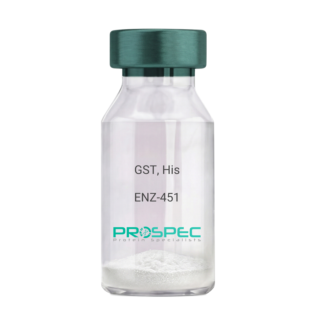
GST, HisDescription:
Glutathione S-Transferase Recombinant, His Tag
Glutathione S-Transferase, GST, Glutathione S-transferase class-mu 28 kDa isozyme, GST 28, EC 2.5.1.18, Sj28GST, Sj28 antigen.
Product # :
ENZ-451Price :
Quantity :
Shipping Method :
Shipped with Ice Packs
More Info
- description
- source
- formulation
- purity
- biological activity
- More Info
Description
Recombinant Schistosoma japonicum GST full length protein contains a total of 244 amino acids (1-218 a.a.) expressed in E.coli, having a molecular mass of 28.3kDa. The GST protein is fused to a 20 amino acids His-Tag at N-terminus. The GST protein is purified by proprietary chromatographic techniques.
Source
Escherichia Coli.
Formulation
GST is supplied in PBS pH 7.4 & 10% glycerol.
Purity
Greater than 90% as determined by SDS-PAGE.
Biological Activity
>10 units/mg, & is defined as the amount of enzyme that conjugate 1.0 u mole of 1-chloro-2,4-dinitrobenzene (CDNB) with reduced glutathione per minute at pH-6.5 at 25C.
More Info
-
Introduction
Antioxidant enzyme Glutathione S- Transferase (GST) is thought to do the primary cellular defense mechanism against reactive oxygen species. GST reduces lipid hydroperoxides through its Se-independent glutathione peroxidase activity. The enzyme also detoxifies lipid peroxidation end products such as 4-hydroxynonenal (4-HNE).
The soluble GST is a 26 kDa protein which occurs as a dimer in all aerobic organisms. Each monomer has two domains, one that binds GSH and is an /-structure similar to thioredoxin and the other, all helical, that binds the hydrophobic substrate. The GST -fusion protein expression system is a widely used recombinant protein expression system that allows a peptide or a regulatory protein domain to be expressed as a fusion to the C-terminus of Schistosoma japonicum GST. Fusion proteins also possess GST -enzymatic activity and can undergo dimerization similar to in vivo. The fusion protein can be purified via GST -affinity column chromatography. In most cases, the desired peptides or domains are removed from GST by applying a specific protease that recognizes and cleaves the linker between the protein domain and GST. The technique has been widely used to generate different kinds of proteins for crystallization, molecular immunology studies, the production of vaccines and studies involving protein-protein and protein-DNA interactions. -
Synonyms
Glutathione S-Transferase, GST, Glutathione S-transferase class-mu 28 kDa isozyme, GST 28, EC 2.5.1.18, Sj28GST, Sj28 antigen.
-
Physical Appearance
Sterile Filtered clear solution.
-
Stability
Store at 4°C if entire vial will be used within 2-4 weeks. Store, frozen at -20°C for longer periods of time. For long term storage it is recommended to add a carrier protein (0.1% HSA or BSA).Avoid multiple freeze-thaw cycles.
-
Amino Acid Sequence
MGSSHHHHHH SSGLVPRGSH MSPILGYWKI KGLVQPTRLL LEYLEEKYEE HLYERDEGDK WRNKKFELGL EFPNLPYYID GDVKLTQSMAIIRYIADKHN MLGGCPKERA EISMLEGAVL DIRYGVSRIA YSKDFETLKV DFLSKLPEML KMFEDRLCHK TYLNGDHVTH PDFMLYDALDVVLYMDPMCL DAFPKLVCFK KRIEAIPQID KYLKSSKYIA WPLQGWQATF GGGDHPPKSD LVPR.
ProSpec's products are furnished for LABORATORY RESEARCH USE ONLY. The product may not be used as drugs, agricultural or pesticidal products, food additives or household chemicals.
- View Data Sheet
Name :
CTRB1 HumanDescription:
Chymotrypsinogen-B1, Human Recombinant
Product # :
ENZ-1016Price :
Quantity :
Shipping Method :
Shipped at Room temp
More Info
- description
- source
- formulation
- purity
- biological activity
- More Info
Description
Recombinant Human CTRB1 expressed in E.coli containing 245 amino acids having a Mw of 27kDa is purified by standard chromatography techniques.
Source
E.coli
Formulation
The Human CTRB1 was lyophilized without any additives.
Purity
Greater than 95% as determined by HPLC.
Biological Activity
1100 units/mg protein.
One unit is defined as the amount of enzyme that will hydrolyze 1.0 μmole of N-alpha-acetyl-L-tyrosine ethyl ester (ATEE) per min at pH 7.0 at 25°C.More Info
-
Introduction
Chymotrypsinogen-B1 (CTRB1) belongs to the serine protease family of enzymes and forms a main precursor of the pancreatic proteolytic enzymes. CTRB1 is located next to a related chymotrypsinogen gene. CTRB1 is a protein coding gene which encodes different isoforms which may undergo similar processing to generate the mature protein.
-
Physical Appearance
Sterile Filtered lyophilized powder.
-
Stability
Recombinant Human CTRB1 although stable at room temp for 1 week, should be stored desiccated below -18°C. For long term storage it is recommended to add a carrier protein (0.1% HSA or BSA).Please prevent freeze-thaw cycles.
-
Solubility
It is recommended to reconstitute the lyophilized Human CTRB1 in 1ml 50mM HAc which can then be further diluted to other aqueous solutions.
-
Amino Acid Sequence
CG VPAIHPVLSG LSRIVNGEDA VPGSWPWQVS LQDKTGFHFC GGSLISEDWV VTAAHCGVRT SDVVVAGEFD QGSDEENIQV LKIAKVFKNP KFSILTVNND ITLLKLATPA RFSQTVSAVC LPSADDDFPAGTLCATTGWG KTKYNANKTP DKLQQAALPL LSNAECKKSW GRRITDVMIC AGASGVSSCM GDSGGPLVCQ KDGAWTLVGI VSWGSDTCST SSPGVYARVTKLIPWVQKIL AAN.
ProSpec's products are furnished for LABORATORY RESEARCH USE ONLY. The product may not be used as drugs, agricultural or pesticidal products, food additives or household chemicals.
- View Data Sheet
Name :
TNFR Human, Sf9Description:
Tumor Necrosis Factor Receptor, sf9 Human Recombinant
Tumor Necrosis Factor Receptor Superfamily Member 9, Tumor Necrosis Factor Receptor Superfamily, Member 9, T-Cell Antigen 4-1BB Homolog, 4-1BB Ligand Receptor, T-Cell Antigen ILA, CD137 Antigen, CDw137, CD137, ILA, Interleukin-Activated Receptor, Homolog Of Mouse Ly63,Induced By Lymphocyte Activation (ILA), Homolog Of Mouse 4-1BB, Receptor Protein 4-1BB, T Cell Antigen ILA, 4-1BB.
Product # :
CYT-951Price :
Quantity :
Shipping Method :
Shipped with Ice Packs
More Info
- description
- source
- formulation
- purity
- More Info
Description
TNFR produced in Sf9 Baculovirus cells is a single, glycosylated polypeptide chain containing 411 amino acids (18-186 a.a.) and having a molecular mass of 45.3kDa. (Migrates at 40-57kDa on SDS-PAGE under reducing conditions). TNFR is expressed with a 239 amino acid hIgG-His-tag at C-Terminus and purified by proprietary chromatographic techniques.
Source
Sf9, Baculovirus cells.
Formulation
TNFR protein solution (0.5mg/ml) contains Phosphate Buffered Saline (pH 7.4) and 10% glycerol.
Purity
Greater than 90.0% as determined by SDS-PAGE.
More Info
-
Introduction
TNFR1 belongs to the TNF-receptor superfamily. TNFR1 is a receptor for TNFSF2/TNF-alpha and homotrimeric TNFSF1/lymphotoxin-alpha.
There are 2 types of soluble TNF receptors: sTNFR-I and sTNFR-II, which act to neutralize the biological activities of TNF alpha and TNF beta. The levels of these soluble receptors seem to increase as a result of shedding of the extracellular domains of the membrane bound receptors. TNF-a, TNFR1 and TNFR2 have roles in cellular differentiation. TNFR1 and TNFR2 function in cell type-specific renal injury.
TNFR1 is capable of signaling both cell survival and apoptosis. TNFR1-induced apoptosis requires 2 sequential signaling complexes. TNFR1 is capable of activating NF-kappaB, mediate apoptosis, and function as a regulator of inflammation. Oxidative stress promotes TNFR1 and TNFR2 self-interaction, ligand-independent and enhanced ligand-dependent TNF signaling. TNFR1 contributes to the induction of non-cytocidal TNF effects including anti-viral state and activation of the acid sphingomyelinase. Human TNFR1 has a major region which controls cell surface expression. High levels of soluble TNF receptors are found in the amniotic fluid of pregnant women.
Germline mutations of the extracellular domains of TNFR1 are linked to the autosomal dominant periodic fever syndrome. The impaired receptor clearance is believed to be a mechanism of the disease. Familial hibernian fever (FHF) is caused by defects in TNFRSF1A gene. -
Synonyms
Tumor Necrosis Factor Receptor Superfamily Member 9, Tumor Necrosis Factor Receptor Superfamily, Member 9, T-Cell Antigen 4-1BB Homolog, 4-1BB Ligand Receptor, T-Cell Antigen ILA, CD137 Antigen, CDw137, CD137, ILA, Interleukin-Activated Receptor, Homolog Of Mouse Ly63,Induced By Lymphocyte Activation (ILA), Homolog Of Mouse 4-1BB, Receptor Protein 4-1BB, T Cell Antigen ILA, 4-1BB.
-
Physical Appearance
Sterile Filtered colorless solution.
-
Stability
Store at 4°C if entire vial will be used within 2-4 weeks. Store, frozen at -20°C for longer periods of time. For long term storage it is recommended to add a carrier protein (0.1% HSA or BSA). Avoid multiple freeze-thaw cycles.
-
Amino Acid Sequence
ADLFERTRSL QDPCSNCPAG TFCDNNRNQI CSPCPPNSFS SAGGQRTCDI CRQCKGVFRT RKECSSTSNA ECDCTPGFHC LGAGCSMCEQ DCKQGQELTK KGCKDCCFGT FNDQKRGICR PWTNCSLDGK SVLVNGTKER DVVCGPSPAD LSPGASSVTP PAPAREPGHS PQLEPKSCDK THTCPPCPAP ELLGGPSVFL FPPKPKDTLM ISRTPEVTCV VVDVSHEDPE VKFNWYVDGV EVHNAKTKPR EEQYNSTYRV VSVLTVLHQD WLNGKEYKCK VSNKALPAPI EKTISKAKGQ PREPQVYTLP PSRDELTKNQ VSLTCLVKGF YPSDIAVEWE SNGQPENNYK TTPPVLDSDG SFFLYSKLTV DKSRWQQGNV FSCSVMHEAL HNHYTQKSLS LSPGKHHHHH H.
ProSpec's products are furnished for LABORATORY RESEARCH USE ONLY. The product may not be used as drugs, agricultural or pesticidal products, food additives or household chemicals.