Search results
1000 results found for “basic transcription factor”
Name
Description
Product #
Price
Quantity
Shipping Method
- View Data Sheet
Name :
GDF7 HumanDescription:
Growth and Differentiation factor 7 Human Recombinant
Growth Differentiation Factor 7, GDF-7, Growth/Differentiation Factor 7, BMP12, GDF7.
Product # :
CYT-870Price :
Quantity :
Shipping Method :
Shipped at Room temp
More Info
- description
- source
- formulation
- purity
- biological activity
- More Info
- Citations
Description
GDF7 Human Recombinant (322-450) produced in E.Coli is a disulfide-linked homodimeric, non-glycosylated, polypeptide chain containing 129 amino acids and having a molecular mass of 28kDa.The GDF-7 is purified by proprietary chromatographic techniques.
Source
Escherichia Coli.
Formulation
Lyophilized from a 0.2µm filtered solution in HCl.
Purity
Greater than 95.0% as determined by SDS-PAGE.
Biological Activity
The ED50, as determined by inducing alkaline phosphatase production by mouse ATDC5 cells, is less than 1.25µg/ml.Citations
More Info
-
Introduction
Growth Differentiation Factor-7 (GDF-7) belongs to the BMP family of TGF-b superfamily proteins. GDF7 elicits its bioactivity via a heterodimeric receptor complex comprised of a type 1 (BMPR-IB) and a type II (BMPR-II or Activin RII) serine/threonine kinase receptor. GDF7 signaling results in the phosphorylation and activation of Smad proteins. GDF-7 is also involved in tendon and ligament formation and repair. In addition, GDF7 regulates bone formation, mesenchymal stem cell differentiation, neuronal differentiation, and axon guidance.
-
Synonyms
Growth Differentiation Factor 7, GDF-7, Growth/Differentiation Factor 7, BMP12, GDF7.
-
Physical Appearance
Sterile Filtered White lyophilized (freeze-dried) powder.
-
Stability
Lyophilized GDF7 although stable at room temperature for 3 weeks, should be stored desiccated below -18°C. Upon reconstitution GDF-7 should be stored at 4°C between 2-7 days and for future use below -18°C.For long term storage it is recommended to add a carrier protein (0.1% HSA or BSA).Please prevent freeze-thaw cycles.
-
Solubility
It is recommended to reconstitute the lyophilized GDF-7 in sterile 18M-cm H2O not less than 100µg/ml, which can then be further diluted to other aqueous solutions.
-
Amino Acid Sequence
TALAGTRTAQ GSGGGAGRGH GRRGRSRCSR KPLHVDFKEL GWDDWIIAPL DYEAYHCEGL CDFPLRSHLE PTNHAIIQTL LNSMAPDAAP ASCCVPARLS PISILYIDAA NNVVYKQYED MVVEACGCR.
-
Background
What is the molecular weight/Mw of GDF7 Protein?
GDF7 Protein has a total Mw of 28kDa.
What is the source or expression system of GDF7 Protein?
Escherichia Coli.
What is the Purity of GDF7 Protein?
GDF7 Protein is >95% pure as determined by SDS-PAGE.
What is the Biological Activity of GDF7 Protein?
The ED50, as determined by inducing alkaline phosphatase production by mouse ATDC5 cells, is less than 1.25µg/ml.
What is the amino acid sequence of GDF7 Protein?
TALAGTRTAQ GSGGGAGRGH GRRGRSRCSR KPLHVDFKEL GWDDWIIAPL DYEAYHCEGL CDFPLRSHLE PTNHAIIQTL LNSMAPDAAP ASCCVPARLS PISILYIDAA NNVVYKQYED MVVEACGCR.
What applications can GDF7 Protein be used in?
GDF7 Protein can probably be used in western blot, ELISA and Lateral Flow.
What is the endotoxin level for GDF7 Protein?
The endotoxin level is minimal, GDF7 Protein was purified using conventional chromatography techniques.
ProSpec's products are furnished for LABORATORY RESEARCH USE ONLY. The product may not be used as drugs, agricultural or pesticidal products, food additives or household chemicals.
- View Data Sheet
Name :
FGFR2 HumanDescription:
Fibroblast Growth Factor Receptor 2 Fc Chimera Human Recombinant
Keratinocyte growth factor receptor 2, CD332, FGFR2.
Product # :
PKA-231Price :
Quantity :
Shipping Method :
Shipped at Room temp
More Info
- description
- source
- formulation
- purity
- biological activity
- More Info
- Citations
Description
Soluble FGFR-2a (IIIc) Fc Chimera Human Recombinant fused with Xa cleavage site with the Fc part of human IgG1 produced in baculovirus is a heterodimeric, glycosylated, Polypeptide chain containing 602 amino acids and having a molecular mass of 170 kDa. The FGFR2 is purified by proprietary chromatographic techniques.
Source
Insect Cells.
Formulation
CD332 was lyophilized from a concentrated (1 mg/ml) sterile solution containing no additives.
Purity
Greater than 90.0% as determined by SDS-PAGE.
Biological Activity
Determined by its ability to inhibit human FGF-2 dependent proliferation on HUVE cells. The ED50 for this effect is typically at 15 - 30ng/ml.
Citations
More Info
-
Introduction
Fibroblast Growth Factors (FGFs) comprise a family of at least eighteen structurally realted proteins that are involved in a multitude of physiological and pathological cellular processes, including cell growth, differentation, angiogenesis, wound healing and tumorgenesis. The biological activities of the FGFs are mediated by a family if type I transmembrane tyrosine kinases which undergo dimerization and autophosphorylation after ligand binding. Four distinct genes encoding closely related FGF receptors, FGFR-1to -4 are known. Multiple forms of FGFR-1 to -3 are generated by alternative splicing of the mRNAs. A frequent splicing event involving FGFR-1 and -2 results in receptors containing all three Ig domains, referred to as the alpha isoform, or only IgII and IgIII, referred to as the ? isoform. Only the alpha isoform has been identified for FGFR-3 and FGFR-4. Additional splicing events for FGFR-1 to -3, involving the C-terminal half of the IgIII domain encoded by two mutually exclusive alternative exons, generate FGF receptors with alternative IgIII domains (IIIb and IIIc). A IIIa isoform which is a secreted FGF binding protein containing only the N-terminal half of the IgIII domain plus some intron sequences has also been reported for FGFR-1. Mutations in FGFR-1 to -3 have been found in patients with birth defects involving craniosynostosis.
-
Synonyms
Keratinocyte growth factor receptor 2, CD332, FGFR2.
-
Physical Appearance
Sterile Filtered White lyophilized (freeze-dried) powder.
-
Stability
Lyophilized FGFR2A although stable at room temperature for 3 weeks, should be stored desiccated below -18°C. Upon reconstitution FGFR2 should be stored at 4°C between 2-7 days and for future use below -18°C. For long term storage it is recommended to add a carrier protein (0.1% HSA or BSA).Please prevent freeze-thaw cycles.
-
Solubility
It is recommended to reconstitute the lyophilized FGFR-2 in sterile PBS not less than 100 µg/ml, which can then be further diluted to other aqueous solutions.
-
Amino Acid Sequence
RPSFSLVEDTTLEPEEPPTKYQISQPEVYVAAPGESLEVRCLLKDAAVISWT KDGVHLGPNNRTVLIGEYLQIKGATPRDSGLYACTASRTVDSETWYFMVNVT DAISSGDDEDDTDGAEDFVSENSNNKRAPYWTNTEKMEKRLHAVPAANTVKF RCPAGGNPMPTMRWLKNGKEFKQEHRIGGYKVRNQHWSLIMESVVPSDKGNY TCVVENEYGSINHTYHLDVVERSPHRPILQAGLPANASTVVGGDVEFVCKVY SDAQPHIQWIKHVEKNGSKYGPDGLPYLKVLKAAGVNTTDKEIEVLYIRNVT FEDAGEYTCLAGNSIGISFHSAWLTVLPAPGREKEITASPDYLEDPRRASIE GRGDPEEPKSCDKTHTCPPCPAPELLGGPSVFLFPPKPKDTLMISRTPEVTC VVVDVSHEDPEVKFNWYVDGVEVHNAKTKPREEQYNSTYRVVSVLTVLHQDW LNGKEYKCKVSNKALPAPIEKTISKAKGQPREPQVYTLPPSRDELTKNQVSL TCLVKGFYPSDIAVEWESNGQPENNYKTTPPVLDSDGSFFLYSKLTVDKSRW QQGNVFSCSVMHEALHNHYTQKSLSLSPGK
ProSpec's products are furnished for LABORATORY RESEARCH USE ONLY. The product may not be used as drugs, agricultural or pesticidal products, food additives or household chemicals.
- View Data Sheet
Name :
TPST1 HumanDescription:
Tyrosylprotein Sulfotransferase 1 Human Recombinant
Tyrosylprotein Sulfotransferase 1, EC 2.8.2.20, TPST-1, Transport And Golgi Organization 13 Homolog A (Drosophila), Transport And Golgi Organization 13 Homolog A, Protein-Tyrosine Sulfotransferase 1, Tyrosylprotein Sulfotransferase-1, TANGO13A, TPST1.
Product # :
ENZ-892Price :
Quantity :
Shipping Method :
Shipped with Ice Packs
More Info
- description
- source
- formulation
- purity
- More Info
- Citations
Description
TPST1 Human Recombinant produced in E.Coli is a single, non-glycosylated polypeptide chain containing 369 amino acids (26-370 a.a) and having a molecular mass of 42kDa.TPST1 is fused to a 24 amino acid His-tag at N-terminus & purified by proprietary chromatographic techniques.
Source
Escherichia Coli.
Formulation
TPST1 protein solution (1mg/ml) containing 20mM Tris-HCl (pH 8.0) and 10% glycerol.
Purity
Greater than 90.0% as determined by SDS-PAGE.
Citations
More Info
-
Introduction
Tyrosylprotein Sulfotransferase 1, also known as TPST1 is the enzyme which catalyzes the sulfation reaction of protein tyrosines, a post-translational modification of proteins. TPST1 belongs to the protein sulfotransferase family. In addition, TPST1 utilizes 3'-Phosphoadenosine-5'-phosphosulfate (PAPS) as the sulfonate donor and also binds proteins with target tyrosineresidues to eventually form the tyrosine O-sulfate ester group in addition to the desulfonated 3’-phosphoadenosine-5’-phosphate.
-
Synonyms
Tyrosylprotein Sulfotransferase 1, EC 2.8.2.20, TPST-1, Transport And Golgi Organization 13 Homolog A (Drosophila), Transport And Golgi Organization 13 Homolog A, Protein-Tyrosine Sulfotransferase 1, Tyrosylprotein Sulfotransferase-1, TANGO13A, TPST1.
-
Physical Appearance
Sterile Filtered colorless solution.
-
Stability
Store at 4°C if entire vial will be used within 2-4 weeks. Store, frozen at -20°C for longer periods of time. For long term storage it is recommended to add a carrier protein (0.1% HSA or BSA).Avoid multiple freeze-thaw cycles.
-
Amino Acid Sequence
MGSSHHHHHH SSGLVPRGSH TGSMQHAMEC HHRIEERSQP VKLESTRTTV RTGLDLKANK TFAYHKDMPL IFIGGVPRSG TTLMRAMLDA HPDIRCGEET RVIPRILALK QMWSRSSKEK IRLDEAGVTD EVLDSAMQAF LLEIIVKHGE PAPYLCNKDP FALKSLTYLS RLFPNAKFLL MVRDGRASVH SMISRKVTIA GFDLNSYRDC LTKWNRAIET MYNQCMEVGY KKCMLVHYEQ LVLHPERWMR TLLKFLQIPW NHSVLHHEEM IGKAGGVSLS KVERSTDQVI KPVNVGALSK WVGKIPPDVL QDMAVIAPML AKLGYDPYAN PPNYGKPDPK IIENTRRVYK GEFQLPDFLK EKPQTEQVE.
ProSpec's products are furnished for LABORATORY RESEARCH USE ONLY. The product may not be used as drugs, agricultural or pesticidal products, food additives or household chemicals.
- View Data Sheet
Name :
TNF a CanineDescription:
Tumor Necrosis Factor-Alpha Canine Recombinant
Tumor necrosis factor, Cachectin, TNF-alpha, Tumor necrosis factor ligand superfamily member 2, TNF-a, TNF, TNFA, TNFSF2.
Product # :
CYT-140Price :
Quantity :
Shipping Method :
Shipped at Room temp
More Info
- description
- source
- formulation
- purity
- biological activity
- More Info
- Citations
Description
TNF-a Canine Recombinant produced in E.Coli is a single, non-glycosylated polypeptide chain containing 157 amino acids and having a molecular mass of 17.3 kDa. The TNF-a is purified by proprietary chromatographic techniques.
Source
Escherichia Coli.
Formulation
Filtered (0.2µm) and lyophilized from a concentrated (1mg/ml) solution in 1×PBS, pH7.4.
Purity
Greater than 95.0% as determined by:
(a) Analysis by RP-HPLC.
(b) Analysis by SDS-PAGE.Biological Activity
The Specific Activity is >3.3×10⁵ IU/mg as determined by the cytolysis of murine L929 cells in the presence of Actinomycin D.Citations
More Info
-
Introduction
Tumor necrosis factor is a cytokine involved in systemic inflammation and is a member of a group of cytokines that all stimulate the acute phase reaction. TNF is mainly secreted by macrophages.
TNF causes apoptotic cell death, cellular proliferation, differentiation, inflammation, tumorigenesis and viral replication, TNF is also involved in lipid metabolism, and coagulation. TNF's primary role is in the regulation of immune cells.
Dysregulation and, in particular, overproduction of TNF have been implicated in a variety of human diseases- autoimmune diseases, insulin resistance, and cancer. -
Synonyms
Tumor necrosis factor, Cachectin, TNF-alpha, Tumor necrosis factor ligand superfamily member 2, TNF-a, TNF, TNFA, TNFSF2.
-
Physical Appearance
Sterile Filtered White lyophilized (freeze-dried) powder.
-
Stability
Lyophilized Tumor Necrosis Factor-a although stable at room temperature for 3 weeks, should be stored desiccated below -18°C. Upon reconstitution TNF-a should be stored at 4°C between 2-7 days and for future use below -18°C.For long term storage it is recommended to add a carrier protein (0.1% HSA or BSA).Please prevent freeze-thaw cycles.
-
Solubility
It is recommended to reconstitute the lyophilized Tumor Necrosis Factor-alpha in sterile 18M-cm H2O not less than 100µg/ml, which can then be further diluted to other aqueous solutions.
-
Amino Acid Sequence
VKSSSRTPSD KPVAHVVANP EAEGQLQWLS RRANALLANG VELTDNQLIV PSDGLYLIYS QVLFKGQGCP STHVLLTHTI SRFAVSYQTK VNLLSAIKSP CQRETPEGTE AKPWYEPIYL GGVFQLEKGD RLSAEINLPN YLDFAESGQV YFGIIAL.
ProSpec's products are furnished for LABORATORY RESEARCH USE ONLY. The product may not be used as drugs, agricultural or pesticidal products, food additives or household chemicals.
- View Data Sheet
Name :
LFA 3 HumanDescription:
Lymphocyte Function Associated Antigen-3 Human Recombinant , Fusion Protein
CD58, LFA-3, Ag3, Surface glycoprotein LFA-3.
Product # :
CYT-423Price :
Quantity :
Shipping Method :
Shipped at Room temp
More Info
- description
- source
- formulation
- purity
- More Info
- Citations
Description
Lymphocyte Function-Associated Antigen-3 Fusion Protein Recombinant Human is produced by recombinant DNA technology in a Chinese Hamster Ovary (CHO) mammalian cell expression system. The molecular weight is 91.4 kDa. Recombinant LFA3 is purified by proprietary chromatographic techniques.
Source
Chinese Hamster Ovary.
Formulation
Each mg of CD58 contains 0.8mg sucrose, 0.3mg glycine, 0.25mg sodium citrate dihydrate, and 4µg citric acid monohydrate.
Purity
Greater than 98.0% as determined by(a) Analysis by RP-HPLC.
(b) Analysis by SDS-PAGE.Citations
More Info
-
Introduction
LFA-3 is ligand of the t-lymphocyte cd2 glycoprotein. This interaction is important in mediating thymocyte interactions with thymic epithelial cells, antigen-independent and dependent interactions of t-lymphocytes with target cells and antigen- presenting cells and the t-lymphocyte rosetting with erythrocytes. In addition, the lfa-3/cd2 interaction may prime response by both the cd2+ and lfa-3+ cells.
-
Synonyms
CD58, LFA-3, Ag3, Surface glycoprotein LFA-3.
-
Physical Appearance
Sterile Filtered White lyophilized (freeze-dried) powder.
-
Stability
Lyophilized LFA3 Human although stable at room temperature for 3 weeks, should be stored desiccated below -18°C. Upon reconstitution Human LFA-3 should be stored at 4°C between 2-7 days and for future use below -18°C. For long term storage it is recommended to add a carrier protein (0.1% HSA or BSA).Please prevent freeze-thaw cycles.
-
Solubility
It is recommended to reconstitute the lyophilized LFA-3 Human in sterile 18MΩ-cm H2O not less than 100µg/ml, which can then be further diluted to other aqueous solutions.
ProSpec's products are furnished for LABORATORY RESEARCH USE ONLY. The product may not be used as drugs, agricultural or pesticidal products, food additives or household chemicals.
- View Data Sheet
Name :
b NGF MouseDescription:
Beta-Nerve Growth Factor Mouse
Beta Polypeptide, NGF, NGFB, HSAN5, Beta-NGF, MGC161426, MGC161428.
Product # :
CYT-440Price :
Quantity :
Shipping Method :
Shipped at Room temp
More Info
- description
- source
- formulation
- purity
- biological activity
- More Info
- Citations
Description
NGF beta Mouse produced in Submaxillary Gland of Grown Mouse is a homodimer, non-glycosylated, polypeptide chain containing 2 identical 120 amino acids and having a molecular mass of 13,471 Dalton each.The NGF beta Mouse is purified by advanced biology purification technology.
Source
Submaxillary Gland of Grown Mouse.
Formulation
The NGF beta Mouse was lyophilized from solution containing 5% mannitol and 1% HSA.
Purity
Greater than 98% as determined by(a) Analysis by RP-HPLC.
(b) Analysis by SDS-PAGE .Biological Activity
The method used to test the bioassay is the NGF-dependent survival of dorsal root ganglia neurons of chick embryo, corresponding to a Specific Activity of 500,000IU/mg.Citations
More Info
-
Introduction
NGF-beta has nerve growth stimulating activity and the complex is involved in the regulation of growth and the differentiation of sympathetic and certain sensory neurons. Mutations in this gene have been associated with hereditary sensory and autonomic neuropathy, type 5 (HSAN5), and dysregulation of this gene's expression is associated with allergic rhinitis.
-
Synonyms
Beta Polypeptide, NGF, NGFB, HSAN5, Beta-NGF, MGC161426, MGC161428.
-
Physical Appearance
Sterile Filtered White lyophilized (freeze-dried) powder.
-
Stability
Lyophilized Mouse Beta-NGF although stable at room temperature for 3 weeks, should be stored desiccated below -18°C. Upon reconstitution Murine NGF-Beta should be stored at 4°C between 2-7 days and for future use below -18°C.Please prevent freeze-thaw cycles.
-
Solubility
It is recommended to reconstitute the lyophilized Murine NGF-beta in sterile 18MΩ-cm H2O not less than 100µg/ml, which can then be further diluted to other aqueous solutions.
-
Amino Acid Sequence
SSTHPVFHMGEF SVCDSVSVWV GDKTTATDIK GKEVTVLAEV NINNSVFRQY FFETKCRASN PVESGCRGID SKHWNSYCTT THTFVKALTT DEKQAAWRFI RIDTACVCVL SRKATRRG.
-
Background
What is the molecular weight/Mw of B NGF Protein?
B NGF Protein has a total Mw of 13kDa.
What is the source or expression system of B NGF Protein?
Submaxillary Gland of Grown Mouse.
What is the Purity of B NGF Protein?
B NGF Protein is >98% pure as determined by SDS-PAGE.
What is the Biological Activity of B NGF Protein?
The method used to test the bioassay is the NGF-dependent survival of dorsal root ganglia neurons of chick embryo, corresponding to a Specific Activity of 500,000IU/mg.
What is the amino acid sequence of B NGF Protein?
SSTHPVFHMGEF SVCDSVSVWV GDKTTATDIK GKEVTVLAEV NINNSVFRQY FFETKCRASN PVESGCRGID SKHWNSYCTT THTFVKALTT DEKQAAWRFI RIDTACVCVL SRKATRRG.
What applications can B NGF Protein be used in?
B NGF Protein can probably be used in western blot, ELISA and Lateral Flow.
What is the endotoxin level for XXX Protein?
The endotoxin level is minimal, XXX Protein was purified using conventional chromatography techniques.
ProSpec's products are furnished for LABORATORY RESEARCH USE ONLY. The product may not be used as drugs, agricultural or pesticidal products, food additives or household chemicals.
- View Data Sheet
Name :
EIF3I HumanDescription:
Eukaryotic Translation Initiation Factor 3I Human Recombinant
Eukaryotic Translation Initiation Factor 3, Subunit I, EIF3S2, Eukaryotic Translation Initiation Factor 3, Subunit 2 Beta, 36kDa, Eukaryotic Translation Initiation Factor 3 Subunit 2, TRIP-1, eIF-3-beta, EIF3 P36, TGF-Beta Receptor-Interacting Protein 1, PRO2242, eIF3-beta, eIF3-p36, Eukaryotic Translation Initiation Factor 3 Subunit I, Eukaryotic Translation Initiation Factor 3, Subunit 2 (Beta, 36kD), Predicted Protein Of HQ2242, TGFbeta Receptor-Interacting Protein 1, eIF3i.
Product # :
PRO-1740Price :
Quantity :
Shipping Method :
Shipped with Ice Packs
More Info
- description
- source
- formulation
- purity
- More Info
- Citations
Description
EIF3I Human Recombinant produced in E.Coli is a single, non-glycosylated polypeptide chain containing 348 amino acids (1-325aa) and having a molecular mass of 38.9kDa.EIF3I is fused to a 23 amino acid His-tag at N-terminus & purified by proprietary chromatographic techniques.
Source
Escherichia Coli.
Formulation
APOBEC4 protein solution (1mg/ml) containing 20mM Tris-HCl buffer (pH 8.0), 0.4M UREA and 10% glycerol.
Purity
Greater than 90% as determined by SDS-PAGE.
Citations
More Info
-
Introduction
Eukaryotic translation initiation factor 3, subunit I (EIF3I) is part of the eukaryotic translation initiation factor 3 (eIF-3) complex, which is essential for numerous steps in the initiation of protein synthesis. The eIF-3 complex links with the 40S ribosome and facilitates the recruitment of eIF-1, eIF-1A, eIF-2: GTP: methionyl-tRNAi and eIF-5 to form the 43S pre-initiation complex (43S PIC). The eIF-3 complex stimulates mRNA recruitment to the 43S PIC and scanning of the mRNA for AUG recognition. The eIF-3 complex is also essential for disassembly and recycling of post-termination ribosomal complexes and subsequently prevents premature joining of the 40S and 60S ribosomal subunits prior to initiation. Among the diseases associated with EIF3I are clonorchiasis, and tonsillitis.
-
Synonyms
Eukaryotic Translation Initiation Factor 3, Subunit I, EIF3S2, Eukaryotic Translation Initiation Factor 3, Subunit 2 Beta, 36kDa, Eukaryotic Translation Initiation Factor 3 Subunit 2, TRIP-1, eIF-3-beta, EIF3 P36, TGF-Beta Receptor-Interacting Protein 1, PRO2242, eIF3-beta, eIF3-p36, Eukaryotic Translation Initiation Factor 3 Subunit I, Eukaryotic Translation Initiation Factor 3, Subunit 2 (Beta, 36kD), Predicted Protein Of HQ2242, TGFbeta Receptor-Interacting Protein 1, eIF3i.
-
Physical Appearance
Sterile Filtered clear solution.
-
Stability
Store at 4°C if entire vial will be used within 2-4 weeks. Store, frozen at -20°C for longer periods of time.For long term storage it is recommended to add a carrier protein (0.1% HSA or BSA).Please avoid freeze thaw cycles.
-
Amino Acid Sequence
MGSSHHHHHH SSGLVPRGSH MGSMKPILLQ GHERSITQIK YNREGDLLFT VAKDPIVNVW YSVNGERLGT YMGHTGAVWC VDADWDTKHV LTGSADNSCR LWDCETGKQL ALLKTNSAVR TCGFDFGGNI IMFSTDKQMG YQCFVSFFDL RDPSQIDNNE PYMKIPCNDS KITSAVWGPL GECIIAGHES GELNQYSAKS GEVLVNVKEH SRQINDIQLS RDMTMFVTAS KDNTAKLFDS TTLEHQKTFR TERPVNSAAL SPNYDHVVLG GGQEAMDVTT TSTRIGKFEA RFFHLAFEEE FGRVKGHFGP INSVAFHPDG KSYSSGGEDG YVRIHYFDPQ YFEFEFEA
ProSpec's products are furnished for LABORATORY RESEARCH USE ONLY. The product may not be used as drugs, agricultural or pesticidal products, food additives or household chemicals.
- View Data Sheet
Name :
TNFR2 Human, Sf9Description:
Tumor Necrosis Factor Receptor Type 2 Human Recombinant, Sf9
Tumor Necrosis Factor Receptor Superfamily, Member 1B, TNFR2, TNFBR, Tumor Necrosis Factor Receptor Type II, Tumor Necrosis Factor Receptor 2, P80 TNF-Alpha Receptor, TNF-RII, TNF-R2, P75, Tumor Necrosis Factor Receptor Superfamily Member 1B, Tumor Necrosis Factor Binding Protein 2, Tumor Necrosis Factor Beta Receptor, Soluble TNFR1B Variant 1, P75 TNF Receptor, CD120b Antigen, Etanercept, TNF-R-II, TNF-R75, P75TNFR, TNFR-II, CD120b, TNFR1B, TNFR80, TBPII.
Product # :
CYT-908Price :
Quantity :
Shipping Method :
Shipped with Ice Packs
More Info
- description
- source
- formulation
- purity
- biological activity
- More Info
- Citations
Description
TNFR2 produced in Sf9 Baculovirus cells is a single, glycosylated polypeptide chain (23-257 a.a.) and fused to a 6 aa His Tag at C-terminus containing a total of 241 amino acids and having a molecular mass of 25.9kDa. TNFR2 is purified by proprietary chromatographic techniques.
Source
Sf9, Baculovirus cells.
Formulation
TNFR2 protein solution (1mg/ml) contains Phosphate buffered saline (pH7.4) and 10% glycerol.
Purity
Greater than 95.0% as determined by SDS-PAGE.
Biological Activity
The ED50 range ≤ 0.2 μg/ml and is measured by its ability to inhibit cytotoxicity using L-929 mouse fibroblast cells in the presence of Human TNF-α.
Citations
More Info
-
Introduction
TNFR2 belongs to the TNF-receptor superfamily. TNFR2 is receptor with high affinity for TNFSF2/TNF-alpha and approximately 5-fold lower affinity for homotrimeric TNFSF1/lymphotoxin-alpha. TNFR2 mediates the majority of the metabolic effects of TNF-alpha. In addition, knockout studies in mice propose a role for TNFR2 in protecting neurons from apoptosis by stimulating antioxidative pathways. TNFR2 expression might have a significant role in the angiogenesis, tumor cell proliferation and metastasis of Invasive micropapillary carcinoma of the breast.
There are 2 types of soluble TNF receptors: sTNFR-I and sTNFR-II, which act to neutralize the biological activities of TNF alpha and TNF beta. The levels of these soluble receptors seem to increase as a result of shedding of the extracellular domains of the membrane bound receptors. High levels of soluble TNF receptors are found in the amniotic fluid of pregnant women. TNFR2 and TNFR1 form a heterocomplex which mediates the recruitment of 2 anti-apoptotic proteins, c-IAP1 and c-IAP2, which possess E3 ubiquitin ligase activity. IAPs’ function in TNF-receptor signaling is unknown; nevertheless, c-IAP1 is believed to potentiate TNF-induced apoptosis by the ubiquitination and degradation of TNF-receptor-associated factor 2, which mediates anti-apoptotic signals. Oxidative stress promotes TNFR1 and TNFR2 self-interaction, ligand-independent and enhanced ligand-dependent TNF signaling. TNF-a, TNFR1 and TNFR2 have roles in cellular differentiation. TNFR1 and TNFR2 function in cell type-specific renal injury. -
Synonyms
Tumor Necrosis Factor Receptor Superfamily, Member 1B, TNFR2, TNFBR, Tumor Necrosis Factor Receptor Type II, Tumor Necrosis Factor Receptor 2, P80 TNF-Alpha Receptor, TNF-RII, TNF-R2, P75, Tumor Necrosis Factor Receptor Superfamily Member 1B, Tumor Necrosis Factor Binding Protein 2, Tumor Necrosis Factor Beta Receptor, Soluble TNFR1B Variant 1, P75 TNF Receptor, CD120b Antigen, Etanercept, TNF-R-II, TNF-R75, P75TNFR, TNFR-II, CD120b, TNFR1B, TNFR80, TBPII.
-
Physical Appearance
Sterile filtered colorless solution.
-
Stability
Store at 4°C if entire vial will be used within 2-4 weeks. Store, frozen at -20°C for longer periods of time. For long term storage it is recommended to add a carrier protein (0.1% HSA or BSA).Avoid multiple freeze-thaw cycles.
-
Amino Acid Sequence
LPAQVAFTPY APEPGSTCRL REYYDQTAQM CCSKCSPGQH AKVFCTKTSD TVCDSCEDST YTQLWNWVPE CLSCGSRCSS DQVETQACTR EQNRICTCRP GWYCALSKQE GCRLCAPLRK CRPGFGVARP GTETSDVVCK PCAPGTFSNT TSSTDICRPH QICNVVAIPG NASMDAVCTS TSPTRSMAPG AVHLPQPVST RSQHTQPTPE PSTAPSTSFL LPMGPSPPAE GSTGDHHHHH H
ProSpec's products are furnished for LABORATORY RESEARCH USE ONLY. The product may not be used as drugs, agricultural or pesticidal products, food additives or household chemicals.
- View Data Sheet
Name :
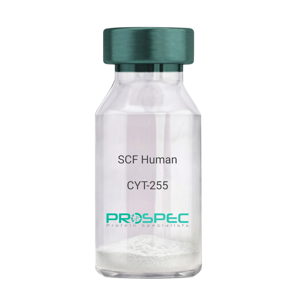
SCF HumanDescription:
Stem Cell Factor Human Recombinant
Kit ligand Precursor, C-kit ligand, SCF, Mast cell growth factor, MGF, SF, KL-1, Kitl, DKFZp686F2250.
Product # :
CYT-255Price :
Quantity :
Shipping Method :
Shipped at Room temp
More Info
- description
- source
- formulation
- purity
- biological activity
- More Info
- Citations
Description
Stem Cell Factor Human Recombinant produced in E.Coli is a single, non-glycosylated polypeptide chain containing 165 amino acids and having a molecular mass of 18409 Dalton. The SCF is purified by proprietary chromatographic techniques.
Source
Escherichia Coli.
Formulation
Lyophilized from a concentrated (1mg/ml) solution in water containing 0.02% NaHCO3.
Purity
Greater than 98.0% as determined by:
(a) Analysis by SEC-HPLC.
(b) Analysis by SDS-PAGE.Biological Activity
The ED50 as determined by the dose-dependant stimulation of Human TF-1 cells is < 2 ng/ml, corresponding to a Specific Activity of 500,000 IU/mg.Citations
More Info
-
Introduction
Stem cell factor / KIT ligand (SCF) is a cytokine which binds CD117(c-Kit). SCF is also known as "steel factor" or "c-kit ligand". SCF exists in two forms, cell surface bound SCF and soluble (or free) SCF. Soluble SCF is produced by the cleavage of surface bound SCF by metalloproteases. SCF is a growth factor important for the survival, proliferation, and differentiation of hematopoietic stem cells and other hematopoietic progenitor cells. One of its roles is to change the BFU-E (burst-forming unit-erythroid) cells, which are the earliest erythrocyte precursors in the erythrocytic series, into the CFU-E (colony-forming unit-erythroid).
-
Synonyms
Kit ligand Precursor, C-kit ligand, SCF, Mast cell growth factor, MGF, SF, KL-1, Kitl, DKFZp686F2250.
-
Physical Appearance
Sterile Filtered White lyophilized (freeze-dried) powder.
-
Stability
Lyophilized KIT ligand although stable at room temperature for 3 weeks, should be stored desiccated below -18°C. Upon reconstitution SCF should be stored at 4°C between 2-7 days and for future use below -18°C.For long term storage it is recommended to add a carrier protein (0.1% HSA or BSA).Please prevent freeze-thaw cycles.
-
Solubility
It is recommended to reconstitute the lyophilized Stem Cell Factor in sterile 18MΩ-cm H2O not less than 100µg/ml, which can then be further diluted to other aqueous solutions.
-
Amino Acid Sequence
The sequence of the first five N-terminal amino acids was determined and was found to be Met-Glu-Gly-Ile-Cys.
-
Protein content
Protein quantitation was carried out by two independent methods1. UV spectroscopy at 280 nm using the absorbency value of 0.52 as the extinction coefficient for a 0.1% (1mg/ml) solution. This value is calculated by the PC GENE computer analysis program of protein sequences (IntelliGenetics). 2. Analysis by RP-HPLC, using a standard solution of Stem Cell Factor as a Reference Standard.
ProSpec's products are furnished for LABORATORY RESEARCH USE ONLY. The product may not be used as drugs, agricultural or pesticidal products, food additives or household chemicals.
- View Data Sheet
Name :
FGF 2 HumanDescription:
Fibroblast Growth Factor-Basic Human Recombinant
Prostatropin, FGF-basic, fgf2, Basic FGF, HBGF-2, FGF-2, FGF-b.
Product # :
CYT-218Price :
Quantity :
Shipping Method :
Shipped at Room temp
More Info
- description
- source
- formulation
- purity
- biological activity
- More Info
- Citations
Description
Fibroblast Growth Factor-2 Human Recombinant (FGF-2) produced in E.Coli is a single, non-glycosylated, polypeptide chain containing 154 amino acids and having a molecular mass of 17.2kDa.The FGF-b is purified by proprietary chromatographic techniques.
Source
Escherichia Coli.
Formulation
The protein was lyophilized from a concentrated (1mg/ml) solution in 20mM Tris-HCl, pH7.4 and 1M NaCl.
Purity
Greater than 98.0% as determined by Analysis by SDS-PAGE.
Biological Activity
The ED50, calculated by the dose-dependant proliferation of murine balb/c 3T3 cells is <0.1ng/ml, corresponding to a specific activity of graeter than 1.0x10⁷ Units/mg.
Citations
More Info
-
Introduction
Basic fibroblast growth factor is a member of the fibroblast growth factor (FGF) family. FGF family members possess broad mitogenic and cell survival activities, and are involved in a variety of biological processes, including embryonic development, cell growth, morphogenesis, tissue repair, tumor growth and invasion. This protein functions as a modifier of endothelial cell migration and proliferation, as well as an angiogenic factor. It acts as a mitogen for a variety of mesoderm- and neuroectoderm-derived cells in vitro, thus is thought to be involved in organogenesis. Three alternatively spliced variants encoding different isoforms have been described. The HPR -binding growth factors are angiogenic agents in vivo and are potent mitogens for a variety of cell types in vitro. There are differences in the tissue distribution and concentration of these 2 growth factors.
-
Synonyms
Prostatropin, FGF-basic, fgf2, Basic FGF, HBGF-2, FGF-2, FGF-b.
-
Physical Appearance
Sterile Filtered White lyophilized (freeze-dried) powder.
-
Stability
Lyophilized Fibroblast Growth Factor-2 although stable at room temperature for 3 weeks, should be stored desiccated below -18°C. Upon reconstitution FGF-b should be stored at 4°C between 2-7 days and for future use below -18°C.For long term storage it is recommended to add a carrier protein (0.1% HSA or BSA).Please prevent freeze-thaw cycles.
-
Solubility
It is recommended to reconstitute the lyophilized Fibroblast Growth Factor Basic in sterile 18MΩ-cm H2O not less than 100µg/ml, which can then be further diluted to other aqueous solutions.
-
Amino Acid Sequence
AAGSITTLPA LPEDGGSGAF PPGHFKDPKR LYCKNGGFFL RIHPDGRVDG VREKSDPHIK LQLQAEERGV VSIKGVCANR YLAMKEDGRL LASKCVTDEC FFFERLESNN YNTYRSRKYT SWYVALKRTG QYKLGSKTGP GQKAILFLPM SAKS.
-
Background
FGF 2 HUMAN: Insights into Fibroblast Growth Factor-2
Basic Fibroblast Growth Factor or FGF 2 HUMAN is a protein with crucial roles in cell growth, tissue repair, and embryonic development. This is part of the larger fibroblast growth factor family and is vital for various biological processes, including the modulation of cell survival activities.
Production and Properties
Produced in E. coli, FGF 2 is a non-glycosylated polypeptide chain possessing 154 amino acids with a molecular weight of about 17.2 kDa. It is purified through advanced chromatographic techniques, ensuring high purity and activity for laboratory use.
Physical Characteristics and Preparation
The physical form of FGF 2 HUMAN is a sterile, white lyophilized powder. For experimental use, it is reconstituted with sterile water to at least 100µg/ml. This reconstitution is crucial for maintaining the integrity and effectiveness of the protein in various research applications.
Storage and Handling
To maintain stability, lyophilized FGF 2 should be stored at -18°C and used within three weeks if kept at room temperature. Once reconstituted, it should be kept at 4°C and used within 2-7 days or stored at -18°C for longer-term storage.
Proper handling and avoiding repeated freeze-thaw cycles are essential to preserve the protein's functionality.
Purity and Biological Activity
FGF 2 is characterized by a purity greater than 98%, verified by SDS-PAGE analysis. Its biological activity is primarily defined by its efficacy in promoting the proliferation of specific cell lines, with an effective dose (ED50) typically below 0.1 ng/ml.
Research Applications and Impact
In the research context, FGF 2 is used extensively to study its effects on cell migration, proliferation, and angiogenesis. Moreover, its role in disease models, particularly in cancer and tissue repair studies, makes it a valuable resource for developing new therapeutic approaches.
Usage Guidelines
FGF 2 HUMAN is strictly for laboratory research use and is not suitable for drug development, food production, or cosmetic applications. Researchers are advised to comply with safety and handling guidelines to ensure that experiments are conducted under optimal conditions.
The Broad Impact on Development and Disease
FGF-2 is known for its multifunctional role across numerous biological processes such as tissue repair, embryonic development, angiogenesis, and even tumorigenesis.
This growth factor, existing in various synonymous forms such as Basic FGF, FGF-b, and HBGF-2, is essential in cellular processes that underpin both health and disease.
Furthermore, FGF-2's ability to bind to cellular receptors triggers a cascade of signaling pathways, including PI3K/Akt, MAPK/ERK, and PLCγ, which in turn influence cell growth, migration, and survival.
These pathways are pivotal in mediating the factor's diverse effects on cell behavior, contributing to its critical roles in wound healing, angiogenesis, and tissue remodeling.
What is the molecular weight/Mw of FGF 2 Protein?
FGF 2 Protein has a total Mw of 17.2kDa.
What is the source or expression system of FGF 2 Protein?
Escherichia Coli.
What is the Purity of FGF 2 Protein?
FGF 2 Protein is >98% pure as determined by SDS-PAGE.
What is the Biological Activity of FGF 2 Protein?
The ED50, calculated by the dose-dependant proliferation of murine balb/c 3T3 cells is <0.1ng/ml, corresponding to a specific activity of graeter than 1.0x10⁷ Units/mg.
What is the amino acid sequence of FGF 2 Protein?
AAGSITTLPA LPEDGGSGAF PPGHFKDPKR LYCKNGGFFL RIHPDGRVDG VREKSDPHIK LQLQAEERGV VSIKGVCANR YLAMKEDGRL LASKCVTDEC FFFERLESNN YNTYRSRKYT SWYVALKRTG QYKLGSKTGP GQKAILFLPM SAKS.
What applications can FGF 2 Protein be used in?
FGF 2 Protein can probably be used in western blot, ELISA and Lateral Flow.
What is the endotoxin level for FGF 2 Protein?
The endotoxin level is minimal, FGF 2 Protein was purified using conventional chromatography techniques.
ProSpec's products are furnished for LABORATORY RESEARCH USE ONLY. The product may not be used as drugs, agricultural or pesticidal products, food additives or household chemicals.
- View Data Sheet
Name :
MED27 HumanDescription:
Mediator Complex Subunit 27 Human Recombinant
Mediator Complex Subunit 27, MED27, CRSP8, Cofactor Required For Sp1 Transcriptional Activation, Subunit 8 34kDa, P37 TRAP/SMCC/PC2 Subunit, Transcriptional Coactivator CRSP34, CRSP34, CRSP Complex Subunit 8, CRAP34, TRAP37, Mediator of RNA Polymerase II Transcription Subunit 27, Cofactor Required For Sp1 Transcriptional Activation Subunit 8.
Product # :
PRO-1946Price :
Quantity :
Shipping Method :
Shipped with Ice Packs
More Info
- description
- source
- formulation
- purity
- More Info
- Citations
Description
MED27 Human Recombinant produced in E.coli is a single, non-glycosylated polypeptide chain containing 334 amino acids (1-311) and having a molecular mass of 37.8 kDa.MED27 is fused to a 23 amino acid His-tag at N-terminus & purified by proprietary chromatographic techniques.
Source
E.coli.
Formulation
The MED27 solution (0.5mg/ml) contains 20mM Tris-HCl buffer (pH 8.0), 0.4M Urea and 10% glycerol.
Purity
Greater than 80% as determined by SDS-PAGE.
Citations
More Info
-
Introduction
Mediator Complex Subunit 27 (MED27) is a subunit of the CRSP (cofactor required for SP1 activation) complex, which, together with TFIID, is necessary for efficient activation by SP1. MED27 is a component of the Mediator complex, a coactivator involved in the regulated transcription of virtually all RNA polymerase II-dependent genes. MED27 protein is also a component of other multisubunit complexes such as the thyroid hormone receptor-(TR-) associated proteins which interact with TR and facilitate TR function on DNA templates in conjunction with initiation factors and cofactors. The activation of MED27 gene transcription is a multistep process, which is triggered by factors that identify transcriptional enhancer sites in DNA.
-
Synonyms
Mediator Complex Subunit 27, MED27, CRSP8, Cofactor Required For Sp1 Transcriptional Activation, Subunit 8 34kDa, P37 TRAP/SMCC/PC2 Subunit, Transcriptional Coactivator CRSP34, CRSP34, CRSP Complex Subunit 8, CRAP34, TRAP37, Mediator of RNA Polymerase II Transcription Subunit 27, Cofactor Required For Sp1 Transcriptional Activation Subunit 8.
-
Physical Appearance
Sterile Filtered colorless solution.
-
Stability
Store at 4°C if entire vial will be used within 2-4 weeks. Store, frozen at -20°C for longer periods of time. For long term storage it is recommended to add a carrier protein (0.1% HSA or BSA).Avoid multiple freeze-thaw cycles.
-
Amino Acid Sequence
MGSSHHHHHH SSGLVPRGSH MGSMADVINV SVNLEAFSQA ISAIQALRSS VSRVFDCLKD GMRNKETLEG REKAFIAHFQ DNLHSVNRDL NELERLSNLV GKPSENHPLH NSGLLSLDPV QDKTPLYSQL LQAYKWSNKL QYHAGLASGL LNQQSLKRSA NQMGVSAKRR PKAQPTTLVL PPQYVDDVIS RIDRMFPEMS IHLSRPNGTS AMLLVTLGKV LKVIVVMRSL FIDRTIVKGY NENVYTEDGK LDIWSKSNYQ VFQKVTDHAT TALLHYQLPQ MPDVVVRSFM TWLRSYIKLF QAPCQRCGKF LQDGLPPTWR DFRTLEAFHD TCRQ.
ProSpec's products are furnished for LABORATORY RESEARCH USE ONLY. The product may not be used as drugs, agricultural or pesticidal products, food additives or household chemicals.
- View Data Sheet
Name :
M CSF Human, Sf9 HisDescription:
Macrophage Colony Stimulating Factor Human Recombinant, Sf9
Macrophage colony-stimulating factor 1, CSF-1, M-CSF, MCSF, Lanimostim.
Product # :
CYT-1015Price :
Quantity :
Shipping Method :
Shipped with Ice Packs
More Info
- description
- source
- formulation
- purity
- biological activity
- More Info
- Citations
Description
MCSF produced in Sf9 Insect cells is a single, glycosylated polypeptide chain containing 231 amino acids (33-255 a.a.) and having a molecular mass of 26.1kDa (Molecular size on SDS-PAGE will appear at approximately 28-40kDa).MCSF is expressed with an 8 amino acid His tag at C-Terminus and purified by proprietary chromatographic techniques.
Source
Sf9, Insect cells.
Formulation
MCSF protein solution (0.25mg/ml) contains Phosphate Buffered Saline (pH 7.4) and 10% glycerol.
Purity
Greater than 95.0% as determined by SDS-PAGE.
Biological Activity
Measured in a cell proliferation assay using M-NFS-60 mouse myelogenous leukemia lymphoblast cell. The ED50 for this effect is less or equal to 3 ng/ml.
Citations
More Info
-
Introduction
Granulocyte/Macrophage Colony-Stimulating Factors are cytokines that act in hematopoiesis by controlling the production, differentiation, and function of 2 related white cell populations of the blood, the granulocytes and the monocytes-macrophages. MCSF induces cells of the monocyte/macrophage lineage. MCSF plays a role in immunological defenses, bone metabolism, lipoproteins clearance, fertility and pregnancy.
-
Synonyms
Macrophage colony-stimulating factor 1, CSF-1, M-CSF, MCSF, Lanimostim.
-
Physical Appearance
Sterile filtered colorless solution.
-
Stability
Store at 4°C if entire vial will be used within 2-4 weeks. Store, frozen at -20°C for longer periods of time. For long term storage it is recommended to add a carrier protein (0.1% HSA or BSA).Avoid multiple freeze-thaw cycles.
-
Amino Acid Sequence
EEVSEYCSHM IGSGHLQSLQ RLIDSQMETS CQITFEFVDQ EQLKDPVCYL KKAFLLVQDI MEDTMRFRDN TPNAIAIVQL QELSLRLKSC FTKDYEEHDK ACVRTFYETP LQLLEKVKNV FNETKNLLDK DWNIFSKNCN NSFAECSSQD VVTKPDCNCL YPKAIPSSDP ASVSPHQPLA PSMAPVAGLT WEDSEGTEGS SLLPGEQPLH TVDPGSAKQR PPRLEHHHHH H.
ProSpec's products are furnished for LABORATORY RESEARCH USE ONLY. The product may not be used as drugs, agricultural or pesticidal products, food additives or household chemicals.
- View Data Sheet
Name :
BDNF Human, CHODescription:
Brain-Derived Neurotrophic Factor Human Recombinant, CHO
Brain-Derived Neurotrophic Factor, BDNF, MGC34632.
Product # :
CYT-1262Price :
Quantity :
Shipping Method :
Shipped at Room temp
More Info
- description
- source
- formulation
- purity
- biological activity
- More Info
- sds-page
- Citations
Description
Brain-derived Neurotrophic Factor Human Recombinant produced in CHO is a homodimer, polypeptide chain containing 2 x 119 amino acids and having a total molecular mass of 27kDa. BDNF Human Recombinant is purified by proprietary chromatographic techniques.
Source
CHO Cells
Formulation
The protein was lyophilized with 5% trehalose and 1x PBS
Purity
Greater than 97.0% as determined by SDS-PAGE and SEC-HPLC analyses.
Biological Activity
BDNF CHO activity is determined by its ability to bind recombinant human TrkB Fc Chimera in a functional ELISA assay.sds-page
Citations
More Info
-
Introduction
BDNF is crucial for the signal survival eukaryotes. BDNF is responsible for the development, repair, adaptation and proper functionality of the nervous system.
BDNF major roles include Neuron survival and development, synaptic flexibility, stress adaptation, Repair and recovery post injury/disease and Pain signaling.
-
Synonyms
Brain-Derived Neurotrophic Factor, BDNF, MGC34632.
-
Physical Appearance
Sterile Filtered White lyophilized (freeze-dried) powder.
-
Stability
Lyophilized BDNF although stable at room temperature for 3 weeks, should be stored desiccated below -18°C. Upon reconstitution BDNF should be stored at 4°C between 2-7 days and for future use below -18°C.For long term storage it is recommended to add a carrier protein (0.1% HSA or BSA).Please prevent freeze-thaw cycles.
-
Solubility
It is recommended to reconstitute the lyophilized BDNF in sterile 18M-cm H2O not less than 100µg/ml, which can then be further diluted to other aqueous solutions.
-
Amino Acid Sequence
HSDPARRGE LSVCDSISEW VTAADKKTAV DMSGGTVTVL EKVPVSKGQL KQYFYETKCN PMGYTKEGCR GIDKRHWNSQ CRTTQSYVRA LTMDSKKRIG WRFIRIDTSC VCTLTIKRGR.
-
Background
Final Thoughts
Although more research is needed on the safety and effectiveness of BDNF human recombinant, trials suggest that this laboratory-produced protein may be effective in managing and treating several neurological and psychiatric disorders. It's important for experts to stay up to date on the latest developments and research to learn more about potential risks and benefits.
What is the molecular weight/Mw of BDNF Protein?
BDNF Protein has a total Mw of 27kDa.
What is the source or expression system of BDNF Protein?
CHO Cells
What is the Purity of BDNF Protein?
BDNF Protein is >97% pure as determined by SDS-PAGE.
What is the Biological Activity of BDNF Protein?
BDNF CHO activity is determined by its ability to bind recombinant human TrkB Fc Chimera in a functional ELISA assay.
What is the amino acid sequence of BDNF Protein?
HSDPARRGE LSVCDSISEW VTAADKKTAV DMSGGTVTVL EKVPVSKGQL KQYFYETKCN PMGYTKEGCR GIDKRHWNSQ CRTTQSYVRA LTMDSKKRIG WRFIRIDTSC VCTLTIKRGR.
What applications can BDNF Protein be used in?
BDNF Protein can probably be used in western blot, ELISA and Lateral Flow.
What is the endotoxin level for BDNF Protein?
The endotoxin level is minimal, BDNF Protein was purified using conventional chromatography techniques.
-
References
Title:Generation of Neurons with Improved Cell Survival and Phenotype Maintenance Using a Degradation-Resistant Nurr1 Mutant†‡
Publication:Article first published online: 11 JUN 2009 DOI: 10.1002/stem.146 Copyright © 2009 AlphaMed Press.
Link:BDNF prospec publication
ProSpec's products are furnished for LABORATORY RESEARCH USE ONLY. The product may not be used as drugs, agricultural or pesticidal products, food additives or household chemicals.
- View Data Sheet
Name :
GM-CSF Human, PichiaDescription:
Granulocyte Macrophage-Colony Stimulating Factor Human Recombinant, Pichia
CSF-2, MGI-1GM, GM-CSF, Pluripoietin-alpha, Molgramostin, Sargramostim, MGC131935, MGC138897.
Product # :
CYT-324Price :
Quantity :
Shipping Method :
Shipped at Room temp
More Info
- description
- source
- formulation
- purity
- biological activity
- More Info
- Citations
Description
Granulocyte Macrophage Colony Stimulating Factor Human Recombinant produced in Yeast is a single, glycosylated, polypeptide chain containing 127 amino acids and having a molecular mass of 26-32 kDa. rhGMCSF differs from the natural human GM-CSF by a substitution of leucine at position 23 (R to L), and the carbohydrate moiety may be different from the native protein. GM-CSF is purified by proprietary chromatographic techniques.
Source
Pichia Pastoris.
Formulation
The protein was lyophilized from a concentrated (1mg/ml) solution containing 10mM phosphate buffer pH 7.0, 40 mg mannitol and 10 mg sucrose.
Purity
Greater than 97.0% as determined by1. Analysis by RP-HPLC.
2. Analysis by SDS-PAGE.Biological Activity
The ED50 as determined by the dose-dependant stimulation of the proliferation of human TF-1 cells (human erythroleukemic indicator cell line) is < 0.183 ng/ml, corresponding to a Specific Activity of 5,500,000IU/mg.Citations
More Info
-
Introduction
GMCSF is a cytokine that controls the production, differentiation, and function of granulocytes and macrophages. The active form of the protein is found extracellularly as a homodimer. This gene has been localized to a cluster of related genes at chromosome region 5q31, which is known to be associated with interstitial deletions in the 5q- syndrome and acute myelogenous leukemia. Other genes in the cluster include those encoding interleukins 4, 5, and 13.
GM-CSF stimulates the growth and differentiation of hematopoietic precursor cells from various lineages, including granulocytes, macrophages, eosinophils and erythrocytes. -
Synonyms
CSF-2, MGI-1GM, GM-CSF, Pluripoietin-alpha, Molgramostin, Sargramostim, MGC131935, MGC138897.
-
Physical Appearance
Sterile Filtered White lyophilized (freeze-dried) powder.
-
Stability
Lyophilized Granulocyte Macrophage Colony Stimulating Factor although stable at room temperature for 3 weeks, should be stored desiccated below -18°C. Upon reconstitution GMCSF should be stored at 4°C between 2-7 days and for future use below -18°C.For long term storage it is recommended to add a carrier protein (0.1% HSA or BSA).Please prevent freeze-thaw cycles.
-
Solubility
It is recommended to reconstitute the lyophilized Granulocyte Macrophage Colony Stimulating Factor in sterile 18MΩ-cm H2O not less than 100µg/ml, which can then be further diluted to other aqueous solutions.
-
Amino Acid Sequence
The sequence of the first five N-terminal amino acids was determined and was found to be Ala-Pro-Ala-Arg-Ser.
-
Background
What is the molecular weight/Mw of GM-CSF HUMAN, PICHIA Protein?
GM-CSF HUMAN, PICHIA Protein has a total Mw of 29kDa.
What is the source or expression system of GM-CSF HUMAN, PICHIA Protein?
Pichia Pastoris.
What is the Purity of GM-CSF HUMAN, PICHIA Protein?
GM-CSF HUMAN, PICHIA Protein is >97% pure as determined by SDS-PAGE.
What is the Biological Activity of GM-CSF HUMAN, PICHIA Protein?
The ED50 as determined by the dose-dependant stimulation of the proliferation of human TF-1 cells (human erythroleukemic indicator cell line) is < 0.183 ng/ml, corresponding to a Specific Activity of 5,500,000IU/mg.
What is the amino acid sequence of GM-CSF HUMAN, PICHIA Protein?
The sequence of the first five N-terminal amino acids was determined and was found to be Ala-Pro-Ala-Arg-Ser.
What applications can GM-CSF HUMAN, PICHIA Protein be used in?
GM-CSF HUMAN, PICHIA Protein can probably be used in western blot, ELISA and Lateral Flow.
What is the endotoxin level for GM-CSF HUMAN, PICHIA Protein?
The endotoxin level is minimal, GM-CSF HUMAN, PICHIA Protein was purified using conventional chromatography techniques.
ProSpec's products are furnished for LABORATORY RESEARCH USE ONLY. The product may not be used as drugs, agricultural or pesticidal products, food additives or household chemicals.
- View Data Sheet
Name :
TNFRSF14 HumanDescription:
HVEM-Fc Human Recombinant
Tumor Necrosis Factor Receptor Superfamily Member 14, HVEM, TR2, Herpes Virus Entry Mediator A, Tumor Necrosis Factor Receptor-Like 2, Herpesvirus Entry Mediator, HVEA, ATAR, CD270, LIGHTR, CD40-Like Protein, Tumor Necrosis Factor Receptor-Like Gene2.
Product # :
CYT-821Price :
Quantity :
Shipping Method :
Shipped at Room temp
More Info
- description
- source
- formulation
- purity
- biological activity
- More Info
- Citations
Description
TNFRSF14 Human Recombinant produced in Pichia Pastoris is a single, glycosylated, polypeptide chain containing 396 amino acids and having a molecular mass of 58.0kDa. However, TNFRSF14 migrates with an apparent molecular mass of 70 kDa in SDS-PAGE under reducing conditions.
Source
Pichia Pastoris.
Formulation
TNFRSF14 protein was lyophilized from a 0.2 µm filtered concentrated solution in PBS, pH7.4, with 3 % Trehalose.
Purity
Greater than 95.0% as determined by: (a) Analysis by RP-HPLC.(b) Analysis by SDS-PAGE.
Biological Activity
Fully biologically active when compared to standard. The biologically active as determined by its ability to inhibit TNF-beta -mediated cytotoxicity using murine L929 cells.Citations
More Info
-
Introduction
TNFRSF14, a member of the TNF receptor superfamily, is a type I transmembrane protein. TNFRSF14 is expressed in peripheral blood T cells, B cells, monocytes and in various tissues enriched in lymphoid cells. TNFRSF14 operates as a co-stimulatory factor for the activation of lymphoid cells and as a deterrent to infection by herpesvirus. Additionally, TNFRSF14 encourages the proliferation of T cells, and triggers apoptosis of various tumor cells.
-
Synonyms
Tumor Necrosis Factor Receptor Superfamily Member 14, HVEM, TR2, Herpes Virus Entry Mediator A, Tumor Necrosis Factor Receptor-Like 2, Herpesvirus Entry Mediator, HVEA, ATAR, CD270, LIGHTR, CD40-Like Protein, Tumor Necrosis Factor Receptor-Like Gene2.
-
Physical Appearance
Sterile Filtered White lyophilized (freeze-dried) powder.
-
Stability
Lyophilized TNFRSF14 although stable at room temperature for 3 weeks, should be stored desiccated below -18°C. Upon reconstitution TNFRSF14 should be stored at 4°C between 2-7 days and for future use below -18°C. For long term storage it is recommended to add a carrier protein (0.1% HSA or BSA). Please prevent freeze-thaw cycles.
-
Solubility
It is recommended to reconstitute the lyophilized TNFRSF14 in sterile 18MΩ-cm H2O not less than 100µg/ml, which can then be further diluted to other aqueous solutions.
-
Amino Acid Sequence
LPSCKEDEYP VGSECCPKCS PGYRVKEACG ELTGTVCEPC PPGTYIAHLN GLSKCLQCQM CDPAMGLRAS RNCSRTENAV CGCSPGHFCI VQDGDHCAAC RAYATSSPGQ RVQKGGTESQ DTLCQNCPPG TFSPNGTLEE CQHQTKCSWL VTKAGAGTSS SHWVEPKSSD KTHTCPPCPA PEFEGAPSVF LFPPKPKDTL MISRTPEVTC VVVDVSHEDP EVKFNWYVDG VEVHNAKTKP REEQYNSTYR VVSVLTVLHQ DWLNGKEYKC KVSNKALPTP IEKTISKAKG QPREPQVYTL PPSRDELTKN QVSLTCLVKG FYPSDIAVEW ESNGQPENNY KTTPPVLDSD GSFFLYSKLT VDKSRWQQGN VFSCSVMHEA LHNHYTQKSL SLSPGK
ProSpec's products are furnished for LABORATORY RESEARCH USE ONLY. The product may not be used as drugs, agricultural or pesticidal products, food additives or household chemicals.
- View Data Sheet
Name :
CNTFR Human, Sf9Description:
Ciliary Neurotrophic Factor Receptor Human Recombinant, Sf9
Ciliary Neurotrophic Factor Receptor, CNTF Receptor Subunit Alpha, CNTFR-Alpha, Ciliary Neurotrophic Factor Receptor Subunit Alpha.
Product # :
CYT-1087Price :
Quantity :
Shipping Method :
Shipped with Ice Packs
More Info
- description
- source
- formulation
- purity
- More Info
- Citations
Description
CTNFR produced in Sf9 Baculovirus cells is a single, glycosylated polypeptide chain containing 329 amino acids (23-342a.a.) and having a molecular mass of 36.9kDa. (Molecular size on SDS-PAGE will appear at approximately 40-57kDa).CTNFR is expressed with a 9 amino acid His tag at C-Terminus and purified by proprietary chromatographic techniques.
Source
Sf9, Baculovirus cells.
Formulation
CTNFR protein solution (1mg/ml) contains 10% glycerol & Phosphate Buffered Saline (pH 7.4).
Purity
Greater than 95.0% as determined by SDS-PAGE.
Citations
More Info
-
Introduction
Ciliary Neurotrophic Factor Receptor (CNTFR) is a member of the type I cytokine receptor family and binds to CNTF. The alpha subunit provides the receptor specificity. Sole nucleotide polymorphisms in CNTFR has been associated with variations in muscle strength, in addition to early onset of eating disorders.
-
Synonyms
Ciliary Neurotrophic Factor Receptor, CNTF Receptor Subunit Alpha, CNTFR-Alpha, Ciliary Neurotrophic Factor Receptor Subunit Alpha.
-
Physical Appearance
Sterile Filtered colorless clear solution.
-
Stability
Store at 4°C if entire vial will be used within 2-4 weeks. Store, frozen at -20°C for longer periods of time. For long term storage it is recommended to add a carrier protein (0.1% HSA or BSA).Avoid multiple freeze-thaw cycles.
-
Amino Acid Sequence
ADPQRHSPQE APHVQYERLG SDVTLPCGTA NWDAAVTWRV NGTDLAPDLL NGSQLVLHGL ELGHSGLYAC FHRDSWHLRH QVLLHVGLPPREPVLSCRSN TYPKGFYCSW HLPTPTYIPN TFNVTVLHGS KIMVCEKDPA LKNRCHIRYM HLFSTIKYKV SISVSNALGH NATAITFDEF TIVKPDPPEN VVARPVPSNP RRLEVTWQTP STWPDPESFP LKFFLRYRPL ILDQWQHVEL SDGTAHTITD AYAGKEYIIQ VAAKDNEIGTWSDWSVAAHA TPWTEEPRHL TTEAQAAETT TSTTSSLAPP PTTKICDPGE LGSHHHHHH.
-
Background
What is the molecular weight/Mw of CNTFR Protein?
CNTFR Protein has a total Mw of 36.9kDa.
What is the source or expression system of CNTFR Protein?
Sf9, Baculovirus cells.
What is the Purity of CNTFR Protein?
CNTFR Protein is >95% pure as determined by SDS-PAGE.
What is the Biological Activity of CNTFR Protein?
The biological functionality of CNTFR Protein will be determined in the future.
What is the amino acid sequence of CNTFR Protein?
ADPQRHSPQE APHVQYERLG SDVTLPCGTA NWDAAVTWRV NGTDLAPDLL NGSQLVLHGL ELGHSGLYAC FHRDSWHLRH QVLLHVGLPPREPVLSCRSN TYPKGFYCSW HLPTPTYIPN TFNVTVLHGS KIMVCEKDPA LKNRCHIRYM HLFSTIKYKV SISVSNALGH NATAITFDEF TIVKPDPPEN VVARPVPSNP RRLEVTWQTP STWPDPESFP LKFFLRYRPL ILDQWQHVEL SDGTAHTITD AYAGKEYIIQ VAAKDNEIGTWSDWSVAAHA TPWTEEPRHL TTEAQAAETT TSTTSSLAPP PTTKICDPGE LGSHHHHHH.
What applications can CNTFR Protein be used in?
CNTFR Protein can probably be used in western blot, ELISA and Lateral Flow.
What is the endotoxin level for CNTFR Protein?
The endotoxin level is minimal, CNTFR Protein was purified using conventional chromatography techniques.
ProSpec's products are furnished for LABORATORY RESEARCH USE ONLY. The product may not be used as drugs, agricultural or pesticidal products, food additives or household chemicals.
- View Data Sheet
Name :
BMP3 HumanDescription:
Bone Morphogenetic protein-3 Human Recombinant
Bone Morphogenetic Protein 3, Osteogenin, Bone Morphogenetic Protein 3 (Osteogenic), Bone Morphogenetic Protein 3A, BMP-3A, BMP-3, Bone Morphogenetic Protein-3, BMP3A, BMP3.
Product # :
CYT-937Price :
Quantity :
Shipping Method :
Shipped at Room temp
More Info
- description
- source
- formulation
- purity
- biological activity
- More Info
- Citations
Description
BMP3 Human Recombinant produced in E.coli is a non-glycosylated disulfide linked homodimer containing 2 chains of 110 amino acids and having a molecular mass of 24.8kDa.The BMP-3 is purified by proprietary chromatographic techniques.
Source
Escherichia Coli.
Formulation
BMP-3 protein was lyophilized from a 0.2µm filtered concentrated solution in 30% Acetonitrile and 0.1% TFA.
Purity
Greater than 95.0% as determined by: (a) Analysis by HPLC. (b) Analysis by SDS-PAGE.
Biological Activity
The ED50 as determined by its ability to inhibit BMP-2-induced activity in murine MC3T3- E1 cells.Citations
More Info
-
Introduction
Bone Morphogenetic Protein 3 (BMP3) is one of the BMPs, some of which are members of the TGF-beta superfamily (BMP2-7). There are more than 13 BMPs, which are involved in inducing cartilage and bone formation, embryogenesis and morphogenesis of various tissues and organs. In addition, BMPs regulate the growth, differentiation, chemotaxis, and apoptosis of various cell types. Akin to most other TGF-beta family proteins, BMPs are extremely conserved across animal species. At the amino acid sequence level, mature human and rat BMP3 are 98% identical.
-
Synonyms
Bone Morphogenetic Protein 3, Osteogenin, Bone Morphogenetic Protein 3 (Osteogenic), Bone Morphogenetic Protein 3A, BMP-3A, BMP-3, Bone Morphogenetic Protein-3, BMP3A, BMP3.
-
Physical Appearance
Sterile Filtered White lyophilized (freeze-dried) powder.
-
Stability
Lyophilized BMP3 although stable at room temperature for 3 weeks, should be stored desiccated below -18°C. Upon reconstitution BMP-3 should be stored at 4°C between 2-7 days and for future use below -18°C. For long term storage it is recommended to add a carrier protein (0.1% HSA or BSA). Please prevent freeze-thaw cycles.
-
Solubility
It is recommended to reconstitute the lyophilized BMP3 in sterile 4mM HCl not less than 100µg/ml, which can then be further diluted to other aqueous solutions.
-
Amino Acid Sequence
QWIEPRNCAR RYLKVDFADI GWSEWIISPK SFDAYYCSGA CQFPMPKSLK PSNHATIQSI VRAVGVVPGI PEPCCVPEKM SSLSILFFDE NKNVVLKVYP NMTVESCACR.
-
Background
Bone Morphogenetic Protein-3 Human Recombinant: Unveiling the Potential of a Key Regulator in Tissue Regeneration
Abstract:
Bone Morphogenetic Protein-3 (BMP-3) human recombinant is a critical member of the bone morphogenetic protein family, known for its role in tissue development, repair, and regeneration. This research paper provides a comprehensive analysis of BMP-3, including its characteristics, signaling pathways, and potential therapeutic applications. Additionally, innovative methodologies for the production and optimization of BMP-3 human recombinant are proposed, shedding light on its future implications in the field of regenerative medicine.
Introduction:
Tissue regeneration is a complex biological process requiring precise molecular cues. BMP-3, a crucial member of the BMP family, plays a significant role in tissue development and regeneration. This paper explores the unique features of BMP-3 and presents novel approaches for its production and optimization, aiming to unlock its therapeutic potential in various regenerative contexts.
Characteristics and Signaling Pathways:
BMP-3 is a secreted protein that binds to cell surface receptors, initiating intracellular signaling cascades. It influences cell differentiation, proliferation, and extracellular matrix synthesis through both Smad-dependent and Smad-independent signaling pathways. BMP-3 signaling regulates critical processes involved in tissue regeneration, including chondrogenesis and osteogenesis.
Production of BMP-3 Human Recombinant:
Efficient production methodologies are essential for harnessing the therapeutic potential of BMP-3 human recombinant. Recombinant protein expression systems, such as Escherichia coli or mammalian cells, have been utilized to produce functional BMP-3. Optimization strategies, including codon optimization, signal peptide engineering, and protein folding optimization, have been employed to enhance the yield and activity of BMP-3 recombinant protein.
Potential Therapeutic Applications:
BMP-3 human recombinant holds significant promise in the field of regenerative medicine. It plays a crucial role in bone and cartilage regeneration, making it a potential candidate for the treatment of skeletal disorders and tissue injuries. Additionally, BMP-3 signaling influences tissue remodeling and wound healing, suggesting its broader therapeutic applications in other regenerative processes.
Conclusion:
BMP-3 human recombinant represents a key regulator in tissue regeneration, with immense potential in regenerative medicine. Optimizing production methodologies and further unraveling its signaling mechanisms will enhance its therapeutic applications. With its implications in bone and cartilage regeneration and its role in tissue remodeling, BMP-3 human recombinant emerges as a promising tool for promoting tissue repair and regeneration.
What is the molecular weight/Mw of BMP3 Protein?
BMP3 Protein has a total Mw of 24.8kDa.
What is the source or expression system of BMP3 Protein?
Escherichia Coli.
What is the Purity of BMP3 Protein?
BMP3 Protein is >95% pure as determined by SDS-PAGE.
What is the Biological Activity of BMP3 Protein?
The ED50 as determined by its ability to inhibit BMP-2-induced activity in murine MC3T3- E1 cells.
What is the amino acid sequence of BMP3 Protein?
QWIEPRNCAR RYLKVDFADI GWSEWIISPK SFDAYYCSGA CQFPMPKSLK PSNHATIQSI VRAVGVVPGI PEPCCVPEKM SSLSILFFDE NKNVVLKVYP NMTVESCACR.
What applications can BMP3 Protein be used in?
BMP3 Protein can probably be used in western blot, ELISA and Lateral Flow.
What is the endotoxin level for BMP3 Protein?
The endotoxin level is minimal, BMP3 Protein was purified using conventional chromatography techniques.
ProSpec's products are furnished for LABORATORY RESEARCH USE ONLY. The product may not be used as drugs, agricultural or pesticidal products, food additives or household chemicals.
- View Data Sheet
Name :
EIF4EBP3 HumanDescription:
Eukaryotic Translation Initiation Factor 4E-Binding Protein 3 Human Recombinant
Eukaryotic translation initiation factor 4E-binding protein 3, 4E-BP3, eIF4E-binding protein 3, EIF4EBP3, 4EBP3.
Product # :
PRO-2054Price :
Quantity :
Shipping Method :
Shipped with Ice Packs
More Info
- description
- source
- formulation
- purity
- More Info
- Citations
Description
EIF4EBP3 Human Recombinant produced in E.coli is a single, non-glycosylated polypeptide chain containing 123 amino acids (1-100) and having a molecular mass of 13.3 kDa.EIF4EBP3 is fused to a 23 amino acid His-Tag at N-terminus and purified by proprietary chromatographic techniques.
Source
E.coli.
Formulation
The EIF4EBP3 solution (0.25mg/ml) contains 20mM Tris-HCl buffer (pH 8.0), 0.15M NaCl, 10% glycerol and 1mM DTT.
Purity
Greater than 85% as determined by SDS-PAGE.
Citations
More Info
-
Introduction
Eukaryotic Translation Initiation Factor 4E-Binding Protein 3 (EIF4EBP3) belongs to the EIF4EBP family whose proteins bind to eukaryotic initiation factor 4E and regulate its assembly into EIF4F, the multi-subunit translation initiation factor that identifies the mRNA cap structure.
-
Synonyms
Eukaryotic translation initiation factor 4E-binding protein 3, 4E-BP3, eIF4E-binding protein 3, EIF4EBP3, 4EBP3.
-
Physical Appearance
Sterile Filtered colorless solution.
-
Stability
Store at 4°C if entire vial will be used within 2-4 weeks. Store, frozen at -20°C for longer periods of time. For long term storage it is recommended to add a carrier protein (0.1% HSA or BSA).Avoid multiple freeze-thaw cycles.
-
Amino Acid Sequence
MGSSHHHHHH SSGLVPRGSH MGSMSTSTSC PIPGGRDQLP DCYSTTPGGT LYATTPGGTR IIYDRKFLLE CKNSPIARTP PCCLPQIPGV TTPPTAPLSK LEELKEQETE EEIPDDAQFE MDI.
ProSpec's products are furnished for LABORATORY RESEARCH USE ONLY. The product may not be used as drugs, agricultural or pesticidal products, food additives or household chemicals.
- View Data Sheet
Name :
PMF1-BGLAP HumanDescription:
PMF1-BGLAP Human Recombinant
PMF1-BGLAP Readthrough, PMF1-BGLAP Protein, Polyamine-Modulated Factor 1, PMF1.
Product # :
PRO-1760Price :
Quantity :
Shipping Method :
Shipped with Ice Packs
More Info
- description
- source
- formulation
- purity
- More Info
- Citations
Description
PMF1-BGLAP Human Recombinant produced in E.Coli is a single, non-glycosylated polypeptide chain containing 234 amino acids (1-211aa) and having a molecular mass of 26.2kDa.PMF1-BGLAP is fused to a 23 amino acid His-tag at N-terminus & purified by proprietary chromatographic techniques.
Source
Escherichia Coli.
Formulation
PMF1-BGLAP protein solution (1mg/ml) containing 20mM Tris-HCl buffer (pH 8.0), 0.4M Urea and 10% glycerol.
Purity
Greater than 85.0% as determined by SDS-PAGE.
Citations
More Info
-
Introduction
PMF1-BGLAP is a locus which represents naturally occurring read-through transcription between the neighboring polyamine-modulated factor 1 (PMF1) and bone gamma-carboxyglutamate Gla protein (BGLAP) genes on chromosome 1. There are multiple transcript variants encoding isoforms as a results of alternative splicing which share sequence identity with the upstream gene product, but they have a different C-termini because of frameshifts as opposed to the downstream gene coding sequence.
-
Synonyms
PMF1-BGLAP Readthrough, PMF1-BGLAP Protein, Polyamine-Modulated Factor 1, PMF1.
-
Physical Appearance
Sterile Filtered clear solution.
-
Stability
Store at 4°C if entire vial will be used within 2-4 weeks. Store, frozen at -20°C for longer periods of time. For long term storage it is recommended to add a carrier protein (0.1% HSA or BSA).Avoid multiple freeze-thaw cycles.
-
Amino Acid Sequence
MGSSHHHHHH SSGLVPRGSH MGSMAEASSA NLGSGCEEKR HEGSSSESVP PGTTISRVKL LDTMVDTFLQ KLVAAGSYQR FTDCYKCFYQ LQPAMTQQIY DKFIAQLQTS IREEISDIKE EGNLEAVLNA LDKIVEEGKV RKEPAWRPSG IPEKDLHSVM APYFLQQRDT LRRHVQKQEA ENQQLADAVL AGRRQVEELQ LQVQAQQQAW QVRSPAVQSP AKVQPLCPSR RAAR
ProSpec's products are furnished for LABORATORY RESEARCH USE ONLY. The product may not be used as drugs, agricultural or pesticidal products, food additives or household chemicals.
- View Data Sheet
Name :
TAF15 HumanDescription:
TAF15 Human Recombinant
Npl3, RBP56, TAF2N, TAFII68, TATA-binding protein-associated factor 2N, 68 kDa TATA-binding protein-associated factor, RNA-binding protein 56, TAF15.
Product # :
PRO-1805Price :
Quantity :
Shipping Method :
Shipped with Ice Packs
More Info
- description
- source
- formulation
- purity
- More Info
- Citations
Description
TAF15 Human Recombinant produced in E. coli is. a single polypeptide chain containing 282 amino acids (148-406) and having a molecular mass of 30kDa. TAF15 is fused to a 23 amino acid His-tag at N-terminus & purified by proprietary chromatographic techniques.
Source
Escherichia Coli.
Formulation
The TAF15 solution (0.25mg/ml) contains 20mM Tris-HCl buffer (pH 8.0), 0.15M NaCl, 30% glycerol and 1mM DTT.
Purity
Greater than 90% as determined by SDS-PAGE.
Citations
More Info
-
Introduction
TAF15 is a member of the TET family of RNA-binding proteins. TAF15 is a component of a subset of multi-subunit transcription initiation factor TFIID complexes which takes part in RNA polymerase II gene transcription. Translocations involving TAF15 are found in severe leukemia and extraskeletal myxoid chondrosarcoma. Mutations in TAF15 take part in amyotrophic lateral sclerosis.
-
Synonyms
Npl3, RBP56, TAF2N, TAFII68, TATA-binding protein-associated factor 2N, 68 kDa TATA-binding protein-associated factor, RNA-binding protein 56, TAF15.
-
Physical Appearance
Sterile Filtered clear solution.
-
Stability
Store at 4°C if entire vial will be used within 2-4 weeks. Store frozen at -20°C for longer periods of time. For long term storage it is recommended to add a carrier protein (0.1% HSA or BSA).Avoid multiple freeze-thaw cycles.
-
Amino Acid Sequence
MGSSHHHHHH SSGLVPRGSH MGSSYHSQRE NYSHHTQDDR RDVSRYGEDN RGYGGSQGGG RGRGGYDKDG RGPMTGSSGG DRGGFKNFGG HRDYGPRTDA DSESDNSDNN TIFVQGLGEG VSTDQVGEFF KQIGIIKTNK KTGKPMINLY TDKDTGKPKG EATVSFDDPP SAKAAIDWFD GKEFHGNIIK VSFATRRPEF MRGGGSGGGR RGRGGYRGRG GFQGRGGDPK SGDWVCPNPS CGNMNFARRN SCNQCNEPRP EDSRPSGGDF RGRGYGGERG YR.
ProSpec's products are furnished for LABORATORY RESEARCH USE ONLY. The product may not be used as drugs, agricultural or pesticidal products, food additives or household chemicals.
- View Data Sheet
Name :
ARF4 HumanDescription:
ADP-Ribosylation Factor 4 Human Recombinant
ADP-ribosylation factor 4, ARF4, ARF2.
Product # :
PRO-066Price :
Quantity :
Shipping Method :
Shipped with Ice Packs
More Info
- description
- source
- formulation
- purity
- More Info
- Citations
Description
ARF4 Human Recombinant fused with a 24 amino acid His tag at N-terminus produced in E.Coli is a single, non-glycosylated, polypeptide chain containing 204 amino acids (1-180 a.a.) and having a molecular mass of 23kDa. The ARF4 is purified by proprietary chromatographic techniques.
Source
Escherichia Coli.
Formulation
The ARF4 solution (0.5 mg/ml) in 20mM Tris-HCl buffer (pH8.0), 0.2M NaCl, 5mM DTT and 20% glycerol.
Purity
Greater than 95.0% as determined by SDS-PAGE.
Citations
More Info
-
Introduction
ARF4 belongs to the ARF gene family whose members encode small guanine nucleotide-binding proteins which stimulate the ADP-ribosyltransferase activity of cholera toxin and have a role in vesicular trafficking and as activators of phospholipase D. The ARF proteins include five ARF proteins and eleven ARF-like proteins and constitute one family of the RAS superfamily. They are classified as class I, class II and class III; the ARF4 gene is a class II member. The members of each class share a common gene organization. The ARF4 gene spans approximately 12kb and contains 6 exons and 5 introns. The ARF4 gene is the most divergent member of the human ARFs.
-
Synonyms
ADP-ribosylation factor 4, ARF4, ARF2.
-
Physical Appearance
Sterile Filtered colorless solution.
-
Stability
Store at 4°C if entire vial will be used within 2-4 weeks.Store, frozen at -20°C for longer periods of time. For long term storage it is recommended to add a carrier protein (0.1% HSA or BSA).Avoid multiple freeze-thaw cycles.
-
Amino Acid Sequence
MGSSHHHHHH SSGLVPRGSH MGSHMGLTIS SLFSRLFGKK QMRILMVGLD AAGKTTILYK LKLGEIVTTI PTIGFNVETV EYKNICFTVW DVGGQDRIRP LWKHYFQNTQ GLIFVVDSND RERIQEVADE LQKMLLVDEL RDAVLLLFAN KQDLPNAMAI SEMTDKLGLQ SLRNRTWYVQ ATCATQGTGL YEGLDWLSNE LSKR.
ProSpec's products are furnished for LABORATORY RESEARCH USE ONLY. The product may not be used as drugs, agricultural or pesticidal products, food additives or household chemicals.
- View Data Sheet
Name :
C1QTNF2 HumanDescription:
Complement C1q Tumor Necrosis Factor-Related Protein 2 Human Recombinant
Complement C1q tumor necrosis factor-related protein 2, CTRP2, zacrp2.
Product # :
PRO-133Price :
Quantity :
Shipping Method :
Shipped at Room temp
More Info
- description
- source
- formulation
- More Info
- Citations
Description
C1QTNF2 Protein is a 29,640 Da protein containing 280 aa fused to a 10 aa N-Terminal His-tag.
Source
E. coli
Formulation
C1QTNF2 Human was filtered (0.4µm) and lyophilized from 0.5 mg/ml supplied in 0.03M Acetate buffer, pH 4.
Citations
More Info
-
Introduction
C1QTNF2 can be found in many different organism parts such as the testis, cervix. C1QTNF2 is located in the extracellular space and has various roles such as activation of MAPK activity, positive regulation of fatty acid oxidation, positive regulation of glycogen biosynthetic process, and positive regulation of glucose import.
-
Synonyms
Complement C1q tumor necrosis factor-related protein 2, CTRP2, zacrp2.
-
Stability
Store lyophilized C1QTNF2 uman at -20°C. Aliquot the product after reconstitution to avoid repeated freezing/thawing cycles. Reconstituted C1QTNF2 an be stored at 4°C for a limited period of time; it does not show any change after two weeks at 4°C.
-
Solubility
Add 0.1M Acetate buffer pH4 to prepare a working stock solution of approximately 0.5 mg/ml and let the lyophilized pellet dissolve completely. For conversion into higher pH value, we recommend intensive dilution by relevant buffer to a concentration of 10?g/ml. In higher concentrations the solubility of this antigen is limited. Product is not sterile! Please filter the product by an appropriate sterile filter before using it in the cell culture.
-
Amino Acid Sequence
MKHHHHHHAS DPLLGAFARR DFRKGSPQLV CSLPGPQGPP GPPGAPGPSG MMGRMGFPGK DGQDGHDGDR GDSGEEGPPG DPLLGAFARR DFRKGSPQLV CSLPGPQGPP GPPGAPGPSG MMGRMGFPGK DGQDGHDGDR GDSGEEGPPG RTGNRGKPGP KGKAGAIGRA GPRGPKGVNG TPGKHGTPGK KGPKGKKGEP GLPGPCSCGS GHTKSAFSVA VTKSYPRERL PIKFDKILMN EGGHYNASSG KFVCGVPGIY YFTYDITLAN KHLAIGLVHN GQYRIRTFDA NTGNHDVASG STILALKQGD EVWLQIFYSE QNGLFYDPYW TDSLFTGFLI YADQDDPNEV.
ProSpec's products are furnished for LABORATORY RESEARCH USE ONLY. The product may not be used as drugs, agricultural or pesticidal products, food additives or household chemicals.
- View Data Sheet
Name :
CTGF Human, HisDescription:
Connective Tissue Growth Factor Human Recombinant, His Tag
CCN2, NOV2, HCS24, IGFBP8, MGC102839, CTGF, Connective Tissue Growth Factor.
Product # :
CYT-438Price :
Quantity :
Shipping Method :
Shipped with Ice Packs
More Info
- description
- source
- formulation
- purity
- More Info
- sds-page
- Citations
Description
CTGF Human Recombinant produced in E.coli is a single, non-glycosylated polypeptide chain containing 344 amino acids (27-349) and having a molecular mass of 37.7kDa.The CTGF is fused to a 21 amino acid His-Tag at N-terminus and purified by proprietary chromatographic techniques.
Source
Escherichia Coli.
Formulation
CTGF protein (1mg/ml) is supplied in 20mM Tris-HCl, pH-8 and 10% Glycerol.
Purity
Greater than 85.0% as determined by Analysis by SDS-PAGE.
sds-page
Citations
More Info
-
Introduction
Connective Tissue Growth Factor belongs to the CCN family of proteins. The CCN family presently consists of six members in human also known as: Cyr61 (Cystein rich 61), CTGF (Connective Tissue Growth Factor), Nov (Nephroblastoma Overexpressed gene), WISP-1, 2 and 3 (Wnt-1 Induced Secreted Proteins). The CCN genes encode secreted proteins associated with the Extracellular Matrix (ECM) and cell membrane. CCN proteins are matricellular proteins which are involved in the regulation of various cellular functions including: proliferation, differentiation, survival, adhesion and migration. They are expressed in derivatives of the three embryonic sheets and are implicated in the development of kidney, nervous system, muscle, bone marrow, cartilage and bone. During adulthood, they are implicated in wound healing, bone fracture repair, and pathologies such as: fibrosis, vascular ailments and tumorigenesis.
Full length secreted CCN proteins can show an antiproliferative activity, whereas truncated isoforms are likely to stimulate proliferation and behave as oncogenes. The full length protein consists of four modulesModule I shares partial identity with the N-terminal part of the Insulin-like Growth Factor Binding Proteins (IGFBPs).
Module II includes a stretch of 70amino acid residues – which shares sequence identity with the Von Willebrand Factor Type C repeat (VWC).
Module III contains sequences sharing identity with the Thrombospondin type 1 repeat (TSP1) (WSXCSXXCG), which is thought to be implicated in the binding of sulfated glycoconjugates and to be important for cell adhesion. Module IV, also designated CT, is encoded by exon5. It is the leasts conserved one of the four domains at the level of nucleotide sequence, but it appears to be critical for several of the biological functions attributed to the CCN proteins. Module IV resembles the CT domain of several extracellular protein including, Von Willebrand's factor and mucins. Sequence similarities to heparin-binding motifs are also found within this domain.
Proteolysis of the secreted full-length CCN proteins that has been reported in the case of CCN2 and CCN3 might result in the production of CCN-derived peptides with high affinity for ligands that full-length CNN proteins bind only poorly. Amino-truncated CCN2 isoforms were biologically active whereas no specific biological activity has been attributed to the truncated CCN3. Although the molecular processes underlying the production of these secreted isoforms is presently unknown, it is important to note that proteolysis occur at the same amino acid residues in both CCN2 and CCN3. An elevated expression of CCN2 has also been detected by Northern blotting in human invasive mammary ductal carcinomas, dermatofibromas, pyogenic granuloma, endothelial cells of angiolipomas and angioleiomyomas, and in pancreatic tumors. A study performed with chondrosarcomas representative of various histological grades established that CCN2 expression was closely correlated with increasing levels of malignancy. In agreement with CCN2 playing a role in brain tumor angiogenesis, immunocytochemistry studies indicated that both glioblastoma tumor cells and proliferating endothelial cells stained positive for CCN2. In astrocytomas, CCN2 expression was particularly elevated in high grade tumors, with a marked effect of CCN2 on cell proliferation. Downregulation of CCN2 expression in these cells was associated with a growth arrest at the G1/S transition while over-expression of CCN2 induced a two-fold increase of the number of cells in the G1 phase. Gene profiling analysis allowed to identify a set of about 50 genes whose expression might account for the proliferative activity of CCN2 in these cells. CCN2 was seen in a higher proportion of mononuclear cells of patients with acute lymphoblastic leukemia. -
Synonyms
CCN2, NOV2, HCS24, IGFBP8, MGC102839, CTGF, Connective Tissue Growth Factor.
-
Physical Appearance
Sterile Filtered clear solution.
-
Stability
Store at 4°C if entire vial will be used within 2-4 weeks. Store, frozen at -20°C for longer periods of time. For long term storage it is recommended to add a carrier protein (0.1% HSA or BSA).Avoid multiple freeze-thaw cycles.
-
Amino Acid Sequence
MGSSHHHHHH SSGLVPRGSH MQNCSGPCRC PDEPAPRCPA GVSLVLDGCG CCRVCAKQLG ELCTERDPCD PHKGLFCDFG SPANRKIGVC TAKDGAPCIF GGTVYRSGES FQSSCKYQCT CLDGAVGCMP LCSMDVRLPS PDCPFPRRVK LPGKCCEEWV CDEPKDQTVV GPALAAYRLE DTFGPDPTMI RANCLVQTTE WSACSKTCGM GISTRVTNDN ASCRLEKQSR LCMVRPCEAD LEENIKKGKK CIRTPKISKP IKFELSGCTS MKTYRAKFCG VCTDGRCCTP HRTTTLPVEF KCPDGEVMKK NMMFIKTCAC HYNCPGDNDI FESLYYRKMY GDMA.
-
Background
What is the molecular weight/Mw of CTGF Protein?
CTGF Protein has a total Mw of 37.7kDa.
What is the source or expression system of CTGF Protein?
Escherichia Coli.
What is the Purity of CTGF Protein?
CTGF Protein is >85% pure as determined by SDS-PAGE.
What is the Biological Activity of CTGF Protein?
The biological functionality of CTGF Protein will be determined in the future.
What is the amino acid sequence of CTGF Protein?
MGSSHHHHHH SSGLVPRGSH MQNCSGPCRC PDEPAPRCPA GVSLVLDGCG CCRVCAKQLG ELCTERDPCD PHKGLFCDFG SPANRKIGVC TAKDGAPCIF GGTVYRSGES FQSSCKYQCT CLDGAVGCMP LCSMDVRLPS PDCPFPRRVK LPGKCCEEWV CDEPKDQTVV GPALAAYRLE DTFGPDPTMI RANCLVQTTE WSACSKTCGM GISTRVTNDN ASCRLEKQSR LCMVRPCEAD LEENIKKGKK CIRTPKISKP IKFELSGCTS MKTYRAKFCG VCTDGRCCTP HRTTTLPVEF KCPDGEVMKK NMMFIKTCAC HYNCPGDNDI FESLYYRKMY GDMA.
What applications can CTGF Protein be used in?
CTGF Protein can probably be used in western blot, ELISA and Lateral Flow.
What is the endotoxin level for CTGF Protein?
The endotoxin level is minimal, CTGF Protein was purified using conventional chromatography techniques.
ProSpec's products are furnished for LABORATORY RESEARCH USE ONLY. The product may not be used as drugs, agricultural or pesticidal products, food additives or household chemicals.
- View Data Sheet
Name :
GM-CSF RatDescription:
Granulocyte Macrophage-Colony Stimulating Factor Rat Recombinant
CSF-2, MGI-1GM, GM-CSF, Pluripoietin-alpha, Molgramostin, Sargramostim.
Product # :
CYT-395Price :
Quantity :
Shipping Method :
Shipped at Room temp
More Info
- description
- source
- formulation
- purity
- biological activity
- More Info
- Citations
Description
Granulocyte Macrophage Colony Stimulating Factor Rat Recombinant produced in E.Coli is a single, non-glycosylated, polypeptide chain containing 128 amino acids and having a molecular mass of 14590.65 Dalton. GM-CSF Rat Recombinant is purified by proprietary chromatographic techniques.
Source
Escherichia Coli.
Formulation
GM-CSF Rat was lyophilized with no additives.
Purity
Greater than 96.0% as determined by:
(a)Analysis by RP-HPLC.
(b)Analysis by SDS-PAGE.Biological Activity
The ED50 as calculated by the dose-dependent stimulation of the proliferation of murine FDC-P1 cells is less than 0.01ng/ml, corresponding to a specific activity of > 1×10⁸units/mg.
Citations
More Info
-
Introduction
GMCSF is a cytokine that controls the production, differentiation, and function of granulocytes and macrophages. The active form of the protein is found extracellularly as a homodimer. This gene has been localized to a cluster of related genes at chromosome region 5q31, which is known to be associated with interstitial deletions in the 5q- syndrome and acute myelogenous leukemia. Other genes in the cluster include those encoding interleukins 4, 5, and 13.
GM-CSF stimulates the growth and differentiation of hematopoietic precursor cells from various lineages, including granulocytes, macrophages, eosinophils and erythrocytes. -
Synonyms
CSF-2, MGI-1GM, GM-CSF, Pluripoietin-alpha, Molgramostin, Sargramostim.
-
Physical Appearance
Sterile Filtered White lyophilized (freeze-dried) powder.
-
Stability
Lyophilized Granulocyte Macrophage Colony Stimulating Factor although stable at room temperature for 3 weeks, should be stored desiccated below -18°C. Upon reconstitution GMCSF should be stored at 4°C between 2-7 days and for future use below -18°C.For long term storage it is recommended to add a carrier protein (0.1% HSA or BSA).Please prevent freeze-thaw cycles.
-
Solubility
It is recommended to reconstitute the lyophilized Granulocyte Macrophage Colony Stimulating Factor in sterile 18MΩ-cm H2O not less than 100µg/ml, which can then be further diluted to other aqueous solutions.
-
Amino Acid Sequence
MAPTRSPNPV TRPWKHVDAI KEALSLLNDM RALENEKNED VDIISNEFSI
QRPTCVQTRL KLYKQGLRGN LTKLNGALTM IASHYQTNCP PTPETDCEIE
VTTFEDFIKN LKGFLFDIPF DCWKPVQK.
-
Background
What is the molecular weight/Mw of GM-CSF RAT Protein?
GM-CSF RAT Protein has a total Mw of 14.59kDa.
What is the source or expression system of GM-CSF RAT Protein?
Escherichia Coli.
What is the Purity of GM-CSF RAT Protein?
GM-CSF RAT Protein is >96% pure as determined by SDS-PAGE.
What is the Biological Activity of GM-CSF RAT Protein?
The ED50 as calculated by the dose-dependent stimulation of the proliferation of murine FDC-P1 cells is less than 0.01ng/ml, corresponding to a specific activity of > 1×10⁸units/mg.
What is the amino acid sequence of GM-CSF RAT Protein?
MAPTRSPNPV TRPWKHVDAI KEALSLLNDM RALENEKNED VDIISNEFSI
QRPTCVQTRL KLYKQGLRGN LTKLNGALTM IASHYQTNCP PTPETDCEIE
VTTFEDFIKN LKGFLFDIPF DCWKPVQK.
What applications can GM-CSF RAT Protein be used in?
GM-CSF RAT Protein can probably be used in western blot, ELISA and Lateral Flow.
What is the endotoxin level for GM-CSF RAT Protein?
The endotoxin level is minimal, GM-CSF RAT Protein was purified using conventional chromatography techniques.
ProSpec's products are furnished for LABORATORY RESEARCH USE ONLY. The product may not be used as drugs, agricultural or pesticidal products, food additives or household chemicals.