Search results
854 results found for “Carbonic Anhydrase”
Name
Description
Product #
Price
Quantity
Shipping Method
- View Data Sheet
Name :
CTSZ HumanDescription:
Cathepsin-Z Human Recombinant
Cathepsin Z preproprotein, Cathepsin Z, CTSX, Cathepsin P, Cathepsin X, CTSZ, Cathepsin-Z.
Product # :
ENZ-748Price :
Quantity :
Shipping Method :
Shipped with Ice Packs
More Info
- description
- source
- formulation
- purity
- More Info
- Citations
Description
CTSZ Human Recombinant produced in E.coli is a single, non-glycosylated polypeptide chain containing 265 amino acids (62-303) and having a molecular mass of 29.5kDa.CTSZ is fused to a 23 amino acid His-tag at N-terminus & purified by proprietary chromatographic techniques.
Source
Escherichia Coli.
Formulation
The CTSZ solution (1mg/ml) contains 20mM Tris-HCl buffer (pH 8.0), 0.4M Urea and 10% glycerol.
Purity
Greater than 90.0% as determined by SDS-PAGE.
Citations
More Info
-
Introduction
Cathepsin-Z (CTSZ) is a lysosomal cysteine proteinase and member of the peptidase C1 family. CTSZ, which has been also known as cathepsin X and cathepsin P, exhibits carboxy-monopeptidase and carboxy-dipeptidase activities. CTSZ is expressed ubiquitously in cancer cell lines and primary tumors and, similar to other members of this family, takes part in tumorigenesis.
-
Synonyms
Cathepsin Z preproprotein, Cathepsin Z, CTSX, Cathepsin P, Cathepsin X, CTSZ, Cathepsin-Z.
-
Physical Appearance
Sterile Filtered colorless solution.
-
Stability
Store at 4°C if entire vial will be used within 2-4 weeks. Store, frozen at -20°C for longer periods of time. For long term storage it is recommended to add a carrier protein (0.1% HSA or BSA).Avoid multiple freeze-thaw cycles.
-
Amino Acid Sequence
MGSSHHHHHH SSGLVPRGSH MGSLPKSWDW RNVDGVNYAS ITRNQHIPQY CGSCWAHAST SAMADRINIK RKGAWPSTLL SVQNVIDCGN AGSCEGGNDL SVWDYAHQHG IPDETCNNYQ AKDQECDKFN QCGTCNEFKE CHAIRNYTLW RVGDYGSLSG REKMMAEIYA NGPISCGIMA TERLANYTGG IYAEYQDTTY INHVVSVAGW GISDGTEYWI VRNSWGEPWG ERGWLRIVTS TYKDGKGARY NLAIEEHCTF GDPIV.
ProSpec's products are furnished for LABORATORY RESEARCH USE ONLY. The product may not be used as drugs, agricultural or pesticidal products, food additives or household chemicals.
- View Data Sheet
Name :
NAA10 HumanDescription:
N Alpha-Acetyltransferase 10, NatA Catalytic Subunit Human Recombinant
N-alpha-acetyltransferase 10, N-terminal acetyltransferase complex ARD1 subunit homolog A, NatA catalytic subunit, NAA10, ARD1, ARD1A, TE2, NATD, DXS707.
Product # :
ENZ-158Price :
Quantity :
Shipping Method :
Shipped with Ice Packs
More Info
- description
- source
- formulation
- purity
- More Info
- Citations
Description
NAA10 Human Recombinant produced in E.Coli is a single, non-glycosylated polypeptide chain containing 255 amino acids (1-235 a.a.) and having a molecular mass of 28.6kDa (the molecular weight on SDS-PAGE will appear higher).NAA10 is fused to a 20 amino acid His-tag at N-terminus & purified by proprietary chromatographic techniques.
Source
Escherichia Coli.
Formulation
NAA10 protein solution (1mg/ml) containing 20mM Tris-HCl buffer (pH 8.0), 5mM DTT, 10% glycerol and 200mM NaCl.
Purity
Greater than 90.0% as determined by SDS-PAGE.
Citations
More Info
-
Introduction
NAA10 is a member of the acetyltransferase family. NAA10 interacts with NAA15, HIF-1 and with the ribosome. In its binding to HIF-1, NAA10 functions as a protein acetyltransferase by regulating its stability. In various cell lines, NAA10 is downregulated in response to hypoxia. NAA10 is expressed during the course of the development of the brain.
-
Synonyms
N-alpha-acetyltransferase 10, N-terminal acetyltransferase complex ARD1 subunit homolog A, NatA catalytic subunit, NAA10, ARD1, ARD1A, TE2, NATD, DXS707.
-
Physical Appearance
Sterile Filtered colorless solution.
-
Stability
Store at 4°C if entire vial will be used within 2-4 weeks. Store, frozen at -20°C for longer periods of time. For long term storage it is recommended to add a carrier protein (0.1% HSA or BSA).Avoid multiple freeze-thaw cycles.
-
Amino Acid Sequence
MGSSHHHHHH SSGLVPRGSH MNIRNARPED LMNMQHCNLL CLPENYQMKY YFYHGLSWPQ LSYIAEDENG KIVGYVLAKM EEDPDDVPHG HITSLAVKRS HRRLGLAQKL MDQASRAMIE NFNAKYVSLH VRKSNRAALH LYSNTLNFQI SEVEPKYYAD GEDAYAMKRD LTQMADELRR HLELKEKGRH VVLGAIENKV ESKGNSPPSS GEACREEKGL AAEDSGGDSK DLSEVSETTE STDVKDSSEA SDSAS.
ProSpec's products are furnished for LABORATORY RESEARCH USE ONLY. The product may not be used as drugs, agricultural or pesticidal products, food additives or household chemicals.
- View Data Sheet
Name :
PPCS HumanDescription:
Phosphopantothenoylcysteine Synthetase Human Recombinant
Phosphopantothenate--cysteine ligase, Phosphopantothenoylcysteine synthetase, PPC synthetase, PPCS, COAB, RP11-163G10.1.
Product # :
ENZ-139Price :
Quantity :
Shipping Method :
Shipped with Ice Packs
More Info
- description
- source
- formulation
- purity
- More Info
- Citations
Description
PPCS produced in E.Coli is a single, non-glycosylated polypeptide chain containing 331 amino acids (1-311 a.a.) and having a molecular mass of 36.1kDa.PPCS is fused to a 20 amino acid His-tag at N-terminus & purified by proprietary chromatographic techniques.
Source
Escherichia Coli.
Formulation
PPCS protein solution (1mg/ml) containing 20mM Tris-HCl buffer (pH8.0) and 10% glycerol.
Purity
Greater than 95.0% as determined by SDS-PAGE.
Citations
More Info
-
Introduction
Phosphopantothenate-cysteine ligase (PPCS) catalyzes the first step in the biosynthesis of coenzyme A (CoA) from pantothenic acid (vitamin B5), which is a vital universal pathway in prokaryotes and eukaryotes. PPCS, being one of the last enzymes in this pathway, converts phosphopantothenate to phosphopantothenoylcysteine.
-
Synonyms
Phosphopantothenate--cysteine ligase, Phosphopantothenoylcysteine synthetase, PPC synthetase, PPCS, COAB, RP11-163G10.1.
-
Physical Appearance
Sterile Filtered colorless solution.
-
Stability
PPCS Human Recombinant although stable at 4°C for 1 week, should be stored below -18°C. Please prevent freeze thaw cycles.
-
Amino Acid Sequence
MGSSHHHHHH SSGLVPRGSH MAEMDPVAEF PQPPGAARWA EVMARFAARL GAQGRRVVLV TSGGTKVPLE ARPVRFLDNF SSGRRGATSA EAFLAAGYGV LFLYRARSAF PYAHRFPPQT WLSALRPSGP ALSGLLSLEA EENALPGFAE ALRSYQEAAA AGTFLAVEFT TLADYLHLLQ AAAQALNPLG PSAMFYLAAA VSDFYVPVSE MPEHKIQSSG GPLQITMKMV PKLLSPLVKD WAPKAFIISF KLETDPAIVI NRARKALEIY QHQVVVANIL ESRQSFVFIV TKDSETKLLL SEEEIEKGVE IEEKIVDNLQ SRHTAFIGDR N.
ProSpec's products are furnished for LABORATORY RESEARCH USE ONLY. The product may not be used as drugs, agricultural or pesticidal products, food additives or household chemicals.
- View Data Sheet
Name :
TrxR YeastDescription:
Thioredoxin Reductase (NADPH) Yeast Recombinant
Thioredoxin Reductase (NADPH), NTR, TrxR.
Product # :
ENZ-278Price :
Quantity :
Shipping Method :
Shipped at Room temp
More Info
- description
- source
- formulation
- purity
- biological activity
- More Info
- Citations
Description
Thioredoxin Reductase (NADPH) Yeast Recombinant produced in E.Coli is a single, non-glycosylated, polypeptide chain having a molecular mass of 36 kDa. Thioredoxin Reductase is purified by proprietary chromatographic techniques.
Source
Escherichia Coli.
Formulation
Each mg of protein contains 20mM phosphate buffer pH 7.4.
Purity
Greater than 95.0% as determined by SDS-PAGE.
Biological Activity
The specific activity was found to be 5.8 IU/mg.
Citations
More Info
-
Introduction
Thioredoxin reductase (TrxR/NTR), an enzyme belonging to the flavoprotein family of pyridine nucleotide-disulfide oxidoreductases. Thioredoxin reductase (TrxR), a component of the thioredoxin system, including thioredoxin (Trx) and NADPH, catalyzes the transfer of electrons from NADPH to Trx, acts as a reductant of disulfide-containing proteins and participates in the defense system against oxidative stresses.
-
Synonyms
Thioredoxin Reductase (NADPH), NTR, TrxR.
-
Physical Appearance
Sterile Filtered White lyophilized (freeze-dried) powder.
-
Stability
NTR although stable at 4°C for 3 weeks, should be stored desiccated below -18°C. Please prevent freeze thaw cycles.
-
Solubility
It is recommended to reconstitute the lyophilized NTR in sterile 18MΩ-cm H2O.
-
Unit Definition
One unit equals the change in absorbance at 412 nm per minute at 25°C using 0.2mM NADPH containing 5mM DTNB (pH 7.0).
ProSpec's products are furnished for LABORATORY RESEARCH USE ONLY. The product may not be used as drugs, agricultural or pesticidal products, food additives or household chemicals.
- View Data Sheet
Name :
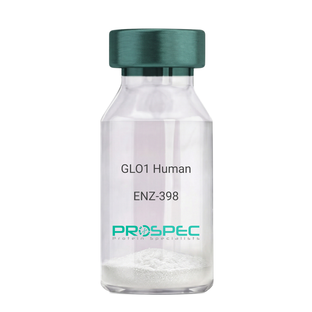
GLO1 HumanDescription:
Glyoxalase-I Human Recombinant
GLYI, GLOD1, GLO1, Glyoxalase-1, Lactoylglutathione lyase, Methylglyoxalase, Aldoketomutase, Ketone-aldehyde mutase, Glyoxalase I, S-D-lactoylglutathione methylglyoxal lyase, Glx I.
Product # :
ENZ-398Price :
Quantity :
Shipping Method :
Shipped with Ice Packs
More Info
- description
- source
- formulation
- purity
- More Info
- Citations
Description
Glyoxalase-I Human Recombinant produced in E.Coli is a single, non-glycosylated, polypeptide chain containing 184 amino acids and having a molecular mass of 20.7 kDa. Glyoxalase-1 is purified by proprietary chromatographic techniques.
Source
Escherichia Coli.
Formulation
Glyoxalase-1 solution containing 20mM Tris-HCl pH-8, 1mM DTT and 10% glycerol.
Purity
Greater than 90.0% as determined by SDS-PAGE.
Citations
More Info
-
Introduction
GLO1 is involved in the catalysis and formation of S-lactoyl-glutathione from methylglyoxal condensation and reduced glutatione. GLO1 is linked to HLA and is localized to 6p21.3-p21.1, between HLA and the centromere. GLO1 enzyme is ubundantly expressed and present in numerous tumor cell lines, in which its concentration is often upregulated ubiquitisly. GLO1 is a major susceptible gene for autism in an ethnic Chinese population from Taiwan. GLO1 might be involved in the pathophysiology of mood disorders. GLO1 plays a role in the pathophysiology of mood disorders. Overexpression of GLO1 is associated with kidney tumor.
-
Synonyms
GLYI, GLOD1, GLO1, Glyoxalase-1, Lactoylglutathione lyase, Methylglyoxalase, Aldoketomutase, Ketone-aldehyde mutase, Glyoxalase I, S-D-lactoylglutathione methylglyoxal lyase, Glx I.
-
Physical Appearance
Sterile filtered colorless solution.
-
Stability
Store at 4°C if entire vial will be used within 2-4 weeks. Store, frozen at -20°C for longer periods of time. For long term storage it is recommended to add a carrier protein (0.1% HSA or BSA).Avoid multiple freeze-thaw cycles.
-
Amino Acid Sequence
MAEPQPPSGG LTDEAALSCC SDADPSTKDF LLQQTMLRVK DPKKSLDFYT RVLGMTLIQK CDFPIMKFSL YFLAYEDKND IPKEKDEKIAWALSRKATLE LTHNWGTEDD ETQSYHNGNS DPRGFGHIGI AVPDVYSACK RFEELGVKFV KKPDDGKMKG LAFIQDPDGY WIEILNPNKM ATLM.
ProSpec's products are furnished for LABORATORY RESEARCH USE ONLY. The product may not be used as drugs, agricultural or pesticidal products, food additives or household chemicals.
- View Data Sheet
Name :
UBE2L3 HumanDescription:
Ubiquitin-Conjugating Enzyme E2L 3 Human Recombinant
Ubiquitin-conjugating enzyme E2 L3, EC 6.3.2.19, Ubiquitin-protein ligase L3,Ubiquitin carrier protein L3, UbcH7, E2-F1, L-UBC, UbcM4.
Product # :
ENZ-342Price :
Quantity :
Shipping Method :
Shipped with Ice Packs
More Info
- description
- source
- formulation
- purity
- More Info
- Citations
Description
Ubiquitin-Conjugating Enzyme E2L 3 Human Recombinant produced in E.Coli is a single, non-glycosylated polypeptide chain containing 154 amino acids & having a molecular mass of 17.9 kDa.
Source
Escherichia Coli.
Formulation
The protein contains 50mM HEPES (pH-7.5) 150mM NaCl, 1mM DTT, and 10% glycerol.
Purity
Greater than 95.0% as determined by SDS-PAGE.
Citations
More Info
-
Introduction
Human Ubquitin-conjugating enzyme 7 (UbcH7) is a ubiquitin-conjugating enzyme (E2) mediating c-fos degradation, transcription factor NF-B maturation, and human papilloma virus-mediated p53 and Myc protein degradation, in vitro. The ubiquitin-conjugating enzymes (E2s) are essential components of the post-translational protein ubiquitination pathway, mediating the transfer of activated ubiquitin to substrate proteins. The human UBE2L1-UBE2L4 gene could potentially encode different isoforms of the UbcH7. UBE2L3 gene, located at chromosome 22q11.2, is the only identical family member with introns and encodes a polypeptide sequence identical to that of UbcH7.
-
Synonyms
Ubiquitin-conjugating enzyme E2 L3, EC 6.3.2.19, Ubiquitin-protein ligase L3,Ubiquitin carrier protein L3, UbcH7, E2-F1, L-UBC, UbcM4.
-
Physical Appearance
Sterile filtered colorless solution.
-
Stability
Store at 4°C if entire vial will be used within 2-4 weeks. Store, frozen at -20°C for longer periods of time. For long term storage it is recommended to add a carrier protein (0.1% HSA or BSA).Avoid multiple freeze-thaw cycles.
-
Amino Acid Sequence
MAASRRLMKE LEEIRKCGMK NFRNIQVDEA NLLTWQGLIV PDNPPYDKGA FRIEINFPAE YPFKPPKITF KTKIYHPNID EKGQVCLPVI SAENWKPATK TDQVIQSLIA LVNDPQPEHP LRADLAEEYS KDRKKFCKNA EEFTKKYGEK RPVD.
ProSpec's products are furnished for LABORATORY RESEARCH USE ONLY. The product may not be used as drugs, agricultural or pesticidal products, food additives or household chemicals.
- View Data Sheet
Name :
pykF E.ColiDescription:
Pyruvate Kinase I E.Coli Recombinant
Pyruvate kinase I, PK-1, pykF, b1676, JW1666.
Product # :
PKA-028Price :
Quantity :
Shipping Method :
Shipped with Ice Packs
More Info
- description
- source
- formulation
- purity
- More Info
- Citations
Description
pykF E.Coli Recombinant produced in E.Coli is a single, non-glycosylated polypeptide chain containing 494 amino acids (1-470 a.a) and having a molecular mass of 53.3kDa.pykF is fused to a 24 amino acid His-tag at N-terminus & purified by proprietary chromatographic techniques.
Source
E.coli.
Formulation
pykF protein solution (1mg/ml) containing 20mM Tris-HCl buffer (pH8.0) and 10% glycerol.
Purity
Greater than 95% as determined by SDS-PAGE.
Citations
More Info
-
Introduction
pykF (pyruvate kinase I) is a member of the pyruvate kinase family. pykF is an enzyme involved in glycolysis. pykF catalyzes the transfer of a phosphate group from phosphoenolpyruvate (PEP) to ADP, yielding one molecule of pyruvate and one molecule of ATP, a process which also requires a Magnesium ion. This is the final step in the glycolytic pathway, which produces pyruvate molecules, the ultimate product of aerobic glycolysis.
-
Synonyms
Pyruvate kinase I, PK-1, pykF, b1676, JW1666.
-
Physical Appearance
Sterile Filtered colorless solution.
-
Stability
Store at 4°C if entire vial will be used within 2-4 weeks. Store, frozen at -20°C for longer periods of time. For long term storage it is recommended to add a carrier protein (0.1% HSA or BSA).Avoid multiple freeze-thaw cycles.
-
Amino Acid Sequence
MGSSHHHHHH SSGLVPRGSH MGSHMKKTKI VCTIGPKTES EEMLAKMLDA GMNVMRLNFS HGDYAEHGQR IQNLRNVMSK TGKTAAILLD TKGPEIRTMK LEGGNDVSLK AGQTFTFTTD KSVIGNSEMV AVTYEGFTTD LSVGNTVLVD DGLIGMEVTA IEGNKVICKV LNNGDLGENK GVNLPGVSIA LPALAEKDKQ DLIFGCEQGV DFVAASFIRK RSDVIEIREH LKAHGGENIH IISKIENQEG LNNFDEILEA SDGIMVARGD LGVEIPVEEV IFAQKMMIEK CIRARKVVIT ATQMLDSMIK NPRPTRAEAG DVANAILDGT DAVMLSGESA KGKYPLEAVS IMATICERTD RVMNSRLEFN NDNRKLRITE AVCRGAVETA EKLDAPLIVV ATQGGKSARA VRKYFPDATI LALTTNEKTA HQLVLSKGVV PQLVKEITST DDFYRLGKEL ALQSGLAHKG DVVVMVSGAL VPSGTTNTAS VHVL.
ProSpec's products are furnished for LABORATORY RESEARCH USE ONLY. The product may not be used as drugs, agricultural or pesticidal products, food additives or household chemicals.
- View Data Sheet
Name :
lldD E. coliDescription:
L-Lactate Dehydrogenase E.Coli Recombinant
L-lactate dehydrogenase [cytochrome], lldD, lctD, b3605, JW3580.
Product # :
ENZ-618Price :
Quantity :
Shipping Method :
Shipped with Ice Packs
More Info
- description
- source
- formulation
- purity
- More Info
- Citations
Description
lldD E.Coli Recombinant produced in E.coli is a single, non-glycosylated polypeptide chain containing 420 amino acids (1-396) and having a molecular mass of 45.3kDa.lldD is fused to a 24 amino acid His-tag at N-terminus & purified by proprietary chromatographic techniques.
Source
E.coli.
Formulation
The lldD solution (1mg/ml) contains 20mM Tris-HCl buffer (pH8.0), 10% glycerol and 1mM DTT.
Purity
Greater than 90% as determined by SDS-PAGE.
Citations
More Info
-
Introduction
L-lactate dehydrogenase (lldD) is present in a various organisms, including plants and animals. lldD is an oxidoreductase which catalyses the interconversion of pyruvate and lactate with concurrent interconversion of NADH and NAD+. Seeing that lldD can catalyze the oxidation of hydroxybutyrate, it is occasionally called Hydroxybutyrate Dehydrogenase (HBD).
-
Synonyms
L-lactate dehydrogenase [cytochrome], lldD, lctD, b3605, JW3580.
-
Physical Appearance
Sterile Filtered colorless solution.
-
Stability
Store at 4°C if entire vial will be used within 2-4 weeks. Store, frozen at -20°C for longer periods of time. For long term storage it is recommended to add a carrier protein (0.1% HSA or BSA).Avoid multiple freeze-thaw cycles.
-
Amino Acid Sequence
MGSSHHHHHH SSGLVPRGSH MGSHMIISAA SDYRAAAQRI LPPFLFHYMD GGAYSEYTLR RNVEDLSEVA LRQRILKNMS DLSLETTLFN EKLSMPVALA PVGLCGMYAR RGEVQAAKAA DAHGIPFTLS TVSVCPIEEV APAIKRPMWF QLYVLRDRGF MRNALERAKA AGCSTLVFTV DMPTPGARYR DAHSGMSGPN AAMRRYLQAV THPQWAWDVG LNGRPHDLGN ISAYLGKPTG LEDYIGWLGN NFDPSISWKD LEWIRDFWDG PMVIKGILDP EDARDAVRFG ADGIVVSNHG GRQLDGVLSS ARALPAIADA VKGDIAILAD SGIRNGLDVV RMIALGADTV LLGRAFLYAL ATAGQAGVAN LLNLIEKEMK VAMTLTGAKS ISEITQDSLV QGLGKELPAA LAPMAKGNAA.
ProSpec's products are furnished for LABORATORY RESEARCH USE ONLY. The product may not be used as drugs, agricultural or pesticidal products, food additives or household chemicals.
- View Data Sheet
Name :
DERA HumanDescription:
Deoxyribose-Phosphate Aldolase Human Recombinant
Putative deoxyribose-phosphate aldolase, DERA, 2-deoxy-D-ribose 5-phosphate aldolase, Phosphodeoxyriboaldolase, Deoxyriboaldolase, DERA, CGI-26.
Product # :
ENZ-170Price :
Quantity :
Shipping Method :
Shipped with Ice Packs
More Info
- description
- source
- formulation
- purity
- More Info
- Citations
Description
DERA Human Recombinant produced in E.coli is a single, non-glycosylated polypeptide chain containing 338 amino acids (1-318) and having a molecular mass of 37.3 kDa.DERA is fused to a 20 amino acid His-tag at N-terminus & purified by proprietary chromatographic techniques.
Source
E.coli.
Formulation
The DERA solution (0.5mg/ml) contains 20mM Tris-HCl buffer (pH 8.0), 0.1M NaCl, 1mM DTT and 20% glycerol.
Purity
Greater than 85% as determined by SDS-PAGE.
Citations
More Info
-
Introduction
Deoxyribose-phosphate aldolase (DERA) is a member of the deoC/fbaB aldolase protein family involved in the carbohydrate degradation pathway. DERA catalyzes the conversion of 2-deoxy-D-ribose 5-phosphate to D-glyceraldehyde 3-phosphate and an acetyldehyde.
-
Synonyms
Putative deoxyribose-phosphate aldolase, DERA, 2-deoxy-D-ribose 5-phosphate aldolase, Phosphodeoxyriboaldolase, Deoxyriboaldolase, DERA, CGI-26.
-
Physical Appearance
Sterile Filtered colorless solution.
-
Stability
Store at 4°C if entire vial will be used within 2-4 weeks. Store, frozen at -20°C for longer periods of time. For long term storage it is recommended to add a carrier protein (0.1% HSA or BSA).Avoid multiple freeze-thaw cycles.
-
Amino Acid Sequence
MGSSHHHHHH SSGLVPRGSH MSAHNRGTEL DLSWISKIQV NHPAVLRRAE QIQARRTVKK EWQAAWLLKA VTFIDLTTLS GDDTSSNIQR LCYKAKYPIR EDLLKALNMH DKGITTAAVC VYPARVCDAV KALKAAGCNI PVASVAAGFP AGQTHLKTRL EEIRLAVEDG ATEIDVVINR SLVLTGQWEA LYDEIRQFRK ACGEAHLKTI LATGELGTLT NVYKASMIAM MAGSDFIKTS TGKETVNATF PVAIVMLRAI RDFFWKTGNK IGFKPAGGIR SAKDSLAWLS LVKEELGDEW LKPELFRIGA STLLSDIERQ IYHHVTGRYA AYHDLPMS
ProSpec's products are furnished for LABORATORY RESEARCH USE ONLY. The product may not be used as drugs, agricultural or pesticidal products, food additives or household chemicals.
- View Data Sheet
Name :
PGLS HumanDescription:
6-Phosphogluconolactonase Human Recombinant
6PGL, 6-Phosphogluconolactonase.
Product # :
ENZ-016Price :
Quantity :
Shipping Method :
Shipped with Ice Packs
More Info
- description
- source
- formulation
- purity
- More Info
- Citations
Description
PGLS produced in E.Coli is a single, non-glycosylated polypeptide chain containing 278 amino acids (1-258a.a.) and having a molecular mass of 29.7kDa.PGLS is fused to a 20 amino acid His-tag at N-terminus & purified by proprietary chromatographic techniques.
Source
Escherichia Coli.
Formulation
The PGLS protein solution (1mg/1ml) is formulated in 20mM Tris-HCl buffer (pH 8.0) 1mM DTT, 100mM NaCl and 10% glycerol.
Purity
Greater than 95% as determined by SDS-PAGE.
Citations
More Info
-
Introduction
PGLS is an enzyme in the second step pentose phosphate pathway. 6-Phosphogluconolactonase is crucial for the synthesis of nucleotide sugars and NADPH, the key cause for decreasing power. PGLS transforms 6-phosphogluconolactone to 6-phosphogluconate.
-
Synonyms
6PGL, 6-Phosphogluconolactonase.
-
Physical Appearance
Sterile Filtered clear solution.
-
Stability
Store at 4°C if entire vial will be used within 2-4 weeks. Store, frozen at -20°C for longer periods of time.Please avoid freeze thaw cycles.
-
Amino Acid Sequence
MGSSHHHHHH SSGLVPRGSH MAAPAPGLIS VFSSSQELGA ALAQLVAQRA ACCLAGARAR FALGLSGGSL VSMLARELPA AVAPAGPASL ARWTLGFCDE RLVPFDHAES TYGLYRTHLL SRLPIPESQV ITINPELPVE EAAEDYAKKL RQAFQGDSIP VFDLLILGVG PDGHTCSLFP DHPLLQEREK IVAPISDSPK PPPQRVTLTL PVLNAARTVI FVATGEGKAA VLKRILEDQE ENPLPAALVQ PHTGKLCWFL DEAAARLLTV PFEKHSTL
ProSpec's products are furnished for LABORATORY RESEARCH USE ONLY. The product may not be used as drugs, agricultural or pesticidal products, food additives or household chemicals.
- View Data Sheet
Name :
GLYAT HumanDescription:
Glycine-N-Acyltransferase Human Recombinant
Glycine-N-acyltransferase, ACGNAT, GAT, CAT, AAc, HRP-1(CLP), Acyl-CoA: glycine amino acid N-acyltransferase, EC 2.3.1.13.
Product # :
ENZ-148Price :
Quantity :
Shipping Method :
Shipped with Ice Packs
More Info
- description
- source
- formulation
- purity
- More Info
- Citations
Description
GLYAT Human Recombinant produced in E.coli is a single, non-glycosylated polypeptide chain containing 316 amino acids (1-296) and having a molecular mass of 36.0 kDa.The GLYAT is fused to a 20 amino acid His-Tag at N-terminus and purified by proprietary chromatographic techniques.
Source
Escherichia Coli.
Formulation
GLYAT protein (0.5mg/ml) is supplied in 20mM Tris-HCl, pH-8, 1mM DTT and 20% Glycerol.
Purity
Greater than 90.0% as determined by SDS-PAGE.
Citations
More Info
-
Introduction
GLYAT is a mitochondrial acyltransferase that conjugates glycine with acyl-CoA substrates in the mitochondria. GLYAT is vital to the detoxification of endogenous and xenobiotic acyl-CoA's.
-
Synonyms
Glycine-N-acyltransferase, ACGNAT, GAT, CAT, AAc, HRP-1(CLP), Acyl-CoA: glycine amino acid N-acyltransferase, EC 2.3.1.13.
-
Physical Appearance
GLYAT is supplied as a sterile filtered clear solution.
-
Stability
Store at 4°C if entire vial will be used within 2-4 weeks. Store, frozen at -20°C for longer periods of time. For long term storage it is recommended to add a carrier protein (0.1% HSA or BSA).Avoid multiple freeze-thaw cycles.
-
Amino Acid Sequence
MGSSHHHHHH SSGLVPRGSH MMLPLQGAQM LQMLEKSLRK SLPASLKVYG TVFHINHGNP FNLKAVVDKW PDFNTVVVCP QEQDMTDDLD HYTNTYQIYS KDPQNCQEFL GSPELINWKQ HLQIQSSQPS LNEAIQNLAA IKSFKVKQTQ RILYMAAETA KELTPFLLKS KILSPSGGKP KAINQEMFKL SSMDVTHAHL VNKFWHFGGN ERSQRFIERC IQTFPTCCLL GPEGTPVCWD LMDQTGEMRM AGTLPEYRLH GLVTYVIYSH AQKLGKLGFP VYSHVDYSNE AMQKMSYTLQ HVPIPRSWNQ WNCVPL.
ProSpec's products are furnished for LABORATORY RESEARCH USE ONLY. The product may not be used as drugs, agricultural or pesticidal products, food additives or household chemicals.
- View Data Sheet
Name :
CMBL AntibodyDescription:
Carboxymethylenebutenolidase, Mouse Anti Human
Carboxymethylenebutenolidase homolog, CMBL, JS-1.
Product # :
ANT-093Price :
Quantity :
Shipping Method :
Shipped with Ice Packs
More Info
- formulation
- More Info
- Citations
Formulation
1mg/ml containing PBS, pH-7.4, & 0.1% Sodium Azide.
Citations
More Info
-
Introduction
Carboxymethylenebutenolidase homolog (CMBL) is a cysteine hydrolase of the dienelactone hydrolase family which is highly expressed in the liver cytosol. CMBL is the human homolog of Pseudomonas dienelactone hydrolase, which is a protein that participates in the bacterial halocatechol degradation pathway. CMBL which preferentially cleaves cyclic esters activates medoxomil-ester prodrugs in which the medoxomil moiety is coupled with an oxygen atom. CMBL is inhibited by PCMB (p-chloromercuribenzoate) and is encoded by a gene which maps to human chromosome 5p15.2. Furthermore, CMBL converts the prodrug olmesartan medoxomil into its pharmacologically active metabolite olmerstatan, which is an angiotensin receptor blocker, in the liver and intestine. CMBL can also activate beta-lactam antibiotics faropenem medoxomil and lenampicillin. CMBL is widely expressed, with the highest levels in the liver, followed by the kidney, small intestine and the colon.
-
Synonyms
Carboxymethylenebutenolidase homolog, CMBL, JS-1.
-
Physical Appearance
Sterile Filtered colorless solution.
-
Immunogen
Anti-human CMBL mAb is derived from hybridization of mouse F0 myeloma cells with spleen cells from BALB/c mice immunized with recombinant human CMBL 1-245 amino acids purified from E. coli.
-
Ig Subclass
Mouse IgG2a heavy chain and lambda light chain.
-
Clone
PAT2B11AT.
-
Applications
CMBL antibody has been tested by ELISA, Western blot analysis to assure specificity and reactivity. Since application varies, however, each investigation should be titrated by the reagent to obtain optimal results. Recommended dilution range for Western blot analysis is 1:500 ~ 1:5000. Recommended starting dilution is 1:500.
-
Type
Mouse Anti Human Monoclonal.
-
Storage Procedures
For periods up to 1 month store at 4°C, for longer periods of time, store at -20°C. Prevent freeze thaw cycles.
-
Purification Method
CMBL antibody was purified from mouse ascitic fluids by protein-G affinity chromatography.
ProSpec's products are furnished for LABORATORY RESEARCH USE ONLY. The product may not be used as drugs, agricultural or pesticidal products, food additives or household chemicals.
- View Data Sheet
Name :
MGAT2 Human, Sf9Description:
Mannoside Acetylglucosaminyltransferase 2 Human Recombinant, Sf9
Alpha-1, 6-mannosyl-glycoprotein 2-beta-N-acetylglucosaminyltransferase, MGAT2, CDG2A, CDGS2, GLCNACTII, GNT-II, GNT2, Beta-1,2-N-acetylglucosaminyltransferase II, GlcNAc-T II, Mannoside acetylglucosaminyltransferase 2, N-glycosyl-oligosaccharide-glycoprotein N-acetylglucosaminyltransferase II.
Product # :
ENZ-1077Price :
Quantity :
Shipping Method :
Shipped with Ice Packs
More Info
- description
- source
- formulation
- purity
- More Info
- Citations
Description
MGAT2 produced in Sf9 Baculovirus cells is a single, glycosylated polypeptide chain containing 427 amino acids (30-447a.a.) and having a molecular mass of 49.3kDa. (Molecular size on SDS-PAGE will appear at approximately 40-57kDa).MGAT2 is expressed with a 6 amino acid His tag at C-Terminus and purified by proprietary chromatographic techniques.
Source
Sf9, Baculovirus cells.
Formulation
MGAT2 protein solution (0.25mg/ml) contains 20mM Tris-HCl (pH 7.5), 10% glycerol, 0.15M NaCl and 1mM DTT.
Purity
Greater than 90.0% as determined by SDS-PAGE.
Citations
More Info
-
Introduction
MGAT2 is an enzyme which takes part in the catalyzation of a crucial step in the reaction of oligomannose which converts to complex N-glycans. MGAT2 has three domains, classic to glycosyltransferase: short N-terminal cytoplasmic domain, a C-terminal catalytic domain and hydrophobic non-cleavable signal-anchor domain. The enzyme MGAT2 is encoded by the MGAT2 gene in humans. There are no introns in the DNA coding the gene, therefore mutations in the MGAT2 will result in carbohydrate-deficient glycoprotein syndrome, type II.
-
Synonyms
Alpha-1, 6-mannosyl-glycoprotein 2-beta-N-acetylglucosaminyltransferase, MGAT2, CDG2A, CDGS2, GLCNACTII, GNT-II, GNT2, Beta-1,2-N-acetylglucosaminyltransferase II, GlcNAc-T II, Mannoside acetylglucosaminyltransferase 2, N-glycosyl-oligosaccharide-glycoprotein N-acetylglucosaminyltransferase II.
-
Physical Appearance
Sterile filtered colorless solution.
-
Stability
Store at 4°C if entire vial will be used within 2-4 weeks. Store, frozen at -20°C for longer periods of time. For long term storage it is recommended to add a carrier protein (0.1% HSA or BSA).Avoid multiple freeze-thaw cycles.
-
Amino Acid Sequence
ADPRQRKNEA LAPPLLDAEP ARGAGGRGGD HPSVAVGIRR VSNVSAASLV PAVPQPEADN LTLRYRSLVY QLNFDQTLRN VDKAGTWAPR ELVLVVQVHN RPEYLRLLLD SLRKAQGIDN VLVIFSHDFW STEINQLIAG VNFCPVLQVF FPFSIQLYPN EFPGSDPRDC PRDLPKNAAL
KLGCINAEYP DSFGHYREAK FSQTKHHWWW KLHFVWERVK ILRDYAGLIL FLEEDHYLAP DFYHVFKKMW KLKQQECPEC DVLSLGTYSA SRSFYGMADK VDVKTWKSTE HNMGLALTRN AYQKLIECTD TFCTYDDYNW DWTLQYLTVS CLPKFWKVLV PQIPRIFHAG DCGMHHKKTC
RPSTQSAQIE SLLNNNKQYM FPETLTISEK FTVVAISPPR KNGGWGDIRD HELCKSYRRL QHHHHHH.
ProSpec's products are furnished for LABORATORY RESEARCH USE ONLY. The product may not be used as drugs, agricultural or pesticidal products, food additives or household chemicals.
- View Data Sheet
Name :
BTD HumanDescription:
Biotinidase Human Recombinant
Biotinidase, EC 3.5.1.12, Biotinase, EC 3.5.1.
Product # :
ENZ-1004Price :
Quantity :
Shipping Method :
Shipped with Ice Packs
More Info
- description
- source
- formulation
- purity
- More Info
- Citations
Description
BTD Human Recombinant produced in Sf9 Baculovirus cells is a single, glycosylated polypeptide chain containing 510 amino acids (44-545a.a) and having a molecular mass of 57.8kDa. (Molecular size on SDS-PAGE will appear at approximately 50-70kDa). BTD is fused to an 8 amino acid His-tag at C-terminus & purified by proprietary chromatographic techniques.
Source
Sf9, Baculovirus cells.
Formulation
BTD protein solution (0.25mg/ml) containing Phosphate Buffered Saline (pH 7.4) and 10% glycerol.
Purity
Greater than 85% as determined by SDS-PAGE.
Citations
More Info
-
Introduction
Biotinidase also known BTD, belongs to the nitrilase superfamily, which contains 12 families of nitrilases, amidases, carbamylases, and N-acyltrasferases. BTD catalyzes the hydrolysis of biocytin, the product of biotin-dependent carboxylase degradation, to biotin and lysine. BTD has a vital regulatory part in chromatin/DNA function. Mutations in BTD protein lead to Biotinidase deficiency.
-
Synonyms
Biotinidase, EC 3.5.1.12, Biotinase, EC 3.5.1.
-
Physical Appearance
Sterile Filtered colorless solution.
-
Stability
Store at 4°C if entire vial will be used within 2-4 weeks. Store, frozen at -20°C for longer periods of time. For long term storage it is recommended to add a carrier protein (0.1% HSA or BSA).Avoid multiple freeze-thaw cycles.
-
Amino Acid Sequence
AHTGEESVAD HHEAEYYVAA VYEHPSILSL NPLALISRQE ALELMNQNLD IYEQQVMTAA QKDVQIIVFP EDGIHGFNFT RTSIYPFLDF MPSPQVVRWN PCLEPHRFND TEVLQRLSCM AIRGDMFLVA NLGTKEPCHS SDPRCPKDGR YQFNTNVVFS NNGTLVDRYR KHNLYFEAAF DVPLKVDLIT FDTPFAGRFG IFTCFDILFF DPAIRVLRDY KVKHVVYPTA WMNQLPLLAA IEIQKAFAVA FGINVLAANV HHPVLGMTGS GIHTPLESFW YHDMENPKSH LIIAQVAKNP VGLIGAENAT GETDPSHSKF LKILSGDPYC EKDAQEVHCD EATKWNVNAP PTFHSEMMYD NFTLVPVWGK EGYLHVCSNG LCCYLLYERP TLSKELYALG VFDGLHTVHG TYYIQVCALV RCGGLGFDTC GQEITEATGI FEFHLWGNFS TSYIFPLFLT SGMTLEVPDQ LGWENDHYFL RKSRLSSGLV TAALYGRLYE RDLEHHHHHH.
ProSpec's products are furnished for LABORATORY RESEARCH USE ONLY. The product may not be used as drugs, agricultural or pesticidal products, food additives or household chemicals.
- View Data Sheet
Name :
MGAT2 HumanDescription:
Mannoside Acetylglucosaminyltransferase 2 Human Recombinant
Mannosyl (Alpha-1,6-)-Glycoprotein Beta-1,2-N-Acetylglucosaminyltransferase, GlcNAc-T II, Mannoside Acetylglucosaminyltransferase 2, GNT-II, Beta-1,2-N-Acetylglucosaminyltransferase II, N-Glycosyl-Oligosaccharide-Glycoprotein N-Acetylglucosaminyltransferase II, EC 2.4.1.143, CDG2A, CDGS2, GLCNACTI, NT2, Alpha-1,6-Mannosyl-Glycoprotein 2-Beta-N-Acetylglucosaminyltransferase, UDP-N-Acetylglucosamine:Alpha-6-D-Mannoside, Beta-1,2-N-Acetylglucosaminyltransferase II.
Product # :
ENZ-781Price :
Quantity :
Shipping Method :
Shipped with Ice Packs
More Info
- description
- source
- formulation
- purity
- More Info
- Citations
Description
MGAT2 Human Recombinant produced in E.Coli is a single, non-glycosylated polypeptide chain containing 439 amino acids (30-447a.a) and having a molecular mass of 50kDa. MGAT2 is fused to a 21 amino acid His-tag at N-terminus & purified by proprietary chromatographic techniques.
Source
Escherichia Coli.
Formulation
MGAT2 protein solution (1mg/ml) containing 20mM Tris-HCl buffer (pH 8.0), 10% glycerol and 0.4M urea.
Purity
Greater than 85.0% as determined by SDS-PAGE.
Citations
More Info
-
Introduction
Mannoside Acetylglucosaminyltransferase 2 (MGAT2) is a golgi enzyme catalyzing an vital step in the conversion of oligomannose to complex N-glycans. MGAT2 enzyme has the characteristic glycosyltransferase domains: a short N-terminal cytoplasmic domain, a hydrophobic non-cleavable signal-anchor domain, and a C-terminal catalytic domain. MGAT2 gene mutations may lead to carbohydrate-deficient glycoprotein syndrome, type II.
-
Synonyms
Mannosyl (Alpha-1,6-)-Glycoprotein Beta-1,2-N-Acetylglucosaminyltransferase, GlcNAc-T II, Mannoside Acetylglucosaminyltransferase 2, GNT-II, Beta-1,2-N-Acetylglucosaminyltransferase II, N-Glycosyl-Oligosaccharide-Glycoprotein N-Acetylglucosaminyltransferase II, EC 2.4.1.143, CDG2A, CDGS2, GLCNACTI, NT2, Alpha-1,6-Mannosyl-Glycoprotein 2-Beta-N-Acetylglucosaminyltransferase, UDP-N-Acetylglucosamine:Alpha-6-D-Mannoside, Beta-1,2-N-Acetylglucosaminyltransferase II.
-
Physical Appearance
Sterile Filtered clear solution.
-
Stability
Store at 4°C if entire vial will be used within 2-4 weeks. Store, frozen at -20°C for longer periods of time.For long term storage it is recommended to add a carrier protein (0.1% HSA or BSA).Please avoid freeze thaw cycles.
-
Amino Acid Sequence
MGSSHHHHHH SSGLVPRGSH MRQRKNEALA PPLLDAEPAR GAGGRGGDHP SVAVGIRRVS NVSAASLVPA VPQPEADNLT LRYRSLVYQL NFDQTLRNVD KAGTWAPREL VLVVQVHNRP EYLRLLLDSL RKAQGIDNVL VIFSHDFWST EINQLIAGVN FCPVLQVFFP FSIQLYPNEF PGSDPRDCPR DLPKNAALKL GCINAEYPDS FGHYREAKFS QTKHHWWWKL HFVWERVKIL RDYAGLILFL EEDHYLAPDF YHVFKKMWKL KQQECPECDV LSLGTYSASR SFYGMADKVD VKTWKSTEHN MGLALTRNAY QKLIECTDTF CTYDDYNWDW TLQYLTVSCL PKFWKVLVPQ IPRIFHAGDC GMHHKKTCRP STQSAQIESL LNNNKQYMFP ETLTISEKFT VVAISPPRKN GGWGDIRDHE LCKSYRRLQ.
ProSpec's products are furnished for LABORATORY RESEARCH USE ONLY. The product may not be used as drugs, agricultural or pesticidal products, food additives or household chemicals.
- View Data Sheet
Name :
IDH1 HumanDescription:
Isocitrate Dehydrogenase-1 Human Recombinant
Isocitrate dehydrogenase [NADP] cytoplasmic, EC 1.1.1.42, Cytosolic NADP-isocitrate dehydrogenase, Oxalosuccinate decarboxylase, IDH, NADP(+)-specific ICDH, IDP, PICD.
Product # :
ENZ-193Price :
Quantity :
Shipping Method :
Shipped with Ice Packs
More Info
- description
- source
- formulation
- purity
- biological activity
- More Info
- Citations
Description
IDH1 Human Recombinant produced in E.coli is a single, non-glycosylated polypeptide chain containing 434 amino acids (1-414) and having a molecular mass of 48.8 kDa.IDH1 is fused to a 20 amino acid His-tag at N-terminus & purified by proprietary chromatographic techniques.
Source
E.coli.
Formulation
The IDH1 solution (1mg/ml) contains 20mM Tris-HCl buffer (pH 8.0), 0.1M NaCl,
1mM DTT, 0.1mM PMSF and 20% glycerol.Purity
Greater than 95% as determined by SDS-PAGE.
Biological Activity
The Specific activity is > 0.7 units/ml. One unit will convert 1.0 umole of isocitrate to alpha-ketoglutarate per minute at pH7.5 at 25C.Citations
More Info
-
Introduction
Isocitrate Dehydrogenase is an enzyme of the oxidoreductase class that catalyzes the conversion of isocitrate and NAD+ to yield 2-ketoglutarate, carbon dioxide, and NADH. It occurs in cell mitochondria. The enzyme requires Mg2+, Mn2+; it is activated by ADP, citrate, and Ca2+, and inhibited by NADH, NADPH, and ATP. The reaction is the key rate-limiting step of the citric acid (tricarboxylic) cycle.
-
Synonyms
Isocitrate dehydrogenase [NADP] cytoplasmic, EC 1.1.1.42, Cytosolic NADP-isocitrate dehydrogenase, Oxalosuccinate decarboxylase, IDH, NADP(+)-specific ICDH, IDP, PICD.
-
Physical Appearance
Sterile Filtered colorless solution.
-
Stability
Store at 4°C if entire vial will be used within 2-4 weeks. Store, frozen at -20°C for longer periods of time. For long term storage it is recommended to add a carrier protein (0.1% HSA or BSA).Avoid multiple freeze-thaw cycles.
-
Amino Acid Sequence
MGSSHHHHHH SSGLVPRGSH MSKKISGGSV VEMQGDEMTR IIWELIKEKL IFPYVELDLH SYDLGIENRD ATNDQVTKDA AEAIKKHNVG VKCATITPDE KRVEEFKLKQ MWKSPNGTIR NILGGTVFRE AIICKNIPRL VSGWVKPIII GRHAYGDQYR ATDFVVPGPG KVEITYTPSD GTQKVTYLVH NFEEGGGVAM GMYNQDKSIE DFAHSSFQMA LSKGWPLYLS TKNTILKKYD GRFKDIFQEI YDKQYKSQFE AQKIWYEHRL IDDMVAQAMK SEGGFIWACK NYDGDVQSDS VAQGYGSLGM MTSVLVCPDG KTVEAEAAHG TVTRHYRMYQ KGQETSTNPI ASIFAWTRGL AHRAKLDNNK ELAFFANALE EVSIETIEAG FMTKDLAACI KGLPNVQRSD YLNTFEFMDK LGENLKIKLA QAKL
ProSpec's products are furnished for LABORATORY RESEARCH USE ONLY. The product may not be used as drugs, agricultural or pesticidal products, food additives or household chemicals.
- View Data Sheet
Name :
BLVRA HumanDescription:
Biliverdin Reductase A Human Recombinant
Biliverdin reductase A, BVR A, Biliverdin-IX alpha-reductase, BLVRA, BLVR, BVR, BVRA.
Product # :
ENZ-446Price :
Quantity :
Shipping Method :
Shipped with Ice Packs
More Info
- description
- source
- formulation
- purity
- More Info
- Citations
Description
BLVRA Human Recombinant produced in E.Coli is a single, non-glycosylated, polypeptide chain containing 295 amino acids (3-296 a.a. and Methionine at N-terminus) and having a molecular mass of 33.3kDa (molecular weight on SDS-PAGE will shift up).The BLVRA is purified by proprietary chromatographic techniques.
Source
Escherichia Coli.
Formulation
The BLVRA solution contains 20mM Tris-HCl buffer (pH8.0) and 10% glycerol.
Purity
Greater than 90.0% as determined by SDS-PAGE.
Citations
More Info
-
Introduction
Biliverdin reductase A (BLVRA) is a member of the gfo/idh/mocA family. BLVRA is an enzyme that converts biliverdin to bilirubin, converting a double-bond between the second and third pyrrole ring into a single-bond. BLVRA reduces the gamma-methene bridge of the open tetrapyrrole, biliverdin IX alpha, to bilirubin with the simultaneous oxidation of a NADH or NADPH cofactor (Bilirubin + NAD(P)+ = biliverdin + NAD(P)H ).
BLVRA is a regulator for induction of activating transcription factor-2 and heme oxygenase-1. Furthermore, BLVRA enhances the role of HO-1 in cytoprotection and provides cytoprotection independent of heme degradation. In addition, Bilirubin while acting as a cytoprotective antioxidant is itself oxidized to biliverdin and subsequently recycled by biliverdin reductase back to bilirubin. -
Synonyms
Biliverdin reductase A, BVR A, Biliverdin-IX alpha-reductase, BLVRA, BLVR, BVR, BVRA.
-
Physical Appearance
Sterile Filtered colorless solution.
-
Stability
Store at 4°C if entire vial will be used within 2-4 weeks. Store, frozen at -20°C for longer periods of time. For long term storage it is recommended to add a carrier protein (0.1% HSA or BSA).Avoid multiple freeze-thaw cycles.
-
Amino Acid Sequence
MAEPERKFGV VVVGVGRAGS VRMRDLRNPH PSSAFLNLIG FVSRRELGSI DGVQQISLED ALSSQEVEVA YICSESSSHE DYIRQFLNAG KHVLVEYPMT LSLAAAQELW ELAEQKGKVL HEEHVELLME EFAFLKKEVV GKDLLKGSLL FTAGPLEEER FGFPAFSGIS RLTWLVSLFG
ELSLVSATLE ERKEDQYMKM TVCLETEKKS PLSWIEEKGP GLKRNRYLSF HFKSGSLENV PNVGVNKNIF LKDQNIFVQK LLGQFSEKEL AAEKKRILHC LGLAEEIQKY CCSRK.
ProSpec's products are furnished for LABORATORY RESEARCH USE ONLY. The product may not be used as drugs, agricultural or pesticidal products, food additives or household chemicals.
- View Data Sheet
Name :
RPE HumanDescription:
Ribulose-5-Phosphate-3-Epimerase Human Recombinant
Ribulose-phosphate 3-epimerase, Ribulose-5-phosphate-3-epimerase, RPE, HUSSY-17, RPE2-1.
Product # :
ENZ-584Price :
Quantity :
Shipping Method :
Shipped with Ice Packs
More Info
- description
- source
- formulation
- purity
- More Info
- Citations
Description
RPE Human Recombinant produced in E.coli is a single, non-glycosylated polypeptide chain containing 252 amino acids (1-228) and having a molecular mass of 27.5kDa.RPE is fused to a 24 amino acid His-tag at N-terminus & purified by proprietary chromatographic techniques.
Source
Escherichia Coli.
Formulation
The RPE solution (0.5mg/ml) contains 20mM Tris-HCl buffer (pH 8.0), 2mM DTT, 30% glycerol and 200mM NaCl.
Purity
Greater than 95% as determined by SDS-PAGE.
Citations
More Info
-
Introduction
Ribulose-phosphate 3-epimerase (RPE) is a member of the ribulose-phosphate 3-epimerase family. RPE is the enzyme which converts D-ribulose 5-phosphate into D-xylulose 5-phosphate in Calvin's reductive pentose phosphate cycle. Therefore, the RPE enzyme has one substrate, D-ribulose 5-phosphate, and one product, D-xylulose 5-phosphate. RPE participates in three metabolic pathways: pentose phosphate pathway, pentose and glucuronate interconversions, and carbon fixation.
-
Synonyms
Ribulose-phosphate 3-epimerase, Ribulose-5-phosphate-3-epimerase, RPE, HUSSY-17, RPE2-1.
-
Physical Appearance
Sterile Filtered clear solution.
-
Stability
Store at 4°C if entire vial will be used within 2-4 weeks.Store, frozen at -20°C for longer periods of time. For long term storage it is recommended to add a carrier protein (0.1% HSA or BSA).Avoid multiple freeze-thaw cycles.
-
Amino Acid Sequence
MGSSHHHHHH SSGLVPRGSH MGSHMASGCK IGPSILNSDL ANLGAECLRM LDSGADYLHL DVMDGHFVPN ITFGHPVVES LRKQLGQDPF FDMHMMVSKP EQWVKPMAVA GANQYTFHLE ATENPGALIK DIRENGMKVG LAIKPGTSVE YLAPWANQID MALVMTVEPG FGGQKFMEDM MPKVHWLRTQ FPSLDIEVDG GVGPDTVHKC AEAGANMIVS GSAIMRSEDP RSVINLLRNV CSEAAQKRSL DR.
ProSpec's products are furnished for LABORATORY RESEARCH USE ONLY. The product may not be used as drugs, agricultural or pesticidal products, food additives or household chemicals.
- View Data Sheet
Name :
PPCDC HumanDescription:
Phosphopantothenoylcysteine Decarboxylase Human Recombinant
FLJ14585, PPC-DC, MDS018, COAC, CoaC, Phosphopantothenoylcysteine Decarboxylase.
Product # :
ENZ-011Price :
Quantity :
Shipping Method :
Shipped with Ice Packs
More Info
- description
- source
- formulation
- purity
- More Info
- Citations
Description
PPCDC produced in E.Coli is a single, non-glycosylated polypeptide chain containing 224 amino acids (1-204a.a.) and having a molecular mass of 24.6kDa.PPCDC is fused to a 20 amino acid His-tag at N-terminus & purified by proprietary chromatographic techniques.
Source
Escherichia Coli.
Formulation
The PPCDC protein solution (1mg/1ml) is formulated in 20mM Tris-HCl buffer(pH 8.0), 1mM DTT, 0.1M NaCl and 10% glycerol.
Purity
Greater than 95% as determined by SDS-PAGE.
Citations
More Info
-
Introduction
PPCDC is a member of the HFCD (homo oligomeric flavin containing Cys decarboxylase) superfamily which takes a part in the biosynthesis of coenzyme A (CoA) from pantothenate (Vitamin B). The biosynthesis of CoA from pantothenate needs several steps: the phosphorylation of pantothenate, the conversion of 4’-hosphopantothenate to 4''-phosphopantetheine, the adenylation by phosphopantetheine adenylyltransferase to form dephospho-CoA and the phosphorylation by dephospho-CoA kinase to form CoA. PPCDC is a key player in this pathway. PPCDC converts 4''-phosphopantothenate into 4''-phosphopantetheine.
-
Synonyms
FLJ14585, PPC-DC, MDS018, COAC, CoaC, Phosphopantothenoylcysteine Decarboxylase.
-
Physical Appearance
Sterile Filtered clear solution.
-
Stability
Store at 4°C if entire vial will be used within 2-4 weeks. Store, frozen at -20°C for longer periods of time.Please avoid freeze thaw cycles.
-
Amino Acid Sequence
MGSSHHHHHH SSGLVPRGSH MEPKASCPAA APLMERKFHV LVGVTGSVAA LKLPLLVSKL LDIPGLEVAV VTTERAKHFY SPQDIPVTLY SDADEWEMWK SRSDPVLHID LRRWADLLLV APLDANTLGK VASGICDNLL TCVMRAWDRS KPLLFCPAMN TAMWEHPITA QQVDQLKAFG YVEIPCVAKK LVCGDEGLGA MAEVGTIVDK VKEVLFQHSG FQQS
ProSpec's products are furnished for LABORATORY RESEARCH USE ONLY. The product may not be used as drugs, agricultural or pesticidal products, food additives or household chemicals.
- View Data Sheet
Name :
UBE2H HumanDescription:
Ubiquitin-Conjugating Enzyme E2H Human Recombinant
Ubiquitin-conjugating enzyme E2 H, UbcH2, Ubiquitin carrier protein H, Ubiquitin-conjugating enzyme E2-20K, Ubiquitin-protein ligase H, UBE2H, GID3, UBC8, UBCH.
Product # :
ENZ-603Price :
Quantity :
Shipping Method :
Shipped with Ice Packs
More Info
- description
- source
- formulation
- purity
- More Info
- Citations
Description
UBE2H Human Recombinant produced in E.coli is a single, non-glycosylated polypeptide chain containing 206 amino acids (1-183) and having a molecular mass of 23.1kDa.UBE2H is fused to a 23 amino acid His-tag at N-terminus & purified by proprietary chromatographic techniques.
Source
Escherichia Coli.
Formulation
The UBE2H solution (1mg/ml) contains 20mM Tris-HCl buffer, pH8.0, 10% glycerol, 1mM DTT and 50mM NaCl.
Purity
Greater than 90.0% as determined by SDS-PAGE.
Citations
More Info
-
Introduction
Ubiquitin-conjugating enzyme E2 H (UBE2H) is a member of the ubiquitin-conjugating enzyme family. Protein modification with ubiquitin is a vital cellular apparatus for directing abnormal or short-lived proteins for degradation. Ubiquitination requires at least 3 classes of enzymes: ubiquitin-activating enzymes (E1s) ubiquitin-conjugating enzymes (E2s) and ubiquitin-protein ligases (E3s). UBE2H receives ubiquitin from the E1 complex and catalyzes its covalent attachment to other proteins. UBE2H protein sequence is 100% identical to the mouse homolog and 98% identical to the frog and zebrafish homologs.
-
Synonyms
Ubiquitin-conjugating enzyme E2 H, UbcH2, Ubiquitin carrier protein H, Ubiquitin-conjugating enzyme E2-20K, Ubiquitin-protein ligase H, UBE2H, GID3, UBC8, UBCH.
-
Physical Appearance
Sterile filtered colorless solution.
-
Stability
Store at 4°C if entire vial will be used within 2-4 weeks. Store, frozen at -20°C for longer periods of time. For long term storage it is recommended to add a carrier protein (0.1% HSA or BSA).Avoid multiple freeze-thaw cycles.
-
Amino Acid Sequence
MGSSHHHHHH SSGLVPRGSH MGSMSSPSPG KRRMDTDVVK LIESKHEVTI LGGLNEFVVK FYGPQGTPYE GGVWKVRVDL PDKYPFKSPS IGFMNKIFHP NIDEASGTVC LDVINQTWTA LYDLTNIFES FLPQLLAYPN PIDPLNGDAA AMYLHRPEEY KQKIKEYIQK YATEEALKEQ EEGTGDSSSE SSMSDFSEDE AQDMEL.
ProSpec's products are furnished for LABORATORY RESEARCH USE ONLY. The product may not be used as drugs, agricultural or pesticidal products, food additives or household chemicals.
- View Data Sheet
Name :
PROK Tritirachium albumDescription:
Tritirachium album Proteinase-K Recombinant
Proteinase K (EC:3.4.21.64), Endopeptidase K, Tritirachium alkaline proteinase, PROK.
Product # :
ENZ-1015Price :
Quantity :
Shipping Method :
Shipped at Room temp
More Info
- description
- source
- formulation
- purity
- biological activity
- More Info
- Citations
Description
Recombinant Tritirachium album Proteinase-K expressed in yeast containing 285 amino acids having a Mw of 29.3 kDa is purified by standard chromatography techniques.
Source
Yeast
Formulation
The Proteinase-K was lyophilized without any additives.
Purity
Greater than 95% as determined by SDS-PAGE.
Biological Activity
36 Units/mg.
One unit is defined as the amount of enzyme that will hydrolyze urea-denatured hemoglobin to produce color equivalent to 1.0 mol tyrosine per min at 37°C, pH 7.5 (color by Folin-Ciocalteu reagent).Citations
More Info
-
Introduction
The Proteinase K enzyme is a member of the Peptidase family S8. Proteinase K is a broad-spectrum serine protease. Proteinase K is capable of digesting hair (keratin), henceforth, the name "Proteinase K". Proteinase K is activated by calcium, the enzyme digests proteins especially after hydrophobic amino acids (aliphatic, aromatic and other hydrophobic amino acids). Proteinase K is frequently utilized in molecular biology to digest protein and remove contamination from preparations of nucleic acid. Addition of Proteinase K to nucleic acid preparations rapidly inactivates nucleases which may otherwise degrade the DNA or RNA during purification. Proteinase K is greatly fitting to this application as the enzyme is active in the presence of chemicals which denature proteins, such as SDS and urea, chelating agents such as EDTA, sulfhydryl reagents, as well as trypsin or chymotrypsin inhibitors. Proteinase K is utilized for the destruction of proteins in cell lysates (tissue, cell culture cells) and for the release of nucleic acids, given that it quite effectively inactivates DNases and RNases.
-
Synonyms
Proteinase K (EC:3.4.21.64), Endopeptidase K, Tritirachium alkaline proteinase, PROK.
-
Physical Appearance
Sterile Filtered lyophilized powder.
-
Stability
Recombinant Proteinase-K although stable at room temperature, should be stored between 2-8°C. Do not freeze!
-
Solubility
It is recommended to reconstitute the lyophilized Proteinase-K in 20mM Tris-HCl (pH 7.4~8.0), 1mM CaCl2, 50% glycerol not less than 100µg/ml, which can then be further diluted to other aqueous solutions.
-
Note
Bulk Proteinase-K recombinant is available 1,000 grams price is $100 per gram
ProSpec's products are furnished for LABORATORY RESEARCH USE ONLY. The product may not be used as drugs, agricultural or pesticidal products, food additives or household chemicals.
- View Data Sheet
Name :
GARS AntibodyDescription:
Glycyl-TRNA Synthetase, Mouse Anti Human
Glycine--tRNA ligase, Diadenosine tetraphosphate synthetase, AP-4-A synthetase, Glycyl-tRNA synthetase, GlyRS, GARS, CMT2D, DSMAV, HMN5, SMAD1.
Product # :
ANT-544Price :
Quantity :
Shipping Method :
Shipped with Ice Packs
More Info
- formulation
- More Info
- Citations
Formulation
1mg/ml containing PBS, pH-7.4, 10% Glycerol and 0.02% Sodium Azide.
Citations
More Info
-
Introduction
GARS is an (alpha)2 dimer which is a member of the class II family of tRNA synthetases. GARS is a glycyl-tRNA synthetase, one of the aminoacyl-tRNA synthetases which charge tRNAs with their cognate amino acids. GARS catalyzes the attachment of glycine to tRNA(Gly). In addition, GARS is able to produce diadenosine tetraphosphate (Ap4A), which is a universal pleiotropic signaling molecule required for cell regulation pathways, by direct condensation of two ATPs. GARS has been demonstrated to be a target of autoantibodies in the human autoimmune diseases, polymyositis or dermatomyositis.
-
Synonyms
Glycine--tRNA ligase, Diadenosine tetraphosphate synthetase, AP-4-A synthetase, Glycyl-tRNA synthetase, GlyRS, GARS, CMT2D, DSMAV, HMN5, SMAD1.
-
Physical Appearance
Sterile filtered colorless solution.
-
Immunogen
Anti-human GARS mAb, is derived from hybridization of mouse F0 myeloma cells with spleen cells from BALB/c mice immunized with recombinant human GARS amino acids 43-289 purified from E. coli.
-
Ig Subclass
Mouse IgG1 heavy chain and k light chain.
-
Clone
PAT4E10AT.
-
Applications
GARS antibody has been tested by ELISA, Western blot analysis to assure specificity and reactivity. Since application varies, however, each investigation should be titrated by the reagent to obtain optimal results. Recommended starting dilution is 1:500.
-
Type
Mouse Anti Human Monoclonal.
-
Storage Procedures
For periods up to 1 month store at 4°C, for longer periods of time, store at -20°C. Prevent freeze thaw cycles.
-
Purification Method
GARS antibody was purified from mouse ascitic fluids by protein-A affinity chromatography.
ProSpec's products are furnished for LABORATORY RESEARCH USE ONLY. The product may not be used as drugs, agricultural or pesticidal products, food additives or household chemicals.
- View Data Sheet
Name :
SUOX HumanDescription:
Sulfite Oxidase Human Recombinant
Sulfite Oxidase, EC 1.8.3.1, Sulfite oxidase, mitochondrial.
Product # :
ENZ-887Price :
Quantity :
Shipping Method :
Shipped with Ice Packs
More Info
- description
- source
- formulation
- purity
- More Info
- Citations
Description
SUOX Human Recombinant produced in E.Coli is a single, non-glycosylated polypeptide chain containing 489 amino acids (80-545 a.a) and having a molecular mass of 53.9kDa. SUOX is fused to a 23 amino acid His-tag at N-terminus & purified by proprietary chromatographic techniques.
Source
E.coli.
Formulation
SUOX protein solution (1mg/ml) containing Phosphate buffered saline (pH7.4), 30% glycerol and 1mM DTT.
Purity
Greater than 90% as determined by SDS-PAGE.
Citations
More Info
-
Introduction
Sulfite oxidase, also known as SUOX is a homodimeric protein localized to the intermembrane space of mitochondria. Each subunit includes a heme domain as well as a molybdopterin-binding domain. The SUOX enzyme catalyzes the oxidation of sulfite to sulfate, the last reaction in the oxidative degradation of the sulfur amino acids cysteine and methionine. In addition, the deficiency of SUOX results in neurological abnormalities which are often fatal at an early age.
-
Synonyms
Sulfite Oxidase, EC 1.8.3.1, Sulfite oxidase, mitochondrial.
-
Physical Appearance
Sterile Filtered colorless solution.
-
Stability
Store at 4°C if entire vial will be used within 2-4 weeks. Store, frozen at -20°C for longer periods of time. For long term storage it is recommended to add a carrier protein (0.1% HSA or BSA).Avoid multiple freeze-thaw cycles.
-
Amino Acid Sequence
MGSSHHHHHH SSGLVPRGSH MGSESTHIYT KEEVSSHTSP ETGIWVTLGS EVFDVTEFVD LHPGGPSKLM LAAGGPLEPF WALYAVHNQS HVRELLAQYK IGELNPEDKV APTVETSDPY ADDPVRHPAL KVNSQRPFNA EPPPELLTEN YITPNPIFFT RNHLPVPNLD PDTYRLHVVG APGGQSLSLS LDDLHNFPRY EITVTLQCAG NRRSEMTQVK EVKGLEWRTG AISTARWAGA RLCDVLAQAG HQLCETEAHV CFEGLDSDPT GTAYGASIPL ARAMDPEAEV LLAYEMNGQP LPRDHGFPVR VVVPGVVGAR HVKWLGRVSV QPEESYSHWQ RRDYKGFSPS VDWETVDFDS APSIQELPVQ SAITEPRDGE TVESGEVTIK GYAWSGGGRA VIRVDVSLDG GLTWQVAKLD GEEQRPRKAW AWRLWQLKAP VPAGQKELNI VCKAVDDGYN VQPDTVAPIW NLRGVLSNAW HRVHVYVSP.
ProSpec's products are furnished for LABORATORY RESEARCH USE ONLY. The product may not be used as drugs, agricultural or pesticidal products, food additives or household chemicals.
- View Data Sheet
Name :
ABHD14B HumanDescription:
Abhydrolase Domain Containing 14B Human Recombinant
Abhydrolase domain containing protein 14B, CIB, CCG1-interacting factor B, cell cycle gene 1-interacting factor B, EC 3.1.11.6, EC 3.1.21.4.
Product # :
ENZ-240Price :
Quantity :
Shipping Method :
Shipped with Ice Packs
More Info
- description
- source
- formulation
- purity
- More Info
- Citations
Description
ABHD14B Human Recombinant produced in E.coli is a single, non-glycosylated polypeptide chain containing 234 amino acids (1-210) and having a molecular mass of 25.0kDa.ABHD14B is fused to a 24 amino acid His-tag at N-terminus & purified by proprietary chromatographic techniques.
Source
E.coli.
Formulation
The ABHD14B solution (0.5mg/ml) contains 20mM Tris-HCl buffer (pH 8.0), 100mM NaCl, 1mM DTT and 10% glycerol.
Purity
Greater than 95% as determined by SDS-PAGE.
Citations
More Info
-
Introduction
ABHD14B is a member of the AB hydrolase superfamily. ABHD14B has an alpha/beta hydrolase fold - a catalytic domain found in a large number of enzymes. In molecular biology, the alpha/beta hydrolase fold is common to a number of hydrolytic enzymes of broad differing phylogenetic source and catalytic function. The Ab hydrolase domain containing gene subfamily includes 15 mostly uncharacterized members. ABHD14B has hydrolase activity with p-nitrophenyl butyrate (in vitro) and is able to activate transcription.
-
Synonyms
Abhydrolase domain containing protein 14B, CIB, CCG1-interacting factor B, cell cycle gene 1-interacting factor B, EC 3.1.11.6, EC 3.1.21.4.
-
Physical Appearance
Sterile Filtered colorless solution.
-
Stability
Store at 4°C if entire vial will be used within 2-4 weeks. Store, frozen at -20°C for longer periods of time. For long term storage it is recommended to add a carrier protein (0.1% HSA or BSA).Avoid multiple freeze-thaw cycles.
-
Amino Acid Sequence
MGSSHHHHHH SSGLVPRGSH MGSHMAASVE QREGTIQVQG QALFFREALP GSGQARFSVL LLHGIRFSSE TWQNLGTLHR LAQAGYRAVA IDLPGLGHSK EAAAPAPIGE LAPGSFLAAV VDALELGPPV VISPSLSGMY SLPFLTAPGS QLPGFVPVAP ICTDKINAAN YASVKTPALI VYGDQDPMGQ TSFEHLKQLP NHRVLIMKGA GHPCYLDKPE EWHTGLLDFL QGLQ
ProSpec's products are furnished for LABORATORY RESEARCH USE ONLY. The product may not be used as drugs, agricultural or pesticidal products, food additives or household chemicals.