Skip to content

High-quality research proteins.

  • 1-800-805-6531

  • Mon - Sat 8.00-18.00, Sun - Closed

  • info@prospecbio.com

  • PO Box 6591 East Brunswick NJ 08816

  • Products
    • Cytokines
    • Growth factors
    • Chemokines
    • Cd antigens
    • Neurotrophins
    • Hormones
    • Enzymes
    • Viral antigens
    • Recombinant proteins
    • Natural proteins
    • Monoclonal antibodies
    • Polyclonal antibodies
    • Thermal Packaging
  • About
    • Company
    • Founder
    • Contribution
  • Customers
    • Eastern Asia
    • Eastern Europe
    • North America
    • South America
    • South East Asia
    • Southern Asia
    • Western Europe
  • Ordering
    • Ordering Method
    • Ordering - Credit Card
    • Ordering - PO
    • Shipping Fees
    • FAQs
  • Services
    • DNA Cloning
    • Protein Expression
    • Monoclonal Antibody
    • Fermentation
    • Technology License
  • Contact us
Log in

Country/region

  • USD $ | Albania
  • USD $ | Andorra
  • USD $ | Argentina
  • USD $ | Armenia
  • USD $ | Australia
  • USD $ | Austria
  • USD $ | Azerbaijan
  • USD $ | Belarus
  • USD $ | Belgium
  • USD $ | Bolivia
  • USD $ | Brazil
  • USD $ | Bulgaria
  • USD $ | Cambodia
  • USD $ | Canada
  • USD $ | Chile
  • USD $ | China
  • USD $ | Colombia
  • USD $ | Costa Rica
  • USD $ | Croatia
  • USD $ | Cyprus
  • USD $ | Czechia
  • USD $ | Denmark
  • USD $ | Estonia
  • USD $ | Finland
  • USD $ | France
  • USD $ | Georgia
  • USD $ | Germany
  • USD $ | Greece
  • USD $ | Hong Kong SAR
  • USD $ | Hungary
  • USD $ | Iceland
  • USD $ | India
  • USD $ | Ireland
  • USD $ | Israel
  • USD $ | Italy
  • USD $ | Japan
  • USD $ | Kazakhstan
  • USD $ | Latvia
  • USD $ | Liechtenstein
  • USD $ | Lithuania
  • USD $ | Madagascar
  • USD $ | Malta
  • USD $ | Mexico
  • USD $ | Moldova
  • USD $ | Monaco
  • USD $ | Netherlands
  • USD $ | New Zealand
  • USD $ | North Macedonia
  • USD $ | Norway
  • USD $ | Panama
  • USD $ | Paraguay
  • USD $ | Peru
  • USD $ | Philippines
  • USD $ | Poland
  • USD $ | Portugal
  • USD $ | Romania
  • USD $ | Singapore
  • USD $ | Slovakia
  • USD $ | Slovenia
  • USD $ | South Africa
  • USD $ | South Korea
  • USD $ | Spain
  • USD $ | Sri Lanka
  • USD $ | Sweden
  • USD $ | Switzerland
  • USD $ | Taiwan
  • USD $ | Thailand
  • USD $ | Türkiye
  • USD $ | Ukraine
  • USD $ | United Kingdom
  • USD $ | United States
  • USD $ | Uruguay
  • USD $ | Vatican City
  • USD $ | Venezuela
  • USD $ | Vietnam
  • Facebook
  • Instagram
logoProSpec - Recombinant Protein Manufacturer for academic and R&D industry
Become a distributor
Account Log in
Wishlist
Cart Cart(0)
  • Products
    +
    • Cytokines
    • Growth factors
    • Chemokines
    • Cd antigens
    • Neurotrophins
    • Hormones
    • Enzymes
    • Viral antigens
    • Recombinant proteins
    • Natural proteins
    • Monoclonal antibodies
    • Polyclonal antibodies
    • Thermal Packaging
  • About
    +
    • Company
    • Founder
    • Contribution
  • Customers
    +
    • Eastern Asia
    • Eastern Europe
    • North America
    • South America
    • South East Asia
    • Southern Asia
    • Western Europe
  • Ordering
    +
    • Ordering Method
    • Ordering - Credit Card
    • Ordering - PO
    • Shipping Fees
    • FAQs
  • Services
    +
    • DNA Cloning
    • Protein Expression
    • Monoclonal Antibody
    • Fermentation
    • Technology License
  • Contact us

Item added to your cart

View cart
View All
  • Cytokines
  • Growth factors
  • Chemokines
  • Cd antigens
  • Neurotrophins
  • Hormones
  • Enzymes
  • Viral antigens
  • Recombinant proteins
  • Natural proteins
  • Monoclonal antibodies
  • Polyclonal antibodies
  • Thermal Packaging
  • Tumor Necrosis Factor

    Tumor Necrosis Factor

    View All
  • 4-1BB

    4-1BB

    View All
  • Adiponectin

    Adiponectin

    View All
  • AIF1

    AIF1

    View All
  • Other Cytokines

    Other Cytokines

    View All
  • AITRL

    AITRL

    View All
  • Angiopoietin

    Angiopoietin

    View All
  • Apolipoprotein

    Apolipoprotein

    View All
  • B-Cell Activating Factor

    B-Cell Activating Factor

    View All
  • Beta Defensin

    Beta Defensin

    View All
  • Bone Morphogenetic Protein

    Bone Morphogenetic Protein

    View All
  • B type Natriuretic Peptide

    B type Natriuretic Peptide

    View All
  • BST

    BST

    View All
  • Betacellulin

    Betacellulin

    View All
  • Cardiotrophin

    Cardiotrophin

    View All
  • Activin

    Activin

    View All
  • Fibroblast Growth Factor

    Fibroblast Growth Factor

    View All
  • Other Growth Factors

    Other Growth Factors

    View All
  • CSF

    CSF

    View All
  • CTGF

    CTGF

    View All
  • IGFBP

    IGFBP

    View All
  • VEGF Protein

    VEGF Protein

    View All
  • EGF

    EGF

    View All
  • Epigen

    Epigen

    View All
  • Erythropoietin

    Erythropoietin

    View All
  • Insulin-Like Growth Factor

    Insulin-Like Growth Factor

    View All
  • Growth Hormone

    Growth Hormone

    View All
  • HDGF

    HDGF

    View All
  • Hepatocyte Growth Factor

    Hepatocyte Growth Factor

    View All
  • Insulin

    Insulin

    View All
  • BCA-1/ BLC (CXCL13)

    BCA-1/ BLC (CXCL13)

    View All
  • BRAK (CXCL14)

    BRAK (CXCL14)

    View All
  • C-10 (CCL6)

    C-10 (CCL6)

    View All
  • Other Chemokines

    Other Chemokines

    View All
  • MEC (CCL28)

    MEC (CCL28)

    View All
  • LD78-beta (CCL3L1)

    LD78-beta (CCL3L1)

    View All
  • CTACK (CCL27)

    CTACK (CCL27)

    View All
  • CXCL16

    CXCL16

    View All
  • CXCL17

    CXCL17

    View All
  • Platelet Factor-4 (CXCL4)

    Platelet Factor-4 (CXCL4)

    View All
  • ENA-78 (CXCL5)

    ENA-78 (CXCL5)

    View All
  • Eotaxin (CCL11,24,26)

    Eotaxin (CCL11,24,26)

    View All
  • Exodus-2 (CCL21)

    Exodus-2 (CCL21)

    View All
  • Fractalkine (CX3CL1)

    Fractalkine (CX3CL1)

    View All
  • CXCL6

    CXCL6

    View All
  • Other CD Antigens

    Other CD Antigens

    View All
  • CD14

    CD14

    View All
  • CD164

    CD164

    View All
  • CD1

    CD1

    View All
  • CD2

    CD2

    View All
  • CD200

    CD200

    View All
  • CD207

    CD207

    View All
  • CD226

    CD226

    View All
  • CD23

    CD23

    View All
  • CD244

    CD244

    View All
  • CD247

    CD247

    View All
  • CD27

    CD27

    View All
  • CD274

    CD274

    View All
  • CD300

    CD300

    View All
  • CD33

    CD33

    View All
  • Other Neurotrophins

    Other Neurotrophins

    View All
  • Beta-NGF

    Beta-NGF

    View All
  • BDNF

    BDNF

    View All
  • CDNF

    CDNF

    View All
  • CNTF

    CNTF

    View All
  • GDNF

    GDNF

    View All
  • Glia Maturation Factor

    Glia Maturation Factor

    View All
  • MANF

    MANF

    View All
  • Midkine

    Midkine

    View All
  • Neuroglobin

    Neuroglobin

    View All
  • Neurotrophic factors

    Neurotrophic factors

    View All
  • Neuregulin

    Neuregulin

    View All
  • Neuropilin

    Neuropilin

    View All
  • Pigment Epithelium-Derived Factor

    Pigment Epithelium-Derived Factor

    View All
  • Pleiotrophin

    Pleiotrophin

    View All
  • Peptide Hormones

    Peptide Hormones

    View All
  • Other Hormones

    Other Hormones

    View All
  • HCG

    HCG

    View All
  • Endothelin

    Endothelin

    View All
  • Exendin

    Exendin

    View All
  • FSH

    FSH

    View All
  • Glucagon

    Glucagon

    View All
  • GHRP

    GHRP

    View All
  • GLP

    GLP

    View All
  • Thyrostimulin

    Thyrostimulin

    View All
  • Inhibin A

    Inhibin A

    View All
  • LHRH

    LHRH

    View All
  • Melanotan

    Melanotan

    View All
  • Procalcitonin

    Procalcitonin

    View All
  • PTH

    PTH

    View All
  • Peroxiredoxin

    Peroxiredoxin

    View All
  • Peptidase

    Peptidase

    View All
  • Synthetase

    Synthetase

    View All
  • Transferase

    Transferase

    View All
  • Hydrolase

    Hydrolase

    View All
  • Dehydrogenase

    Dehydrogenase

    View All
  • ACE2

    ACE2

    View All
  • Esterase

    Esterase

    View All
  • Protein Kinases

    Protein Kinases

    View All
  • Other Enzymes

    Other Enzymes

    View All
  • Phosphatase

    Phosphatase

    View All
  • Deaminase

    Deaminase

    View All
  • Oxygenase

    Oxygenase

    View All
  • Adenylate Kinase

    Adenylate Kinase

    View All
  • Lyase

    Lyase

    View All
  • Chagas

    Chagas

    View All
  • SARS

    SARS

    View All
  • Influenza

    Influenza

    View All
  • ASFV

    ASFV

    View All
  • Astrovirus

    Astrovirus

    View All
  • Borrelia

    Borrelia

    View All
  • Helicobacter Pylori

    Helicobacter Pylori

    View All
  • Chikungunya

    Chikungunya

    View All
  • Chlamydia

    Chlamydia

    View All
  • Cytomegalo

    Cytomegalo

    View All
  • Dengue

    Dengue

    View All
  • EBV

    EBV

    View All
  • Ebola

    Ebola

    View All
  • Feline Leukemia Virus

    Feline Leukemia Virus

    View All
  • Hepatitis A

    Hepatitis A

    View All
  • Other

    Other

    View All
  • Actin

    Actin

    View All
  • ADAM

    ADAM

    View All
  • Complement Component

    Complement Component

    View All
  • Ag85

    Ag85

    View All
  • Eukaryotic Translation Initiation Factor

    Eukaryotic Translation Initiation Factor

    View All
  • Anterior Gradient Protein

    Anterior Gradient Protein

    View All
  • Heat Shock Protein

    Heat Shock Protein

    View All
  • Alpha-2-HS-Glycoprotein

    Alpha-2-HS-Glycoprotein

    View All
  • Hemoglobin

    Hemoglobin

    View All
  • Allergy

    Allergy

    View All
  • Anaplasma

    Anaplasma

    View All
  • Angiogenin

    Angiogenin

    View All
  • Ankyrin Repeat Domain

    Ankyrin Repeat Domain

    View All
  • Annexin

    Annexin

    View All
  • Other Natural Proteins

    Other Natural Proteins

    View All
  • Aprotinin

    Aprotinin

    View All
  • Transferrin

    Transferrin

    View All
  • Avidin

    Avidin

    View All
  • Anti Coagulation Factors

    Anti Coagulation Factors

    View All
  • Natural Albumin

    Natural Albumin

    View All
  • Natural Coagulation Factors

    Natural Coagulation Factors

    View All
  • Fibronectin

    Fibronectin

    View All
  • Thrombin

    Thrombin

    View All
  • Anti Human Enzyme

    Anti Human Enzyme

    View All
  • Other Monoclonal Antibodies

    Other Monoclonal Antibodies

    View All
  • Anti Human Cytokine

    Anti Human Cytokine

    View All
  • Anti Human Heat Shock Protein

    Anti Human Heat Shock Protein

    View All
  • Anti Mouse Lymphocyte

    Anti Mouse Lymphocyte

    View All
  • Anti Human Chemokine

    Anti Human Chemokine

    View All
  • Anti Human Lymphocyte

    Anti Human Lymphocyte

    View All
  • Anti Viral Monoclonal

    Anti Viral Monoclonal

    View All
  • Anti Mouse Cytokine

    Anti Mouse Cytokine

    View All
  • Anti-GST Monoclonal

    Anti-GST Monoclonal

    View All
  • Other Polyclonal Antibodies

    Other Polyclonal Antibodies

    View All
  • Anti Viral

    Anti Viral

    View All
  • Anti-GST Monoclonal

    Anti-GST Monoclonal

    View All
Back

Search results

458 results found for “tissue factor pathway inhibitor”

Name

Description

Product #

Price

Quantity

Shipping Method

  • View Data Sheet

    Name :

    GDF6 Human

    Description:

    Bone Morphogenetic protein-13 Human Recombinant

    Growth Differentiation Factor 6, Growth/Differentiation Factor 16, Bone Morphogenetic Protein 13, BMP-13, BMP13, GDF-6, Klippel-Feil Malformation, Segmentation Syndrome 1, Klip-Feil Malformation, Klippel-Feil Syndrome, MCOPCB6, SCDO4, CDMP2, LCA17, MCOP4, GDF16, KFS1, KFSL, SGM1, KFM, KFS, GDF6.

    Product # :

    CYT-938

    Price :

    Quantity :

    Shipping Method :

    Room Temp Icon

    Shipped at Room temp

    Add To Cart

    More Info

    • description
    • source
    • formulation
    • purity
    • biological activity
    • More Info

    Description

    BMP13 Human Recombinant produced in E.coli is a non-glycosylated disulfide linked homodimer containing 2 chains of 120 amino acids and having a molecular mass of 27.1kDa.The BMP-13 is purified by proprietary chromatographic techniques.

    Source

    Escherichia Coli.

    Formulation

    BMP-13 protein was lyophilized from a 0.2µm filtered concentrated solution in 30% Acetonitrile and 0.1% TFA.

    Purity

    Greater than 95.0% as determined by: (a) Analysis by HPLC. (b) Analysis by SDS-PAGE.

    Biological Activity

    The ED50 as determined by inducing alkaline phosphatase production of murine ATDC5 cells is less than 2.0µg/ml, corresponding to a specific activity of > 500IU/mg.

    More Info

    • Introduction

      Growth/differentiation factors (GDF1-GDF15) belong to the BMP family of TGF-beta superfamily proteins. These factors are produced as inactive preproproteins which are subsequently cleaved and assembled into active secreted homodimers. BMP13 is a growth factor which controls proliferation and cellular differentiation in the retina and bone formation. BMP13 has a central role in regulating apoptosis during retinal development. GDF proteins are vital during embryonic development, particularly in the skeletal, nervous, and muscular systems. BMP13 gene mutations result in colobomata, which are congenital abnormalities in ocular development, and in Klippel-Feil syndrome (KFS), which is a congenital disorder of spinal segmentation.

    • Synonyms

      Growth Differentiation Factor 6, Growth/Differentiation Factor 16, Bone Morphogenetic Protein 13, BMP-13, BMP13, GDF-6, Klippel-Feil Malformation, Segmentation Syndrome 1, Klip-Feil Malformation, Klippel-Feil Syndrome, MCOPCB6, SCDO4, CDMP2, LCA17, MCOP4, GDF16, KFS1, KFSL, SGM1, KFM, KFS, GDF6.

    • Physical Appearance

      Sterile Filtered White lyophilized (freeze-dried) powder.

    • Stability

      Lyophilized BMP13 although stable at room temperature for 3 weeks, should be stored desiccated below -18°C. Upon reconstitution BMP-13 should be stored at 4°C between 2-7 days and for future use below -18°C. For long term storage it is recommended to add a carrier protein (0.1% HSA or BSA). Please prevent freeze-thaw cycles.

    • Solubility

      It is recommended to reconstitute the lyophilized BMP13 in sterile 18M-cm H2O not less than 100µg/ml, which can then be further diluted to other aqueous solutions.

    • Amino Acid Sequence

      TAFASRHGKR HGKKSRLRCS KKPLHVNFKE LGWDDWIIAP LEYEAYHCEG VCDFPLRSHL EPTNHAIIQT LMNSMDPGST PPSCCVPTKL TPISILYIDA GNNVVYKQYE DMVVESCGCR.

    • Background

      Bone Morphogenetic Protein-13 Human Recombinant: Unraveling its Potential in Tissue Engineering and Regenerative Medicine

      Abstract:

      Bone Morphogenetic Protein-13 (BMP-13) human recombinant is a pivotal member of the bone morphogenetic protein family, known for its crucial role in tissue development, regeneration, and repair. This research paper aims to provide a comprehensive analysis of BMP-13, including its characteristics, signaling pathways, and potential therapeutic applications. Furthermore, innovative methodologies for the production and optimization of BMP-13 human recombinant are proposed, shedding light on its future implications in the field of tissue engineering and regenerative medicine.

      Introduction:

      Tissue engineering and regenerative medicine hold great promise in addressing tissue repair and regeneration challenges. BMP-13, a prominent member of the BMP family, plays a vital role in orchestrating cellular responses during tissue development and healing. This paper explores the distinctive features of BMP-13 and presents novel approaches for the production and optimization of BMP-13 human recombinant, aiming to unlock its therapeutic potential in various regenerative contexts.

      Characteristics and Signaling Pathways:

      BMP-13 is a secreted growth factor belonging to the transforming growth factor-beta (TGF-β) superfamily. It exerts its biological effects by binding to specific cell surface receptors, initiating intricate intracellular signaling cascades. BMP-13 signaling pathways, including Smad-dependent and Smad-independent pathways, regulate critical processes such as cell differentiation, proliferation, and extracellular matrix synthesis, influencing tissue development and repair.

      Production of BMP-13 Human Recombinant:

      Efficient production methodologies are crucial for harnessing the therapeutic potential of BMP-13 human recombinant. Various recombinant protein expression systems, such as mammalian cells or baculovirus-insect cell systems, have been utilized for the production of functional BMP-13. Optimization strategies, including codon optimization, signal peptide engineering, and protein folding enhancement, have been employed to improve the yield and bioactivity of BMP-13 recombinant protein.

      Potential Therapeutic Applications:

      BMP-13 human recombinant holds immense promise in the field of tissue engineering and regenerative medicine. Its involvement in cartilage formation, osteogenesis, and tissue repair makes it a potential candidate for the treatment of musculoskeletal disorders, joint injuries, and cartilage defects. Furthermore, the ability of BMP-13 to modulate cell behavior and tissue remodeling indicates its wider therapeutic applications in diverse regenerative processes.

      Conclusion:

      BMP-13 human recombinant emerges as a crucial regulator in tissue engineering and regenerative medicine, offering significant potential for tissue repair and regeneration. Optimizing production methodologies and further unraveling its signaling mechanisms will undoubtedly enhance its therapeutic applications. Given its involvement in cartilage and bone formation, as well as tissue repair, BMP-13 human recombinant represents a valuable tool for promoting tissue regeneration and addressing the unmet clinical needs in regenerative medicine.

      What is the molecular weight/Mw of GDF6 Protein?
      GDF6 Protein has a total Mw of 27.1kDa.

      What is the source or expression system of GDF6 Protein?
      Escherichia Coli.

      What is the Purity of GDF6 Protein?
      GDF6 Protein is >95% pure as determined by SDS-PAGE.

      What is the Biological Activity of GDF6 Protein?
      The ED50 as determined by inducing alkaline phosphatase production of murine ATDC5 cells is less than 2.0µg/ml, corresponding to a specific activity of > 500IU/mg.

      What is the amino acid sequence of GDF6 Protein?
      TAFASRHGKR HGKKSRLRCS KKPLHVNFKE LGWDDWIIAP LEYEAYHCEG VCDFPLRSHL EPTNHAIIQT LMNSMDPGST PPSCCVPTKL TPISILYIDA GNNVVYKQYE DMVVESCGCR.

      What applications can GDF6 Protein be used in?
      GDF6 Protein can probably be used in western blot, ELISA and Lateral Flow.

      What is the endotoxin level for GDF6 Protein?

      The endotoxin level is minimal, GDF6 Protein was purified using conventional chromatography techniques.

    ProSpec's products are furnished for LABORATORY RESEARCH USE ONLY. The product may not be used as drugs, agricultural or pesticidal products, food additives or household chemicals.

    Bmp13 Human
  • View Data Sheet

    Name :

    AIFM1 Human

    Description:

    Apoptosis-Inducing Factor, Mitochondrion-Associated, 1 Human Recombinant

    Apoptosis-inducing factor 1, mitochondrial, Programmed cell death protein 8, AIFM1, AIF, PDCD8, CMTX4, COWCK, COXPD6, isoform 2 precursor, Apoptosis-Inducing Factor, Mitochondrion-Associated, 1.

    Product # :

    PRO-1898

    Price :

    Quantity :

    Shipping Method :

    Ice Icon

    Shipped with Ice Packs

    Add To Cart

    More Info

    • description
    • source
    • formulation
    • purity
    • More Info

    Description

    AIFM1 Human Recombinant produced in E.coli is a single, non-glycosylated polypeptide chain containing 537 amino acids (98-609) and having a molecular mass of 58.5 kDa.AIFM1 is fused to a 25 amino acid His-Tag at N-terminus and purified by proprietary chromatographic techniques.

    Source

    Escherichia Coli.

    Formulation

    The AIFM1 solution (0.5mg/ml) contains 20mM Tris-HCl buffer (pH 8.0), 0.1M NaCl and 10% glycerol.

    Purity

    Greater than 95.0% as determined by SDS-PAGE.

    More Info

    • Introduction

      Apoptosis-Inducing Factor, Mitochondrion-Associated, 1 (AIFM1) is a mitochondrial protein which translocates to the nucleus once apoptosis has been initiated. AIFM1 causes DNA fragmentation and chromatin condensation and also triggers the release of cytochrome c and caspase-9 from mitochondria. Bcl-2 overexpression prevents the release of AIFM1 from mitochondria, but doesn’t block its apoptogenic activity.

    • Synonyms

      Apoptosis-inducing factor 1, mitochondrial, Programmed cell death protein 8, AIFM1, AIF, PDCD8, CMTX4, COWCK, COXPD6, isoform 2 precursor, Apoptosis-Inducing Factor, Mitochondrion-Associated, 1.

    • Physical Appearance

      Sterile Filtered colorless solution.

    • Stability

      Store at 4°C if entire vial will be used within 2-4 weeks. Store, frozen at -20°C for longer periods of time. For long term storage it is recommended to add a carrier protein (0.1% HSA or BSA).Avoid multiple freeze-thaw cycles.

    • Amino Acid Sequence

      MGSSHHHHHH SSGLVPRGSH MGSEFLGLTP EQKQKKAALS ASEGEEVPQD KAPSHVPFLL IGGGTAAFAA ARSIRARDPG ARVLIVSEDP ELPYMRPPLS KELWFSDDPN VTKTLRFKQW NGKERSIYFQ PPSFYVSAQD LPHIENGGVA VLTGKKVVQL DVRDNMVKLN DGSQITYEKC LIATGGTPRS LSAIDRAGAE VKSRTTLFRK IGDFRSLEKI SREVKSITII GGGFLGSELA CALGRKARAL GTEVIQLFPE KGNMGKILPE YLSNWTMEKV RREGVKVMPN AIVQSVGVSS GKLLIKLKDG RKVETDHIVA AVGLEPNVEL AKTGGLEIDS DFGGFRVNAE LQARSNIWVA GDAACFYDIK LGRRRVEHHD HAVVSGRLAG ENMTGAAKPY WHQSMFWSDL GPDVGYEAIG LVDSSLPTVG VFAKATAQDN PKSATEQSGT GIRSESETES EASEITIPPS TPAVPQAPVQ GEDYGKGVIF YLRDKVVVGI VLWNIFNRMP IARKIIKDGE QHEDLNEVAK LFNIHED.

    ProSpec's products are furnished for LABORATORY RESEARCH USE ONLY. The product may not be used as drugs, agricultural or pesticidal products, food additives or household chemicals.

    Aifm1 Human
  • View Data Sheet

    Name :

    OX40L Human

    Description:

    OX40 Ligand Human Recombinant

    Tumor necrosis factor ligand superfamily member 4, OX40 ligand, OX40L, OX-40L, CD252, Tnfsf4, Ox40l, Txgp1l, gp3, OX4, Ath-1, Ath1, CD134L, gp34, Tnlg2b, Txgp1l.

    Product # :

    CYT-1226

    Price :

    Quantity :

    Shipping Method :

    Ice Icon

    Shipped with Ice Packs

    Add To Cart

    More Info

    • description
    • source
    • formulation
    • purity
    • biological activity
    • More Info

    Description

    OX40L Human Recombinant is a single, glycosylated, polypeptide chain (51-183 a.a) containing a total of139 amino acids and having a molecular mass of 16.2 kDa. OX40L is fused to a 6 a.a his-Tag at C-terminus and is purified by proprietary chromatographic techniques.

    Source

    HEK293 Cells.

    Formulation

    The OX40L solution (1mg/ml) contains 10% Glycerol and Phosphate-Buffered Saline (pH 7.4).

    Purity

    Greater than 90.0% as determined by SDS-PAGE.

    Biological Activity

    Measured by its binding ability in a functional ELISA with Human OX40/TNFRSF4.

    More Info

    • Synonyms

      Tumor necrosis factor ligand superfamily member 4, OX40 ligand, OX40L, OX-40L, CD252, Tnfsf4, Ox40l, Txgp1l, gp3, OX4, Ath-1, Ath1, CD134L, gp34, Tnlg2b, Txgp1l.

    • Physical Appearance

      Sterile Filtered colorless solution.

    • Stability

      Store at 4°C if entire vial will be used within 2-4 weeks. Store, frozen at -20°C for longer periods of time. For long term storage it is recommended to add a carrier protein (0.1% HSA or BSA). Avoid multiple freeze-thaw cycles.

    • Amino Acid Sequence

      QVSHRYPRIQ SIKVQFTEYK KEKGFILTSQ KEDEIMKVQN NSVIINCDGF YLISLKGYFS QEVNISLHYQ KDEEPLFQLK KVRSVNSLMV ASLTYKDKVY LNVTTDNTSL DDFHVNGGEL ILIHQNPGEF CVLHHHHHH.

    • Background

      OX40 Ligand (OX40L), a member of the tumor necrosis factor (TNF) superfamily, plays a pivotal role in regulating immune responses and orchestrating the delicate balance between activation and tolerance. The human recombinant form of OX40L has emerged as a potent tool in immunology, offering insights into its molecular intricacies and potential applications in therapeutic interventions. This research embarks on a journey to unravel the multifaceted role of OX40L Human Recombinant, shedding light on its structural attributes, signaling pathways, and its promising avenues in immunotherapy. By delving into the properties of OX40L, scientists aim to expand our understanding of immune modulation and open new frontiers in the treatment of immune-related disorders.

      Structural Insights into OX40L Human Recombinant:

      OX40L, as a trimeric transmembrane protein, exhibits a unique structural configuration that governs its interactions with the OX40 receptor on T cells. The human recombinant form, engineered for controlled study, provides a window into the three-dimensional intricacies of the ligand. Understanding its structure is pivotal for deciphering how OX40L engages with its receptor and modulates immune responses.

      Immunomodulatory Signaling Pathways:

      OX40L binding to its cognate receptor OX40 on T cells triggers intricate signaling cascades that impact immune cell activation, proliferation, and cytokine production. The OX40-OX40L axis is a crucial regulator of T cell function, influencing both effector and regulatory T cell responses. Unraveling the specific pathways activated by OX40L Human Recombinant provides valuable insights into the modulation of immune responses in health and disease.

      Applications in Immunotherapy:

      The immunomodulatory properties of OX40L make it an attractive candidate for therapeutic interventions. OX40L Human Recombinant, in preclinical and clinical studies, is being explored for its potential in enhancing antitumor immune responses. By harnessing the ligand's ability to stimulate effector T cells and memory T cell formation, researchers aim to develop novel immunotherapies for cancer and other immune-related disorders.

      OX40L in Autoimmune Diseases:

      Conversely, OX40L's role in autoimmune diseases has spurred investigations into its inhibition as a therapeutic strategy. Blocking the OX40-OX40L interaction has shown promise in mitigating autoimmune responses, presenting a potential avenue for the development of treatments for conditions such as rheumatoid arthritis and inflammatory bowel disease.

      While the potential of OX40L Human Recombinant in immunotherapy is promising, challenges persist. Fine-tuning its applications, understanding potential side effects, and optimizing dosages are critical considerations for translational success. Additionally, comprehending the context-dependent nature of OX40L signaling is essential for tailoring therapeutic strategies to specific diseases and patient profiles.

      OX40L Human Recombinant stands at the forefront of immunomodulation research, offering a lens through which we can unravel the complexities of immune responses. Its structural insights, signaling pathways, and therapeutic applications position it as a key player in the evolving landscape of immunotherapy. As researchers continue to dissect the molecular nuances of OX40L, they not only expand our understanding of immune regulation but also pave the way for transformative advancements in the treatment of cancer and autoimmune diseases, shaping the future of precision medicine and immunotherapy.

    ProSpec's products are furnished for LABORATORY RESEARCH USE ONLY. The product may not be used as drugs, agricultural or pesticidal products, food additives or household chemicals.

    Ox40L Human
  • View Data Sheet

    Name :

    TANK Human (1-119)

    Description:

    TRAF Family Member-Associated NFKB Activator Human Recombinant (1-119 a.a.)

    TRAF2, I-TRAF, TANK, TRAF family member-associated NF-kappa-B activator, TRAF-interacting protein, ITRAF.

    Product # :

    PRO-685

    Price :

    Quantity :

    Shipping Method :

    Ice Icon

    Shipped with Ice Packs

    Add To Cart

    More Info

    • description
    • source
    • formulation
    • purity
    • More Info

    Description

    TANK produced in E.Coli is a single, non-glycosylated polypeptide chain containing 119 amino acids (1-119 a.a.) and having a molecular mass of 13.6 kDa.TANK is purified by proprietary chromatographic techniques.

    Source

    Escherichia Coli.

    Formulation

    The TANK protein solution contains 20mM Tris, pH-8.

    Purity

    Greater than 90.0% as determined by SDS-PAGE.

    More Info

    • Introduction

      TRAF2 protein is related with transduce signals from members of the TNFR superfamily. TRAF2 is located in the cytoplasm binds to TRAF1, TRAF2, or TRAF3, thus inhibiting TRAF function by sequestering the TRAFs in a suppressed state in the cytoplasm. Overexpression of TANK inhibits TRAF2-mediated NF-Kappa-B activation signaled by CD40 and both TNF receptors and inhibits LMP1-mediated NF-kappa-B activation by blocking the association of TRAF2 with LMP1. TRAF2 is necessary for the cellular response to TNF-alpha by connecting upstream signalling molecules to the IKKs and p65.

    • Synonyms

      TRAF2, I-TRAF, TANK, TRAF family member-associated NF-kappa-B activator, TRAF-interacting protein, ITRAF.

    • Physical Appearance

      Sterile filtered colorless solution.

    • Stability

      Store at 4°C if entire vial will be used within 2-4 weeks. Store, frozen at -20°C for longer periods of time. For long term storage it is recommended to add a carrier protein (0.1% HSA or BSA).Avoid multiple freeze-thaw cycles.

    • Amino Acid Sequence

      MDKNIGEQLN KAYEAFRQAC MDRDSAVKEL QQKTENYEQR IREQQEQLSL QQTIIDKLKS QLLLVNSTQD NNYGCVPLLE DSETRKNNLT LDQPQDKVIS GIAREKLPKV DIASAESSI.

    ProSpec's products are furnished for LABORATORY RESEARCH USE ONLY. The product may not be used as drugs, agricultural or pesticidal products, food additives or household chemicals.

    Traf2 Human
  • View Data Sheet

    Name :

    TNFR Mouse

    Description:

    Tumor Necrosis Factor Receptor Mouse Recombinant

    Tumor necrosis factor receptor superfamily member 1A, Tumor necrosis factor receptor 1, TNF-R1, Tumor necrosis factor receptor type I, TNF-RI, TNFR-I, p55, p60, CD120a, Tnfrsf1a, Tnfr-1, Tnfr1, FPF, TNF-R, TNFAR, TNFRI, p55-R, TNFR60, Tnfr-2, TNF-R-I, TNF-R55, TNFRp55, TNF-alphaR1, TNFalpha-R1.

    Product # :

    CYT-774

    Price :

    Quantity :

    Shipping Method :

    Room Temp Icon

    Shipped at Room temp

    Add To Cart

    More Info

    • description
    • source
    • formulation
    • purity
    • biological activity
    • More Info

    Description

    TNFR Mouse Recombinant produced in E.coli is a single, non-glycosylated polypeptide chain containing 191 amino acids and having a molecular mass of 21.1kDa.The TNFR is purified by proprietary chromatographic techniques.

    Source

    Escherichia Coli.

    Formulation

    TNFR protein was lyophilized from a 0.2µm filtered concentrated solution in PBS, pH 7.4.

    Purity

    Greater than 97.0% as determined by SDS-PAGE.

    Biological Activity

    The ED50 as determined by its ability to inhibit the TNF-α mediated cytotoxicity in the L-929 cells is less than 1.0 µg/ml, corresponding to a specific activity of > 1000 IU/mg in the presence of 0.1 ng/mL of rMuTNF-a.

    More Info

    • Introduction

      TNFR1 belongs to the TNF-receptor superfamily. TNFR1 is a receptor for TNFSF2/TNF-alpha and homotrimeric TNFSF1/lymphotoxin-alpha.
      There are 2 types of soluble TNF receptors: sTNFR-I and sTNFR-II, which act to neutralize the biological activities of TNF alpha and TNF beta. The levels of these soluble receptors seem to increase as a result of shedding of the extracellular domains of the membrane bound receptors. TNF-a, TNFR1 and TNFR2 have roles in cellular differentiation. TNFR1 and TNFR2 function in cell type-specific renal injury.
      TNFR1 is capable of signaling both cell survival and apoptosis. TNFR1-induced apoptosis requires 2 sequential signaling complexes. TNFR1 is capable of activating NF-kappaB, mediate apoptosis, and function as a regulator of inflammation. Oxidative stress promotes TNFR1 and TNFR2 self-interaction, ligand-independent and enhanced ligand-dependent TNF signaling. TNFR1 contributes to the induction of non-cytocidal TNF effects including anti-viral state and activation of the acid sphingomyelinase. Human TNFR1 has a major region which controls cell surface expression. High levels of soluble TNF receptors are found in the amniotic fluid of pregnant women.
      Germline mutations of the extracellular domains of TNFR1 are linked to the autosomal dominant periodic fever syndrome. The impaired receptor clearance is believed to be a mechanism of the disease. Familial hibernian fever (FHF) is caused by defects in TNFRSF1A gene.

    • Synonyms

      Tumor necrosis factor receptor superfamily member 1A, Tumor necrosis factor receptor 1, TNF-R1, Tumor necrosis factor receptor type I, TNF-RI, TNFR-I, p55, p60, CD120a, Tnfrsf1a, Tnfr-1, Tnfr1, FPF, TNF-R, TNFAR, TNFRI, p55-R, TNFR60, Tnfr-2, TNF-R-I, TNF-R55, TNFRp55, TNF-alphaR1, TNFalpha-R1.

    • Physical Appearance

      Sterile Filtered White lyophilized (freeze-dried) powder.

    • Stability

      Lyophilized TNFR although stable at room temperature for 3 weeks, should be stored desiccated below -18C. Upon reconstitution TNFR should be stored at 4C between 2-7 days and for future use below -18C. For long term storage it is recommended to add a carrier protein (0.1% HSA or BSA). Please prevent freeze-thaw cycles.

    • Solubility

      It is recommended to reconstitute the lyophilized TNFR in sterile 18M-cm H2O not less than 100µg/ml, which can then be further diluted to other aqueous solutions.

    • Amino Acid Sequence

      IHPSGVTGLV PSLGDREKRD SLCPQGKYVH SKNNSICCTK CHKGTYLVSD CPSPGRDTVC RECEKGTFTA SQNYLRQCLS CKTCRKEMSQ VEISPCQADK DTVCGCKENQ FQRYLSETHF QCVDCSPCFN GTVTIPCKET QNTVCNCHAG FFLRESECVP CSHCKKNEEC MKLCLPPPLA NVTNPQDSGT A.

    ProSpec's products are furnished for LABORATORY RESEARCH USE ONLY. The product may not be used as drugs, agricultural or pesticidal products, food additives or household chemicals.

    Tnfr Mouse
  • View Data Sheet

    Name :

    VEGF Equine

    Description:

    Vascular Endothelial Growth Factor Equine Recombinant

    VEGF-A, VPF, glioma-derived endothelial cell mitogen.

    Product # :

    CYT-1200

    Price :

    Quantity :

    Shipping Method :

    Room Temp Icon

    Shipped at Room temp

    Add To Cart

    More Info

    • description
    • source
    • formulation
    • purity
    • biological activity
    • More Info

    Description

    VEGF Equine Recombinant produced in E.Coli is a single, non-glycosylated, polypeptide chain containing 2 x 165 amino acids and having a total molecular mass of 38.6 kDa.The VEGF is purified by proprietary chromatographic techniques.

    Source

    Escherichia Coli.

    Formulation

    Lyophilized from a sterile (0.2 micron) filtered aqueous solution containing 10 mM Sodium Phosphate, pH 7.5.

    Purity

    Greater than 95.0% as determined by SDS-PAGE.

    Biological Activity

    The ED50 as determined by HUVEC Proliferation is ≤ 10 ng/mL, corresponding to a specific activity of ≥ 1.0 x 10^5 units/mg.

    More Info

    • Introduction

      Vascular endothelial growth factor is an important signaling protein involved in both vasculogenesis and angiogenesis. As its name implies, VEGF activity has been mostly studied on cells of the vascular endothelium, although it does have effects on a number of other cell types (e.g. stimulation monocyte/macrophage migration, neurons, cancer cells, kidney epithelial cells ).VEGF mediates increased vascular permeability, induces angiogenesis, vasculogenesis and endothelial cell growth, promotes cell migration, and inhibits apoptosis. In vitro, VEGF has been shown to stimulate endothelial cell mitogenesis and cell migration. VEGF is also a vasodilator and increases microvascular permeability and was originally referred to as vascular permeability factor. Elevated levels of this protein is linked to POEMS syndrome, also known as Crow-Fukase syndrome. Mutations in this gene have been associated with proliferative and nonproliferative diabetic retinopathy.

    • Synonyms

      VEGF-A, VPF, glioma-derived endothelial cell mitogen.

    • Physical Appearance

      Sterile Filtered White lyophilized (freeze-dried) powder.

    • Stability

      Lyophilized VEGF although stable at room temperature for 3 weeks, should be stored desiccated below -18°C. Upon reconstitution VEGF Human should be stored at 4°C between 2-7 days and for future use below -18°C.For long term storage it is recommended to add a carrier protein (0.1% HSA or BSA).Please prevent freeze-thaw cycles.

    • Solubility

      It is recommended to reconstitute the lyophilized VEGF in sterile water at a concentration of 0.1 mg/ml, which can then be further diluted to other aqueous solutions.

    • Amino Acid Sequence

      MAPMAEGEHK THEVVKFMDV YQRSYCRPIE TLVDIFQEYP DEIEYIFKPS CVPLMRCGGC CNDEGLECVP TAEFNITMQI MRIKPHQSQH IGEMSFLQHS KCECRPKKDK ARQENPCGPC SERRKHLFVQ DPQTCKCSCK NTDSRCKARQ LELNERTCRC DKPRR

    ProSpec's products are furnished for LABORATORY RESEARCH USE ONLY. The product may not be used as drugs, agricultural or pesticidal products, food additives or household chemicals.

    Vegf Equine
  • View Data Sheet

    Name :

    MIF Rat

    Description:

    Macrophage Migration Inhibitory Factor Rat Recombinant

    Macrophage migration inhibitory factor, MIF, Glutathione-binding 13 kDa protein, L-dopachrome isomerase, L-dopachrome tautomerase, Phenylpyruvate tautomerase.

    Product # :

    CYT-193

    Price :

    Quantity :

    Shipping Method :

    Room Temp Icon

    Shipped at Room temp

    Add To Cart

    More Info

    • description
    • source
    • formulation
    • purity
    • More Info

    Description

    MIF Rat Recombinant produced in E.Coli is a single, non-glycosylated polypeptide chain containing 115 amino acids and having a molecular mass of 12.5kDa. The MIF is purified by proprietary chromatographic techniques.

    Source

    Escherichia Coli.

    Formulation

    Lyophilized from a 0.2µm filtered concentrated solution in 1×PBS, pH 7.4 and 5% trehalose.

    Purity

    Greater than 95.0% as determined by:
    (a) Analysis by RP-HPLC.
    (b) Analysis by SDS-PAGE.

    More Info

    • Introduction

      The cytokine Macrophage migration inhibitory factor (MIF) has been identified to be secreted by the pituitary gland and the monocyte/macrophage and to play an important role in endotoxic shock. MIF has the unique property of being released from macrophages and T cells in response to physiological concentrations of glucocorticoids. The secretion of MIF is tightly regulated and decreases at high, anti-inflammatory steroid concentration.

    • Synonyms

      Macrophage migration inhibitory factor, MIF, Glutathione-binding 13 kDa protein, L-dopachrome isomerase, L-dopachrome tautomerase, Phenylpyruvate tautomerase.

    • Physical Appearance

      Sterile Filtered White lyophilized (freeze-dried) powder.

    • Stability

      Lyophilized MIF although stable at room temperature for 3 weeks, should be stored desiccated below -18°C. Upon reconstitution MIF should be stored at 4°C between 2-7 days and for future use below -18°C. For long term storage it is recommended to add a carrier protein (0.1% HSA or BSA).Please prevent freeze-thaw cycles.

    • Solubility

      It is recommended to reconstitute the lyophilized MIF in sterile 18MΩ-cm H2O not less than 100µg/ml, which can then be further diluted to other aqueous solutions.

    • Amino Acid Sequence

      MPMFIVNTNV PRASVPEGFL SELTQQLAQA TGKPAQYIAV HVVPDQLMTF SGTSDPCALC SLHSIGKIGG AQNRNYSKLL CGLLSDRLHI SPDRVYINYY DMNAANVGWN GSTFA.

    ProSpec's products are furnished for LABORATORY RESEARCH USE ONLY. The product may not be used as drugs, agricultural or pesticidal products, food additives or household chemicals.

    Mif Rat
  • View Data Sheet

    Name :

    TNFSF14 Human

    Description:

    LIGHT Human Recombinant

    Tumor necrosis factor ligand superfamily member 14, CD258, Tnfsf14, Light

    Product # :

    CYT-1202

    Price :

    Quantity :

    Shipping Method :

    Ice Icon

    Shipped with Ice Packs

    Add To Cart

    More Info

    • description
    • source
    • formulation
    • purity
    • More Info

    Description

    Recombinant Human LIGHT (74-240 aa) having a Mw of 23kDa was purified from E. coli.The Recombinant Human LIGHT is purified by proprietary chromatographic technique.

    Source

    Escherichia Coli.

    Formulation

    TNFSF14 solution contains PBS & 25mM K2CO3.

    Purity

    Protein is >95% pure as determined by 10% PAGE (coomassie staining).

    More Info

    • Introduction

      TNFRSF14, a member of the TNF receptor superfamily, is a type I transmembrane protein. TNFRSF14 is expressed in peripheral blood T cells, B cells, monocytes and in various tissues enriched in lymphoid cells. TNFRSF14 operates as a co-stimulatory factor for the activation of lymphoid cells and as a deterrent to infection by herpesvirus. Additionally, TNFRSF14 encourages the proliferation of T cells, and triggers apoptosis of various tumor cells.

    • Synonyms

      Tumor necrosis factor ligand superfamily member 14, CD258, Tnfsf14, Light

    • Physical Appearance

      Sterile Filtered clear solution.

    • Stability

      Store at 4°C if entire vial will be used within 2-4 weeks.Store, frozen at -20°C for longer periods of time.For long term storage it is recommended to add a carrier protein (0.1% HSA or BSA).Avoid multiple freeze-thaw cycles.

    • Amino Acid Sequence

      DGPAGSWEQL IQERRSHEVN PAAHLTGANS SLTGSGGPLL WETQLGLAFL RGLSYHDGAL VVTKAGYYYI YSKVQLGGVG CPLGLASTIT HGLYKRTPRY PEELELLVSQ QSPCGRATSS SRVWWDSSFL GGVVHLEAGE KVVVRVLDER LVRLRDGTRS YFGAFMV

    ProSpec's products are furnished for LABORATORY RESEARCH USE ONLY. The product may not be used as drugs, agricultural or pesticidal products, food additives or household chemicals.

    Tnfsf14 Human
  • View Data Sheet

    Name :

    DBI Mouse

    Description:

    Diazepam Binding Inhibitor Mouse Recombinant

    Acyl-CoA-binding protein, ACBP, Diazepam-binding inhibitor, DBI, Endozepine, EP, Dbi.  

    Product # :

    PRO-2527

    Price :

    Quantity :

    Shipping Method :

    Ice Icon

    Shipped with Ice Packs

    Add To Cart

    More Info

    • description
    • source
    • formulation
    • purity
    • More Info

    Description

    DBI Mouse Recombinant produced in E.Coli is a single, non-glycosylated polypeptide chain containing 110 amino acids (1-87 a.a) and having a molecular mass of 12.4kDa. DBI is fused to a 23 amino acid His-tag at N-terminus & purified by proprietary chromatographic techniques.

    Source

    Escherichia Coli.

    Formulation

    DBI protein solution (1mg/ml) contains Phosphate buffer saline (pH7.4).

    Purity

    Greater than 95.0% as determined by SDS-PAGE.

    More Info

    • Introduction

      DBI, also known as Acyl-CoA-binding protein isoform 2, is a diazepam binding inhibitor, which is regulated by hormones. DBI participates in lipid metabolism and in the displacement of beta-carbolines & benzodiazepines, which modulate signal transduction at type A gamma-aminobutyric acid receptors located in brain synapses. Moreover, during adipocyte differentiation the expression of DBI is significantly induced. DBI preforms as an acyl-CoA pool former and regulates LCFA (long-chain fatty acids) metabolism in peripheral tissues.

    • Synonyms

      Acyl-CoA-binding protein, ACBP, Diazepam-binding inhibitor, DBI, Endozepine, EP, Dbi.

    • Physical Appearance

      Sterile Filtered clear solution.

    • Stability

      Store at 4°C if entire vial will be used within 2-4 weeks. Store, frozen at -20°C for longer periods of time. For long term storage it is recommended to add a carrier protein (0.1% HSA or BSA).Avoid multiple freeze-thaw cycles.

    • Amino Acid Sequence

      MGSSHHHHHH SSGLVPRGSH MGSMSQAEFD KAAEEVKRLK TQPTDEEMLF IYSHFKQATV GDVNTDRPGL LDLKGKAKWD SWNKLKGTSK ESAMKTYVEK VDELKKKYGI

    ProSpec's products are furnished for LABORATORY RESEARCH USE ONLY. The product may not be used as drugs, agricultural or pesticidal products, food additives or household chemicals.

    Dbi Mouse
  • View Data Sheet

    Name :

    SDF 1g Human

    Description:

    Stromal Cell-Derived Factor-1 gamma Human Recombinant (CXCL12)

    Stromal cell-derived factor 1, SDF-1, hSDF-1, C-X-C motif chemokine 12, Intercrine reduced in hepatomas, IRH, hIRH, Pre-B cell growth-stimulating factor, PBSF, CXCL12, SDF1, hSDF-1gamma, SDF-1g, TLSF, TPAR1, SCYB12.

    Product # :

    CHM-020

    Price :

    Quantity :

    Shipping Method :

    Room Temp Icon

    Shipped at Room temp

    Add To Cart

    More Info

    • description
    • source
    • formulation
    • purity
    • biological activity
    • More Info

    Description

    SDF-1 gamma Human Recombinant produced in E.coli is a single, non-glycosylated polypeptide chain containing 99 amino acids and having a molecular mass of 11.6kDa.The SDF-1g is purified by proprietary chromatographic techniques.

    Source

    Escherichia Coli.

    Formulation

    SDF-1g protein was lyophilized from a 0.2µm filtered concentrated solution in PBS, pH 7.4.

    Purity

    Greater than 96.0% as determined by:
    (a) Analysis by RP-HPLC.
    (b) Analysis by SDS-PAGE.

    Biological Activity

    The biological activity determined by a chemotaxis bioassay using PHA and rHuIL-2 activated human peripheral blood T-lymphocytes is in a concentration range of 30-100 ng/ml.

    More Info

    • Introduction

      SDF-1 (stromal cell-derived factor-1) is small cytokine belonging to the chemokine family that is officially designated Chemokine (C-X-C motif) ligand 12 (CXCL12). It is produced in two forms, SDF-1a/CXCL12a and SDF-1b/CXCL12b, by alternate splicing of the same gene. Chemokines are characterized by the presence of four conserved cysteines, which form two disulfide bonds. The CXCL12 proteins belong to the group of CXC chemokines, whose initial pair of cysteines are separated by one intervening amino acid. CXCL12 is strongly chemotactic for lymphocytes and has been implicated as an important cell co-ordinator during development. During embryogenesis it directs the migration of hematopoietic cells from foetal liver to bone marrow. Mice which were knocked-out for CXCL12 gene were lethal before the birth or within just 1 hour of life. As another role, CXCL12a alters also the electrophysiology of neurons. CXCL12 was shown to be expressend in many tissues in mice (including brain, thymus, heart, lung, liver, kidney, spleen and bone marrow).
      The receptor for this chemokine is CXCR4, which was previously called fusin. This CXCL12-CXCR4 interaction used to be considered exclusive (unlike for other chemokines and their receptors), but recently it was suggested that CXCL12 is also bound by CXCR7 receptor.
      The gene for CXCL12 is located on human chromosome 10. In human and mouse both CXCL12 and CXCR4 show high identity of sequence: 99% and 90%, respectively.

    • Synonyms

      Stromal cell-derived factor 1, SDF-1, hSDF-1, C-X-C motif chemokine 12, Intercrine reduced in hepatomas, IRH, hIRH, Pre-B cell growth-stimulating factor, PBSF, CXCL12, SDF1, hSDF-1gamma, SDF-1g, TLSF, TPAR1, SCYB12.

    • Physical Appearance

      Sterile Filtered White lyophilized (freeze-dried) powder.

    • Stability

      Lyophilized SDF-1g although stable at room temperature for 3 weeks, should be stored desiccated below -18°C. Upon reconstitution SDF-1 gamma should be stored at 4°C between 2-7 days and for future use below -18°C. For long term storage it is recommended to add a carrier protein (0.1% HSA or BSA). Please prevent freeze-thaw cycles.

    • Solubility

      It is recommended to reconstitute the lyophilized SDF-1g in sterile 18M-cm H2O not less than 100µg/ml, which can then be further diluted to other aqueous solutions.

    • Amino Acid Sequence

      GKPVSLSYRC PCRFFESHVA RANVKHLKIL NTPNCALQIV ARLKNNNRQV CIDPKLKWIQ EYLEKALNKG RREEKVGKKE KIGKKKRQKK RKAAQKRKN.

    ProSpec's products are furnished for LABORATORY RESEARCH USE ONLY. The product may not be used as drugs, agricultural or pesticidal products, food additives or household chemicals.

    Sdf 1G Human
  • View Data Sheet

    Name :

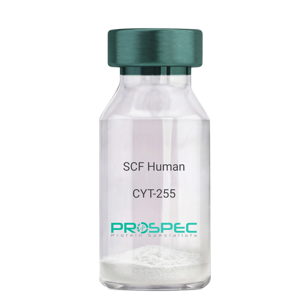
    SCF Human

    Description:

    Stem Cell Factor Human Recombinant

    Kit ligand Precursor, C-kit ligand, SCF, Mast cell growth factor, MGF, SF, KL-1, Kitl, DKFZp686F2250.

    Product # :

    CYT-255

    Price :

    Quantity :

    Shipping Method :

    Room Temp Icon

    Shipped at Room temp

    Add To Cart

    More Info

    • description
    • source
    • formulation
    • purity
    • biological activity
    • More Info

    Description

    Stem Cell Factor Human Recombinant produced in E.Coli is a single, non-glycosylated polypeptide chain containing 165 amino acids and having a molecular mass of 18409 Dalton. The SCF is purified by proprietary chromatographic techniques.

    Source

    Escherichia Coli.

    Formulation

    Lyophilized from a concentrated (1mg/ml) solution in water containing 0.02% NaHCO3.

    Purity

    Greater than 98.0% as determined by:
    (a) Analysis by SEC-HPLC.
    (b) Analysis by SDS-PAGE.

    Biological Activity

    The ED50 as determined by the dose-dependant stimulation of Human TF-1 cells is < 2 ng/ml, corresponding to a Specific Activity of 500,000 IU/mg.

    More Info

    • Introduction

      Stem cell factor / KIT ligand (SCF) is a cytokine which binds CD117(c-Kit). SCF is also known as "steel factor" or "c-kit ligand". SCF exists in two forms, cell surface bound SCF and soluble (or free) SCF. Soluble SCF is produced by the cleavage of surface bound SCF by metalloproteases. SCF is a growth factor important for the survival, proliferation, and differentiation of hematopoietic stem cells and other hematopoietic progenitor cells. One of its roles is to change the BFU-E (burst-forming unit-erythroid) cells, which are the earliest erythrocyte precursors in the erythrocytic series, into the CFU-E (colony-forming unit-erythroid).

    • Synonyms

      Kit ligand Precursor, C-kit ligand, SCF, Mast cell growth factor, MGF, SF, KL-1, Kitl, DKFZp686F2250.

    • Physical Appearance

      Sterile Filtered White lyophilized (freeze-dried) powder.

    • Stability

      Lyophilized KIT ligand although stable at room temperature for 3 weeks, should be stored desiccated below -18°C. Upon reconstitution SCF should be stored at 4°C between 2-7 days and for future use below -18°C.For long term storage it is recommended to add a carrier protein (0.1% HSA or BSA).Please prevent freeze-thaw cycles.

    • Solubility

      It is recommended to reconstitute the lyophilized Stem Cell Factor in sterile 18MΩ-cm H2O not less than 100µg/ml, which can then be further diluted to other aqueous solutions.

    • Amino Acid Sequence

      The sequence of the first five N-terminal amino acids was determined and was found to be Met-Glu-Gly-Ile-Cys.

    • Protein content

      Protein quantitation was carried out by two independent methods1. UV spectroscopy at 280 nm using the absorbency value of 0.52 as the extinction coefficient for a 0.1% (1mg/ml) solution. This value is calculated by the PC GENE computer analysis program of protein sequences (IntelliGenetics). 2. Analysis by RP-HPLC, using a standard solution of Stem Cell Factor as a Reference Standard.

    ProSpec's products are furnished for LABORATORY RESEARCH USE ONLY. The product may not be used as drugs, agricultural or pesticidal products, food additives or household chemicals.

    Scf Human
  • View Data Sheet

    Name :

    tPA Human, Sf9

    Description:

    Tissue Plasminogen Activator Human Recombinant, Sf9

    Tissue-type plasminogen activator, EC 3.4.21.68, tPA, t-PA, t-plasminogen activator, TPA, T-PA, DKFZp686I03148, PLAT and tPA.

    Product # :

    ENZ-1011

    Price :

    Quantity :

    Shipping Method :

    Ice Icon

    Shipped with Ice Packs

    Add To Cart

    More Info

    • description
    • source
    • formulation
    • purity
    • More Info

    Description

    tPA Human Recombinant produced in Sf9 Baculovirus cells is a single, glycosylated polypeptide chain containing 545 amino acids (24-562 a.a) and having a molecular mass of 61.3kDa (Molecular size on SDS-PAGE will appear at approximately 50-70kDa).tPA is fused to a 6 amino acid His-tag at C-terminus & purified by proprietary chromatographic techniques.

    Source

    Sf9, Baculovirus cells.

    Formulation

    tPA protein solution (0.25mg/ml) containing 50mM MES buffer(pH 5.5 ), 40% glycerol, 5mM CaCl2, 1mM DTT and 0.5M NaCl.

    Purity

    Greater than 90.0% as determined by SDS-PAGE.

    More Info

    • Introduction

      Tissue plasminogen activator (abbreviated PLAT or tPA) is a secreted serine proteasewhich converts the proenzymeplasminogento plasmin, a fibrinolyticenzyme. Plasminogen is synthesized as a single chain which is cleaved by PLAT into the two chain disulfide linked plasmin.
      This enzyme plays a role in cell migrationand tissue remodeling. Increased enzymatic activity causes hyperfibrinolysis, which manifests as excessive bleeding; decreased activity leads to hypofibrinolysiswhich can result in thrombosisor embolism.

    • Synonyms

      Tissue-type plasminogen activator, EC 3.4.21.68, tPA, t-PA, t-plasminogen activator, TPA, T-PA, DKFZp686I03148, PLAT and tPA.

    • Physical Appearance

      Sterile Filtered colorless solution.

    • Stability

      Store at 4°C if entire vial will be used within 2-4 weeks. Store, frozen at -20°C for longer periods of time. For long term storage it is recommended to add a carrier protein (0.1% HSA or BSA).Avoid multiple freeze-thaw cycles.

    • Amino Acid Sequence

      QEIHARFRRG ARSYQVICRD EKTQMIYQQH QSWLRPVLRS NRVEYCWCNS GRAQCHSVPV KSCSEPRCFN GGTCQQALYF SDFVCQCPEG FAGKCCEIDT RATCYEDQGI SYRGTWSTAE SGAECTNWNS SALAQKPYSG RRPDAIRLGL GNHNYCRNPD RDSKPWCYVF KAGKYSSEFC STPACSEGNS DCYFGNGSAY RGTHSLTESG ASCLPWNSMI LIGKVYTAQN PSAQALGLGK HNYCRNPDGD AKPWCHVLKN RRLTWEYCDV PSCSTCGLRQ YSQPQFRIKG GLFADIASHP WQAAIFAKHR RSPGERFLCG GILISSCWIL SAAHCFQERF PPHHLTVILG RTYRVVPGEE EQKFEVEKYI VHKEFDDDTY DNDIALLQLK SDSSRCAQES SVVRTVCLPP ADLQLPDWTE CELSGYGKHE ALSPFYSERL KEAHVRLYPS SRCTSQHLLN RTVTDNMLCA GDTRSGGPQA NLHDACQGDS GGPLVCLNDG RMTLVGIISW GLGCGQKDVP GVYTKVTNYL DWIRDNMRPHHHHHH.

    ProSpec's products are furnished for LABORATORY RESEARCH USE ONLY. The product may not be used as drugs, agricultural or pesticidal products, food additives or household chemicals.

    Tpa Human Sf9
  • View Data Sheet

    Name :

    FLT1 D3 Human

    Description:

    Vascular Endothelial Growth Factor Receptor-1 D3 Human Recombinant

    FLT-1, FLT1, Tyrosine-protein kinase receptor FLT, Flt-1, Tyrosine-protein kinase FRT, Fms-like tyrosine kinase 1, VEGFR-1.

    Product # :

    PKA-234

    Price :

    Quantity :

    Shipping Method :

    Room Temp Icon

    Shipped at Room temp

    Add To Cart

    More Info

    • description
    • source
    • formulation
    • purity
    • biological activity
    • More Info

    Description

    FLT1 D1-3 Human Recombinant produced in baculovirus is monomeric, glycosylated, polypeptide containing 327 amino acids and having a molecular mass of 45 kDa. The soluble receptor protein contains only the first 3 extracellular domains, which contain all the information necessary for binding of VEGF.The FLT1 is purified by proprietary chromatographic techniques.

    Source

    Insect Cells.

    Formulation

    FLT1 D1-3 was lyophilized from a concentrated (1mg/ml) sterile solution containing 1xPBS.

    Purity

    Greater than 90.0% as determined by SDS-PAGE.

    Biological Activity

    The activity of FLT1D1-3 was determined by its ability to inhibit the VEGF-165-induced proliferation of HUVE cells.

    More Info

    • Introduction

      Endothelial cells express three different vascular endothelial growth factor (VEGF) receptors, belonging to the family of receptor tyrosine kinases (RTKs). They are named VEGFR-1 (Flt-1), VEGFR-2 (KDR/Flk-1), VEGFR-3 (Flt-4). Their expression is almost exclusively restricted to endothelial cells, but VEGFR-1 can also be found on monocytes, dendritic cells and on trophoblast cells. The flt-1 gene was first described in 1990. The receptor contains seven immunoglobulin-like extracellular domains, a single transmembrane region and an intracellular splited tyrosine kinase domain. Compared to VEGFR-2 the Flt-1 receptor has a higher affinity for VEGF but a weaker signaling activity. VEGFR-1 thus leads not to proliferation of endothelial cells, but mediates signals for differentiation. Interestingly a naturally occuring soluble variant of VEGFR-1 (sVEGFR-1) was found in HUVE supernatants in 1996, which is generated by alternative splicing of the flt-1 mRNA. The biological functions of sVEGFR-1 still are not clear, but it seems to be an endogenous regulator of angiogenesis, binding VEGF with the same affinity as the full-length receptor.

    • Synonyms

      FLT-1, FLT1, Tyrosine-protein kinase receptor FLT, Flt-1, Tyrosine-protein kinase FRT, Fms-like tyrosine kinase 1, VEGFR-1.

    • Physical Appearance

      Sterile Filtered White lyophilized (freeze-dried) powder.

    • Stability

      Lyophilized FLT-1 although stable at room temperature for 3 weeks, should be stored desiccated below -18°C. Upon reconstitution FLT1 should be stored at 4°C between 2-7 days and for future use below -18°C.For long term storage it is recommended to add a carrier protein (0.1% HSA or BSA).Please prevent freeze-thaw cycles.

    • Solubility

      It is recommended to reconstitute the lyophilized FLT1 D3 in sterile water not less than 100 µg/ml, which can then be further diluted to other aqueous solutions.

    • Amino Acid Sequence

      SKLKDPELSLKGTQHIMQAGQTLHLQCRGEAAHKWSLPEMVSKESERLSI TKSACGRNGKQFCSTLTLNTAQANHTGFYSCKYLAVPTSKKKETESAIYI FISDTGRPFVEMYSEIPEIIHMTEGRELVIPCRVTSPNITVTLKKFPLDT LIPDGKRIIWDSRKGFIISNATYKEIGLLTCEATVNGHLYKTNYLTHRQT NTIIDVQISTPRPVKLLRGHTLVLNCTATTPLNTRVQMTWSYPDEKNKRA SVRRRIDQSNSHANIFYSVLTIDKMQNKDKGLYTCRVRSGPSFKSVNTSV HIYDKAFITVKHRKQQVLETVAGKRSY.

    ProSpec's products are furnished for LABORATORY RESEARCH USE ONLY. The product may not be used as drugs, agricultural or pesticidal products, food additives or household chemicals.

    Flt1 D3 Human
  • View Data Sheet

    Name :

    RARRES2 Human, Sf9

    Description:

    Retinoic Acid Receptor Responder 2 Human Recombinant, Sf9

    Retinoic Acid Receptor Responder 2, Chemerin, Retinoic Acid Receptor Responder (Tazarotene Induced) 2, Tazarotene-Induced Gene 2 Protein, RAR-Responsive Protein TIG2, TIG2, Retinoic Acid Receptor Responder Protein 2, HP10433, Retinoic acid receptor responder protein 2, Chemerin, RAR-responsive protein TIG2, Tazarotene-induced gene 2 protein.

    Product # :

    PRO-2382

    Price :

    Quantity :

    Shipping Method :

    Ice Icon

    Shipped with Ice Packs

    Add To Cart

    More Info

    • description
    • source
    • formulation
    • purity
    • More Info

    Description

    RARRES2 produced in Sf9 Baculovirus cells is a single, glycosylated polypeptide chain containing 146 amino acids (21-157a.a.) and having a molecular mass of 16.9kDa. (Molecular size on SDS-PAGE will appear at approximately 18-28kDa). RARRES2 is expressed with a 6 amino acid His tag at C-Terminus and purified by proprietary chromatographic techniques.

    Source

    Sf9, Baculovirus cells.

    Formulation

    RARRES2 protein solution (0.5mg/ml) contains Phosphate Buffered Saline (pH 7.4) and 10% glycerol.

    Purity

    Greater than 90.0% as determined by SDS-PAGE.

    More Info

    • Introduction

      RARRES2 is a secreted chemotactic protein that initiates chemotaxis through the ChemR23 G protein-coupled seven-transmembrane domain ligand. RARRES2 is upregulated by the synthetic retinoid tazarotene and found in a vast variety of tissues. RARRES2 acts as an adipokine, and is truncated on both termini from the proprotein. RARRES2 is structurally related to the cathelicidin precursors, cystatin C and kininogens. RARRES2 promotes calcium mobilization and chemotaxis of immature dendritic cells and macrophages. RARRES2 is secreted as a precursor of little biological activity, which requires proteolytic cleavage of its COOH-terminal domain to be exchangeed into a potent and highly specific agonist of ChemR23. RARRES2 signals via its receptor, ChemR23 (CMKLR1), as a positive regulator of adipocyte differentiation and metabolic function. The Chemerin receptor acts as a coreceptor for SIV and some primary HIV-1 strains. The Chemerin receptor has another ligand, called tazarotene-induced gene.

    • Synonyms

      Retinoic Acid Receptor Responder 2, Chemerin, Retinoic Acid Receptor Responder (Tazarotene Induced) 2, Tazarotene-Induced Gene 2 Protein, RAR-Responsive Protein TIG2, TIG2, Retinoic Acid Receptor Responder Protein 2, HP10433, Retinoic acid receptor responder protein 2, Chemerin, RAR-responsive protein TIG2, Tazarotene-induced gene 2 protein.

    • Physical Appearance

      Sterile Filtered colorless solution.

    • Stability

      Store at 4°C if entire vial will be used within 2-4 weeks. Store, frozen at -20°C for longer periods of time. For long term storage it is recommended to add a carrier protein (0.1% HSA or BSA).Avoid multiple freeze-thaw cycles.

    • Amino Acid Sequence

      ADPELTEAQR RGLQVALEEF HKHPPVQWAF QETSVESAVD TPFPAGIFVR LEFKLQQTSC RKRDWKKPEC KVRPNGRKRK CLACIKLGSE DKVLGRLVHC PIETQVLREA EEHQETQCLR VQRAGEDPHS FYFPGQFAFS HHHHHH.

    ProSpec's products are furnished for LABORATORY RESEARCH USE ONLY. The product may not be used as drugs, agricultural or pesticidal products, food additives or household chemicals.

    Rarres2 Human Sf9
  • View Data Sheet

    Name :

    VEGFR2 Human, His

    Description:

    Vascular Endothelial Growth Factor receptor-2 Human Recombinant, His Tag

    KDR D1-7, sKDR D1-7, Kinase insert domain receptor, Protein-tyrosine kinase receptor Flk-1, CD309, type III receptor tyrosine kinase, FLK1, VEGFR-2. 

    Product # :

    PKA-099

    Price :

    Quantity :

    Shipping Method :

    Ice Icon

    Shipped with Ice Packs

    Add To Cart

    More Info

    • description
    • source
    • formulation
    • purity
    • More Info

    Description

    VEGFR2 Human Recombinant produced in Sf9 Baculovirus cells is a single, glycosylated polypeptide chain containing 987 amino acids (20-764a.a) and having a molecular mass of 110.5kDa (Molecular size on SDS-PAGE will appear at approximately 100-150kDa). VEGFR2 is fused to a 239 amino acids hIgG-His-tag at C-terminus & purified by proprietary chromatographic techniques.

    Source

    Sf9, Baculovirus cells.

    Formulation

    VEGFR2 solution (0.5mg/ml) contains Phosphate Buffered Saline (pH 7.4) and 10% glycerol.

    Purity

    Greater than 90% as determined by SDS-PAGE.

    More Info

    • Introduction

      Endothelial cells express three different vascular endothelial growth factor (VEGF) receptors, belonging to the family of receptor tyrosine kinases (RTKs). They are named VEGFR-1 (Flt-1), VEGFR-2 (KDR/Flk-1), VEGFR-3 (Flt-4). Their expression is almost exclusively restricted to endothelial cells, but VEGFR-1 can also be found on monocytes. All VEGF-receptors have seven immunoglobulin-like extracellular domains, a single transmembrane region and an intracellular split tyrosine kinase domain. VEGFR-2 has a lower affinity for VEGF than the Flt-1 receptor, but a higher signaling activity. Mitogenic activity in endothelial cells is mainly mediated by VEGFR-2 leading to their proliferation.
      Differential splicing of the flt-1 gene leads to the formation of a secreted, soluble variant of VEGFR-1 (sVEGFR-1). No naturally occuring, secreted forms of VEGFR-2 have so far been reported. The binding of VEGF165 to VEGFR-2 is dependent on heparin.

    • Synonyms

      KDR D1-7, sKDR D1-7, Kinase insert domain receptor, Protein-tyrosine kinase receptor Flk-1, CD309, type III receptor tyrosine kinase, FLK1, VEGFR-2.

    • Physical Appearance

      Sterile Filtered colorless solution.

    • Stability

      Store at 4°C if entire vial will be used within 2-4 weeks. Store, frozen at -20°C for longer periods of time. For long term storage it is recommended to add a carrier protein (0.1% HSA or BSA).Avoid multiple freeze-thaw cycles.

    • Amino Acid Sequence

      ADPASVGLPS VSLDLPRLSI QKDILTIKAN TTLQITCRGQ RDLDWLWPNN QSGSEQRVEV TECSDGLFCK TLTIPKVIGN DTGAYKCFYR ETDLASVIYV YVQDYRSPFI ASVSDQHGVV YITENKNKTV VIPCLGSISN LNVSLCARYP EKRFVPDGNR ISWDSKKGFT IPSYMISYAG MVFCEAKIND ESYQSIMYIV VVVGYRIYDV VLSPSHGIEL SVGEKLVLNC TARTELNVGI DFNWEYPSSK HQHKKLVNRD LKTQSGSEMK KFLSTLTIDG VTRSDQGLYT CAASSGLMTK KNSTFVRVHE KPFVAFGSGM ESLVEATVGE RVRIPAKYLG YPPPEIKWYK NGIPLESNHT IKAGHVLTIM EVSERDTGNY TVILTNPISK EKQSHVVSLV VYVPPQIGEK SLISPVDSYQ YGTTQTLTCT VYAIPPPHHI HWYWQLEEEC ANEPSQAVSV TNPYPCEEWR SVEDFQGGNK IEVNKNQFAL IEGKNKTVST LVIQAANVSA LYKCEAVNKV GRGERVISFH VTRGPEITLQ PDMQPTEQES VSLWCTADRS TFENLTWYKL GPQPLPIHVG ELPTPVCKNL DTLWKLNATM FSNSTNDILI MELKNASLQD QGDYVCLAQD RKTKKRHCVV RQLTVLERVA PTITGNLENQ TTSIGESIEV SCTASGNPPP QIMWFKDNET LVEDSGIVLK DGNRNLTIRR VRKEDEGLYT CQACSVLGCA KVEAFFIIEG AQEKTNLELE PKSCDKTHTC PPCPAPELLG GPSVFLFPPK PKDTLMISRT PEVTCVVVDV SHEDPEVKFN WYVDGVEVHN AKTKPREEQY NSTYRVVSVL TVLHQDWLNG KEYKCKVSNK ALPAPIEKTI SKAKGQPREP QVYTLPPSRD ELTKNQVSLT CLVKGFYPSD IAVEWESNGQ PENNYKTTPP VLDSDGSFFL YSKLTVDKSR WQQGNVFSCS VMHEALHNHY TQKSLSLSPG KHHHHHH.

    ProSpec's products are furnished for LABORATORY RESEARCH USE ONLY. The product may not be used as drugs, agricultural or pesticidal products, food additives or household chemicals.

    Human Vegfr2
  • View Data Sheet

    Name :

    APRIL Human

    Description:

    APRIL Human Recombinant

    Tumor necrosis factor ligand superfamily member 13, A proliferation-inducing ligand, APRIL, TNF- and APOL-related leukocyte expressed ligand 2, TALL-2, TNF-related death ligand 1, TRDL-1, CD256, TNFSF13, TALL2, ZTNF2, UNQ383/PRO715.

    Product # :

    CYT-815

    Price :

    Quantity :

    Shipping Method :

    Ice Icon

    Shipped with Ice Packs

    Add To Cart

    More Info

    • description
    • source
    • formulation
    • purity
    • More Info
    • sds-page

    Description

    APRIL Human Recombinant produced in E.coli is a single, non-glycosylated polypeptide chain containing 159 amino acids (105-247) and having a molecular mass of 17.6kDa.APRIL is fused to a 16 amino acid T7-tag at N-terminus and purified by proprietary chromatographic techniques.

    Source

    Escherichia Coli.

    Formulation

    APRIL protein solution (1mg/ml) containing 20mM Tris-HCl buffer (pH 8.0), 0.4M UREA and 10% glycerol.

    Purity

    Greater than 85.0% as determined by SDS-PAGE.

    sds-page

    APRIL-sds-page - Product image 1

    More Info

    • Introduction

      APRIL which is a part of the TNF ligand superfamily (TNFSF13) is a type II transmembrane protein. Normally, APRIL expression is low in tissues, but is elevated in numerous types of tumors and transformed cell lines.

    • Synonyms

      Tumor necrosis factor ligand superfamily member 13, A proliferation-inducing ligand, APRIL, TNF- and APOL-related leukocyte expressed ligand 2, TALL-2, TNF-related death ligand 1, TRDL-1, CD256, TNFSF13, TALL2, ZTNF2, UNQ383/PRO715.

    • Physical Appearance

      Sterile filtered colorless solution.

    • Stability

      Store at 4°C if entire vial will be used within 2-4 weeks. Store, frozen at -20°C for longer periods of time. For long term storage it is recommended to add a carrier protein (0.1% HSA or BSA).Avoid multiple freeze-thaw cycles.

    • Amino Acid Sequence

      MASMTGGQQM GRGSHMAVLT QKQKKQHSVL HLVPINATSK DDSDVTEVMW QPALRRGRGL QAQGYGVRIQ DAGVYLLYSQ VLFQDVTFTM GQVVSREGQG RQETLFRCIR SMPSHPDRAY NSCYSAGVFH LHQGDILSVI IPRARAKLNL SPHGTFLGL.

    • Background

      APRIL Human Recombinant: Expanding the Horizons of Immunotherapy

      Abstract:


      APRIL (A Proliferation-Inducing Ligand) is a promising molecule within the tumor necrosis factor (TNF) superfamily that plays a pivotal role in immune regulation. This research paper provides an overview of APRIL human recombinant, exploring its potential applications in immunotherapy. Understanding the mechanisms and therapeutic implications of APRIL holds promise in the field of immune-related disorders. This article presents a concise analysis of APRIL, highlighting its potential as a therapeutic target.

      Introduction:


      Immunotherapy has revolutionized the treatment of various diseases by harnessing the power of the immune system. APRIL, a member of the TNF superfamily, has emerged as a potential candidate for immunotherapeutic interventions. This paper provides an overview of APRIL, shedding light on its structure, function, and potential applications in immunotherapy.

      APRIL Structure and Function:


      APRIL is a transmembrane protein that can be proteolytically cleaved, leading to the generation of soluble forms. It interacts with its receptors, such as BCMA and TACI, to regulate immune responses. APRIL influences B-cell activation, proliferation, and antibody production, making it a compelling target for immunotherapeutic strategies.

      Immunotherapeutic Applications of APRIL Human Recombinant:


      APRIL human recombinant holds great potential in immunotherapy. By modulating APRIL activity, it may be possible to enhance immune responses against cancer cells or dampen immune dysregulation in autoimmune disorders. Additionally, APRIL-based therapeutics could be developed to target specific immune cell populations or enhance the efficacy of existing immunotherapies.

      Challenges and Future Directions:


      Although APRIL shows promise, there are challenges to overcome. Further research is needed to elucidate the precise mechanisms of APRIL-mediated immune regulation and identify optimal therapeutic approaches. Additionally, safety considerations and potential side effects must be thoroughly evaluated.

      Conclusion:


      APRIL human recombinant represents a valuable tool for advancing immunotherapy. Understanding the structure, function, and therapeutic potential of APRIL opens up new avenues for treating immune-related disorders. Continued research and development in this field have the potential to revolutionize the landscape of immunotherapy, improving patient outcomes and expanding the possibilities for personalized medicine.

      What is the molecular weight/Mw of APRIL Protein?
      APRIL Protein has a total Mw of 17.6kDa.

      What is the source or expression system of APRIL Protein?
      Escherichia Coli.

      What is the Purity of APRIL Protein?
      APRIL Protein is >85% pure as determined by SDS-PAGE.

      What is the Biological Activity of APRIL Protein?
      The biological functionality of APRIL Protein will be determined in the future.

      What is the amino acid sequence of APRIL Protein?
      MASMTGGQQM GRGSHMAVLT QKQKKQHSVL HLVPINATSK DDSDVTEVMW QPALRRGRGL QAQGYGVRIQ DAGVYLLYSQ VLFQDVTFTM GQVVSREGQG RQETLFRCIR SMPSHPDRAY NSCYSAGVFH LHQGDILSVI IPRARAKLNL SPHGTFLGL.

      What applications can APRIL Protein be used in?
      APRIL Protein can probably be used in western blot, ELISA and Lateral Flow.

      What is the endotoxin level for APRIL Protein?
      The endotoxin level is minimal, APRIL Protein was purified using conventional chromatography techniques.

    ProSpec's products are furnished for LABORATORY RESEARCH USE ONLY. The product may not be used as drugs, agricultural or pesticidal products, food additives or household chemicals.

    April Human
  • View Data Sheet

    Name :

    FAS Antibody (CD95)

    Description:

    FAS Antibody (CD95), Mouse Anti Human

    FASLG receptor, Apoptosis-mediating surface antigen FAS, Apo-1 antigen, CD95, Tumor necrosis factor receptor superfamily member 6 TNR6, APT1, FAS1, TNFRSF6.

    Product # :

    ANT-735

    Price :

    Quantity :

    Shipping Method :

    Ice Icon

    Shipped with Ice Packs

    Add To Cart

    More Info

    • formulation
    • More Info

    Formulation

    1X PBS pH 7.4, 0.5% Saccharose, 0.2% gelatin and 0.1% NaN3.

    More Info

    • Introduction

      The Fas receptor (CD95) mediates apoptotic signaling by Fas-ligand expressed on the surface of other cells. The Fas-FasL interaction plays an important role in the immune system and lack of this system leads to autoimmunity, indicating that Fas-mediated apoptosis removes self-reactive lymphocytes. Fas signaling is also involved in immune surveillance to remove transformed cells and virus infected cells. Binding of FAS to oligimerized FasL on another cell activates apoptotic signaling through a cytoplasmic domain termed the death domain that interacts with signaling adaptors including FAF, FADD and DAX to activate the caspase proteolytic cascade. Caspase-8 and caspase-10 are first activated, to then cleave and activate downstream caspases, and a variety of cellular substrates that lead to cell death. Caspases cleave nuclear lamins, causing the nucleus to break down and lose its normal structure and another caspase substrate is DFF, inducing cleavage and degradation of the genome. Other caspase substrates are involved in cytoskeletal structure, cell cycle regulation and signaling pathways. Activation of JNK kinase, activation of Jun, and production of ceramide may also play roles in Fas-mediated apoptosis. Activation of fas-mediated apoptosis is opposed by I-FLICE and FAP. Viruses and tumors may escape immune surveillance in part through suppression of fas-mediated apoptosis using similar mechanisms.

    • Synonyms

      FASLG receptor, Apoptosis-mediating surface antigen FAS, Apo-1 antigen, CD95, Tumor necrosis factor receptor superfamily member 6 TNR6, APT1, FAS1, TNFRSF6.

    • Physical Appearance

      Sterile filtered colorless solution.

    • Immunogen

      Recombinant Human FAS.

    • Ig Subclass

      Mouse IgG2a

    • Clone

      pprro-160

    • Applications

      FAS Antibody can be used in Flow cytometry.

    • Type

      Mouse Anti Human Monoclonal.

    • Storage Procedures

      For periods up to 1 month store at 4°C, for longer periods, store at -20°C. Prevent freeze thaw cycles.

    • Purification Method

      Protein-A column.

    ProSpec's products are furnished for LABORATORY RESEARCH USE ONLY. The product may not be used as drugs, agricultural or pesticidal products, food additives or household chemicals.

    Fas Antibody
  • View Data Sheet

    Name :

    EGF Antibody

    Description:

    Endothelial Growth Factor, Mouse Anti-Human

    Urogastrone, URG, EGF.

    Product # :

    ANT-169

    Price :

    Quantity :

    Shipping Method :

    Room Temp Icon

    Shipped at Room temp

    Add To Cart

    More Info

    • formulation
    • More Info

    Formulation

    1mg/ml in PBS (after reconstitution).

    More Info

    • Introduction

      Epidermal growth factor has a profound effect on the differentiation of specific cells in vivo and is a potent mitogenic factor for a variety of cultured cells of both ectodermal and mesodermal origin. The EGF precursor is believed to exist as a membrane-bound molecule which is proteolytically cleaved to generate the 53-amino acid peptide hormone that stimulates cells to divide.
      EGF stimulates the growth of various epidermal and epithelial tissues in vivo and in vitro and of some fibroblasts in cell culture.

    • Synonyms

      Urogastrone, URG, EGF.

    • Solubility

      Reconstitute with sterile H20. Mix gently, wash the sides of the vial and wait 30-60 seconds before use.

    • Immunogen

      r.HumanEGF.

    • Ig Subclass

      Mouse IgG2b.

    • Clone

      NYRhEGF.

    • Applications

      Direct ELISA, Western Blot, Immuneprecipitation.

    • Titer

      By direct ELISA, 1:10,000 dilution will yield 0.3 O.D using alkaline phosphatase conjugated rabbit anti-mouse Ig (Jackson Laboratories).

    • Shipping Conditions

      Antibody is shipped lyophilized at ambient temperature.

    • Type

      Mouse Anti Human Monoclonal.

    • Storage Procedures

      In lyophilized form, for long periods, store at 4oC in a dry environment. After reconstitution, if not intended for use within a month, aliquot and store at -20oC.

    • Purification Method

      Ion exchange.

    ProSpec's products are furnished for LABORATORY RESEARCH USE ONLY. The product may not be used as drugs, agricultural or pesticidal products, food additives or household chemicals.

    Egf Antibody
  • View Data Sheet

    Name :

    IGF1 PAT6F8AT Antibody

    Description:

    Insulin-Like Growth Factor-1 Clone PAT6F8AT, Mouse Anti Human

    Somatomedin C, IGF-I, IGFI, IGF1, IGF-IA, Mechano growth factor, MGF.

    Product # :

    ANT-538

    Price :

    Quantity :

    Shipping Method :

    Ice Icon

    Shipped with Ice Packs

    Add To Cart

    More Info

    • formulation
    • More Info

    Formulation

    1mg/ml containing PBS, pH-7.4, 10% Glycerol and 0.02% Sodium Azide.

    More Info

    • Introduction

      The somatomedins, or insulin-like growth factors (IGFs), comprise a family of peptides that play important roles in mammalian growth and development. IGF1 mediates many of the growth-promoting effects of growth hormone (GH; MIM 139250). Early studies showed that growth hormone did not directly stimulate the incorporation of sulfate into cartilage, but rather acted through a serum factor, termed 'sulfation factor,' which later became known as 'somatomedin' (Daughaday et al., 1972). Three main somatomedins have been characterized: somatomedin C (IGF1), somatomedin A (IGF2; MIM 147470), and somatomedin B (MIM 193190) (Rotwein, 1986; Rosenfeld, 2003).

    • Synonyms

      Somatomedin C, IGF-I, IGFI, IGF1, IGF-IA, Mechano growth factor, MGF.

    • Physical Appearance

      Sterile filtered colorless solution.

    • Immunogen

      Anti-human IGF1 mAb, clone PAT6F8AT, is derived from hybridization of mouse F0 myeloma cells with spleen cells from BALB/c mice immunized with a recombinant human IGF1 protein 49-118 amino acids purified from E. coli.

    • Ig Subclass

      Mouse IgG2b heavy chain and k light chain.

    • Clone

      PAT6F8AT.

    • Applications

      HSPA5 antibody has been tested by ELISA, Western blot analysis to assure specificity and reactivity. Since application varies, however, each investigation should be titrated by the reagent to obtain optimal results. Recommended starting dilution is 1:1000.

    • Type

      Mouse Anti Human Monoclonal.

    • Storage Procedures

      For periods up to 1 month store at 4°C, for longer periods of time, store at -20°C. Prevent freeze thaw cycles.

    • Purification Method

      IGF1 antibody was purified from mouse ascitic fluids by protein-A affinity chromatography.

    ProSpec's products are furnished for LABORATORY RESEARCH USE ONLY. The product may not be used as drugs, agricultural or pesticidal products, food additives or household chemicals.

    Igf1 Pat6F8At Antibody
  • View Data Sheet

    Name :

    TNFA Rat, His Active

    Description:

    Tumor Necrosis Factor-alpha Rat Recombinant, His Tag Active

    Tumor Necrosis Factor-alpha, TNF a His,  Cachectin, TNF-alpha, Tumor necrosis factor ligand superfamily member 2, TNF-a, N-terminal fragment, NTF, Intracellular domain 1, Intracellular domain 2, ICD2, C-domain 1, C-domain 2, Tumor necrosis factor, soluble form, Tnfa, Tnfsf2, RATTNF, Tnfa.

    Product # :

    CYT-1057

    Price :

    Quantity :

    Shipping Method :

    Ice Icon

    Shipped with Ice Packs

    Add To Cart

    More Info

    • description
    • source
    • formulation
    • purity
    • biological activity
    • More Info

    Description

    TNFA Rat Recombinant produced in E.Coli is a single, non-glycosylated polypeptide chain containing 181 amino acids (80-235 a.a) and having a molecular mass of 19.9kDa.TNFA Rat is expressed with an 25 amino acid His tag at C-Terminus and purified by proprietary chromatographic techniques.

    Source

    Escherichia Coli.

    Formulation

    TNFA Rat protein solution (1mg/ml) contains Phosphate Buffered Saline (pH 7.4), 10% glycerol and 1mM DTT.

    Purity

    Greater than 90.0% as determined by SDS-PAGE.

    Biological Activity

    Measured in a cytotoxicity assay using L929 mouse fibrosarcoma cells in the presence of the metabolic inhibitor actinomycin D.
    The ED50 for this effect is ≤ to 0.2 ng/ml.

    More Info

    • Introduction

      Tumor necrosis factor is a cytokine involved in systemic inflammation and is a member of a group of cytokines that all stimulate the acute phase reaction. TNF is mainly secreted by macrophages.
      TNF causes apoptotic cell death, cellular proliferation, differentiation, inflammation, tumorigenesis and viral replication, TNF is also involved in lipid metabolism, and coagulation. TNF's primary role is in the regulation of immune cells.
      Dysregulation and, in particular, overproduction of TNF have been implicated in a variety of human diseases- autoimmune diseases, insulin resistance, and cancer.

    • Synonyms

      Tumor Necrosis Factor-alpha, TNF a His, Cachectin, TNF-alpha, Tumor necrosis factor ligand superfamily member 2, TNF-a, N-terminal fragment, NTF, Intracellular domain 1, Intracellular domain 2, ICD2, C-domain 1, C-domain 2, Tumor necrosis factor, soluble form, Tnfa, Tnfsf2, RATTNF, Tnfa.

    • Physical Appearance

      Sterile Filtered colorless liquid.

    • Stability

      Store at 4°C if entire vial will be used within 2-4 weeks. Store, frozen at -20°C for longer periods of time. For long term storage it is recommended to add a carrier protein (0.1% HSA or BSA).Avoid multiple freeze-thaw cycles.

    • Amino Acid Sequence

      MGSSHHHHHH SSGLVPRGSH MGSHMLRSSS QNSSDKPVAH VVANHQAEEQ LEWLSQRANA LLANGMDLKD NQLVVPADGL YLIYSQVLFK GQGCPDYVLL THTVSRFAIS YQEKVSLLSA IKSPCPKDTP EGAELKPWYE PMYLGGVFQL EKGDLLSAEV NLPKYLDITE SGQVYFGVIA L.

    • Background

      Tumor Necrosis Factor-alpha Rat Recombinant, His Tag Active: An In-Depth Analysis

      Abstract:


      Tumor Necrosis Factor-alpha (TNF-α) is a cytokine that plays a significant role in various physiological and pathological processes. This human research paper provides an in-depth analysis of TNF-α Rat Recombinant with a His Tag, focusing on its structure, signaling pathways, and diverse functions. Additionally, the paper explores the potential applications of TNF-α Rat Recombinant in human research.

      Introduction:


      TNF-α is a key mediator of inflammation and immune responses in humans. This research paper aims to provide a comprehensive analysis of TNF-α Rat Recombinant with a His Tag, highlighting its significance in human physiology and its potential applications in human research.

      Structure and Function of TNF-α:


      TNF-α is a homotrimeric protein that binds to two distinct receptors, TNFR1 and TNFR2, initiating downstream signaling cascades. It regulates immune cell activation, cytokine production, and cellular responses, influencing diverse biological processes.

      Signaling Pathways:


      Upon binding to its receptors, TNF-α activates various signaling pathways, including the NF-κB pathway, MAPK pathway, and cell death pathways. These pathways regulate gene expression and mediate cellular responses, impacting inflammation, apoptosis, and tissue homeostasis.

      Functions of TNF-α:


      TNF-α plays a crucial role in immune responses, inflammation, and tissue homeostasis. It regulates the activation and migration of immune cells, promotes cytokine production, and modulates cell survival and death. Dysregulation of TNF-α is implicated in the pathogenesis of various human diseases, making it an attractive target for research and therapeutic interventions.

      Applications in Human Research:


      TNF-α Rat Recombinant with a His Tag has diverse applications in human research. It can be used to investigate TNF-α signaling pathways, study its effects on immune cell functions, and explore its role in disease pathogenesis. Additionally, this recombinant protein can be utilized for in vitro and in vivo studies aimed at developing novel therapeutic strategies.

      Future Directions:


      Further research is necessary to unravel the intricate mechanisms of TNF-α signaling and its contributions to human diseases. Continued investigations will enable the development of targeted therapies and personalized medicine approaches. Future studies should also focus on optimizing the use of TNF-α Rat Recombinant in preclinical and clinical research settings.

    ProSpec's products are furnished for LABORATORY RESEARCH USE ONLY. The product may not be used as drugs, agricultural or pesticidal products, food additives or household chemicals.

    Tnfa Rat 2
  • View Data Sheet

    Name :

    TNFRSF14 Mouse

    Description:

    HVEM Mouse Recombinant

    Tumor Necrosis Factor Receptor Superfamily Member 14, HVEM, TR2, Herpes Virus Entry Mediator A, Tumor Necrosis Factor Receptor-Like 2, Herpesvirus Entry Mediator, HVEA, ATAR, CD270, LIGHTR, CD40-Like Protein, Tumor Necrosis Factor Receptor-Like Gene2.

    Product # :

    CYT-1145

    Price :

    Quantity :

    Shipping Method :

    Ice Icon

    Shipped with Ice Packs

    Add To Cart

    More Info

    • description
    • source
    • formulation
    • purity
    • More Info

    Description

    TNFRSF14 Mouse produced in Sf9 Baculovirus cells is a single, glycosylated polypeptide chain containing 407 amino acids (39-206 aa) and having a molecular mass of 45.3kDa.TNFRSF14 is fused to a 239 amino acid hIgG-His-Tag at C-terminus and purified by proprietary chromatographic techniques.

    Source

    Sf9, Baculovirus cells.

    Formulation

    The TNFRSF14 solution (0.25mg/ml) contains 10% glycerol and Phosphate-Buffered Saline (pH 7.4).

    Purity

    Greater than 90.0% as determined by SDS-PAGE.

    More Info

    • Introduction

      Herpesvirus entry mediator or HVEM or tumour necrosis factor receptor superfamily member 14 or TNFRSF14, is part of the TNF receptors family that is a receptor located on the cell surface. The cytoplasmic area of this receptor can bind all sorts of TNF receptor associated factor (TRAF) protein. Those proteins can mediate pathways activating an immune response. The MITF is regulating TNFRSF14 gene expression.

    • Synonyms

      Tumor Necrosis Factor Receptor Superfamily Member 14, HVEM, TR2, Herpes Virus Entry Mediator A, Tumor Necrosis Factor Receptor-Like 2, Herpesvirus Entry Mediator, HVEA, ATAR, CD270, LIGHTR, CD40-Like Protein, Tumor Necrosis Factor Receptor-Like Gene2.

    • Physical Appearance

      Sterile Filtered colorless solution.

    • Stability

      Store at 4°C if entire vial will be used within 2-4 weeks. Store, frozen at -20°C for longer periods of time. For long term storage it is recommended to add a carrier protein (0.1% HSA or BSA).Avoid multiple freeze-thaw cycles.

    • Amino Acid Sequence

      QPSCRQEEFL VGDECCPMCN PGYHVKQVCS EHTGTVCAPC PPQTYTAHAN GLSKCLPCGV
      CDPDMGLLTW QECSSWKDTV CRCIPGYFCE NQDGSHCSTC LQHTTCPPGQ RVEKRGTHDQ
      DTVCADCLTG TFSLGGTQEE CLPWTNCSAF QQEVRRGTNS TDTTCSSQLE PKSCDKTHTC
      PPCPAPELLG GPSVFLFPPK PKDTLMISRT PEVTCVVVDV SHEDPEVKFN WYVDGVEVHN
      AKTKPREEQY NSTYRVVSVL TVLHQDWLNG KEYKCKVSNK ALPAPIEKTI SKAKGQPREP
      QVYTLPPSRD ELTKNQVSLT CLVKGFYPSD IAVEWESNGQ PENNYKTTPP VLDSDGSFFL
      YSKLTVDKSR WQQGNVFSCS VMHEALHNHY TQKSLSLSPG KHHHHHH

    ProSpec's products are furnished for LABORATORY RESEARCH USE ONLY. The product may not be used as drugs, agricultural or pesticidal products, food additives or household chemicals.

    Tnfrsf14 Mouse
  • View Data Sheet

    Name :

    FASLG Human, HEK

    Description:

    FAS Ligand Human Recombinant, HEK

    Fas ligand (TNF superfamily, member 6), APT1LG1, FASL, TNFSF6, CD178, tumor necrosis factor (ligand) superfamily member 6, Apoptosis antigen ligand, Fas antigen ligand, APTL, CD95-L.

    Product # :

    CYT-051

    Price :

    Quantity :

    Shipping Method :

    Ice Icon

    Shipped with Ice Packs

    Add To Cart

    More Info

    • description
    • source
    • formulation
    • purity
    • biological activity
    • More Info

    Description

    Recombinant Human FAS Ligand produced in HEK293 cells is a polypeptide chain containing 147 amino acids (134-281a.a).FASLG is fused to a 6 amino acid His-tag at N-terminus and purified by proprietary chromatographic techniques.

    Source

    HEK293 cells.

    Formulation

    The FASLG solution (0.6mg/ml) contains 1xPBS.

    Purity

    Greater than 95.0% as determined by:
    (a) Analysis by SEC-HPLC.
    (b) Analysis by SDS-PAGE.

    Biological Activity

    Fas ligand is biologically active as determined by its ability to induce cytotoxicity in Jurkat cells in the absence of any cross-linking. The expected ED50< 10 ng/ml, corresponding to a specific activity of 1x10⁵ units/mg.

    More Info

    • Introduction

      The type II transmembrane protein FASLG is a member of the tumor necrosis factor (TNF) superfamily. A fas ligand/receptor interaction has a significant part in the regulation of the immune system and the advancement of cancer. FASLG is expressed on the activated T cell surface as a nondisulfidelinked homotrimer. FASLG binding to Fas/CD95/TNFRSF6 on a nearby cell prompts apoptosis in the Fas expressing cell. FASLG is released from the cell surface by metalloproteinases as a soluble molecule that stays trimeric and is able to bind with Fas, but its capability to activate apoptosis is radically reduced. In addition, FASLG binds to DcR3 - a soluble trap receptor with no signal transduction capabilities. Flawed Fas-mediated apoptosis causes oncogenesis in addition to drug resistance in existing tumors. Constitutive expression of FASLG in a variety of tumors enables their immune evasion. Both mouse and human FASLG are active on mouse and human cells.

    • Synonyms

      Fas ligand (TNF superfamily, member 6), APT1LG1, FASL, TNFSF6, CD178, tumor necrosis factor (ligand) superfamily member 6, Apoptosis antigen ligand, Fas antigen ligand, APTL, CD95-L.

    • Physical Appearance

      Sterile Filtered colorless solution.

    • Stability

      FASLG Human Recombinant although stable at 4°C for 1 week, should be stored below -18°C. Please prevent freeze thaw cycles.

    • Background

      What is the source or expression system of FASL Protein?
      HEK293 cells.

      What is the Purity of FASL Protein?
      FASL Protein is >95% pure as determined by SDS-PAGE.

      What is the Biological Activity of FASL Protein?
      Fas ligand is biologically active as determined by its ability to induce cytotoxicity in Jurkat cells in the absence of any cross-linking. The expected ED50< 10 ng/ml, corresponding to a specific activity of 1x10⁵ units/mg.

      What is the amino acid sequence of FASL Protein?
      FASL Protein is composed from 147 amino acids.

      What applications can FASL Protein be used in?
      FASL Protein can probably be used in western blot, ELISA and Lateral Flow.

      What is the endotoxin level for FASL Protein?
      The endotoxin level is minimal, FASL Protein was purified using conventional chromatography techniques.

    ProSpec's products are furnished for LABORATORY RESEARCH USE ONLY. The product may not be used as drugs, agricultural or pesticidal products, food additives or household chemicals.

    Faslg Human Hek
  • View Data Sheet

    Name :

    VEGF E (Orf Virus)

    Description:

    Vascular Endothelial Growth Factor-E Recombinant (Orf Virus)

    Product # :

    CYT-263

    Price :

    Quantity :

    Shipping Method :

    Room Temp Icon

    Shipped at Room temp

    Add To Cart

    More Info

    • description
    • source
    • formulation
    • purity
    • biological activity
    • More Info

    Description

    A DNA sequence encoding the mature variant of ovVEGF-E isolate D1701 (Dehio et al., 1999; GenBank accession No. AF106020) was expressed in E. coli as a 132 amino acid residue fusion protein with an N-terminal His-tag sequence and a thrombin cleavage site. Recombinant VEGF-E homodimer was dimerized in vitro and has a predicted mass of approximately 35 kDa.

    Source

    Escherichia Coli.

    Formulation

    The protein was lyophilized from a concentrated (1mg/ml) solution containing PBS.

    Purity

    Greater than 90.0% as determined by SDS-PAGE.

    Biological Activity

    The biological activity was determined (1) by the ability to induce VEGFR-2/KDR receptor phosphorylation in PAE/KDR cells and (2) in a cell proliferation assay using primary HUVECs. The ED50 for this effect is typically 1-5ng/ml.

    More Info

    • Introduction

      Based on sequence similarity to VEGF-A, a gene encoding a VEGF homologue has recently been discovered in the genome of Orf virus (OV) (Lyttle et al., 1994). Different isolates of Orf virus show significant amino acid sequence similarity to VEGF-A and described as a viral virulence factor that appears to be derived from captured host genes. All eight cysteine residues of the central cysteine knot motif characteristic of members of the VEGF family are conserved among other residues in the VEGF-E proteins (Dehio et al., 1999; Wise et al., 1999). Alignment of all mammalian VEGF sequences indicated that VEGF-E is distinct from the previously described VEGFs but most closely related to VEGF-A. Like VEGF-A, VEGF-E was found to bind with high affinity to VEGF receptor-2 (KDR) resulting in receptor autophosphorylation, whilst in contrast to VEGF-A, VEGF-E can not bind to VEGF receptor-1 (Flt-1). Furthermore VEGF-E can also not bind to VEGF receptor-3 (FLT-4). Therefore VEGF-E is a potent angiog

    • Physical Appearance

      Sterile Filtered White lyophilized (freeze-dried) powder.

    • Stability

      Lyophilized Vascular Endothelial Growth Factor-E Orf Virus although stable at room temperature for 3 weeks, should be stored desiccated below -18°C. Upon reconstitution VEGF E -OV should be stored at 4°C between 2-7 days and for future use below -18°C.Please prevent freeze-thaw cycles.

    • Solubility

      The lyophilized oVEGF-E Orf Virus should be reconstituted in water or medium to a concentration not lower than 50µg/ml. For long term storage we would recommend to add at least 0.1% human or bovine serum albumin.

    • Amino Acid Sequence

      MGSSHHHHHH SSGLVPRGSH DSTKTWSEVF ENSGCKPRPM VFRVHDEHPE LTSQRFNPPC VTLMRCGGCC NDESLECVPT EEANVTMQLM GASVSGGNGM QHLSFVEHKK CDCKPPLTTT PPTTTRPPRR RR

    ProSpec's products are furnished for LABORATORY RESEARCH USE ONLY. The product may not be used as drugs, agricultural or pesticidal products, food additives or household chemicals.

    Vegf E Orf Virus
  • View Data Sheet

    Name :

    SDF 1a Rat

    Description:

    Stromal Cell-Derived Factor-1 alpha Rat Recombinant (CXCL12)

    SDF-1, CXCL12, Pre-B cell growth-stimulating factor, PBSF, hIRH, chemokine (C-X-C motif) ligand 12, SDF1, SDF1A, TPAR1, SCYB12, SDF-1a, TLSF-a, 12-O-tetradecanoylphorbol 13-acetate repressed protein 1, Thymic lymphoma cell-stimulating factor, TLSF.

    Product # :

    CHM-354

    Price :

    Quantity :

    Shipping Method :

    Room Temp Icon

    Shipped at Room temp

    Add To Cart

    More Info

    • description
    • source
    • formulation
    • purity
    • biological activity
    • More Info

    Description

    Stromal Cell-Derived Factor-1 alpha Rat Recombinant produced in E.Coli is a non-glycosylated, Polypeptide chain containing 68 amino acids and having a molecular mass of 7.9 kDa. The SDF-1a is purified by proprietary chromatographic techniques.

    Source

    Escherichia Coli.

    Formulation

    The CXCL12 protein was lyophilized from a concentrated (1 mg/ml) sterile solution containing 20mM Phosphate buffer pH-7.4 and 150mM NaCl.

    Purity

    Greater than 97.0% as determined by:
    (a) Analysis by RP-HPLC.
    (b) Analysis by SDS-PAGE.

    Biological Activity

    The specific activity as determined by its ability to chemoattract human peripheral blood monocytes at 50-100ng/ml corresponding to a Specific Activity of 10,000-20,000IU/mg.

    More Info

    • Introduction

      SDF-1 (stromal cell-derived factor-1) is small cytokine belonging to the chemokine family that is officially designated Chemokine (C-X-C motif) ligand 12 (CXCL12). It is produced in two forms, SDF-1?/CXCL12a and SDF-1?/CXCL12b, by alternate splicing of the same gene. Chemokines are characterized by the presence of four conserved cysteines, which form two disulfide bonds. The CXCL12 proteins belong to the group of CXC chemokines, whose initial pair of cysteines are separated by one intervening amino acid. CXCL12 is strongly chemotactic for lymphocytes and has been implicated as an important cell co-ordinator during development. During embryogenesis it directs the migration of hematopoietic cells from foetal liver to bone marrow. Mice which were knocked-out for CXCL12 gene were lethal before the birth or within just 1 hour of life. As another role, CXCL12a alters also the electrophysiology of neurons. CXCL12 was shown to be expressend in many tissues in mice (including brain, thymus, heart, lung, liver, kidney, spleen and bone marrow).
      The receptor for this chemokine is CXCR4, which was previously called fusin. This CXCL12-CXCR4 interaction used to be considered exclusive (unlike for other chemokines and their receptors), but recently it was suggested that CXCL12 is also bound by CXCR7 receptor.
      The gene for CXCL12 is located on human chromosome 10. In human and mouse both CXCL12 and CXCR4 show high identity of sequence: 99% and 90%, respectively.

    • Synonyms

      SDF-1, CXCL12, Pre-B cell growth-stimulating factor, PBSF, hIRH, chemokine (C-X-C motif) ligand 12, SDF1, SDF1A, TPAR1, SCYB12, SDF-1a, TLSF-a, 12-O-tetradecanoylphorbol 13-acetate repressed protein 1, Thymic lymphoma cell-stimulating factor, TLSF.

    • Physical Appearance

      Sterile Filtered White lyophilized (freeze-dried) powder.

    • Stability

      Lyophilized SDF-1a although stable at room temperature for 3 weeks, should be stored desiccated below -18°C. Upon reconstitution CXCL12 should be stored at 4°C between 2-7 days and for future use below -18°C.For long term storage it is recommended to add a carrier protein (0.1% HSA or BSA).Please prevent freeze-thaw cycles.

    • Solubility

      It is recommended to reconstitute the lyophilized SDF-1a in sterile 18MΩ-cm H2O not less than 100µg/ml, which can then be further diluted to other aqueous solutions.

    • Amino Acid Sequence

      KPVSLSYRCP CRFFESHVAR ANVKHLKILN TPNCALQIVA RLKSNNRQVC IDPKLKWIQE YLDKALNK.

    ProSpec's products are furnished for LABORATORY RESEARCH USE ONLY. The product may not be used as drugs, agricultural or pesticidal products, food additives or household chemicals.

    Cxcl12 Rat
  • 1
  • …
  • 10
  • 11
  • 12
  • 13
  • 14
  • …
  • 20
Your browser does not support the video tag.

Products

  • Cytokines
  • Growth Factors
  • Chemokines
  • CD Antigens
  • Neurotrophins
  • Hormones
  • Enzymes
  • Viral Antigens
  • Recombinant Proteins
  • Natural Proteins
  • Monoclonal Antibodies
  • Polyclonal Antibodies
  • Thermal Packaging

About

  • Company
  • Founder
  • Contribution
  • Contact Us
  • My Account

Ordering

  • Ordering Method
  • Ordering - Credit Card
  • Ordering PO
  • Shipping Fees
  • FAQ's

Legal

  • Terms & Conditions
  • Privacy
  • Site Map
  • info@prospecbio.com
  • Facebook
  • Instagram
  • Linkedin Profile

© 2026 ProSpec-Tany TechnoGene Ltd. All Rights Reserved.

  • Choosing a selection results in a full page refresh.
  • Opens in a new window.