Search results
1000 results found for “ferritin”
Name
Description
Product #
Price
Quantity
Shipping Method
- View Data Sheet
Name :
GDF6 HumanDescription:
Bone Morphogenetic protein-13 Human Recombinant
Growth Differentiation Factor 6, Growth/Differentiation Factor 16, Bone Morphogenetic Protein 13, BMP-13, BMP13, GDF-6, Klippel-Feil Malformation, Segmentation Syndrome 1, Klip-Feil Malformation, Klippel-Feil Syndrome, MCOPCB6, SCDO4, CDMP2, LCA17, MCOP4, GDF16, KFS1, KFSL, SGM1, KFM, KFS, GDF6.
Product # :
CYT-938Price :
Quantity :
Shipping Method :
Shipped at Room temp
More Info
- description
- source
- formulation
- purity
- biological activity
- More Info
Description
BMP13 Human Recombinant produced in E.coli is a non-glycosylated disulfide linked homodimer containing 2 chains of 120 amino acids and having a molecular mass of 27.1kDa.The BMP-13 is purified by proprietary chromatographic techniques.
Source
Escherichia Coli.
Formulation
BMP-13 protein was lyophilized from a 0.2µm filtered concentrated solution in 30% Acetonitrile and 0.1% TFA.
Purity
Greater than 95.0% as determined by: (a) Analysis by HPLC. (b) Analysis by SDS-PAGE.
Biological Activity
The ED50 as determined by inducing alkaline phosphatase production of murine ATDC5 cells is less than 2.0µg/ml, corresponding to a specific activity of > 500IU/mg.More Info
-
Introduction
Growth/differentiation factors (GDF1-GDF15) belong to the BMP family of TGF-beta superfamily proteins. These factors are produced as inactive preproproteins which are subsequently cleaved and assembled into active secreted homodimers. BMP13 is a growth factor which controls proliferation and cellular differentiation in the retina and bone formation. BMP13 has a central role in regulating apoptosis during retinal development. GDF proteins are vital during embryonic development, particularly in the skeletal, nervous, and muscular systems. BMP13 gene mutations result in colobomata, which are congenital abnormalities in ocular development, and in Klippel-Feil syndrome (KFS), which is a congenital disorder of spinal segmentation.
-
Synonyms
Growth Differentiation Factor 6, Growth/Differentiation Factor 16, Bone Morphogenetic Protein 13, BMP-13, BMP13, GDF-6, Klippel-Feil Malformation, Segmentation Syndrome 1, Klip-Feil Malformation, Klippel-Feil Syndrome, MCOPCB6, SCDO4, CDMP2, LCA17, MCOP4, GDF16, KFS1, KFSL, SGM1, KFM, KFS, GDF6.
-
Physical Appearance
Sterile Filtered White lyophilized (freeze-dried) powder.
-
Stability
Lyophilized BMP13 although stable at room temperature for 3 weeks, should be stored desiccated below -18°C. Upon reconstitution BMP-13 should be stored at 4°C between 2-7 days and for future use below -18°C. For long term storage it is recommended to add a carrier protein (0.1% HSA or BSA). Please prevent freeze-thaw cycles.
-
Solubility
It is recommended to reconstitute the lyophilized BMP13 in sterile 18M-cm H2O not less than 100µg/ml, which can then be further diluted to other aqueous solutions.
-
Amino Acid Sequence
TAFASRHGKR HGKKSRLRCS KKPLHVNFKE LGWDDWIIAP LEYEAYHCEG VCDFPLRSHL EPTNHAIIQT LMNSMDPGST PPSCCVPTKL TPISILYIDA GNNVVYKQYE DMVVESCGCR.
-
Background
Bone Morphogenetic Protein-13 Human Recombinant: Unraveling its Potential in Tissue Engineering and Regenerative Medicine
Abstract:
Bone Morphogenetic Protein-13 (BMP-13) human recombinant is a pivotal member of the bone morphogenetic protein family, known for its crucial role in tissue development, regeneration, and repair. This research paper aims to provide a comprehensive analysis of BMP-13, including its characteristics, signaling pathways, and potential therapeutic applications. Furthermore, innovative methodologies for the production and optimization of BMP-13 human recombinant are proposed, shedding light on its future implications in the field of tissue engineering and regenerative medicine.
Introduction:
Tissue engineering and regenerative medicine hold great promise in addressing tissue repair and regeneration challenges. BMP-13, a prominent member of the BMP family, plays a vital role in orchestrating cellular responses during tissue development and healing. This paper explores the distinctive features of BMP-13 and presents novel approaches for the production and optimization of BMP-13 human recombinant, aiming to unlock its therapeutic potential in various regenerative contexts.
Characteristics and Signaling Pathways:
BMP-13 is a secreted growth factor belonging to the transforming growth factor-beta (TGF-β) superfamily. It exerts its biological effects by binding to specific cell surface receptors, initiating intricate intracellular signaling cascades. BMP-13 signaling pathways, including Smad-dependent and Smad-independent pathways, regulate critical processes such as cell differentiation, proliferation, and extracellular matrix synthesis, influencing tissue development and repair.
Production of BMP-13 Human Recombinant:
Efficient production methodologies are crucial for harnessing the therapeutic potential of BMP-13 human recombinant. Various recombinant protein expression systems, such as mammalian cells or baculovirus-insect cell systems, have been utilized for the production of functional BMP-13. Optimization strategies, including codon optimization, signal peptide engineering, and protein folding enhancement, have been employed to improve the yield and bioactivity of BMP-13 recombinant protein.
Potential Therapeutic Applications:
BMP-13 human recombinant holds immense promise in the field of tissue engineering and regenerative medicine. Its involvement in cartilage formation, osteogenesis, and tissue repair makes it a potential candidate for the treatment of musculoskeletal disorders, joint injuries, and cartilage defects. Furthermore, the ability of BMP-13 to modulate cell behavior and tissue remodeling indicates its wider therapeutic applications in diverse regenerative processes.
Conclusion:
BMP-13 human recombinant emerges as a crucial regulator in tissue engineering and regenerative medicine, offering significant potential for tissue repair and regeneration. Optimizing production methodologies and further unraveling its signaling mechanisms will undoubtedly enhance its therapeutic applications. Given its involvement in cartilage and bone formation, as well as tissue repair, BMP-13 human recombinant represents a valuable tool for promoting tissue regeneration and addressing the unmet clinical needs in regenerative medicine.
What is the molecular weight/Mw of GDF6 Protein?
GDF6 Protein has a total Mw of 27.1kDa.
What is the source or expression system of GDF6 Protein?
Escherichia Coli.
What is the Purity of GDF6 Protein?
GDF6 Protein is >95% pure as determined by SDS-PAGE.
What is the Biological Activity of GDF6 Protein?
The ED50 as determined by inducing alkaline phosphatase production of murine ATDC5 cells is less than 2.0µg/ml, corresponding to a specific activity of > 500IU/mg.
What is the amino acid sequence of GDF6 Protein?
TAFASRHGKR HGKKSRLRCS KKPLHVNFKE LGWDDWIIAP LEYEAYHCEG VCDFPLRSHL EPTNHAIIQT LMNSMDPGST PPSCCVPTKL TPISILYIDA GNNVVYKQYE DMVVESCGCR.
What applications can GDF6 Protein be used in?
GDF6 Protein can probably be used in western blot, ELISA and Lateral Flow.
What is the endotoxin level for GDF6 Protein?
The endotoxin level is minimal, GDF6 Protein was purified using conventional chromatography techniques.
ProSpec's products are furnished for LABORATORY RESEARCH USE ONLY. The product may not be used as drugs, agricultural or pesticidal products, food additives or household chemicals.
- View Data Sheet
Name :
EREG HumanDescription:
Epiregulin Human Recombinant
EREG, Epiregulin, ER.
Product # :
CYT-609Price :
Quantity :
Shipping Method :
Shipped at Room temp
More Info
- description
- source
- formulation
- purity
- biological activity
- More Info
Description
Epiregulin Human Recombinant produced in E.Coli is a single, non-glycosylated, polypeptide chain containing 49 amino acids and having a molecular mass of 5.6 kDa. Epiregulin is purified by proprietary chromatographic techniques.
Source
Escherichia Coli.
Formulation
Epiregulin was lyophilized from 0.5mg/ml solution ciontaing 20mM PBS buffer pH-7.4 containing 20mM sodium chloride.
Purity
Greater than 97.0% as determined by:
(a) Analysis by RP-HPLC.
(b) Analysis by SDS-PAGE.Biological Activity
The ED50 was determined by the dose-dependent stimulation of the proliferation of murine Balb/3T3 cells is < 2.0 ng/ml, corresponding to a specific activity of > 500,000 units/mg.More Info
-
Introduction
Epiregulin is part of the EGF family. Epiregulin functions as a ligand of EGFR, as well as a ligand of most members of the ERBB (v-erb-b2 oncogene homolog) family of tyrosine-kinase receptors.Epiregulin is expressed mostly in the placenta and peripheral blood leukocytes and in specific carcinomas of the bladder, lung, kidney and colon. Epiregulin stimulates the proliferation of keratinocytes, hepatocytes, fibroblasts and vascular smooth muscle cells. Epiregulin inhibits the growth of several tumor-derived epithelial cell lines. Human Epiregulin is initially synthesized as a glycosylated 19.0 kDa transmembrane precursor protein, which is processed by proteolytic cleavage to produce a 6.0 kDa mature secreted sequence.
-
Synonyms
EREG, Epiregulin, ER.
-
Physical Appearance
Sterile Filtered White lyophilized (freeze-dried) powder.
-
Stability
Lyophilized Epiregulin although stable at room temperature for 3 weeks, should be stored desiccated below -18°C. Upon reconstitution Epiregulin should be stored at 4°C between 2-7 days and for future use below -18°C.For long term storage it is recommended to add a carrier protein (0.1% HSA or BSA).Please prevent freeze-thaw cycles.
-
Solubility
It is recommended to reconstitute the lyophilized Epiregulin in sterile 18MΩ-cm H2O not less than 100µg/ml, which can then be further diluted to other aqueous solutions.
-
Amino Acid Sequence
VAQVSITKC SSDMNGYCLH GQCIYLVDMS QNYCRCEVGY TGVRCEHFFL.
-
Background
What is the molecular weight/Mw of EREG Protein?
EREG Protein has a total Mw of 5.6kDa.
What is the source or expression system of EREG Protein?
Escherichia Coli.
What is the Purity of EREG Protein?
EREG Protein is >97% pure as determined by SDS-PAGE.
What is the Biological Activity of EREG Protein?
The ED50 was determined by the dose-dependent stimulation of the proliferation of murine Balb/3T3 cells is < 2.0 ng/ml, corresponding to a specific activity of > 500,000 units/mg.
What is the amino acid sequence of EREG Protein?
VAQVSITKC SSDMNGYCLH GQCIYLVDMS QNYCRCEVGY TGVRCEHFFL.
What applications can EREG Protein be used in?
EREG Protein can probably be used in western blot, ELISA and Lateral Flow.
What is the endotoxin level for EREG Protein?
The endotoxin level is minimal, EREG Protein was purified using conventional chromatography techniques.
ProSpec's products are furnished for LABORATORY RESEARCH USE ONLY. The product may not be used as drugs, agricultural or pesticidal products, food additives or household chemicals.
- View Data Sheet
Name :
MIF HumanDescription:
Macrophage Migration Inhibitory Factor Human Recombinant
Phenylpyruvate tautomerase, Glycosylation-inhibiting factor, GIF, MMIF, MIF.
Product # :
CYT-575Price :
Quantity :
Shipping Method :
Shipped with Ice Packs
More Info
- description
- source
- formulation
- purity
- More Info
Description
Macrophage Inducing Factor Human Recombinant produced in E. coli is a single, non-glycosylated, polypeptide chain containing 115 amino acids (1-115aa) and having a molecular mass of 12kDa. MIF human Recombinant is purified by proprietary chromatographic techniques.
Source
Escherichia Coli.
Formulation
1mg/ml solution containing 50mM Tris-HCl pH-8, 0.5mM DTT & 10% glycerol.
Purity
Greater than 98.0% as determined by SDS-PAGE.
More Info
-
Introduction
The cytokine Macrophage migration inhibitory factor (MIF) has been identified to be secreted by the pituitary gland and the monocyte/macrophage and to play an important role in endotoxic shock. MIF has the unique property of being released from macrophages and T cells in response to physiological concentrations of glucocorticoids. The secretion of MIF is tightly regulated and decreases at high, anti-inflammatory steroid concentration.
-
Synonyms
Phenylpyruvate tautomerase, Glycosylation-inhibiting factor, GIF, MMIF, MIF.
-
Physical Appearance
Sterile Filtered clear solution.
-
Stability
Store at 4°C if entire vial will be used within 2-4 weeks. Store, frozen at -20°C for longer periods of time. For long term storage it is recommended to add a carrier protein (0.1% HSA or BSA).Avoid multiple freeze-thaw cycles.
-
Amino Acid Sequence
MPMFIVNTNV PRASVPDGFL SELTQQLAQA TGKPPQYIAV HVVPDQLMAF GGSSEPCALC SLHSIGKIGG AQNRSYSKLL CGLLAERLRI SPDRVYINYY DMNAANVGWN NSTFA.
ProSpec's products are furnished for LABORATORY RESEARCH USE ONLY. The product may not be used as drugs, agricultural or pesticidal products, food additives or household chemicals.
- View Data Sheet
Name :
S100A8 HumanDescription:
S100 Calcium Binding Protein A8 Human Recombinant
Calgranulin A, MRP8, CAGA, CGLA, CFAG, Protein S100-A8, S100 calcium-binding protein A8, Migration inhibitory factor-related protein 8, MRP-8, p8, Cystic fibrosis antigen, Leukocyte L1 complex light chain, Calprotectin L1L subunit, Urinary stone protein band A, S100A8, MIF, NIF, L1Ag, CP-10, MA387, 60B8AG.
Product # :
PRO-800Price :
Quantity :
Shipping Method :
Shipped with Ice Packs
More Info
- description
- source
- formulation
- purity
- More Info
Description
S100A8 Human Recombinant produced in E.Coli is a single, non-glycosylated, polypeptide chain containing 93 amino acids (1-93 a.a.) and having a molecular mass of 10.8 kDa. The S100A8 is purified by proprietary chromatographic techniques.
Source
Escherichia Coli.
Formulation
The S100A8 solution (0.5mg/ml) contains 20mM Tris-HCl pH-8, 1mM DTT and 10% glycerol.
Purity
Greater than 90% as determined by SDS-PAGE.
More Info
-
Introduction
S100A8 is a part of the S100 family of proteins containing 2 EF-hand calcium-binding motifs. S100 proteins are localized in the cytoplasm and/or nucleus of a broad range of cells, and participate in the regulation of cellular processes such as cell cycle progression and differentiation. S100A8 plays a role in the inhibition of casein kinase and as a cytokine. S100A8 altered expression is related with cystic fibrosis disease. S100A8 is a calcium-binding protein that has antimicrobial activity against bacteria and fungi.S100A8 is crucial for resistance towards invasion by pathogenic bacteria. S100A8 up-regulates transcription of genes that are under the control of NF-kappa-B. S100A8 plays a role in the development of endotoxic shock in response to bacterial lipopolysaccharide. S100A8 endorses tubulin polymerization and promotes phagocyte migration and infiltration of granulocytes at sites of wounding. S100A8 takes part as a pro-inflammatory mediator in acute and chronic inflammation and up-regulates the release of IL8 and cell-surface expression of ICAM1.
-
Synonyms
Calgranulin A, MRP8, CAGA, CGLA, CFAG, Protein S100-A8, S100 calcium-binding protein A8, Migration inhibitory factor-related protein 8, MRP-8, p8, Cystic fibrosis antigen, Leukocyte L1 complex light chain, Calprotectin L1L subunit, Urinary stone protein band A, S100A8, MIF, NIF, L1Ag, CP-10, MA387, 60B8AG.
-
Physical Appearance
Sterile Filtered clear colorless solution.
-
Stability
Store at 4°C if entire vial will be used within 2-4 weeks.Store, frozen at -20°C for longer periods of time. For long term storage it is recommended to add a carrier protein (0.1% HSA or BSA).Avoid multiple freeze-thaw cycles.
-
Amino Acid Sequence
MLTELEKALN SIIDVYHKYS LIKGNFHAVY RDDLKKLLET ECPQYIRKKG ADVWFKELDI NTDGAVNFQE FLILVIKMGV AAHKKSHEES HKE.
ProSpec's products are furnished for LABORATORY RESEARCH USE ONLY. The product may not be used as drugs, agricultural or pesticidal products, food additives or household chemicals.
- View Data Sheet
Name :
MMACHC HumanDescription:
Methylmalonic Aciduria cblC type, with Homocystinuria Human Recombinant
Methylmalonic aciduria and homocystinuria type C protein, MMACHC, cblC, RP11-291L19.3.
Product # :
PRO-1119Price :
Quantity :
Shipping Method :
Shipped with Ice Packs
More Info
- description
- source
- formulation
- purity
- More Info
Description
MMACHC Human Recombinant produced in E.Coli is a single, non-glycosylated polypeptide chain containing 306 amino acids (1-282 a.a) and having a molecular mass of 34.3kDa.MMACHC is fused to a 24 amino acid His-tag at N-terminus & purified by proprietary chromatographic techniques.
Source
Escherichia Coli.
Formulation
MMACHC protein solution (0.5mg/ml) containing 20mM Tris-HCl buffer (pH 8.0), 0.15M NaCl, 10% glycerol and 1mM DTT.
Purity
Greater than 90.0% as determined by SDS-PAGE.
More Info
-
Introduction
Though the exact role of MMACHC is unknown, its C-terminal region shows similarity to TonB, which is a bacterial protein involved in energy transduction for cobalamin (vitamin B12) uptake. Therefore, it is suggested that MMACHC may have a role in the binding and intracellular trafficking of cobalamin. MMACHC mutations are linked with methylmalonic aciduria and homocystinuria type cblC. MMACHC is widely expressed, it is expressed at higher level in the fetal liver, however it is also expressed in the spleen, lymph node, thymus and bone marrow. MMACHC is weakly or not expressed in the peripheral blood leukocytes.
-
Synonyms
Methylmalonic aciduria and homocystinuria type C protein, MMACHC, cblC, RP11-291L19.3.
-
Physical Appearance
Sterile Filtered colorless solution.
-
Stability
Store at 4°C if entire vial will be used within 2-4 weeks. Store, frozen at -20°C for longer periods of time. For long term storage it is recommended to add a carrier protein (0.1% HSA or BSA).Avoid multiple freeze-thaw cycles.
-
Amino Acid Sequence
MGSSHHHHHH SSGLVPRGSH MGSHMEPKVA ELKQKIEDTL CPFGFEVYPF QVAWYNELLP PAFHLPLPGP TLAFLVLSTP AMFDRALKPF LQSCHLRMLT DPVDQCVAYH LGRVRESLPE LQIEIIADYE VHPNRRPKIL AQTAAHVAGA AYYYQRQDVE ADPWGNQRIS GVCIHPRFGG
WFAIRGVVLL PGIEVPDLPP RKPHDCVPTR ADRIALLEGF NFHWRDWTYR DAVTPQERYS EEQKAYFSTP PAQRLALLGL AQPSEKPSSP SPDLPFTTPA PKKPGNPSRA RSWLSPRVSP PASPGP.
ProSpec's products are furnished for LABORATORY RESEARCH USE ONLY. The product may not be used as drugs, agricultural or pesticidal products, food additives or household chemicals.
- View Data Sheet
Name :
AHSP HumanDescription:
Alpha Hemoglobin Stabilizing Protein Human Recombinant
Alpha-hemoglobin-stabilizing protein, Erythroid-associated factor, Erythroid differentiation-related factor, AHSP, EDRF, ERAF.
Product # :
PRO-720Price :
Quantity :
Shipping Method :
Shipped with Ice Packs
More Info
- description
- source
- formulation
- purity
- More Info
Description
AHSP Human Recombinant produced in E.Coli is a single, non-glycosylated, polypeptide chain containing 102 amino acids (1-102 a.a.) and having a molecular mass of 11.8kDa.The AHSP is purified by proprietary chromatographic techniques.
Source
Escherichia Coli.
Formulation
The AHSP protein solution (1mg/ml) contains 20mM Tris-HCl buffer (pH8.0) and 10% glycerol.
Purity
Greater than 95.0% as determined by SDS-PAGE.
More Info
-
Introduction
Alpha-hemoglobin stabilizing protein (AHSP) is an erythroid-specific protein that acts as a chaperone to prevent the aggregation of A-hemoglobin during normal erythroid cell development. AHSP specifically protects free A-hemoglobin from precipitation in live cells and in solution. AHSP is expected to modulate pathological states of alpha-hemoglobin excess such as beta-thalassemia. Furthermore, AHSP promotes alpha globin chain stability in human erythropoiesis. In addition, the AHSP stabilizes the alpha-Hb chain, thus avoiding its precipitation and its ability to generate ROS, which is implicated in cell death. AHSP is expressed in blood and bone marrow. AHSP subunit is a monomer, it forms a heterodimer with free alpha-hemoglobin. On the other hand, AHSP does not bind beta-hemoglobin nor alpha2beta2 hemoglobin A. AHSP is downregulated in TSEs (transmissible spongiform encephalopathies).
-
Synonyms
Alpha-hemoglobin-stabilizing protein, Erythroid-associated factor, Erythroid differentiation-related factor, AHSP, EDRF, ERAF.
-
Physical Appearance
Sterile Filtered colorless solution.
-
Stability
Store at 4°C if entire vial will be used within 2-4 weeks.Store, frozen at -20°C for longer periods of time. For long term storage it is recommended to add a carrier protein (0.1% HSA or BSA).Avoid multiple freeze-thaw cycles.
-
Amino Acid Sequence
MALLKANKDL ISAGLKEFSV LLNQQVFNDP LVSEEDMVTV VEDWMNFYIN YYRQQVTGEP QERDKALQEL RQELNTLANP FLAKYRDFLK SHELPSHPPP SS.
ProSpec's products are furnished for LABORATORY RESEARCH USE ONLY. The product may not be used as drugs, agricultural or pesticidal products, food additives or household chemicals.
- View Data Sheet
Name :
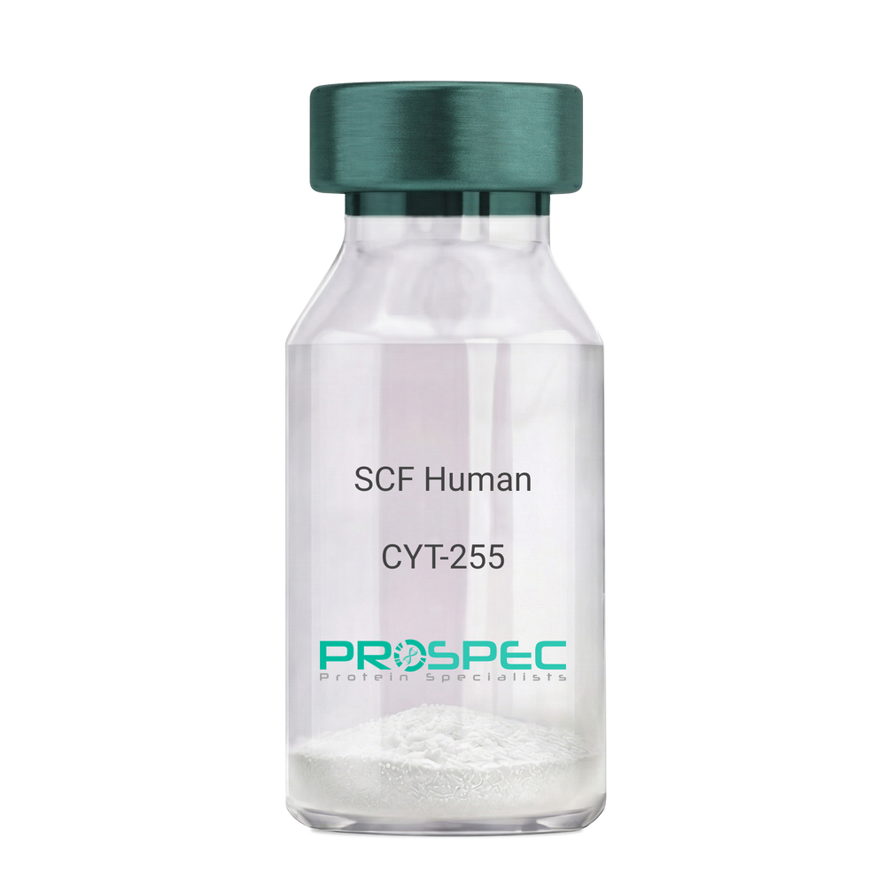
SCF HumanDescription:
Stem Cell Factor Human Recombinant
Kit ligand Precursor, C-kit ligand, SCF, Mast cell growth factor, MGF, SF, KL-1, Kitl, DKFZp686F2250.
Product # :
CYT-255Price :
Quantity :
Shipping Method :
Shipped at Room temp
More Info
- description
- source
- formulation
- purity
- biological activity
- More Info
Description
Stem Cell Factor Human Recombinant produced in E.Coli is a single, non-glycosylated polypeptide chain containing 165 amino acids and having a molecular mass of 18409 Dalton. The SCF is purified by proprietary chromatographic techniques.
Source
Escherichia Coli.
Formulation
Lyophilized from a concentrated (1mg/ml) solution in water containing 0.02% NaHCO3.
Purity
Greater than 98.0% as determined by:
(a) Analysis by SEC-HPLC.
(b) Analysis by SDS-PAGE.Biological Activity
The ED50 as determined by the dose-dependant stimulation of Human TF-1 cells is < 2 ng/ml, corresponding to a Specific Activity of 500,000 IU/mg.More Info
-
Introduction
Stem cell factor / KIT ligand (SCF) is a cytokine which binds CD117(c-Kit). SCF is also known as "steel factor" or "c-kit ligand". SCF exists in two forms, cell surface bound SCF and soluble (or free) SCF. Soluble SCF is produced by the cleavage of surface bound SCF by metalloproteases. SCF is a growth factor important for the survival, proliferation, and differentiation of hematopoietic stem cells and other hematopoietic progenitor cells. One of its roles is to change the BFU-E (burst-forming unit-erythroid) cells, which are the earliest erythrocyte precursors in the erythrocytic series, into the CFU-E (colony-forming unit-erythroid).
-
Synonyms
Kit ligand Precursor, C-kit ligand, SCF, Mast cell growth factor, MGF, SF, KL-1, Kitl, DKFZp686F2250.
-
Physical Appearance
Sterile Filtered White lyophilized (freeze-dried) powder.
-
Stability
Lyophilized KIT ligand although stable at room temperature for 3 weeks, should be stored desiccated below -18°C. Upon reconstitution SCF should be stored at 4°C between 2-7 days and for future use below -18°C.For long term storage it is recommended to add a carrier protein (0.1% HSA or BSA).Please prevent freeze-thaw cycles.
-
Solubility
It is recommended to reconstitute the lyophilized Stem Cell Factor in sterile 18MΩ-cm H2O not less than 100µg/ml, which can then be further diluted to other aqueous solutions.
-
Amino Acid Sequence
The sequence of the first five N-terminal amino acids was determined and was found to be Met-Glu-Gly-Ile-Cys.
-
Protein content
Protein quantitation was carried out by two independent methods1. UV spectroscopy at 280 nm using the absorbency value of 0.52 as the extinction coefficient for a 0.1% (1mg/ml) solution. This value is calculated by the PC GENE computer analysis program of protein sequences (IntelliGenetics). 2. Analysis by RP-HPLC, using a standard solution of Stem Cell Factor as a Reference Standard.
ProSpec's products are furnished for LABORATORY RESEARCH USE ONLY. The product may not be used as drugs, agricultural or pesticidal products, food additives or household chemicals.
- View Data Sheet
Name :
SERPINB8 HumanDescription:
Serpin Peptidase Inhibitor, Clade B Member 8 Human Recombinant
CAP2, PI8, Serpin B8, Cytoplasmic antiproteinase 2, Peptidase inhibitor 8, CAP-2.
Product # :
PRO-2142Price :
Quantity :
Shipping Method :
Shipped with Ice Packs
More Info
- description
- source
- formulation
- purity
- More Info
Description
SERPINB8 Human Recombinant produced in E.Coli is a single, non-glycosylated polypeptide chain containing 397 amino acids (1-374 a.a) and having a molecular mass of 45.2kDa.SERPINB8 is fused to a 23 amino acid His-tag at N-terminus & purified by proprietary chromatographic techniques.
Source
Escherichia Coli.
Formulation
SERPINB8 protein solution (0.5mg/ml) containing 20mM Tris-HCl buffer (pH 8.0), 0.15M NaCl, 30% glycerol and 1mM DTT.
Purity
Greater than 95.0% as determined by SDS-PAGE.
More Info
-
Introduction
Serpin Peptidase Inhibitor, Clade B Member 8 (SERPINB8) belongs to the ov-serpin subfamily, which, compared with the archetypal serpin PI1, is characterized by a high degree of homology to chicken ovalbumin, absence of N- and C-terminal extensions, lack of a signal peptide, and a serine instead of an asparagine residue at the penultimate position. The superfamily of high molecular weight serine proteinase inhibitors (serpins) control various intracellular and extracellular processes such as complement activation, fibrinolysis, coagulation, cellular differentiation, tumor suppression, apoptosis, and cell migration.
-
Synonyms
CAP2, PI8, Serpin B8, Cytoplasmic antiproteinase 2, Peptidase inhibitor 8, CAP-2.
-
Physical Appearance
Sterile Filtered clear solution.
-
Stability
Store at 4°C if entire vial will be used within 2-4 weeks. Store, frozen at -20°C for longer periods of time. For long term storage it is recommended to add a carrier protein (0.1% HSA or BSA).Avoid multiple freeze-thaw cycles.
-
Amino Acid Sequence
MGSSHHHHHH SSGLVPRGSH MGSMDDLCEA NGTFAISLFK ILGEEDNSRN VFFSPMSISS ALAMVFMGAK GSTAAQMSQA LCLYKDGDIH RGFQSLLSEV NRTGTQYLLR TANRLFGEKT CDFLPDFKEY CQKFYQAELE ELSFAEDTEE CRKHINDWVA EKTEGKISEV LDAGTVDPLT KLVLVNAIYF KGKWNEQFDR KYTRGMLFKT NEEKKTVQMM FKEAKFKMGY ADEVHTQVLE LPYVEEELSM VILLPDDNTD LAVVEKALTY EKFKAWTNSE KLTKSKVQVF LPRLKLEESY DLEPFLRRLG MIDAFDEAKA DFSGMSTEKN VPLSKVAHKC FVEVNEEGTE AAAATAVVRN SRCSRMEPRF CADHPFLFFI RHHKTNCILF CGRFSSP.
ProSpec's products are furnished for LABORATORY RESEARCH USE ONLY. The product may not be used as drugs, agricultural or pesticidal products, food additives or household chemicals.
- View Data Sheet
Name :
ANXA11 HumanDescription:
Annexin A11 Human Recombinant
Annexin A11, 56 kDa autoantigen, Annexin XI, Annexin-11, Calcyclin-associated annexin 50, CAP-50, ANXA11, ANX11, CAP50.
Product # :
PRO-278Price :
Quantity :
Shipping Method :
Shipped with Ice Packs
More Info
- description
- source
- formulation
- purity
- More Info
Description
ANXA11 Human Recombinant produced in E.coli is a single, non-glycosylated polypeptide chain containing 525 amino acids (1-505) and having a molecular mass of 56.6 kDa. The ANXA11 is fused to a 20 amino acid His-Tag at N-terminus and purified by proprietary chromatographic techniques.
Source
Escherichia Coli.
Formulation
The ANXA11 solution (0.25mg/ml) contains 20mM MES buffer (Ph 5.0), 20% glycerol, 2mM DTT and 100mM NaCl.
Purity
Greater than 95.0% as determined by SDS-PAGE.
More Info
-
Introduction
Annexin A11 (ANXA11) belongs to the annexin family, which is a group of calcium-dependent phospholipid-binding proteins. Annexins have distinctive N-terminal domains and conserved C-terminal domains, which contain the calcium-dependent phospholipid-binding sites. Annexin A11 binds specifically to calcyclin in a calcium-dependent manner. Moreover, ANXA11 is required for midbody formation and completion of the terminal phase of cytokinesis. The ANXA11 protein is a 56kDa antigen recognized by sera from patients with various autoimmune diseases.
-
Synonyms
Annexin A11, 56 kDa autoantigen, Annexin XI, Annexin-11, Calcyclin-associated annexin 50, CAP-50, ANXA11, ANX11, CAP50.
-
Physical Appearance
Sterile Filtered colorless solution.
-
Stability
Store at 4°C if entire vial will be used within 2-4 weeks. Store, frozen at -20°C for longer periods of time. For long term storage it is recommended to add a carrier protein (0.1% HSA or BSA).Avoid multiple freeze-thaw cycles.
-
Amino Acid Sequence
MGSSHHHHHH SSGLVPRGSH MSYPGYPPPP GGYPPAAPGG GPWGGAAYPP PPSMPPIGLD NVATYAGQFN QDYLSGMAAN MSGTFGGANM PNLYPGAPGA GYPPVPPGGF GQPPSAQQPV PPYGMYPPPG NPPSRMPSY PPYPGAPVPG QPMPPPGQQP PGAYPGQPPV TYPGQPPVPL PGQQQPVPSY PGYPGSGTVT PAVPPTQFGS RGTITDAPGF DPLRDAEVLR KAMKGFGTDE QAIIDCLGSR SNKQRQQILL SFKTAYGKDL IKDLKSELSG NFEKTILALM KTPVLFDIYE IKEAIKGVGT DEACLIEILA SRSNEHIREL NRAYKAEFKK TLEEAIRSDT GHFQRLLIS LSQGNRDEST NVDMSLAQRD AQELYAAGEN RLGTDESKFN AVLCSRSRAH LVAVFNEYQR MTGRDIEKSI CREMSGDLEE GMLAVVKCLK NTPAFFAERL NKAMRGAGTK DRTLIRIMVS RSETDLLDIR SEYKRMYGKS LYHDISGDTS GDYRKILLKI CGGND.
ProSpec's products are furnished for LABORATORY RESEARCH USE ONLY. The product may not be used as drugs, agricultural or pesticidal products, food additives or household chemicals.
- View Data Sheet
Name :
ANXA8 HumanDescription:
Annexin A8 Human Recombinant
ANX8, Annexin-8, Annexin VII, Vascular anticoagulant-beta, VAC-beta.
Product # :
PRO-548Price :
Quantity :
Shipping Method :
Shipped with Ice Packs
More Info
- description
- source
- formulation
- purity
- More Info
Description
ANXA8 Human Recombinant produced in E.Coli is a signle, non-glycosylated, polypeptide chain containing 347 amino acids (1-327 a.a.)and having a total molecular mass of 39 kDa. ANXA8 is fused to a 20 amino acid His Tag at N-terminus and is purified by proprietary chromatographic techniques.
Source
Escherichia Coli.
Formulation
The ANXA8 1mg/ml protein solution contains 20mM Tris-HCL pH-8, 1mM DTT,1mM EDTA, 0.1M NaCl and 10% Glycerol.
Purity
Greater than 90.0% as determined by SDS-PAGE.
More Info
-
Introduction
ANXA8 is part of the annexin family of evolutionarily conserved Ca2+ and phospholipid binding proteins. ANXA8 is an anticoagulant protein that functions as an indirect inhibitor of the thromboplastin-specific complex, which takes part in the blood coagulation cascade. Where co-expressed in the same tissues, ANXA8 is expressed at a 100-fold lower level than Annexin A5. Though, ANXA8 is preferentially expressed in acute promyelocytic leukemia (APL) cells which suggests its involvement in hematopoietic cell differentiation.
-
Synonyms
ANX8, Annexin-8, Annexin VII, Vascular anticoagulant-beta, VAC-beta.
-
Physical Appearance
Sterile Filtered colorless clear solution.
-
Stability
Store at 4°C if entire vial will be used within 2-4 weeks. Store, frozen at -20°C for longer periods of time. Please avoid freeze thaw cycles.
-
Amino Acid Sequence
MGSSHHHHHH SSGLVPRGSH MAWWKSWIEQ EGVTVKSSSH FNPDPDAETL YKAMKGIGTN EQAIIDVLTK RSNTQRQQIA KSFKAQFGKD
LTETLKSELS GKFERLIVAL MYPPYRYEAK ELHDAMKGLG TKEGVIIEIL ASRTKNQLRE IMKAYEEDYG SSLEEDIQAD TSGYLERILV
CLLQGSRDDV SSFVDPGLAL QDAQDLYAAG EKIRGTDEMK FITILCTRSA THLLRVFEEY EKIANKSIED SIKSETHGSL EEAMLTVVKC
TQNLHSYFAE RLYYAMKGAG TRDGTLIRNI VSRSEIDLNL IKCHFKKMYG KTLSSMIMED TSGDYKNALL SLVGSDP.
ProSpec's products are furnished for LABORATORY RESEARCH USE ONLY. The product may not be used as drugs, agricultural or pesticidal products, food additives or household chemicals.
- View Data Sheet
Name :
SNF8 HumanDescription:
SNF8, ESCRT-II Complex Subunit Human Recombinant
SNF8 ESCRT-II complex subunit homolog (S. cerevisiae), ESCRT-II complex subunit VPS22, ELL-associated protein of 30 kDa, EAP30 subunit of ELL complex, vacuolar-sorting protein SNF8, EAP30, VPS22, Dot3.
Product # :
PRO-1135Price :
Quantity :
Shipping Method :
Shipped with Ice Packs
More Info
- description
- source
- formulation
- purity
- More Info
Description
SNF8 Human Recombinant produced in E. coli is a single polypeptide chain containing 282 amino acids (1-258) and having a molecular mass of 31.4 kDa.SNF8 is fused to a 24 amino acid His-tag at N-terminus & purified by proprietary chromatographic techniques.
Source
E.coli.
Formulation
The SNF8 solution (0.25mg/ml) contains 20mM Tris-HCl buffer (pH 8.0), 0.2M NaCl, 2mM DTT and 50% glycerol.
Purity
Greater than 90% as determined by SDS-PAGE.
More Info
-
Introduction
SNF8 belongs to the SNF8 family of vacuolar sorting proteins and is restricts to both the nucleus and the cytoplasm. SNF8 is a subunit of the endosomal sorting complex essential for transport II (ESCRT-II), which is necessary for multivesicular body (MVB) formation and sorting of endosomal cargo proteins into MVBs. The MVB pathway facilitates transfer of transmembrane proteins into the lumen of the lysosome for degradation. Additionally, the ESCRT-II complex takes part in transcription regulation by contributing to derepression of transcription by RNA polymerase II, probably by its interface with ELL.
-
Synonyms
SNF8 ESCRT-II complex subunit homolog (S. cerevisiae), ESCRT-II complex subunit VPS22, ELL-associated protein of 30 kDa, EAP30 subunit of ELL complex, vacuolar-sorting protein SNF8, EAP30, VPS22, Dot3.
-
Physical Appearance
Sterile Filtered colorless solution.
-
Stability
Store at 4°C if entire vial will be used within 2-4 weeks. Store, frozen at -20°C for longer periods of time. For long term storage it is recommended to add a carrier protein (0.1% HSA or BSA).Avoid multiple freeze-thaw cycles.
-
Amino Acid Sequence
MGSSHHHHHH SSGLVPRGSH MGSHMHRRGV GAGAIAKKKL AEAKYKERGT VLAEDQLAQM SKQLDMFKTN LEEFASKHKQ EIRKNPEFRV QFQDMCATIG VDPLASGKGF WSEMLGVGDF YYELGVQIIE VCLALKHRNG GLITLEELHQ QVLKGRGKFA QDVSQDDLIR AIKKLKALGT GFGIIPVGGT YLIQSVPAEL NMDHTVVLQL AEKNGYVTVS EIKASLKWET ERARQVLEHL LKEGLAWLDL QAPGEAHYWL PALFTDLYSQ EITAEEAREA LP
ProSpec's products are furnished for LABORATORY RESEARCH USE ONLY. The product may not be used as drugs, agricultural or pesticidal products, food additives or household chemicals.
- View Data Sheet
Name :
IFNW1 Human, HEKDescription:
Interferon-Omega 1 Human Recombinant, HEK
IFN omega-1, IFN alpha-II-1, IFNW1.
Product # :
CYT-1225Price :
Quantity :
Shipping Method :
Shipped with Ice Packs
More Info
- description
- source
- formulation
- purity
- biological activity
- More Info
Description
IFNW1 Human Recombinant is a single, glycosylated, polypeptide chain (22-195 a.a) containing a total of 180 amino acids and having a molecular mass of 20.9 kDa. IFNW1 is fused to a 6 a.a his-Tag at C-terminus and is purified by proprietary chromatographic techniques.
Source
HEK293 Cells.
Formulation
The IFNW1 solution (1mg/ml) contains 10% Glycerol and Phosphate-Buffered Saline (pH 7.4).
Purity
Greater than 95.0% as determined by SDS-PAGE.
Biological Activity
The ED50 is ≤0.07 ng/ml, measured in a cytotoxicity assay using TF-1 human erythroleukemic cells .
More Info
-
Synonyms
IFN omega-1, IFN alpha-II-1, IFNW1.
-
Physical Appearance
Sterile Filtered colorless solution.
-
Stability
Store at 4°C if entire vial will be used within 2-4 weeks. Store, frozen at -20°C for longer periods of time. For long term storage it is recommended to add a carrier protein (0.1% HSA or BSA).Avoid multiple freeze-thaw cycles.
-
Amino Acid Sequence
LGCDLPQNHG LLSRNTLVLL HQMRRISPFL CLKDRRDFRF PQEMVKGSQL QKAHVMSVLH EMLQQIFSLF HTERSSAAWN MTLLDQLHTG LHQQLQHLET CLLQVVGEGE SAGAISSPAL TLRRYFQGIR VYLKEKKYSD CAWEVVRMEI MKSLFLSTNM QERLRSKDRD LGSSHHHHHH.
-
Background
Interferons, a family of signaling proteins, play a pivotal role in the immune system’s defense against viral infections and other threats. Among these, Interferon W1 (IFNW1), a member of the Type I interferon family, has emerged as a key player in orchestrating antiviral responses and modulating immune reactions. This research embarks on a detailed exploration of the IFNW1 protein, unveiling its structural intricacies, signaling pathways, and its broader implications in immune regulation and disease. By delving into IFNW1, scientists aim to comprehend the nuances of its functions, decipher its interactions within the complex interferon network, and explore its potential applications in therapeutic interventions and beyond.
Structural Insights into IFNW1:
IFNW1, like other Type I interferons, exhibits a unique tertiary structure that enables it to interact with specific cell surface receptors. This interaction triggers a cascade of events, leading to the activation of various antiviral genes and immune modulatory pathways. Understanding the structural basis of IFNW1 is crucial for elucidating its binding affinities, biological activities, and its significance in immune responses.
Signaling Pathways and Antiviral Defense:
IFNW1 engages with its cognate receptors, initiating Janus kinase (JAK)-Signal Transducer and Activator of Transcription (STAT) signaling pathways. This activation leads to the transcription of interferon-stimulated genes (ISGs) with potent antiviral properties. IFNW1’s ability to induce an antiviral state in infected and neighboring cells is fundamental for restricting viral replication and curtailing the spread of infections. Additionally, IFNW1 plays a role in modulating adaptive immune responses, contributing to the broader immune defense mechanisms.
IFNW1 in Immunomodulation and Disease:
Beyond its antiviral functions, IFNW1 is implicated in immunomodulation and disease pathogenesis. Dysregulation of IFNW1 signaling is associated with autoimmune disorders, including lupus and rheumatoid arthritis, highlighting its involvement in immune-related diseases. Moreover, IFNW1 is being explored in cancer immunotherapy, where its ability to modulate the tumor microenvironment and enhance immune surveillance presents opportunities for novel treatment strategies.
Therapeutic Potential and Future Prospects:
The unique properties of IFNW1, particularly its role in immune regulation and antiviral defense, position it as a potential therapeutic target. Research efforts are directed towards harnessing its immunomodulatory functions for developing therapies against infectious diseases, autoimmune disorders, and certain cancers. Additionally, understanding IFNW1’s interactions with other components of the immune system opens avenues for innovative approaches in personalized medicine and targeted immunotherapies.
IFNW1 Protein, as an integral component of the interferon network, stands as a sentinel in the body’s defense against viral invasions and immune dysregulations. Its multifaceted roles in antiviral defense, immune modulation, and disease pathogenesis underscore its significance in biology and medicine. As researchers delve deeper into the intricacies of IFNW1, they pave the way for innovative therapies, immunomodulatory interventions, and a deeper understanding of immune responses. This research not only illuminates the pivotal role of IFNW1 but also holds the promise of transformative advancements in medicine, shaping the future of immunology and disease therapeutics.
What is the molecular weight/Mw of IFNW1 HUMAN, HEK Protein?
IFNW1 HUMAN, HEK Protein has a total Mw of 20.9kDa.
What is the source or expression system of IFNW1 HUMAN, HEK Protein?
HEK293 Cells.
What is the Purity of IFNW1 HUMAN, HEK Protein?
IFNW1 HUMAN, HEK Protein is >95% pure as determined by SDS-PAGE.
What is the Biological Activity of IFNW1 HUMAN, HEK Protein?
The ED50 is ≤0.07 ng/ml, measured in a cytotoxicity assay using TF-1 human erythroleukemic cells .
What is the amino acid sequence of IFNW1 HUMAN, HEK Protein?
LGCDLPQNHG LLSRNTLVLL HQMRRISPFL CLKDRRDFRF PQEMVKGSQL QKAHVMSVLH EMLQQIFSLF HTERSSAAWN MTLLDQLHTG LHQQLQHLET CLLQVVGEGE SAGAISSPAL TLRRYFQGIR VYLKEKKYSD CAWEVVRMEI MKSLFLSTNM QERLRSKDRD LGSSHHHHHH.
What applications can IFNW1 HUMAN, HEK Protein be used in?
IFNW1 HUMAN, HEK Protein can probably be used in western blot, ELISA and Lateral Flow.
What is the endotoxin level for IFNW1 HUMAN, HEK Protein?
The endotoxin level is minimal, IFNW1 HUMAN, HEK Protein was purified using conventional chromatography techniques.
ProSpec's products are furnished for LABORATORY RESEARCH USE ONLY. The product may not be used as drugs, agricultural or pesticidal products, food additives or household chemicals.
- View Data Sheet
Name :
OLR1 HumanDescription:
Oxidized Low Density Lipoprotein Receptor 1 Human Recombinant
Oxidized low density lipoprotein (lectin-like) receptor 1, CLEC8A, hLOX1, SCARE1, Lectin-type oxidized LDL receptor 1, Lectin-like oxidized LDL receptor 1, C-type lectin domain family 8 member A, LOXIN, SLOX1, ox LDL receptor 1, Oxidized low-density lipoprotein receptor 1 soluble form, scavenger receptor class E member 1.
Product # :
PRO-923Price :
Quantity :
Shipping Method :
Shipped with Ice Packs
More Info
- description
- source
- formulation
- purity
- More Info
Description
OLR1 Human Recombinant produced in E.coli is a single, non-glycosylated polypeptide chain containing 216 amino acids (58-273) and having a molecular mass of 24.7 kDa.The OLR1 is purified by proprietary chromatographic techniques.
Source
E.coli.
Formulation
The OLR1 solution (1mg/ml) contains 20mM Tris-HCl buffer (pH 8.0), 0.4M Urea and 5% glycerol.
Purity
Greater than 90% as determined by SDS-PAGE.
More Info
-
Introduction
OLR1 is a type II membrane protein which belongs to the C-type lectin family and performs as a cell-surface receptor for Ox-LDL. Ox-LDL has a part in early ather-osclerosis, which includes the transformation of monocyte-derived macro-phages to foam cells in atherosclerotic lesions. In addition, OLR1 protein triggers the activation of the NF?B signal transduction pathway.
-
Synonyms
Oxidized low density lipoprotein (lectin-like) receptor 1, CLEC8A, hLOX1, SCARE1, Lectin-type oxidized LDL receptor 1, Lectin-like oxidized LDL receptor 1, C-type lectin domain family 8 member A, LOXIN, SLOX1, ox LDL receptor 1, Oxidized low-density lipoprotein receptor 1 soluble form, scavenger receptor class E member 1.
-
Physical Appearance
Sterile Filtered colorless solution.
-
Stability
Store at 4°C if entire vial will be used within 2-4 weeks. Store, frozen at -20°C for longer periods of time. For long term storage it is recommended to add a carrier protein (0.1% HSA or BSA).Avoid multiple freeze-thaw cycles.
-
Amino Acid Sequence
MQLSQVSDLL TQEQANLTHQ KKKLEGQISA RQQAEEASQE SENELKEMIE TLARKLNEKS KEQMELHHQN LNLQETLKRV ANCSAPCPQD WIWHGENCYL FSSGSFNWEK SQEKCLSLDA KLLKINSTAD LDFIQQAISY SSFPFWMGLS RRNPSYPWLW EDGSPLMPHL FRVRGAVSQT YPSGTCAYIQ RGAVYAENCI LAAFSICQKK ANLRAQ
ProSpec's products are furnished for LABORATORY RESEARCH USE ONLY. The product may not be used as drugs, agricultural or pesticidal products, food additives or household chemicals.
- View Data Sheet
Name :
STX11 HumanDescription:
Syntaxin-11 Human Recombinant
Syntaxin-11, STX11, FHL4, HLH4, HPLH4.
Product # :
PRO-1111Price :
Quantity :
Shipping Method :
Shipped with Ice Packs
More Info
- description
- source
- formulation
- purity
- More Info
Description
STX11 Human Recombinant produced in E.Coli is a single, non-glycosylated polypeptide chain containing 311 amino acids (1-287 a.a) and having a molecular mass of 35.8kDa.STX11 is fused to a 24 amino acid His-tag at N-terminus & purified by proprietary chromatographic techniques.
Source
E.coli.
Formulation
STX11 protein solution (1mg/ml) containing 20mM Tris-HCl buffer (pH8.0), 10% glycerol and 0.4M Urea.
Purity
Greater than 95% as determined by SDS-PAGE.
More Info
-
Introduction
Syntaxin-11 (STX11) belongs to the t-SNARE family. Syntaxin-11 regulates protein transport between late endosomes and the trans-Golgi network. STX11 interacts with the SNARE proteins SNAP-23 and VAMP. STX11 gene mutations are linked with familial hemophagocytic lymphohistiocytosis.
-
Synonyms
Syntaxin-11, STX11, FHL4, HLH4, HPLH4.
-
Physical Appearance
Sterile Filtered colorless solution.
-
Stability
Store at 4°C if entire vial will be used within 2-4 weeks. Store, frozen at -20°C for longer periods of time. For long term storage it is recommended to add a carrier protein (0.1% HSA or BSA).Avoid multiple freeze-thaw cycles.
-
Amino Acid Sequence
MGSSHHHHHH SSGLVPRGSH MGSHMKDRLA ELLDLSKQYD QQFPDGDDEF DSPHEDIVFE TDHILESLYR DIRDIQDENQ LLVADVKRLG KQNARFLTSM RRLSSIKRDT NSIAKAIKAR GEVIHCKLRA MKELSEAAEA QHGPHSAVAR ISRAQYNALT LTFQRAMHDY NQAEMKQRDN CKIRIQRQLE IMGKEVSGDQ IEDMFEQGKW DVFSENLLAD VKGARAALNE IESRHRELLR LESRIRDVHE LFLQMAVLVE KQADTLNVIE LNVQKTVDYT GQAKAQVRKA VQYEEKNPCR TLCCFCCPCL K.
ProSpec's products are furnished for LABORATORY RESEARCH USE ONLY. The product may not be used as drugs, agricultural or pesticidal products, food additives or household chemicals.
- View Data Sheet
Name :
FAS Human, HisDescription:
sFas Receptor Human Recombinant, His Tag
CD95, CD-95, Tumor necrosis factor receptor superfamily member 6, FASLG receptor, Apoptosis-mediating surface antigen FAS, Apo-1 antigen, CD95 antigen, FAS, APT1, FAS1, TNFRSF6, APO-1, FASTM, ALPS1A.
Product # :
PRO-594Price :
Quantity :
Shipping Method :
Shipped with Ice Packs
More Info
- description
- source
- formulation
- purity
- More Info
Description
Recombinant FAS antigen/ CD95 purified from E. coli is a single non-glycosilated polypeptide chain containing amino acids 157-335 of Fas antigen. The recombinant CD95 is fused to C-terminal 6-histidine amino acids. The FAS antigen is purified by proprietary chromatographic techniques.
Source
Escherichia Coli.
Formulation
10mM Sodium Phosphate, pH 8.0.
Purity
Greater than 95% as determined by:
(a) Analysis by RP-HPLC.
(b) Analysis by SDS-PAGE.More Info
-
Introduction
FAS / CD95 is a 36kDa transmembrane type I receptor, which belongs to the NF/NGF receptor super-family. It is a potent inducer of apoptosis in cells of the immune system upon interaction with its natural ligand, CD95L. Although the CD95/CD95L system could play a role in tumor regression, tumor cells seem to down-regulate CD95 expression as a mechanism of resistance to CD95L-induced killing by T lymphocytes and NK cells. The Fas antigen is expressed on the surface of various cell types, including activated T and B lymphocytes and T lymphoblastoid cell line. CD95 is also expressed in a broad panel of non-transformed cells outside the immune system.
-
Synonyms
CD95, CD-95, Tumor necrosis factor receptor superfamily member 6, FASLG receptor, Apoptosis-mediating surface antigen FAS, Apo-1 antigen, CD95 antigen, FAS, APT1, FAS1, TNFRSF6, APO-1, FASTM, ALPS1A.
-
Physical Appearance
Sterile liquid formulation.
-
Stability
CD95 although stable at 10°C for 5 days, should be stored below -18°C. Please prevent freeze-thaw cycles.
-
Background
What is the source or expression system of FAS Protein?
Escherichia Coli.
What is the Purity of FAS Protein?
FAS Protein is >95% pure as determined by SDS-PAGE.
What is the Biological Activity of FAS Protein?
The biological functionality of FAS Protein will be determined in the future.
What is the amino acid sequence of FAS Protein?
FAS Protein is composed from 179 amino acids.
What applications can FAS Protein be used in?
FAS Protein can probably be used in western blot, ELISA and Lateral Flow.
What is the endotoxin level for FAS Protein?
The endotoxin level is minimal, FAS Protein was purified using conventional chromatography techniques.
ProSpec's products are furnished for LABORATORY RESEARCH USE ONLY. The product may not be used as drugs, agricultural or pesticidal products, food additives or household chemicals.
- View Data Sheet
Name :
FASLG HumanDescription:
FAS Ligand Human Recombinant
Tumor necrosis factor ligand superfamily member 6, Apoptosis antigen ligand, APTL, CD95 ligand, CD95-L, Fas antigen ligand, Fas ligand, FasL, CD178, FASLG, APT1LG1, CD95L, TNFSF6, ALPS1B.
Product # :
CYT-031Price :
Quantity :
Shipping Method :
Shipped with Ice Packs
More Info
- description
- source
- formulation
- purity
- More Info
- sds-page
Description
FASLG Human Recombinant produced in E.Coli is a single, non-glycosylated polypeptide chain containing 173 amino acids (130-281 a.a.) and having a molecular mass of 19.6kDa.FASLG is fused to a 21 amino acid His-tag at N-terminus & purified by proprietary chromatographic techniques.
Source
Escherichia Coli.
Formulation
FASLG protein solution (1mg/ml) containing 20mM Tris-HCl buffer (pH 8.0), 0.4M urea and 10% glycerol.
Purity
Greater than 90% as determined by SDS-PAGE.
sds-page
More Info
-
Introduction
The type II transmembrane protein FASLG is a member of the tumor necrosis factor (TNF) superfamily. A fas ligand/receptor interaction has a significant part in the regulation of the immune system and the advancement of cancer. FASLG is expressed on the activated T cell surface as a nondisulfidelinked homotrimer. FASLG binding to Fas/CD95/TNFRSF6 on a nearby cell prompts apoptosis in the Fas expressing cell. FASLG is released from the cell surface by metalloproteinases as a soluble molecule that stays trimeric and is able to bind with Fas, but its capability to activate apoptosis is radically reduced. In addition, FASLG binds to DcR3 - a soluble trap receptor with no signal transduction capabilities. Flawed Fas-mediated apoptosis causes oncogenesis in addition to drug resistance in existing tumors. Constitutive expression of FASLG in a variety of tumors enables their immune evasion. Both mouse and human FASLG are active on mouse and human cells.
-
Synonyms
Tumor necrosis factor ligand superfamily member 6, Apoptosis antigen ligand, APTL, CD95 ligand, CD95-L, Fas antigen ligand, Fas ligand, FasL, CD178, FASLG, APT1LG1, CD95L, TNFSF6, ALPS1B.
-
Physical Appearance
Sterile Filtered colorless solution.
-
Stability
Store at 4°C if entire vial will be used within 2-4 weeks. Store, frozen at -20°C for longer periods of time. For long term storage it is recommended to add a carrier protein (0.1% HSA or BSA).Avoid multiple freeze-thaw cycles.
-
Amino Acid Sequence
MGSSHHHHHH SSGLVPRGSH MQIGHPSPPP EKKELRKVAH LTGKSNSRSM PLEWEDTYGI VLLSGVKYKK GGLVINETGL YFVYSKVYFR GQSCNNLPLS HKVYMRNSKY PQDLVMMEGK MMSYCTTGQM WARSSYLGAV FNLTSADHLY VNVSELSLVN FEESQTFFGL YKL.
-
Background
What is the molecular weight/Mw of FASL Protein?
FASL Protein has a total Mw of 19.6kDa.
What is the source or expression system of FASL Protein?
Escherichia Coli.
What is the Purity of FASL Protein?
FASL Protein is >90% pure as determined by SDS-PAGE.
What is the Biological Activity of FASL Protein?
The biological functionality of FASL Protein will be determined in the future.
What is the amino acid sequence of FASL Protein?
MGSSHHHHHH SSGLVPRGSH MQIGHPSPPP EKKELRKVAH LTGKSNSRSM PLEWEDTYGI VLLSGVKYKK GGLVINETGL YFVYSKVYFR GQSCNNLPLS HKVYMRNSKY PQDLVMMEGK MMSYCTTGQM WARSSYLGAV FNLTSADHLY VNVSELSLVN FEESQTFFGL YKL.
What applications can FASL Protein be used in?
FASL Protein can probably be used in western blot, ELISA and Lateral Flow.
What is the endotoxin level for FASL Protein?
The endotoxin level is minimal, FASL Protein was purified using conventional chromatography techniques.
ProSpec's products are furnished for LABORATORY RESEARCH USE ONLY. The product may not be used as drugs, agricultural or pesticidal products, food additives or household chemicals.
- View Data Sheet
Name :
PEX26 HumanDescription:
Peroxisomal Biogenesis Factor 26 Human Recombinant
PBD7A, PBD7B, PEX26M1T, Pex26pM1T, Peroxisome assembly protein 26, PEX26.
Product # :
PRO-1544Price :
Quantity :
Shipping Method :
Shipped with Ice Packs
More Info
- description
- source
- formulation
- purity
- More Info
Description
PEX26 Human Recombinant produced in E. coli is a single polypeptide chain containing 269 amino acids (1-246) and having a molecular mass of 29.3kDa. PEX26 is fused to a 23 amino acid His-tag at N-terminus & purified by proprietary chromatographic techniques.
Source
Escherichia Coli.
Formulation
The PEX26 solution (0.5mg/ml) contains 20mM Tris-HCl buffer (pH 8.0), 0.15M NaCl, 20% glycerol and 1mM DTT.
Purity
Greater than 90% as determined by SDS-PAGE.
More Info
-
Introduction
Peroxisomal Biogenesis Factor 26 (PEX26) which is a part of the peroxin-26 gene family is probably required for protein import into peroxisomes. PEX26 attaches PEX1 and PEX6 to peroxisome membranes to form heteromeric AAA ATPase complexes needed for the import of proteins into peroxisomes. Deficiencies in this gene are the cause of peroxisome biogenesis disorder complementation group 8. PBD is a group of peroxisomal disorders evolving from a failure of protein import into the peroxisomal membrane or matrix.
-
Synonyms
PBD7A, PBD7B, PEX26M1T, Pex26pM1T, Peroxisome assembly protein 26, PEX26.
-
Physical Appearance
Sterile Filtered clear solution.
-
Stability
Store at 4°C if entire vial will be used within 2-4 weeks. Store frozen at -20°C for longer periods of time. For long term storage it is recommended to add a carrier protein (0.1% HSA or BSA).Avoid multiple freeze-thaw cycles.
-
Amino Acid Sequence
MGSSHHHHHH SSGLVPRGSH MGSMKSDSST SAAPLRGLGG PLRSSEPVRA VPARAPAVDL LEEAADLLVV HLDFRAALET CERAWQSLAN HAVAEEPAGT SLEVKCSLCV VGIQALAEMD RWQEVLSWVL QYYQVPEKLP PKVLELCILL YSKMQEPGAV LDVVGAWLQD PANQNLPEYG ALAEFHVQRV LLPLGCLSEA EELVVGSAAF GEERRLDVLQ AIHTARQQQK QEHSGSEEAQ KPNLEGSVSH KFLSLPMLVR QLWDSAVSH.
ProSpec's products are furnished for LABORATORY RESEARCH USE ONLY. The product may not be used as drugs, agricultural or pesticidal products, food additives or household chemicals.
- View Data Sheet
Name :
PFN1 HumanDescription:
Profilin-1 Human Recombinant
Profilin-1, Profilin I, PFN1.
Product # :
PRO-528Price :
Quantity :
Shipping Method :
Shipped with Ice Packs
More Info
- description
- source
- formulation
- purity
- More Info
Description
PFN1 Human Recombinant produced in E.Coli is a single, non-glycosylated, polypeptide chain containing 140 amino acids (1-140 a.a.) and having a molecular mass of 15kDa.The PFN1 is purified by proprietary chromatographic techniques.
Source
Escherichia Coli.
Formulation
The PFN1 protein solution contains 20mM Tris-HCl buffer (pH8.0) and 10% glycerol.
Purity
Greater than 95.0% as determined by SDS-PAGE.
More Info
-
Introduction
Profilin1 (PFN1) is a ubiquitous actin monomer-binding protein which is a member of the profilin family. PFN1 significantly boosts skin wound healing in-vitro and in-vivo which may be mediated by purinergic receptors. PFN1 is also active in endothelial cell migration and vessel sprouting. PFN1 is thought to control actin polymerization in response to extracellular signals. PFN1 binds to actin and affects the formation of the cytoskeleton. In addition, PFN1 has an important role in the regulation of epithelial cell-cell adhesion. At high concentrations, profilin averts the polymerization of actin, while at low concentrations it enhances the polymerization. PFN1 gene deletion is linked to Miller-Dieker syndrome.
-
Synonyms
Profilin-1, Profilin I, PFN1.
-
Physical Appearance
Sterile Filtered colorless solution.
-
Stability
Store at 4°C if entire vial will be used within 2-4 weeks. Store, frozen at -20°C for longer periods of time. For long term storage it is recommended to add a carrier protein (0.1% HSA or BSA).Avoid multiple freeze-thaw cycles.
-
Amino Acid Sequence
MAGWNAYIDN LMADGTCQDA AIVGYKDSPS VWAAVPGKTF VNITPAEVGV LVGKDRSSFY VNGLTLGGQK CSVIRDSLLQ DGEFSMDLRT KSTGGAPTFN VTVTKTDKTL VLLMGKEGVH GGLINKKCYE MASHLRRSQY.
ProSpec's products are furnished for LABORATORY RESEARCH USE ONLY. The product may not be used as drugs, agricultural or pesticidal products, food additives or household chemicals.
- View Data Sheet
Name :
TXN1 Human, HisDescription:
Thioredoxin Human Recombinant, His Tag
Thioredoxin, ATL-derived factor, ADF, Surface-associated sulphydryl protein, SASP, TXN, TRDX, TRX, TRX1, MGC61975, DKFZp686B1993.
Product # :
PRO-804Price :
Quantity :
Shipping Method :
Shipped with Ice Packs
More Info
- description
- source
- formulation
- purity
- biological activity
- More Info
Description
Thioredoxin Human Recombinant produced in E.Coli is a single, non-glycosylated, polypeptide chain containing 125 amino acids (1-105 a.a.) and having a molecular mass of 13.9 kDa (Molecular weight on SDS-PAGE will appear higher). TXN protein is fused to a 20 amino acid His-Tag at N-terminus and purified by standard chromatography.
Source
Escherichia Coli.
Formulation
TXN1 solution containing 1x PBS pH 7.4.
Purity
Greater than 95.0% as determined by SDS-PAGE.
Biological Activity
Specific activity is 7-10 A650/min/mg, obtained by measuring the increase of insulin precipitation in absorbance at 650 nm resulting from the reduction of insulin.More Info
-
Introduction
Thioredoxins are small disulphide-containing redox proteins (within the conserved Cys-Gly-Pro-Cys active site) that have been found in all the kingdoms of living organisms. Thioredoxin contains a single disulfide active site and serves as a general protein disulphide oxidoreductase. Thioredoxins are involved in the first unique step in DNA synthesis. It interacts with a broad range of proteins by a redox mechanism based on reversible oxidation of two cysteine thiol groups to a disulphide, accompanied by the transfer of two electrons and two protons. The net result is the covalent interconversion of a disulphide and a dithiol. It has been suggested that thioredoxin may catalyze the formation of correct disulfides during protein folding because of its ability to act as an efficient oxidoreductant. Trx also provides control over a number of transcription factors affecting cell proliferation and death through a mechanism referred to as redox regulation.
-
Synonyms
Thioredoxin, ATL-derived factor, ADF, Surface-associated sulphydryl protein, SASP, TXN, TRDX, TRX, TRX1, MGC61975, DKFZp686B1993.
-
Physical Appearance
Sterile filtered colorless solution.
-
Stability
Store at 4°C if entire vial will be used within 2-4 weeks. Store, frozen at -20°C for longer periods of time. For long term storage it is recommended to add a carrier protein (0.1% HSA or BSA).Avoid multiple freeze-thaw cycles.
-
Amino Acid Sequence
MGSSHHHHHH SSGLVPRGSH MVKQIESKTA FQEALDAAGD KLVVVDFSAT WCGPCKMIKP FFHSLSEKYS NVIFLEVDVD DCQDVASECE VKCMPTFQFF KKGQKVGEFS GANKEKLEAT INELV.
ProSpec's products are furnished for LABORATORY RESEARCH USE ONLY. The product may not be used as drugs, agricultural or pesticidal products, food additives or household chemicals.
- View Data Sheet
Name :
CIAO1 HumanDescription:
Cytosolic Iron-Sulfur Protein Assembly 1 Human Recombinant
CIA1, WDR39, Probable cytosolic iron-sulfur protein assembly protein CIAO1, WD repeat-containing protein 39, CIAO1.
Product # :
PRO-1396Price :
Quantity :
Shipping Method :
Shipped with Ice Packs
More Info
- description
- source
- formulation
- purity
- More Info
Description
CIAO1 Human Recombinant produced in E.Coli is a single, non-glycosylated polypeptide chain containing 362 amino acids (1-339a.a) and having a molecular mass of 40kDa. CIAO1 is fused to a 23 amino acid His-tag at N-terminus & purified by proprietary chromatographic techniques.
Source
E.coli.
Formulation
CIAO1 protein solution (1mg/ml) containing 20mM Tris-HCl buffer (pH 8.0), 0.15M NaCl, 20% glycerol and 1mM DTT.
Purity
Greater than 95% as determined by SDS-PAGE.
More Info
-
Introduction
CIAO1 is a vital component of the cytosolic iron-sulfur (Fe/S) protein assembly machinery. CIAO1 protein is required for the maturation of extra mitochondrial Fe/S proteins and seems to specifically modulate the trans-activation activity of WT1. CIAO1 participates in chromosome segregation as a part of the mitotic spindle-associated MMXD complex.
-
Synonyms
CIA1, WDR39, Probable cytosolic iron-sulfur protein assembly protein CIAO1, WD repeat-containing protein 39, CIAO1.
-
Physical Appearance
Sterile Filtered colorless solution.
-
Stability
Store at 4°C if entire vial will be used within 2-4 weeks. Store, frozen at -20°C for longer periods of time. For long term storage it is recommended to add a carrier protein (0.1% HSA or BSA).Avoid multiple freeze-thaw cycles.
-
Amino Acid Sequence
MGSSHHHHHH SSGLVPRGSH MGSMKDSLVL LGRVPAHPDS RCWFLAWNPA GTLLASCGGD RRIRIWGTEG DSWICKSVLS EGHQRTVRKV AWSPCGNYLA SASFDATTCI WKKNQDDFEC VTTLEGHENE VKSVAWAPSG NLLATCSRDK SVWVWEVDEE DEYECVSVLN SHTQDVKHVV WHPSQELLAS ASYDDTVKLY REEEDDWVCC ATLEGHESTV WSLAFDPSGQ RLASCSDDRT VRIWRQYLPG NEQGVACSGS DPSWKCICTL SGFHSRTIYD IAWCQLTGAL ATACGDDAIR VFQEDPNSDP QQPTFSLTAH LHQAHSQDVN CVAWNPKEPG LLASCSDDGE VAFWKYQRPE GL.
ProSpec's products are furnished for LABORATORY RESEARCH USE ONLY. The product may not be used as drugs, agricultural or pesticidal products, food additives or household chemicals.
- View Data Sheet
Name :
VAMP8 HumanDescription:
Endobrevin Human Recombinant
VAMP8, VAMP-8, Endobrevin, Vesicle-Associated Membrane Protein 8, EDB.
Product # :
PRO-660Price :
Quantity :
Shipping Method :
Shipped with Ice Packs
More Info
- description
- source
- formulation
- purity
- More Info
Description
VAMP8 Human Recombinant produced in E.Coli is a single, non-glycosylated polypeptide chain containing 96 amino acids (1-76 a.a.) and having a molecular mass of 10.9 kDa. The VAMP8 is fused to a 20 amino acid His Tag at N-terminus and purified by proprietary chromatography techniques.
Source
Escherichia Coli.
Formulation
The VAMP8 protein solution (0.25mg/ml) contains 20mM Tris pH-8, 0.1mM PMSF, 0.2M NaCl and 50% glycerol.
Purity
Greater than 85.0% as determined by SDS-PAGE.
More Info
-
Introduction
VAMP8 also called endobrevin, is the main component of a SNARE complex involved in the docking and fusion of synaptic vesicles with the presynaptic membrane. VAMP8 protein is involved in the regulatation of enzyme secretion in pancreatic acinar cells and plays a part in the abscission of the midbody during cell division, which leads to completely separate daughter cells. VAMP8 is essential for dense-granule secretion in platelets. VAMP8 is related with the perinuclear vesicular structures of the early endocytic compartment. VAMP8 interacts particularly with the soluble NSF-attachment protein (alpha-SNAP), through an VAMP8-containing SNARE complex.
-
Synonyms
VAMP8, VAMP-8, Endobrevin, Vesicle-Associated Membrane Protein 8, EDB.
-
Physical Appearance
Sterile Filtered colorless solution.
-
Stability
Store at 4°C if entire vial will be used within 2-4 weeks. Store, frozen at -20°C for longer periods of time. For long term storage it is recommended to add a carrier protein (0.1% HSA or BSA).Please prevent freeze-thaw cycles.
-
Amino Acid Sequence
MGSSHHHHHH SSGLVPRGSH MEEASEGGGN DRVRNLQSEV EGVKNIMTQN VERILARGEN LEHLRNKTED LEATSEHFKT TSQKVARKFW WKNVKM.
ProSpec's products are furnished for LABORATORY RESEARCH USE ONLY. The product may not be used as drugs, agricultural or pesticidal products, food additives or household chemicals.
- View Data Sheet
Name :
Epoetin HumanDescription:
Erythropoietin-Alpha Human Recombinant
Erythropoietin-Alpha, EPO-a, EPO-alpha, EP, MGC138142.
Product # :
CYT-201Price :
Quantity :
Shipping Method :
Shipped at Room temp
More Info
- description
- source
- formulation
- purity
- biological activity
- More Info
Description
Erythropoietin-alpha Human Recombinant is produced in Chinese hamster ovary (CHO) cells by recombinant DNA technology is a single, polypeptide chain containing 166 amino acids and having a predicted molecular mass of 21,000 Dalton and apparent glycosylated molecular mass of 36-40kDa. EPO-a is purified by proprietary chromatographic techniques.
Source
Chinese Hamster Ovary Cells(CHO).
Formulation
Each mg of lyophilized powder contains 0.59 mg sodium citrate, 0.58 mg sodium chloride and 0.006 mg citric acid.
Purity
Greater than 98.0% as determined by:
(a) Analysis by RP-HPLC.
(b) Analysis by SDS-PAGE.Biological Activity
The Specific Activity was measured by Normocyth -aemic mice and was found to be 150,000 IU/mg.More Info
-
Introduction
This gene is a member of the EPO/TPO family and encodes a secreted, glycosylated cytokine composed of four alpha helical bundles. The protein is found in the plasma and regulates red cell production by promoting erythroid differentiation and initiating hemoglobin synthesis. This protein also has neuroprotective activity against a variety of potential brain injuries and antiapoptotic functions in several tissue types.
-
Synonyms
Erythropoietin-Alpha, EPO-a, EPO-alpha, EP, MGC138142.
-
Physical Appearance
Sterile Filtered White lyophilized (freeze-dried) powder.
-
Stability
Lyophilized Erythropoietin-a although stable at room temperature for 3 weeks, should be stored desiccated below -18°C. Upon reconstitution EPO-alpha should be stored at 4°C between 2-7 days and for future use below -18°C.For long term storage it is recommended to add a carrier protein (0.1% HSA or BSA).Please prevent freeze-thaw cycles.
-
Solubility
It is recommended to reconstitute the lyophilized EPO-alpha in sterile 18MΩ-cm H2O not less than 100µg/ml, which can then be further diluted to other aqueous solutions.
-
Amino Acid Sequence
APPRLICDSR VLERYLLEAK EAENITTGCA EHCSLNENIT VPDTKVNFYA WKRMEVGQQA VEVWQGLALL SEAVLRGQAL LVNSSQPWEP LQLHVDKAVS GLRSLTTLLR ALGAQKEAIS PPDAASAAPL RTITADTFRK LFRVYSNFLR GKLKLYTGEA CRTGDR.
-
Background
What is the molecular weight/Mw of EPOETIN Protein?
EPOETIN Protein has a total Mw of 38kDa.
What is the source or expression system of EPOETIN Protein?
Chinese Hamster Ovary Cells(CHO).
What is the Purity of EPOETIN Protein?
EPOETIN Protein is >98% pure as determined by SDS-PAGE.
What is the Biological Activity of EPOETIN Protein?
The Specific Activity was measured by Normocyth -aemic mice and was found to be 150,000 IU/mg.
What is the amino acid sequence of EPOETIN Protein?
APPRLICDSR VLERYLLEAK EAENITTGCA EHCSLNENIT VPDTKVNFYA WKRMEVGQQA VEVWQGLALL SEAVLRGQAL LVNSSQPWEP LQLHVDKAVS GLRSLTTLLR ALGAQKEAIS PPDAASAAPL RTITADTFRK LFRVYSNFLR GKLKLYTGEA CRTGDR.
What applications can EPOETIN Protein be used in?
EPOETIN Protein can probably be used in western blot, ELISA and Lateral Flow.
What is the endotoxin level for EPOETIN Protein?
The endotoxin level is minimal, EPOETIN Protein was purified using conventional chromatography techniques.
ProSpec's products are furnished for LABORATORY RESEARCH USE ONLY. The product may not be used as drugs, agricultural or pesticidal products, food additives or household chemicals.
- View Data Sheet
Description:
Leptin Super Antagonist Human Recombinant
Product # :
CYT-1238Price :
Quantity :
Shipping Method :
Shipped at Room temp
More Info
- description
- source
- formulation
- purity
- biological activity
- More Info
Description
Super Leptin Antagonist Human Recombinant is a single polypeptide chain containing 146 amino acids. Super Human Leptin Antagonist was mutated, resulting in D23L/L39A/D40A/F41A super human leptin antagonist that was purified by proprietary chromatographic techniques.
Source
Escherichia coli.
Formulation
The protein was lyophilized from a concentrated (1mg/ml) solution with 0.003mM NaHCO3.
Purity
Greater than 98.0% as determined by:
(a) Gel filtration analysis.
(b) Analysis by SDS-PAGE.
Biological Activity
ProSpec’s super human leptin antagonist is capable of inhibiting leptin-induced proliferation of BAF/3 cells stably transfected with the long form of human leptin.
More Info
-
Physical Appearance
White lyophilized (freeze-dried) powder.
-
Stability
Lyophilized Super Leptin Antagonist although stable at room temperature for several weeks, should be stored desiccated below -18°C. Upon reconstitution of super human leptin antagonist at > 0.1 mg/ml and up to 2 mg/ml and filter sterilization super human leptin antagonist can be stored at 4C or even room temperature for several weeks making it suitable for long term infusion studies using osmotic pumps. At lower concentration addition of a carrier protein (0.1% HSA or BSA) is suggested. Please prevent freeze-thaw cycles.
-
Solubility
It is recommended to reconstitute the lyophilized Super Leptin Antagonist in sterile 0.4% NaHCO3 adjusted to pH 8-9, not less than 100µg/ml, which can then be further diluted to other aqueous solutions.
-
Amino Acid Sequence
The sequence of the first five N-terminal amino acids was determined and was found to be Ala-Val-Pro-Ile-Gln.
-
Background
Leptin is a hormone which takes part in regulating body weight, metabolism and reproductive function. Leptin is a~16 kDa protein which is encoded by the obese gene. leptin is expressed predominantly by adipocytes, which fits with the idea that body weight is sensed as the total mass of fat in the body. Smaller amounts of leptin are also secreted by cellsin the epithelium of the stomach and in the placenta. Leptin receptors are highly expressed in areas of the hypothalamus which regulates body weight, as well as in T lymphocytes and vascular endothelial cells.
ProSpec's products are furnished for LABORATORY RESEARCH USE ONLY. The product may not be used as drugs, agricultural or pesticidal products, food additives or household chemicals.
- View Data Sheet
Name :
NDUFS2 HumanDescription:
Histidine NADH Dehydrogenase Fe-S Protein 2 Human Recombinant
CI-49 , NADH dehydrogenase [ubiquinone] iron-sulfur protein 2, mitochondrial, Complex I-49kD, CI-49kD, NADH-ubiquinone oxidoreductase 49 kDa subunit, NDUFS2.
Product # :
ENZ-737Price :
Quantity :
Shipping Method :
Shipped with Ice Packs
More Info
- description
- source
- formulation
- purity
- More Info
Description
NDUFS2 Human Recombinant produced in E.coli is a single, non-glycosylated polypeptide chain containing 410 amino acids (77-463a.a) and having a molecular mass of 46.5kDa. NDUFS2 is fused to a 23 amino acid His-tag at N-terminus & purified by proprietary chromatographic techniques.
Source
Escherichia Coli.
Formulation
The NDUFS2 solution (0.25mg/ml) contains 20mM Tris-HCl buffer (pH 8.0), 0.4M Urea and 10% glycerol.
Purity
Greater than 80% as determined by SDS-PAGE.
More Info
-
Introduction
NDUFS2 is a core subunit of the mitochondrial membrane respiratory chain NADH dehydrogenase (Complex I) which is a part of the minimal assembly required for catalysis. Complex I takes part in the transfer of electrons from NADH to the respiratory chain. Histidine NADH Dehydrogenase Fe-S Protein 2 (NDUFS2) is required for catalytic activity. Imperfections in NDUFS2 are the source of complex I mitochondrial respiratory chain deficiency, which is characterized by many symptoms including liver failure, cardiomyopathy and neurodegeneration.
-
Synonyms
CI-49 , NADH dehydrogenase [ubiquinone] iron-sulfur protein 2, mitochondrial, Complex I-49kD, CI-49kD, NADH-ubiquinone oxidoreductase 49 kDa subunit, NDUFS2.
-
Physical Appearance
Sterile Filtered clear solution.
-
Stability
Store at 4°C if entire vial will be used within 2-4 weeks. Store, frozen at -20°C for longer periods of time. For long term storage it is recommended to add a carrier protein (0.1% HSA or BSA).Avoid multiple freeze-thaw cycles.
-
Amino Acid Sequence
MGSSHHHHHH SSGLVPRGSH MGSVKNITLN FGPQHPAAHG VLRLVMELSG EMVRKCDPHI GLLHRGTEKL IEYKTYLQAL PYFDRLDYVS MMCNEQAYSL AVEKLLNIRP PPRAQWIRVL FGEITRLLNH IMAVTTHALD LGAMTPFFWL FEEREKMFEF YERVSGARMH AAYIRPGGVH QDLPLGLMDD IYQFSKNFSL RLDELEELLT NNRIWRNRTI DIGVVTAEEA LNYGFSGVML RGSGIQWDLR KTQPYDVYDQ VEFDVPVGSR GDCYDRYLCR VEEMRQSLRI IAQCLNKMPP GEIKVDDAKV SPPKRAEMKT SMESLIHHFK LYTEGYQVPP GATYTAIEAP KGEFGVYLVS DGSSRPYRCK IKAPGFAHLA GLDKMSKGHM LADVVAIIGT QDIVFGEVDR.
ProSpec's products are furnished for LABORATORY RESEARCH USE ONLY. The product may not be used as drugs, agricultural or pesticidal products, food additives or household chemicals.