Search results
1000 results found for “chitinase”
Name
Description
Product #
Price
Quantity
Shipping Method
- View Data Sheet
Name :
ALPL HumanDescription:
Alkaline Phosphatase Human Recombinant
Alkaline phosphatase liver/bone/kidney isozyme, phosphoamidase, Phosphocreatine phosphatase, aalkaline phosphatase, tissue-nonspecific isozyme isoform 1, ALPL, AP-TNAP, APTNAP, HOPS, HPPA, HPPC, HPPI, HPPO, TNALP, TNAP, TNS-ALP, TNSALP.
Product # :
ENZ-1190Price :
Quantity :
Shipping Method :
Shipped with Ice Packs
More Info
- description
- source
- formulation
- purity
- biological activity
- More Info
Description
ALPL Human Recombinant produced in HEK293 is a single, glycosylated polypeptide chain containing 493 amino acids (18-501 a.a) and having a molecular mass of 54.3kDa. ALPL is fused to a 6 amino acid His-tag at C-terminus & purified by proprietary chromatographic techniques.
Source
HEK293.
Formulation
ALPL protein solution (1mg/ml) containing Phosphate buffered saline (pH7.4) & 10% glycerol.
Purity
Greater than 95.0% as determined by SDS-PAGE.
Biological Activity
Specific activity is > 40,000 pmol/min/ug and is defined as the amount of enzyme that hydrolyze 1pmole of 4-Methylumbelliferyl phosphate to phosphate and 4-Methylumbelliferone per minute at pH 8.8 at 37C.
More Info
-
Synonyms
Alkaline phosphatase liver/bone/kidney isozyme, phosphoamidase, Phosphocreatine phosphatase, aalkaline phosphatase, tissue-nonspecific isozyme isoform 1, ALPL, AP-TNAP, APTNAP, HOPS, HPPA, HPPC, HPPI, HPPO, TNALP, TNAP, TNS-ALP, TNSALP.
-
Physical Appearance
Sterile Filtered colorless solution.
-
Stability
Store at 4°C if entire vial will be used within 2-4 weeks. Store, frozen at -20°C for longer periods of time. For long term storage it is recommended to add a carrier protein (0.1% HSA or BSA). Avoid multiple freeze-thaw cycles.
-
Amino Acid Sequence
DGSLVPEKEK DPKYWRDQAQ ETLKYALELQ KLNTNVAKNV IMFLGDGMGV STVTAARILK GQLHHNPGEE TRLEMDKFPF VALSKTYNTN AQVPDSAGTA TAYLCGVKAN EGTVGVSAAT ERSRCNTTQG NEVTSILRWA KDAGKSVGIV TTTRVNHATP SAAYAHSADR DWYSDNEMPP EALSQGCKDI AYQLMHNIRD IDVIMGGGRK YMYPKNKTDV EYESDEKARG TRLDGLDLVD TWKSFKPRYK HSHFIWNRTE LLTLDPHNVD YLLGLFEPGD MQYELNRNNV TDPSLSEMVV VAIQILRKNP KGFFLLVEGG RIDHGHHEGK AKQALHEAVE MDRAIGQAGS LTSSEDTLTV VTADHSHVFT FGGYTPRGNS IFGLAPMLSD TDKKPFTAIL YGNGPGYKVV GGERENVSMV DYAHNNYQAQ SAVPLRHETH GGEDVAVFSK GPMAHLLHGV HEQNYVPHVM AYAACIGANL GHCAPAS HHHHHH.
-
Background
The ALPL human recombinant, a variant of the alkaline phosphatase enzyme, has emerged as a significant focus of biomedical research due to its diverse biological functions and potential therapeutic applications. Alkaline phosphatase (ALPL) is an essential enzyme involved in various physiological processes, including bone mineralization, liver function, and immune regulation. The ALPL human recombinant, generated through recombinant DNA technology, offers a unique platform to explore the molecular complexity and therapeutic implications of this enzyme.
Understanding the molecular characteristics of ALPL is crucial to unravel its functional diversity. ALPL belongs to a family of enzymes that hydrolyze phosphate esters under alkaline conditions. The structural features, post-translational modifications, and molecular interactions of ALPL contribute to its complexity and enable its participation in multiple biological processes.
ALPL plays diverse roles in different tissues and physiological contexts. In bone, ALPL is involved in the regulation of mineralization, ensuring proper skeletal development and maintenance. In the liver, ALPL participates in bile acid metabolism and detoxification processes. Furthermore, ALPL has been implicated in immune regulation and inflammation modulation.
The therapeutic potential of the ALPL human recombinant is vast, offering opportunities for the diagnosis, treatment, and management of various diseases. ALPL-based therapies hold promise for addressing skeletal disorders, such as hypophosphatasia, where ALPL deficiency leads to impaired bone mineralization. ALPL's involvement in liver function also presents avenues for therapeutic interventions in liver diseases. Moreover, the immunomodulatory properties of ALPL highlight its potential role in immune-related disorders.
This research aims to provide a comprehensive analysis of the ALPL human recombinant, focusing on its molecular characteristics, biological functions, and therapeutic implications. By exploring the intricate nature of ALPL, we aim to shed light on its therapeutic potential and pave the way for future research in this exciting field.
ProSpec's products are furnished for LABORATORY RESEARCH USE ONLY. The product may not be used as drugs, agricultural or pesticidal products, food additives or household chemicals.
- View Data Sheet
Name :
StreptokinaseDescription:
Streptokinase Recombinant
Streptokinase, SK.
Product # :
ENZ-315Price :
Quantity :
Shipping Method :
Shipped at Room temp
More Info
- description
- source
- formulation
- purity
- biological activity
- More Info
- sds-page
Description
Streptokinase Recombinant produced in E.Coli is a non-glycosylated polypeptide chain containing 414 amino acids and having a molecular weight of 47.3kDa.The Streptokinase is purified by proprietary chromatographic techniques.
Source
Escherichia Coli.
Formulation
Lyophilized from a 0.2µm filtered concentrated (1mg/ml) solution in PBS, pH 7.4.
Purity
Greater than 97.0% as determined by:
(a) Analysis by RP-HPLC.
(b) Analysis by SDS-PAGE.Biological Activity
The specific biological activity measured by the ability of fibrin lysis in agarose plate was found to be 80000IU/mg.
sds-page
More Info
-
Introduction
Streptokinase is an extracellular metallo-enzymeproduced by beta-haemolytic streptococcusand is used as an effective and cheap clot-dissolving medicationin some cases of myocardial infarction(heart attack) and pulmonary embolism.
It belongs to a group of medications known as fibrinolytics, and works by activating plasminogenthrough cleavage to produce plasmin. -
Synonyms
Streptokinase, SK.
-
Physical Appearance
Sterile Filtered White lyophilized (freeze-dried) powder.
-
Stability
Lyophilized Streptokinase although stable at room temperature for 3 weeks, should be stored desiccated below -18°C. Upon reconstitution Streptokinase should be stored at 4°C between 2-7 days and for future use below -18°C.Please prevent freeze-thaw cycles.
-
Solubility
It is recommended to reconstitute the lyophilized Streptokinase in sterile 18M-cm H2O not less than 100µg/ml, which can then be further diluted to other aqueous solutions.
-
Amino Acid Sequence
IAGPEWLLDR PSVNNSQLVV SVAGTVEGTN QDISLKFFEI DLTSRPAHGG KTEQGLSPKS KLFATDSGAM PHKLEKADLL KAIQEQLIAN VHSNDDYFEV IDFASDATIT DRNGKVYFAD KDGSVTLPIQ PVQEFLLKGH VRVRPYKEKP VQNQAKSVDV EYTVQFTPLN PDDDFRPALK DTKLLKTLAI GDTITSQELL AQAQSILNKN HPGYTIYERD SSIVTHDNDI FRTILPMDQE FTYHVKNREQ AYRINKKSGL NEEINNTDLI SEKYYVLKKG EKPYDPFDRS HLKLFTIKYV DVNTNELLKS EQLLTASERN LDFRDLYDPR DKAKLLYNNL DAFGIMDYTL TGKVEDNHDD TNRIITVYMG KRPEGENASY HLAYDKDRYT EEEREVYSYL RYTGTPIPDN PNDK.
ProSpec's products are furnished for LABORATORY RESEARCH USE ONLY. The product may not be used as drugs, agricultural or pesticidal products, food additives or household chemicals.
- View Data Sheet
Name :
Luciferase Firefly, ActiveDescription:
Luciferin 4-Monooxygenase Firefly Recombinant, Active
Luciferase-like monooxygenase, LUC, EC 1.13.12.7.
Product # :
ENZ-1035Price :
Quantity :
Shipping Method :
Shipped with Ice Packs
More Info
- description
- source
- formulation
- purity
- biological activity
- More Info
Description
Luciferase produced in E.Coli is a single, non-glycosylated polypeptide chain containing 335 amino acids (1-311 a.a) and having a molecular mass of 38.5kDa. Luciferase is fused to a 24 amino acid His-tag at N-terminus & purified by proprietary chromatographic techniques.
Source
Escherichia Coli.
Formulation
The Luciferase solution (0.5mg/ml) contains 20mM Tris-HCl (pH8.0), 1mM DTT and 10% glycerol.
Purity
Greater than 95.0% as determined by SDS-PAGE.
Biological Activity
Specific activity is >1x10⁹ light units/mg. One luciferase enzyme units will produce one Relative Light Unit (RLU) at pH7.5 at 25°C.
More Info
-
Introduction
Luciferase is a general term for the class of oxidative enzymes used in bioluminescence and is distinct from a photoprotein. Luciferase catalyzes a bioluminescent reaction which involves the substrate luciferin as well as Mg2+ and ATP, produces green light with a wavelength of 562 nm. Luciferase from firefly is broadly used as a reporter for studying gene regulation and function, and for pharmaceutical screening.
-
Synonyms
Luciferase-like monooxygenase, LUC, EC 1.13.12.7.
-
Physical Appearance
Sterile Filtered colorless solution.
-
Stability
Store at 4°C if entire vial will be used within 2-4 weeks. Store, frozen at -20°C for longer periods of time. For long term storage it is recommended to add a carrier protein (0.1% HSA or BSA).Avoid multiple freeze-thaw cycles.
-
Amino Acid Sequence
MGSSHHHHHH SSGLVPRGSH MGSHMTSKVY DPEQRKRMIT GPQWWARCKQ MNVLDSFINY YDSEKHAENA VIFLHGNAAS SYLWRHVVPH IEPVARCIIP DLIGMGKSGK SGNGSYRLLD HYKYLTAWFE LLNLPKKIIF VGHDWGACLA FHYSYEHQDK IKAIVHAESV VDVIESWDEW PDIEEDIALI KSEEGEKMVL ENNFFVETML PSKIMRKLEP EEFAAYLEPF KEKGEVRRPT LSWPREIPLV KGGKPDVVQI VRNYNAYLRA SDDLPKMFIE SDPGFFSNAI VEGAKKFPNT EFVKVKGLHF SQEDAPDEMG KYIKSFVERV LKNEQ.
ProSpec's products are furnished for LABORATORY RESEARCH USE ONLY. The product may not be used as drugs, agricultural or pesticidal products, food additives or household chemicals.
- View Data Sheet
Name :
CAT HumanDescription:
Catalase Human Recombinant
Catalase, CAT.
Product # :
ENZ-629Price :
Quantity :
Shipping Method :
Shipped with Ice Packs
More Info
- description
- source
- formulation
- purity
- biological activity
- More Info
Description
CAT Human Recombinant produced in E.coli is a single, non-glycosylated polypeptide chain containing 547 amino acids (1-527) and having a molecular mass of 61.9kDa.CAT is fused to a 20 amino acid His-tag at N-terminus & purified by proprietary chromatographic techniques.
Source
Escherichia Coli.
Formulation
The CAT solution (1mg/ml) contains 20mM Tris-HCl buffer (pH 8.0), 0.15M NaCl and 10% glycerol.
Purity
Greater than 90.0% as determined by SDS-PAGE.
Biological Activity
Specific activity is >30,000 unit/mg.More Info
-
Introduction
Catalase (CAT) is a key antioxidant enzyme in the body’s defense against oxidative stress. Furthermore, Catalase is a heme enzyme which is present in the peroxisome of virtually all aerobic cells. Catalase converts the reactive oxygen species hydrogen peroxide to water and oxygen and thus diminishes the toxic effects of hydrogen peroxide. Catalase stimulates growth of cells including T-cells, B-cells, myeloid leukemia cells, melanoma cells, mastocytoma cells and normal and transformed fibroblast cells. Catalase gene polymorphisms are linked with decreases in catalase activity nevertheless, to date, acatalasemia is the only disease known to be caused by the CAT gene.
-
Synonyms
Catalase, CAT.
-
Physical Appearance
Sterile filtered yellowish solution.
-
Stability
Store at 4°C if entire vial will be used within 2-4 weeks. Store, frozen at -20°C for longer periods of time. For long term storage it is recommended to add a carrier protein (0.1% HSA or BSA).Avoid multiple freeze-thaw cycles.
-
Amino Acid Sequence
MGSSHHHHHH SSGLVPRGSH MADSRDPASD QMQHWKEQRA AQKADVLTTG AGNPVGDKLN VITVGPRGPL LVQDVVFTDE MAHFDRERIP ERVVHAKGAG AFGYFEVTHD ITKYSKAKVF EHIGKKTPIA VRFSTVAGES GSADTVRDPR GFAVKFYTED GNWDLVGNNT PIFFIRDPIL FPSFIHSQKR NPQTHLKDPD MVWDFWSLRP ESLHQVSFLF SDRGIPDGHR HMNGYGSHTF KLVNANGEAV YCKFHYKTDQ GIKNLSVEDA ARLSQEDPDY GIRDLFNAIA TGKYPSWTFY IQVMTFNQAE TFPFNPFDLT KVWPHKDYPL IPVGKLVLNR NPVNYFAEVE QIAFDPSNMP PGIEASPDKM LQGRLFAYPD THRHRLGPNY LHIPVNCPYR ARVANYQRDG PMCMQDNQGG APNYYPNSFG APEQQPSALE HSIQYSGEVR RFNTANDDNV TQVRAFYVNV LNEEQRKRLC ENIAGHLKDA QIFIQKKAVK NFTEVHPDYG SHIQALLDKY NAEKPKNAIH TFVQSGSHLA AREKANL.
-
Unit Definition
One unit will decompose 1.0 umole of H2O2 per minute at pH 8.0 at 25°C.
ProSpec's products are furnished for LABORATORY RESEARCH USE ONLY. The product may not be used as drugs, agricultural or pesticidal products, food additives or household chemicals.
- View Data Sheet
Name :
ALPP HumanDescription:
Alkaline Phosphatase Placental Human Recombinant
ALP, PLAP, Alkaline phosphatase placental type, EC 3.1.3.1, PLAP-1, Alkaline phosphatase Regan isozyme.
Product # :
ENZ-333Price :
Quantity :
Shipping Method :
Shipped with Ice Packs
More Info
- description
- source
- formulation
- More Info
Description
Placental Alkaline Phosphatase Human Recombinant encoding 154-287 amino acids expressed in E.coli, shows a 41kDa band on SDS-PAGE (including GST tag).PLAP Human Recombinant is purified by proprietary chromatographic techniques.
Source
Escherichia Coli.
Formulation
PLAP in 50mM Tris-Acetate, pH7.5, 1mM EDTA and 20% Glycerol.
More Info
-
Introduction
Placental alkaline phosphatase (PLAP) is a membrane-associated siaglycoprotein enzyme normally present at high concentration in syncytiotrophoblasts within the placenta during the third trimester of gestation. The expression of PLAP was originally thought to be restricted to term placenta but a human PLAP-like variant has been described which shares more than 85% homology with PLAP itself. PLAP is expressed only in normal term placenta, endocervix and fallopian tube and also in ovarian and proximal gastrointestinal tumors. It is also commonly expressed in germ cell tumors and more recently described in seminomas.
-
Synonyms
ALP, PLAP, Alkaline phosphatase placental type, EC 3.1.3.1, PLAP-1, Alkaline phosphatase Regan isozyme.
-
Physical Appearance
Sterile filtered liquid.
-
Stability
Store vial at -20°C to -80°C. When stored at the recommended temperature, this protein is stable for 12 months. Please prevent freeze-thaw cycles.
ProSpec's products are furnished for LABORATORY RESEARCH USE ONLY. The product may not be used as drugs, agricultural or pesticidal products, food additives or household chemicals.
- View Data Sheet
Name :
MPO HumanDescription:
Myeloperoxidase Human
Myeloperoxidase, EC 1.11.1.7, MPO.
Product # :
ENZ-074Price :
Quantity :
Shipping Method :
Shipped with Ice Packs
More Info
- description
- source
- formulation
- purity
- More Info
Description
MPO is a natural protein having a molecular mass of 150kDa containing 2 subunits each of a heavy chain with 64kDa and a light chain with 13kDa. MPO is isolated from human peripheral blood polymorphonuclear leukocytes.
Source
Human peripheral blood polymorphonuclear leukocytes.
Formulation
MPO solution is supplied in 20mM HEPES buffer pH-7.5, 200mM NaCl and 20% glycerol.
Purity
Greater than 80% as determined by SDS-PAGE.
More Info
-
Introduction
Myeloperoxidase is an important enzyme used by granulocytes during phagocytic lysis of foreign particles engulfed. In normal tissues and in a variety of myeloproliferative disorders myeloid cells of both neutrophilic and eosinophilic types, at all stages of maturation, exhibit strong cytoplasmic reactivity for MPO. Erythroid precursors, megakaryocytes, lymphoid cells, mast cells, and plasma cells are nonreactive. MPO is not observed in the neoplastic cells of a wide variety of epithelial tumors and sarcomas. MPO is useful in differentiating between myeloid and lymphoid leukemias.
-
Synonyms
Myeloperoxidase, EC 1.11.1.7, MPO.
-
Physical Appearance
Sterile filtered colorless solution.
-
Stability
Store at 4°C if entire vial will be used within 2-4 weeks.Store, frozen at -20°C for longer periods of time. Avoid multiple freeze-thaw cycles.
ProSpec's products are furnished for LABORATORY RESEARCH USE ONLY. The product may not be used as drugs, agricultural or pesticidal products, food additives or household chemicals.
- View Data Sheet
Name :
ARSA MouseDescription:
Arylsulfatase A Mouse Recombinant
Arylsulfatase A, ASA, Cerebroside-sulfatase.
Product # :
ENZ-814Price :
Quantity :
Shipping Method :
Shipped with Ice Packs
More Info
- description
- source
- formulation
- purity
- More Info
Description
ARSA Mouse Recombinant produced in Sf9 Baculovirus cells is a single, glycosylated polypeptide chain containing 498 amino acids (18-506 a.a.) and having a molecular mass of 53.2kDa (Molecular size on SDS-PAGE will appear at approximately 50-70kDa). ARSA is expressed with a 6 amino acid His tag at C-Terminus and purified by proprietary chromatographic techniques.
Source
Sf9, Baculovirus cells.
Formulation
ARSA protein solution (0.5mg/ml) containing Phosphate Buffered Saline (pH 7.4) and 10% glycerol.
Purity
Greater than 95% as determined by SDS-PAGE.
More Info
-
Introduction
Arylsulfatase A (ARSA) hydrolyzes cerebrosidesulfate to cerebroside and sulfate. ARSA is inhibited by phosphate. The phosphate develops a covalent bond with the active site 3-oxoalanine. ARSA gene defects cause metachromatic leucodystrophy (MLD), a progressive demyelination disease which results in various neurological symptoms and ultimately death.
-
Synonyms
Arylsulfatase A, ASA, Cerebroside-sulfatase.
-
Physical Appearance
Sterile Filtered clear solution.
-
Stability
Store at 4°C if entire vial will be used within 2-4 weeks. Store, frozen at -20°C for longer periods of time. For long term storage it is recommended to add a carrier protein (0.1% HSA or BSA).Avoid multiple freeze-thaw cycles.
-
Amino Acid Sequence
ADPSPPNILL IFADDLGYGD LGSYGHPSST TPNLDQLAEG GLRFTDFYVP VSLCTPSRAA LLTGRLPVRS GMYPGVLGPS SQGGLPLEEV TLAEVLAARG YLTGMAGKWH LGVGPEGAFL PPHQGFHRFL GIPYSHDQGP CQNLTCFPPD IPCKGGCDQG LVPIPLLANL TVEAQPPWLP GLEARYVSFS RDLMADAQRQ GRPFFLYYAS HHTHYPQFSG QSFTKRSGRG PFGDSLMELD GAVGALMTTV GDLGLLEETL VIFTADNGPE LMRMSNGGCS GLLRCGKGTT FEGGVREPAL VYWPGHITPG VTHELASSLD LLPTLAALTG APLPNVTLDG VDISPLLLGT GKSPRKSVFF YPPYPDEIHG VFAVRNGKYK AHFFTQGSAH SDTTSDPACH AANRLTAHEP PLLYDLSQDP GENYNVLESI EGVSPEALQA LKHIQLLKAQ YDAAMTFGPS QIAKGEDPAL QICCQPSCTP HPVCCHCPGS QSHHHHHH.
ProSpec's products are furnished for LABORATORY RESEARCH USE ONLY. The product may not be used as drugs, agricultural or pesticidal products, food additives or household chemicals.
- View Data Sheet
Name :
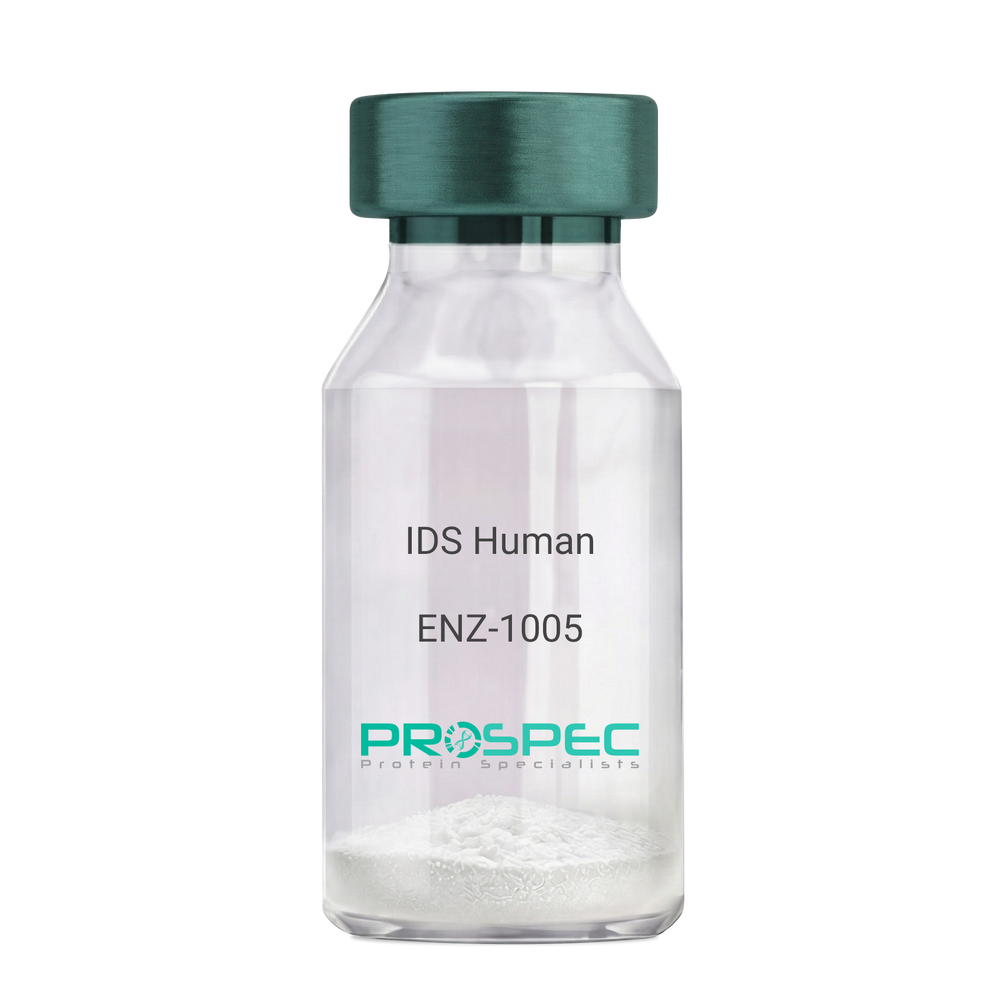
IDS HumanDescription:
Iduronate 2-Sulfatase Human Recombinant
Iduronate 2-Sulfatase, Alpha-L-Iduronate Sulfate Sulfatase, SIDS, Iduronate 2-Sulfatase 14 KDa Chain, Iduronate 2-Sulfatase 42 KDa Chain, Hunter Syndrome, EC 3.1.6.13, MPS2, Iduronate 2-sulfatase, Alpha-L-iduronate sulfate sulfatase.
Product # :
ENZ-1005Price :
Quantity :
Shipping Method :
Shipped with Ice Packs
More Info
- description
- source
- formulation
- purity
- More Info
Description
IDS Human Recombinant produced in Sf9 Baculovirus cells is a single, glycosylated polypeptide chain containing 533 amino acids (26-550a.a) and having a molecular mass of 60.3kDa. (Molecular size on SDS-PAGE will appear at approximately 35-70kDa). IDS is fused to an 8 amino acid His-tag at C-terminus & purified by proprietary chromatographic techniques.
Source
Sf9, Baculovirus cells.
Formulation
IDS protein solution (0.25mg/ml) containing Phosphate Buffered Saline (pH 7.4) and 10% glycerol.
Purity
Greater than 90% as determined by SDS-PAGE.
More Info
-
Introduction
Iduronate 2-Sulfatase also known as IDS, belongs to the highly-conserved sulfatase family of enzymes which catalyze the hydrolysis of O-sulfate and N-salfate esters from a variety of substrates. IDS is essential for the lysosomal degradation of the glycosaminoglycans (GAG) heparan sulfate as well as dermatan sulfate. Furthermore, IDS hydrolyzes the 2-sulfate group of the IDS units of the GAG.
-
Synonyms
Iduronate 2-Sulfatase, Alpha-L-Iduronate Sulfate Sulfatase, SIDS, Iduronate 2-Sulfatase 14 KDa Chain, Iduronate 2-Sulfatase 42 KDa Chain, Hunter Syndrome, EC 3.1.6.13, MPS2, Iduronate 2-sulfatase, Alpha-L-iduronate sulfate sulfatase.
-
Physical Appearance
Sterile Filtered clear solution.
-
Stability
Store at 4°C if entire vial will be used within 2-4 weeks. Store, frozen at -20°C for longer periods of time. For long term storage it is recommended to add a carrier protein (0.1% HSA or BSA).Avoid multiple freeze-thaw cycles.
-
Amino Acid Sequence
SETQANSTTD ALNVLLIIVD DLRPSLGCYG DKLVRSPNID QLASHSLLFQ NAFAQQAVCA PSRVSFLTGR RPDTTRLYDF NSYWRVHAGN FSTIPQYFKE NGYVTMSVGK VFHPGISSNH TDDSPYSWSF PPYHPSSEKY ENTKTCRGPD GELHANLLCP VDVLDVPEGT LPDKQSTEQA IQLLEKMKTS ASPFFLAVGY HKPHIPFRYP KEFQKLYPLE NITLAPDPEV PDGLPPVAYN PWMDIRQRED VQALNISVPY GPIPVDFQRK IRQSYFASVS YLDTQVGRLL SALDDLQLAN STIIAFTSDH GWALGEHGEW AKYSNFDVAT HVPLIFYVPG RTASLPEAGE KLFPYLDPFD SASQLMEPGR QSMDLVELVS LFPTLAGLAG LQVPPRCPVP SFHVELCREG KNLLKHFRFR DLEEDPYLPG NPRELIAYSQ YPRPSDIPQW NSDKPSLKDI KIMGYSIRTI DYRYTVWVGF NPDEFLANFS DIHAGELYFV DSDPLQDHNM YNDSQGGDLF QLLMPLEHHH HHH.
ProSpec's products are furnished for LABORATORY RESEARCH USE ONLY. The product may not be used as drugs, agricultural or pesticidal products, food additives or household chemicals.
- View Data Sheet
Name :
PGAM1 HumanDescription:
Phosphoglycerate Mutase 1 Human Recombinant
Phosphoglycerate mutase isozyme B, PGAM-B, PGAMA.
Product # :
ENZ-337Price :
Quantity :
Shipping Method :
Shipped with Ice Packs
More Info
- description
- source
- formulation
- purity
- More Info
Description
PGAM1 Human Recombinant produced in E.Coli is a single, non-glycosylated, polypeptide chain containing 274 amino acids (1-254 a.a.) and having a molecular mass of 30.9 kDa. The PGAM1 is fused to a 20 amino acid His Tag at N-Terminus and purified by proprietary chromatographic techniques.
Source
Escherichia Coli.
Formulation
The PGAM1 1mg/ml protein solution contains 20mM Tris-HCl pH-8, 1mM DTT, and 10% glycerol.
Purity
Greater than 90% as determined by SDS-PAGE.
More Info
-
Introduction
PGAM1 is part of the phosphoglycerate mutase family. PGAM1 is an essential component of glucose and 2,3-BPGA (2,3-bisphosphoglycerate) metabolism and catalyzes the reversible reaction of 3-phosphoglycerate (3-PGA) to 2-phosphoglycerate (2-PGA) in the glycolytic pathway. PGAM1 is a dimeric enzyme containing, in different tissues, different proportions of a slow-migrating muscle (MM) isozyme, a fast-migrating brain (BB) isozyme, and a hybrid form (MB). PGAM1 mutations lead to muscle phosphoglycerate mutase deficiency, a.k.a. glycogen storage disease X.
-
Synonyms
Phosphoglycerate mutase isozyme B, PGAM-B, PGAMA.
-
Physical Appearance
Sterile Filtered clear colorless solution.
-
Stability
Store at 4°C if entire vial will be used within 2-4 weeks. Store, frozen at -20°C for longer periods of time. For long term storage it is recommended to add a carrier protein (0.1% HSA or BSA).Avoid multiple freeze-thaw cycles.
-
Amino Acid Sequence
MGSSHHHHHH SSGLVPRGSH MAAYKLVLIR HGESAWNLEN RFSGWYDADL SPAGHEEAKR GGQALRDAGY EFDICFTSVQ KRAIRTLWTV LDAIDQMWLP VVRTWRLNER HYGGLTGLNK AETAAKHGEA QVKIWRRSYD VPPPPMEPDH PFYSNISKDR RYADLTEDQL PSCESLKDTI ARALPFWNEE IVPQIKEGKR VLIAAHGNSL RGIVKHLEGL SEEAIMELNL PTGIPIVYEL DKNLKPIKPM QFLGDEETVR KAMEAVAAQG KAKK.
ProSpec's products are furnished for LABORATORY RESEARCH USE ONLY. The product may not be used as drugs, agricultural or pesticidal products, food additives or household chemicals.
- View Data Sheet
Name :
IDE HumanDescription:
Insulin-Degrading Enzyme Human Recombinant
Insulin-Degrading Enzyme, Abeta-Degrading Protease, Insulin Protease, EC 3.4.24.56, Insulinase, INSULYSIN, Insulysin, EC 3.4.24, IDE.
Product # :
ENZ-813Price :
Quantity :
Shipping Method :
Shipped with Ice Packs
More Info
- description
- source
- formulation
- purity
- More Info
Description
IDE Human Recombinant produced in E.Coli is a single, non-glycosylated, polypeptide chain (Met1-Leu1019) containing 1026 amino acids including a 7 aa His tag at C-terminus. The total calculated molecular mass is 119kDa.
Source
Escherichia Coli.
Formulation
IDE filtered (0.4µm) in 20mM Tris buffer, 50mM NaCl, pH 8.0 and 10% (w/v) glycerol.
Purity
Greater than 95.0% as determined by SDS-PAGE.
More Info
-
Introduction
Insulin-Degrading Enzyme (IDE) is a zinc metallopeptidase which degrades intracellular insulin, and thus terminates insulins activity, as well as playing a part in intercellular peptide signaling by degrading various peptides such as amylin, bradykinin, and kallidin. The preferential affinity of the IDE enzyme for insulin results in insulin-mediated inhibition of the degradation of additional peptides such as beta-amyloid. Deficiencies in IDE protein's function are linked with Alzheimer's disease and type 2 diabetes mellitus nevertheless mutations in the IDE gene have not been demonstrated to be causative for these diseases. Insulin-Degrading Enzyme localizes mainly to the cytoplasm however in some cell types it localizes to the extracellular space, cell membrane, peroxisome, and mitochondrion. In addition, IDE degrades amyloid formed by APP and IAPP. Furthermore, IDE plays a part in the degradation and clearance of naturally secreted amyloid beta-protein by neurons and microglia.
-
Synonyms
Insulin-Degrading Enzyme, Abeta-Degrading Protease, Insulin Protease, EC 3.4.24.56, Insulinase, INSULYSIN, Insulysin, EC 3.4.24, IDE.
-
Physical Appearance
Filtered colorless solution.
-
Stability
Store at 4°C if entire vial will be used within 2-4 weeks. Store, frozen at -20°C for longer periods of time. For long term storage it is recommended to add a carrier protein (0.1% HSA or BSA).Avoid multiple freeze-thaw cycles.
-
Amino Acid Sequence
MRYRLAWLLH PALPSTFRSV LGARLPPPER LCGFQKKTYS KMNNPAIKRI GNHITKSPED KREYRGLELA NGIKVLLISD PTTDKSSAAL DVHIGSLSDP PNIAGLSHFC EHMLFLGTKK YPKENEYSQF LSEHAGSSNA FTSGEHTNYY FDVSHEHLEG ALDRFAQFFL CPLFDESCKD REVNAVDSEH EKNVMNDAWR LFQLEKATGN PKHPFSKFGT GNKYTLETRP NQEGIDVRQE LLKFHSAYYS SNLMAVCVLG RESLDDLTNL VVKLFSEVEN KNVPLPEFPE HPFQEEHLKQ LYKIVPIKDI RNLYVTFPIP DLQKYYKSNP GHYLGHLIGH EGPGSLLSEL KSKGWVNTLV GGQKEGARGF MFFIINVDLT EEGLLHVEDI ILHMFQYIQK LRAEGPQEWV FQECKDLNAV AFRFKDKERP RGYTSKIAGI LHYYPLEEVL TAEYLLEEFR PDLIEMVLDK LRPENVRVAI VSKSFEGKTD RTEEWYGTQY KQEAIPDEVI KKWQNADLNG KFKLPTKNEF IPTNFEILPL EKEATPYPAL IKDTAMSKLW FKQDDKFFLP KACLNFEFFS PFAYVDPLHC NMAYLYLELL KDSLNEYAYA AELAGLSYDL QNTIYGMYLS VKGYNDKQPI LLKKIIEKMA TFEIDEKRFE IIKEAYMRSL NNFRAEQPHQ HAMYYLRLLM TEVAWTKDEL KEALDDVTLP RLKAFIPQLL SRLHIEALLH GNITKQAALG IMQMVEDTLI EHAHTKPLLP SQLVRYREVQ LPDRGWFVYQ QRNEVHNNCG IEIYYQTDMQ STSENMFLEL FCQIISEPCF NTLRTKEQLG YIVFSGPRRA NGIQGLRFII QSEKPPHYLE SRVEAFLITM EKSIEDMTEE AFQKHIQALA IRRLDKPKKL SAECAKYWGE IISQQYNFDR DNTEVAYLKT LTKEDIIKFY KEMLAVDAPR RHKVSVHVLA REMDSCPVVG EFPCQNDINL SQAPALPQPE VIQNMTEFKR GLPLFPLVKP HINFMAAKL E HHHHHH.
ProSpec's products are furnished for LABORATORY RESEARCH USE ONLY. The product may not be used as drugs, agricultural or pesticidal products, food additives or household chemicals.
- View Data Sheet
Name :
GOT2 MouseDescription:
Glutamic-Oxaloacetic Transaminase 2 Mouse Recombinant
Transaminase A, KAT4, KATIV, KAT-4, KAT-IV,Kynurenine Aminotransferase 4.
Product # :
ENZ-1095Price :
Quantity :
Shipping Method :
Shipped with Ice Packs
More Info
- description
- source
- formulation
- purity
- biological activity
- More Info
Description
GOT2 Recombinant Mouse produced in E.Coli is a single, non-glycosylated polypeptide chain containing 424 amino acids (30-430 a.a.) and having a molecular mass of 46.8kDa. Mouse GOT2 is fused to a 21 amino acid His-tag at N-terminus & purified by proprietary chromatographic techniques.
Source
Escherichia Coli.
Formulation
The GOT2 solution (0.5mg/ml) contains PBS, pH 7.4 and 10% glycerol.
Purity
Greater than 90.0% as determined by SDS-PAGE.
Biological Activity
Activity is greater than 20 units/mg, and is defined as the amount of enzyme that converts 1umole of alpha-ketoglutarate to L-Glutamate per minute at pH 8.0 at 25℃.
More Info
-
Introduction
GOT2 is a pyridoxal phosphate-dependent enzyme which is found in cytoplasmic and inner-membrane mitochondrial forms, GOT1 and GOT2. GOT2 takes part in amino acid metabolism and the urea and tricarboxylic acid cycles. Both enzymes are homodimeric and demonstrate close homology.
-
Synonyms
Transaminase A, KAT4, KATIV, KAT-4, KAT-IV,Kynurenine Aminotransferase 4.
-
Physical Appearance
Sterile filtered colorless solution.
-
Stability
Store at 4°C if entire vial will be used within 2-4 weeks. Store, frozen at -20°C for longer periods of time. For long term storage it is recommended to add a carrier protein (0.1% HSA or BSA).Avoid multiple freeze-thaw cycles.
-
Amino Acid Sequence
MGSSHHHHHH SSGLVPRGSH MSSWWTHVEM GPPDPILGVT EAFKRDTNSK KMNLGVGAYR DDNGKPYVLP SVRKAEAQIA AKNLDKEYLP IGGLAEFCKA SAELALGENN EVLKSGRFVT VQTISGTGAL RVGASFLQRF FKFSRDVFLP KPSWGNHTPI FRDAGMQLQG YRYYDPKTCG FDFSGALEDI SKIPEQSVLL LHACAHNPTG VDPRPEQWKE IASVVKKKNL FAFFDMAYQG FASGDGDKDA WAVRHFIEQG INVCLCQSYA KNMGLYGERV GAFTVVCKDA EEAKRVESQL KILIRPLYSN PPLNGARIAA TILTSPDLRK QWLQEVKGMA DRIISMRTQL VSNLKKEGSS HNWQHITDQI GMFCFTGLKP EQVERLTKEF SVYMTKDGRI SVAGVTSGNV GYLAHAIHQV.
ProSpec's products are furnished for LABORATORY RESEARCH USE ONLY. The product may not be used as drugs, agricultural or pesticidal products, food additives or household chemicals.
- View Data Sheet
Name :
CAIII Human, HisDescription:
Carbonic Anhydrase III Human Recombinant, His Tag
Car3, CAIII, Carbonic anhydrase 3, EC 4.2.1.1, Carbonic anhydrase III, Carbonate dehydratase III, CA-III.
Product # :
ENZ-270Price :
Quantity :
Shipping Method :
Shipped with Ice Packs
More Info
- description
- source
- formulation
- purity
- More Info
Description
Carbonic anhydrase III Human Recombinant produced in E.Coli, and having a molecular mass of 33.9 kDa. CAIII is expressed with an amino-terminal hexahistidine tag.The CA-III is purified by proprietary chromatographic techniques.
Source
Escherichia Coli.
Formulation
Supplied in 10mM Tris-HCl (pH 8), 250mM NaCl, 0.5mM DTT, 1.5mM Cysteine, and 50% Glycerol.
Purity
Greater than 95.0% as determined by SDS-PAGE.
More Info
-
Introduction
Carbonic anhydrase (carbonate dehydratase) is a family of metalloenzymes (enzymes that contain one or more metal atoms as a functional component of the enzyme) that catalyze the rapid (and reversible) conversion of carbon dioxide to bicarbonate and protons, a reaction that occurs rather slowly in the absence of a catalyst. Carbonic anhydrase greatly increases the rate of the reaction, with typical catalytic rates of the different forms of this enzyme ranging between 104 and 106 reactions per second. The active site of most carbonic anhydrases contains a zincion. CAIII is a cytoplasmic isoenzyme, but is released into the circulation following injury.
-
Synonyms
Car3, CAIII, Carbonic anhydrase 3, EC 4.2.1.1, Carbonic anhydrase III, Carbonate dehydratase III, CA-III.
-
Physical Appearance
Sterile Filtered blue solution.
-
Stability
Store at 4°C if entire vial will be used within 2-4 weeks. Store, frozen at -20°C for longer periods of time. For long term storage it is recommended to add a carrier protein (0.1% HSA or BSA).Avoid multiple freeze-thaw cycles.
ProSpec's products are furnished for LABORATORY RESEARCH USE ONLY. The product may not be used as drugs, agricultural or pesticidal products, food additives or household chemicals.
- View Data Sheet
Name :
XYLT2 HumanDescription:
Xylosyltransferase 2 Human Recombinant
Xylosyltransferase 2, Peptide O-xylosyltransferase 1, Xylosyltransferase II, XT-II, XylT-II, XYLT2, XT2.
Product # :
ENZ-1086Price :
Quantity :
Shipping Method :
Shipped at Room temp
More Info
- description
- source
- formulation
- purity
- More Info
Description
XYLT2 Human Recombinant is a single, glycosylated polypeptide chain containing 839 amino acids (Gly37-Arg865, luminal domain, isoform 1, natural variant with Thr305) and having a molecular mass of 94.0kDa. XYLT2 is fused to an N-terminal linker (2 extra a.a), C-terminal linker (2 extra a.a) and C-terminal His-tag (6 extra a.a).
Source
HEK293 Cells.
Formulation
XYLT2 filtered (0.4 µm) and lyophilized in 0.05 M PBS and 0.075 M NaCl, pH 7.4.
Purity
Greater than 95.0% as determined by SDS-PAGE.
More Info
-
Introduction
XYLT2 or Xylosyltransferase 2 is an enzyme which is expressed in ubiquitous and is part of the glycosyltransfe-rases family. XYLT2 promotes proteoglycans formation by attaching GAG chains to the substrate protein via transfer of xylose molecule from the donor (nucleoside diphosphate) to the protein’s serine residues. XYLT2 is present in the ER and the cis part of the Golgi, furthermore the protein is released to the extracellular matrix.
-
Synonyms
Xylosyltransferase 2, Peptide O-xylosyltransferase 1, Xylosyltransferase II, XT-II, XylT-II, XYLT2, XT2.
-
Physical Appearance
Filtered White lyophilized (freeze-dried) powder.
-
Stability
Store lyophilized protein at -20°C. Aliquot the product after reconstitution to avoid repeated freezing/thawing cycles. Reconstituted protein can be stored at 4°C for a limited period of time; it does not show any change after two weeks at 4°C.
-
Solubility
It is recommended to add deionized water to prepare a working stock solution of approximately 0.5mg/ml and let the lyophilized pellet dissolve completely. XYLT2 is not sterile! Please filter the product by an appropriate sterile filter before using it in the cell culture.
-
Amino Acid Sequence
ASGLEEDEAG EKGRQRKPRP LDPGEGSKDT DSSAGRRGST GRRHGRWRGR AESPGVPVAK VVRAVTSRQR ASRRVPPAPP PEAPGRQNLS GAAAGEALVG AAGFPPHGDT GSVEGAPQPT DNGFTPKCEI VGKDALSALA RASTKQCQQE IANVVCLHQA GSLMPKAVPR HCQLTGKMSP GIQWDESQAQ QPMDGPPVRI AYMLVVHGRA IRQLKRLLKA VYHEQHFFYI HVDKRSDYLH REVVELAQGY DNVRVTPWRM VTIWGGASLL TMYLRSMRDL LEVPGWAWDF FINLSATDYP TRTNEELVAF LSKNRDKNFL KSHGRDNSRF IKKQGLDRLF HECDSHMWRL GERQIPAGIV VDGGSDWFVL TRSFVEYVVY TDDPLVAQLR QFYTYTLLPA ESFFHTVLEN SLACETLVDN NLRVTNWNRK LGCKCQYKHI VDWCGCSPND FKPQDFLRLQ QVSRPTFFAR KFESTVNQEV LEILDFHLYG SYPPGTPALK AYWENTYDAA DGPSGLSDVM LTAYTAFARL SLHHAATAAP PMGTPLCRFE PRGLPSSVHL YFYDDHFQGY LVTQAVQPSA QGPAETLEMW LMPQGSLKLL GRSDQASRLQ SLEVGTDWDP KERLFRNFGG LLGPLDEPVA VQRWARGPNL TATVVWIDPT YVVATSYDIT VDTETEVTQY KPPLSRPLRP GPWTVRLLQF WEPLGETRFL VLPLTFNRKL PLRKDDASWL HAGPPHNEYM EQSFQGLSSI LNLPQPELAE EAAQRHTQLT GPALEAWTDR ELSSFWSVAG LCAIGPSPCP SLEPCRLTSW SSLSPDPKSE LGPVKADGRL RKLHHHHHH.
ProSpec's products are furnished for LABORATORY RESEARCH USE ONLY. The product may not be used as drugs, agricultural or pesticidal products, food additives or household chemicals.
- View Data Sheet
Name :
CASP3 Human, Sf9Description:
Caspase 3 Apoptosis-Related Cysteine Peptidase Human Recombinant, Sf9
CASP3, CPP32, CPP32B, SCA-1, CASP-3, Apopain, Cysteine protease CPP32, CPP-32, Protein Yama, SREBP cleavage activity 1.
Product # :
ENZ-1106Price :
Quantity :
Shipping Method :
Shipped with Ice Packs
More Info
- description
- source
- formulation
- purity
- biological activity
- More Info
Description
CASP3 Human produced in Sf9 Baculovirus cells is a single, glycosylated polypeptide chain containing 256 amino acids (29-277 a.a.) and having a molecular mass of 29.4kDa (Migrates at 13.5-18kDa on SDS-PAGE under reducing conditions). CASP3 is expressed with a 6 amino acid His tag at C-Terminus and purified by proprietary chromatographic techniques.
Source
Sf9, Baculovirus cells.
Formulation
CASP3 protein solution (0.5mg/ml) containing 20mM HEPES buffer (pH 7.5), 0.1M NaCl, 1mM EDTA, 20% Glycerol and 1mM DTT.
Purity
Greater than 90.0% as determined by SDS-PAGE.
Biological Activity
Specific activity is greater than 5,000 pmol/min/ug. One unit will liberate 1 pmoles of Ac-DEVD-AFC to Ac-DEVD and AFC per minute at pH7.5 at 25C.
More Info
-
Introduction
Caspase 3 Apoptosis-Related Cysteine Peptidase (CASP3) belongs to the cysteine-aspartic acid protease (caspase) family. Sequential activation of caspases plays a key role in the execution-phase of cell apoptosis. Caspases exist as inactive proenzymes which undergo proteolytic processing at conserved aspartic residues to generate 2 subunits, large and small, that dimerize to create the active enzyme. CASP3 protein cleaves and activates caspases 6, 7 and 9, and the protein itself is processed by caspases 8, 9 and 10. CASP3 is the leading caspase involved in the cleavage of amyloid-beta 4A precursor protein, which is linked with neuronal death in Alzheimer's disease. In addition, CASP3 is involved in the cleavage of huntingtin. CASP3 also cleaves and activates sterol regulatory element binding proteins (SREBPs) between the basic helix-loop-helix leucine zipper domain and the membrane attachment domain. CASP3 initiates cell adhesion in sympathetic neurons through RET cleavage.
-
Synonyms
CASP3, CPP32, CPP32B, SCA-1, CASP-3, Apopain, Cysteine protease CPP32, CPP-32, Protein Yama, SREBP cleavage activity 1.
-
Physical Appearance
Sterile Filtered colorless solution.
-
Stability
Store at 4°C if entire vial will be used within 2-4 weeks. Store, frozen at -20°C for longer periods of time. For long term storage it is recommended to add a carrier protein (0.1% HSA or BSA).Avoid multiple freeze-thaw cycles.
-
Amino Acid Sequence
MSGISLDNSY KMDYPEMGLC IIINNKNFHK STGMTSRSGT DVDAANLRET FRNLKYEVRN
KNDLTREEIV ELMRDVSKED HSKRSSFVCV LLSHGEEGII FGTNGPVDLK KITNFFRGDR
CRSLTGKPKL FIIQACRGTE LDCGIETDSG VDDDMACHKI PVEADFLYAY STAPGYYSWR
NSKDGSWFIQ SLCAMLKQYA DKLEFMHILT RVNRKVATEF ESFSFDATFH AKKQIPCIVS MLTKELYFYH HHHHHH
ProSpec's products are furnished for LABORATORY RESEARCH USE ONLY. The product may not be used as drugs, agricultural or pesticidal products, food additives or household chemicals.
- View Data Sheet
Name :
B3GNT2 HumanDescription:
Beta-1,3-N-Acetylglucosaminyltransferase 2 Human Recombinant
B3GNT2, B3GN-T2, B3GNT, B3GNT-2, B3GNT1, BETA3GNT, BGnT-2, BGNT2, N-acetyllactosaminide beta-1,3-N-acetylglucosaminyltransferase 2, Beta-1,3-N-acetylglucosaminyltransferase 1, BGnT-1, Beta-1,3-Gn-T1, Beta3Gn-T1, Beta-1,3-galactosyltransferase 7, Beta-1,3-GalTase 7, Beta3Gal-T7, Beta3GalT7, b3Gal-T7, Beta-3-Gx-T7, beta-GlcNAc beta-1,3-galactosyltransferase 7, betaGal beta-1,3-N-acetylglucosaminyltransferase 2, BGnT-2, Beta-1,3-Gn-T2, Beta-1,3-N-acetylglucosaminyltransferase 2, Beta3Gn-T2, beta-N-acetylglucosamine beta-1,3-galactosyltransferase 7.
Product # :
ENZ-973Price :
Quantity :
Shipping Method :
Shipped with Ice Packs
More Info
- description
- source
- formulation
- purity
- More Info
Description
B3GNT2 produced in Sf9 Baculovirus cells is a single, glycosylated polypeptide chain containing 375 amino acids (29-397a.a.) and having a molecular mass of 43.5kDa (Molecular size on SDS-PAGE will appear at approximately 40-57kDa). B3GNT2 is expressed with a 6 amino acid His tag at C-Terminus and purified by proprietary chromatographic techniques.
Source
Sf9, Baculovirus cells.
Formulation
B3GNT2 protein solution (0.5mg/ml) contains Phosphate Buffered Saline (pH 7.4) and 10% glycerol.
Purity
Greater than 90.0% as determined by SDS-PAGE.
More Info
-
Introduction
Beta-1,3-N-Acetylglucosaminyltransferase 2 (B3GNT2) is a part of the beta-1,3-N-acetylglucosaminyltransferase family which takes part in the synthesis of poly-N-acetyllactosamine. B3GNT2 is a type II transmembrane protein which prefers the substrate of lacto-N-neotetraose. B3GNT2 catalyzes the initiation and elongation of poly-N-acetyllactosamine chains and comprises the main polylactosamine synthase.
-
Synonyms
B3GNT2, B3GN-T2, B3GNT, B3GNT-2, B3GNT1, BETA3GNT, BGnT-2, BGNT2, N-acetyllactosaminide beta-1,3-N-acetylglucosaminyltransferase 2, Beta-1,3-N-acetylglucosaminyltransferase 1, BGnT-1, Beta-1,3-Gn-T1, Beta3Gn-T1, Beta-1,3-galactosyltransferase 7, Beta-1,3-GalTase 7, Beta3Gal-T7, Beta3GalT7, b3Gal-T7, Beta-3-Gx-T7, beta-GlcNAc beta-1,3-galactosyltransferase 7, betaGal beta-1,3-N-acetylglucosaminyltransferase 2, BGnT-2, Beta-1,3-Gn-T2, Beta-1,3-N-acetylglucosaminyltransferase 2, Beta3Gn-T2, beta-N-acetylglucosamine beta-1,3-galactosyltransferase 7.
-
Physical Appearance
Sterile Filtered colorless solution.
-
Stability
Store at 4°C if entire vial will be used within 2-4 weeks. Store, frozen at -20°C for longer periods of time. For long term storage it is recommended to add a carrier protein (0.1% HSA or BSA). Avoid multiple freeze-thaw cycles.
-
Amino Acid Sequence
ADPKSSSQEK NGKGEVIIPK EKFWKISTPP EAYWNREQEK LNRQYNPILS MLTNQTGEAG RLSNISHLNY CEPDLRVTSV VTGFNNLPDR FKDFLLYLRC RNYSLLIDQP DKCAKKPFLL LAIKSLTPHF ARRQAIRESW GQESNAGNQT VVRVFLLGQT PPEDNHPDLS DMLKFESEKH QDILMWNYRD TFFNLSLKEV LFLRWVSTSC PDTEFVFKGD DDVFVNTHHI LNYLNSLSKT KAKDLFIGDV IHNAGPHRDK KLKYYIPEVV YSGLYPPYAG GGGFLYSGHL ALRLYHITDQ VHLYPIDDVY TGMCLQKLGL VPEKHKGFRT FDIEEKNKNN ICSYVDLMLV HSRKPQEMID IWSQLQSAHL KCHHHHHH.
ProSpec's products are furnished for LABORATORY RESEARCH USE ONLY. The product may not be used as drugs, agricultural or pesticidal products, food additives or household chemicals.
- View Data Sheet
Name :
ALDH2 Mouse, ActiveDescription:
Aldehyde Dehydrogenase 2 Mouse Recombinant, Active
Aldehyde dehydrogenase, mitochondrial, AHD-M1, ALDH class 2, ALDH-E2, ALDHI.
Product # :
ENZ-1093Price :
Quantity :
Shipping Method :
Shipped with Ice Packs
More Info
- description
- source
- formulation
- purity
- biological activity
- More Info
Description
ALDH2 Mouse Recombinant produced in E.Coli is a single, non-glycosylated polypeptide chain containing 523 amino acids (20-519) and having a molecular mass of 56.8kDa. ALDH2 Mouse is fused to a 23 amino acid His-Tag at N-terminus and purified by proprietary chromatographic techniques.
Source
Escherichia Coli.
Formulation
ALDH2 Mouse protein (0.5mg/ml) is formulated in Phosphate buffered saline (pH7.4), 20% glycerol and 1mM DTT.
Purity
Greater than 95% as determined by SDS-PAGE.
Biological Activity
Specific activity is > 180 pmol/min/ug, and was obtained by measuring the increase of NADH in absorbance at 340 nm resulting from the reduction of NAD at pH 8.0 at 25C.
More Info
-
Introduction
ALDH2 is part of the aldehyde dehydrogenase family of proteins which catalyze the chemical transformation from acetaldehyde to acetic acid. ALDH2 is the second enzyme of the major oxidative pathway of alcohol metabolism. ALDH2 has 2 major liver isoforms: cytosolic and mitochondrial, which differ by their electrophoretic mobilities, kinetic properties, and subcellular localizations. Nearly all Caucasians have 2 major isozymes, whereas roughly 50% of Orientals have only the cytosolic isozyme, omitting the mitochondrial isozyme. The extremely higher rate of acute alcohol intoxication with Orientals compared to Caucasians is due to the fact of the absence of mitochondrial isozyme. ALDH2 has a low Km for acetaldehydes, and is localized in mitochondrial matrix.
-
Synonyms
Aldehyde dehydrogenase, mitochondrial, AHD-M1, ALDH class 2, ALDH-E2, ALDHI.
-
Physical Appearance
Sterile Filtered clear solution.
-
Stability
Store at 4°C if entire vial will be used within 2-4 weeks. Store, frozen at -20°C for longer periods of time. For long term storage it is recommended to add a carrier protein (0.1% HSA or BSA).Avoid multiple freeze-thaw cycles.
-
Amino Acid Sequence
MGSSHHHHHH SSGLVPRGSH MGSSAAATSA VPAPNHQPEV FCNQIFINNE WHDAVSRKTF PTVNPSTGEV ICQVAEGNKE DVDKAVKAARAAFQLGSPWR RMDASDRGRL LYRLADLIER DRTYLAALET LDNGKPYVIS YLVDLDMVLK CLRYYAGWAD KYHGKTIPID GDFFSYTRHEPVGVCGQIIP WNFPLLMQAW KLGPALATGN VVVMKVAEQT PLTALYVANL IKEAGFPPGV VNIVPGFGPT AGAAIASHEG VDKVAFTGSTEVGHLIQVAA GSSNLKRVTL ELGGKSPNII MSDADMDWAV EQAHFALFFN QGQCCCAGSR TFVQENVYDE FVERSVARAK SRVVGNPFDSRTEQGPQVDE TQFKKILGYI KSGQQEGAKL LCGGGAAADR GYFIQPTVFG DVKDGMTIAK EEIFGPVMQI LKFKTIEEVV GRANDSKYGLAAAVFTKDLD KANYLSQALQ AGTVWINCYD VFGAQSPFGG YKMSGSGREL GEYGLQAYTE VKTVTVKVPQ KNS.
ProSpec's products are furnished for LABORATORY RESEARCH USE ONLY. The product may not be used as drugs, agricultural or pesticidal products, food additives or household chemicals.
- View Data Sheet
Name :
NANS HumanDescription:
N-acetylneuraminic acid synthase Human Recombinant
Sialic acid synthase, N-acetylneuraminate synthase, N-acetylneuraminate-9-phosphate synthase, N-acetylneuraminic acid phosphate synthase, N-acetylneuraminic acid synthase, NANS, SAS.
Product # :
ENZ-024Price :
Quantity :
Shipping Method :
Shipped with Ice Packs
More Info
- description
- source
- formulation
- purity
- More Info
Description
NANS Human Recombinant fused with a 20 amino acid His tag at N-terminus produced in E.Coli is a single, non-glycosylated, polypeptide chain containing 379 amino acids (1-359 a.a.) and having a molecular mass of 42.4kDa. The NANS is purified by proprietary chromatographic techniques.
Source
Escherichia Coli.
Formulation
The NANS solution (0.5 mg/ml) contains 20mM Tris-HCl buffer (pH8.0), 0.1M NaCl, 1mM DTT and 10% glycerol.
Purity
Greater than 95.0% as determined by SDS-PAGE.
More Info
-
Introduction
NANS is a 359 amino acid protein that contains one AFP (antifreeze proteins)-like domain and functions in the biosynthesis of sialic acids. The ubiquitously expressed NANS enzymatically catalyzes the H2O-dependent formation of N-acetylneuraminic acid (Neu5Ac) and 2-keto-3-deoxy-D-glycero-D-galacto-nononic acid (KDN), both of which are sialic acids. NANS uses N-acetylmannosamine 6-phosphate as a substrate for Neu5Ac synthesis and mannose 6-phosphate as a substrate for KDN synthesis.
-
Synonyms
Sialic acid synthase, N-acetylneuraminate synthase, N-acetylneuraminate-9-phosphate synthase, N-acetylneuraminic acid phosphate synthase, N-acetylneuraminic acid synthase, NANS, SAS.
-
Physical Appearance
Sterile Filtered colorless solution.
-
Stability
Store at 4°C if entire vial will be used within 2-4 weeks. Store, frozen at -20°C for longer periods of time. For long term storage it is recommended to add a carrier protein (0.1% HSA or BSA).Avoid multiple freeze-thaw cycles.
-
Amino Acid Sequence
MGSSHHHHHH SSGLVPRGSH MPLELELCPG RWVGGQHPCF IIAEIGQNHQ GDLDVAKRMI RMAKECGADC AKFQKSELEF KFNRKALERP YTSKHSWGKT YGEHKRHLEF SHDQYRELQR YAEEVGIFFT ASGMDEMAVE FLHELNVPFF KVGSGDTNNF PYLEKTAKKG RPMVISSGMQ SMDTMKQVYQ IVKPLNPNFC FLQCTSAYPL QPEDVNLRVI SEYQKLFPDI PIGYSGHETG IAISVAAVAL GAKVLERHIT LDKTWKGSDH SASLEPGELA ELVRSVRLVE RALGSPTKQL LPCEMACNEK LGKSVVAKVK IPEGTILTMD MLTVKVGEPK GYPPEDIFNL VGKKVLVTVE EDDTIMEELV DNHGKKIKS.
ProSpec's products are furnished for LABORATORY RESEARCH USE ONLY. The product may not be used as drugs, agricultural or pesticidal products, food additives or household chemicals.
- View Data Sheet
Name :
PGAM2 Human, ActiveDescription:
Phosphoglycerate Mutase 2 Human Recombinant, Active
Phosphoglycerate mutase 2, BPG-dependent PGAM 2, Muscle-specific phosphoglycerate mutase, Phosphoglycerate mutase isozyme M, PGAM-M, PGAM2, PGAMM, GSD10.
Product # :
ENZ-981Price :
Quantity :
Shipping Method :
Shipped with Ice Packs
More Info
- description
- source
- formulation
- purity
- biological activity
- More Info
Description
PGAM2 Human Recombinant produced in E.coli is a single, non-glycosylated polypeptide chain containing 273 amino acids (1-253) and having a molecular mass of 30.9kDa.PGAM2 is fused to a 20 amino acid His-tag at N-terminus & purified by proprietary chromatographic techniques.
Source
Escherichia Coli.
Formulation
The PGAM2 solution (1mg/ml) contains 20mM Tris-HCl buffer (pH8.0), 20% glycerol, 0.1M NaCl and 1mM DTT.
Purity
Greater than 95.0% as determined by SDS-PAGE.
Biological Activity
Specific activity is > 100units/mg, in which One unit will convert 1.0 umole of 3-phosphoglycerate to 2-phosphoglcerate per minute at pH 7.6 at 37C.More Info
-
Introduction
Phosphoglycerate mutase 2 (PGAM2) is a member of the phosphoglycerate mutase family. PGAM is a dimeric enzyme which contains in separate tissues, different proportions of a slow-migrating muscle (MM) isozyme, a fast-migrating brain (BB) isozyme, and a hybrid form (MB). PGAM (Phosphoglycerate mutase) catalyzes the reversible reaction of 3-phosphoglycerate (3-PGA) to 2-phosphoglycerate (2-PGA) in the glycolytic pathway. PGAM2 gene mutations cause muscle phosphoglycerate mutase efficiency, otherwise known as glycogen storage disease X.
-
Synonyms
Phosphoglycerate mutase 2, BPG-dependent PGAM 2, Muscle-specific phosphoglycerate mutase, Phosphoglycerate mutase isozyme M, PGAM-M, PGAM2, PGAMM, GSD10.
-
Physical Appearance
Sterile Filtered colorless solution.
-
Stability
Store at 4°C if entire vial will be used within 2-4 weeks. Store, frozen at -20°C for longer periods of time. For long term storage it is recommended to add a carrier protein (0.1% HSA or BSA).Avoid multiple freeze-thaw cycles.
-
Amino Acid Sequence
MGSSHHHHHH SSGLVPRGSH MATHRLVMVR HGESTWNQEN RFCGWFDAEL SEKGTEEAKR GAKAIKDAKM EFDICYTSVL KRAIRTLWAI LDGTDQMWLP VVRTWRLNER HYGGLTGLNK AETAAKHGEE QVKIWRRSFD IPPPPMDEKH PYYNSISKER RYAGLKPGEL PTCESLKDTI ARALPFWNEE IVPQIKAGKR VLIAAHGNSL RGIVKHLEGM SDQAIMELNL PTGIPIVYEL NKELKPTKPM QFLGDEETVR KAMEAVAAQG KAK.
ProSpec's products are furnished for LABORATORY RESEARCH USE ONLY. The product may not be used as drugs, agricultural or pesticidal products, food additives or household chemicals.
- View Data Sheet
Name :
IDE Human, ActiveDescription:
Insulin-Degrading Enzyme Human Recombinant
Insulin-Degrading Enzyme, Abeta-Degrading Protease, Insulysin, EC 3.4.24.56, Insulinase, Insulin Protease, INSULYSIN, EC 3.4.24, IDE, insulin-degrading enzyme isoform 1.
Product # :
ENZ-1192Price :
Quantity :
Shipping Method :
Shipped with Ice Packs
More Info
- description
- source
- formulation
- purity
- biological activity
- More Info
Description
IDE Human, Active Human Recombinant produced in E.Coli is a single, non-glycosylated, polypeptide chain (42-1019 a.a) containing a total of 984 amino acids, having a molecular mass of 114 kDa. IDE is fused to a 6 amino acid His-tag at C-terminus and is purified by proprietary chromatographic techniques.
Source
Escherichia Coli.
Formulation
The IDE solution (0.5mg/ml) contains 10% Glycerol, 100mM NaCl, 0.05% Brij35 and 20mM Tris-HCl buffer (pH 7.5).
Purity
Greater than 90.0% as determined by SDS-PAGE.
Biological Activity
Specific activity is greater than 3,000 pmol/min/ug in which 1 unit will convert 1.0 pmole of Mca-RPPGFSAFK(Dnp)-OH to MCA-Pro-Leu-OH per minute at pH 7.5 at 25°C.
More Info
-
Synonyms
Insulin-Degrading Enzyme, Abeta-Degrading Protease, Insulysin, EC 3.4.24.56, Insulinase, Insulin Protease, INSULYSIN, EC 3.4.24, IDE, insulin-degrading enzyme isoform 1.
-
Physical Appearance
Filtered colorless solution.
-
Stability
Store at 4°C if entire vial will be used within 2-4 weeks. Store, frozen at -20°C for longer periods of time. For long term storage it is recommended to add a carrier protein (0.1% HSA or BSA). Avoid multiple freeze-thaw cycles.
-
Amino Acid Sequence
MNNPAIKRIG NHITKSPEDK REYRGLELAN GIKVLLISDP TTDKSSAALD VHIGSLSDPP NIAGLSHFCE HMLFLGTKKY PKENEYSQFL SEHAGSSNAF TSGEHTNYYF DVSHEHLEGA LDRFAQFFLC PLFDESCKDR EVNAVDSEHE KNVMNDAWRL FQLEKATGNP KHPFSKFGTG NKYTLETRPN QEGIDVRQEL LKFHSAYYSS NLMAVCVLGR ESLDDLTNLV VKLFSEVENK NVPLPEFPEH PFQEEHLKQL YKIVPIKDIR NLYVTFPIPD LQKYYKSNPG HYLGHLIGHE GPGSLLSELK SKGWVNTLVG GQKEGARGFM FFIINVDLTE EGLLHVEDII LHMFQYIQKL RAEGPQEWVF QECKDLNAVA FRFKDKERPR GYTSKIAGIL HYYPLEEVLT AEYLLEEFRP DLIEMVLDKL RPENVRVAIV SKSFEGKTDR TEEWYGTQYK QEAIPDEVIK KWQNADLNGK FKLPTKNEFI PTNFEILPLE KEATPYPALI KDTAMSKLWF KQDDKFFLPK ACLNFEFFSP FAYVDPLHCN MAYLYLELLK DSLNEYAYAA ELAGLSYDLQ NTIYGMYLSV KGYNDKQPIL LKKIIEKMAT FEIDEKRFEI IKEAYMRSLN NFRAEQPHQH AMYYLRLLMT EVAWTKDELK EALDDVTLPR LKAFIPQLLS RLHIEALLHG NITKQAALGI MQMVEDTLIE HAHTKPLLPS
QLVRYREVQL PDRGWFVYQQ RNEVHNNCGI EIYYQTDMQS TSENMFLELF CQIISEPCFN TLRTKEQLGY IVFSGPRRAN GIQGLRFIIQ SEKPPHYLES RVEAFLITME KSIEDMTEEA FQKHIQALAI RRLDKPKKLS AECAKYWGEI ISQQYNFDRD NTEVAYLKTL TKEDIIKFYK EMLAVDAPRR HKVSVHVLAR EMDSCPVVGE FPCQNDINLS QAPALPQPEV IQNMTEFKRG LPLFPLVKPH INFMAAKLHH HHHH. -
Background
Insulin-degrading enzyme (IDE) is a crucial protease that plays a significant role in maintaining glucose homeostasis by degrading insulin and other bioactive peptides. Dysregulation of IDE has been implicated in various metabolic disorders, particularly type 2 diabetes mellitus. IDE is also associated with the clearance of amyloid-beta peptides in the brain, making it relevant to Alzheimer's disease pathology. Studying the recombinant form of IDE is fundamental to understanding its functional mechanisms and exploring potential avenues for therapeutic interventions.
The primary goal of this research is to express and purify recombinant IDE using diverse expression systems. Recombinant DNA techniques will be employed to construct expression vectors containing the IDE gene, followed by expression in bacterial, yeast, or mammalian cell-based systems. The recombinant IDE will be purified using affinity chromatography or other appropriate methods, facilitating subsequent biochemical and biophysical characterization.
The second objective is to investigate the substrate specificity and catalytic activity of the purified IDE. In vitro enzymatic assays will be conducted to analyse the ability of the recombinant IDE to degrade insulin and other potential substrates. The effects of various factors, such as pH, temperature, and potential modulators, on IDE activity will be evaluated. Additionally, the interactions between IDE and its substrates will be explored using binding assays.
The third objective is to elucidate the three-dimensional structure of the IDE recombinant using techniques like X-ray crystallography or nuclear magnetic resonance (NMR) spectroscopy. Structural insights into the active site and binding pockets of IDE will provide valuable information for understanding its substrate recognition and catalytic mechanisms. This knowledge could be instrumental in designing targeted therapeutic compounds.
By characterizing the IDE recombinant, this research aims to contribute to our understanding of its role in insulin metabolism, glucose regulation, and potential therapeutic applications. The findings from this study may have implications for the development of novel treatments for diabetes and other related disorders.
ProSpec's products are furnished for LABORATORY RESEARCH USE ONLY. The product may not be used as drugs, agricultural or pesticidal products, food additives or household chemicals.
- View Data Sheet
Name :
AHCY HumanDescription:
Adenosylhomocysteinase Human Recombinant
EC 3.3.1.1, SAHH, AdoHcyase, S-adenosyl-L-homocysteine hydrolase, Adenosylhomocysteinase.
Product # :
ENZ-532Price :
Quantity :
Shipping Method :
Shipped with Ice Packs
More Info
- description
- source
- formulation
- purity
- More Info
Description
AHCY Recombinant Human produced in E.Coli is a single, non-glycosylated polypeptide chain containing 452 amino acids (1-432 a.a.) and having a molecular mass of 49.8 kDa. The AHCY is fused to a 20 amino acids His-Tag at N-terminus and purified by proprietary chromatographic techniques.
Source
Escherichia Coli.
Formulation
AHCY Human solution containing 20mM Tris pH-8, & 10% glycerol.
Purity
Greater than 95.0% as determined by SDS-PAGE.
More Info
-
Introduction
AHCY is an enzyme that catalyzes the reversible hydrolysis of S-adenosylhomocysteine (AdoHcy) to adenosine (Ado) and L-homocysteine (Hcy). AHCY controls the intracellular S-adenosylhomocysteine (SAH) concentration that is crucial for transmethylation reactions. AHCY deficiency causes hypermethioninemia.
-
Synonyms
EC 3.3.1.1, SAHH, AdoHcyase, S-adenosyl-L-homocysteine hydrolase, Adenosylhomocysteinase.
-
Physical Appearance
Sterile filtered colorless solution.
-
Stability
Store at 4°C if entire vial will be used within 2-4 weeks. Store, frozen at -20°C for longer periods of time. For long term storage it is recommended to add a carrier protein (0.1% HSA or BSA).Avoid multiple freeze-thaw cycles.
-
Amino Acid Sequence
MGSSHHHHHH SSGLVPRGSH MSDKLPYKVA DIGLAAWGRK ALDIAENEMP GLMRMRERYS ASKPLKGARI AGCLHMTVET AVLIETLVTL GAEVQWSSCN IFSTQDHAAA AIAKAGIPVY AWKGETDEEY LWCIEQTLYF KDGPLNMILD DGGDLTNLIH TKYPQLLPGI RGISEETTTG VHNLYKMMAN GILKVPAINV NDSVTKSKFD NLYGCRESLI DGIKRATDVM IAGKVAVVAG YGDVGKGCAQ ALRGFGARVI ITEIDPINAL QAAMEGYEVT TMDEACQEGN IFVTTTGCID IILGRHFEQM KDDAIVCNIG HFDVEIDVKW LNENAVEKVN IKPQVDRYRL KNGRRIILLA EGRLVNLGCA MGHPSFVMSN SFTNQVMAQI ELWTHPDKYP VGVHFLPKKL DEAVAEAHLG KLNVKLTKLT EKQAQYLGMS CDGPFKPDHY RY.
ProSpec's products are furnished for LABORATORY RESEARCH USE ONLY. The product may not be used as drugs, agricultural or pesticidal products, food additives or household chemicals.
- View Data Sheet
Name :
FOLH1 HumanDescription:
Folate Hydrolase 1 Human Recombinant
Glutamate carboxypeptidase 2 isoform 1,Cell growth-inhibiting gene 27 protein, Folate hydrolase 1, Folylpolygamma-glutamate carboxypeptidase, Glutamate carboxypeptidase II, Membrane glutamate carboxypeptidase, Nacetylated-alpha-linked acidic dipeptidase I, Prostate-specific membrane antigen, Pteroylpoly-gamma glutamate carboxypeptidase, Folh1, FGCP, FOLH, GCP2, GCPII, mGCP, NAALAD1, NAALAdase, PSM, PSMA
Product # :
ENZ-1170Price :
Quantity :
Shipping Method :
Shipped with Ice Packs
More Info
- description
- source
- formulation
- purity
- More Info
Description
FOLH1 produced in Sf9 Baculovirus cells is a single, glycosylated polypeptide chain containing 717 amino acids (44-750 a.a) and having a molecular mass of 80.7kDa.FOLH1 is expressed with a 6 amino acid His tag at C-Terminus and purified by proprietary chromatographic techniques.
Source
Sf9, Baculovirus cells.
Formulation
FOLH1 protein solution (0.25mg/ml) contains Phosphate buffered saline (pH 7.4) and 20% glycerol.
Purity
Greater than 90.0% as determined by SDS-PAGE.
More Info
-
Introduction
FOLH1, also known as glutamate carboxypeptidase 2 (GCPII), is a single pass type 2 membrane protein which belongs to the peptidase M28 family. FOLH1 is highly produced in prostate epithelium. FOLH1 is also found in ovary, live, stomach, small intestine colon, urinary bladder, kidney, testis, and the capillary endothelium of a variety of tumours. Therefore, it plays a role in directed imaging and therapy of recurrent of metastatic disease. FOLH1 is a zinc metalloenzyme that resides in membranes and catalyses the hydrolysis of N-acetylaspartylglutamate to glutamate and N-acetylaspartate.
-
Synonyms
Glutamate carboxypeptidase 2 isoform 1,Cell growth-inhibiting gene 27 protein, Folate hydrolase 1, Folylpolygamma-glutamate carboxypeptidase, Glutamate carboxypeptidase II, Membrane glutamate carboxypeptidase, Nacetylated-alpha-linked acidic dipeptidase I, Prostate-specific membrane antigen, Pteroylpoly-gamma glutamate carboxypeptidase, Folh1, FGCP, FOLH, GCP2, GCPII, mGCP, NAALAD1, NAALAdase, PSM, PSMA
-
Physical Appearance
Sterile Filtered colorless solution.
-
Stability
Store at 4°C if entire vial will be used within 2-4 weeks.Store, frozen at -20°C for longer periods of time.For long term storage it is recommended to add a carrier protein (0.1% HSA or BSA).Avoid multiple freeze-thaw cycles.
-
Amino Acid Sequence
ADPMKSSNEA TNITPKHNMK AFLDELKAEN IKKFLYNFTQ IPHLAGTEQN FQLAKQIQSQ WKEFGLDSVE LAHYDVLLSY PNKTHPNYIS IINEDGNEIF NTSLFEPPPP GYENVSDIVP PFSAFSPQGM PEGDLVYVNY ARTEDFFKLE RDMKINCSGK IVIARYGKVF RGNKVKNAQL AGAKGVILYS DPADYFAPGV KSYPDGWNLP GGGVQRGNIL NLNGAGDPLT PGYPANEYAY RRGIAEAVGL PSIPVHPIGY YDAQKLLEKM GGSAPPDSSW RGSLKVPYNV GPGFTGNFST QKVKMHIHST NEVTRIYNVI GTLRGAVEPD RYVILGGHRD SWVFGGIDPQ SGAAVVHEIV RSFGTLKKEG WRPRRTILFA SWDAEEFGLL GSTEWAEENS RLLQERGVAY INADSSIEGN YTLRVDCTPL MYSLVHNLTK ELKSPDEGFE GKSLYESWTK KSPSPEFSGM PRISKLGSGN DFEVFFQRLG IASGRARYTK NWETNKFSGY PLYHSVYETY ELVEKFYDPM FKYHLTVAQV RGGMVFELAN SIVLPFDCRD YAVVLRKYAD KIYSISMKHP QEMKTYSVSF DSLFSAVKNF TEIASKFSER LQDFDKSNPI VLRMMNDQLM FLERAFIDPL GLPDRPFYRH VIYAPSSHNK YAGESFPGIY DALFDIESKV DPSKAWGEVK RQIYVAAFTV QAAAETLSEV AHHHHHH
ProSpec's products are furnished for LABORATORY RESEARCH USE ONLY. The product may not be used as drugs, agricultural or pesticidal products, food additives or household chemicals.
- View Data Sheet
Name :
HMGCL HumanDescription:
3-Hydroxymethyl-3-Methylglutaryl-CoA Lyase Human Recombinant
Hydroxymethylglutaryl-CoA lyase mitochondrial, HL, HMG-CoA lyase, 3-hydroxy-3-methylglutarate-CoA lyase, HMGCL.
Product # :
ENZ-218Price :
Quantity :
Shipping Method :
Shipped with Ice Packs
More Info
- description
- source
- formulation
- purity
- More Info
Description
HMGCL Human Recombinant produced in E.coli is a single, non-glycosylated polypeptide chain containing 323 amino acids (28-325) and having a molecular mass of 34.2kDa.HMGCL is fused to a 25 amino acid His-tag at N-terminus & purified by proprietary chromatographic techniques.
Source
Escherichia Coli.
Formulation
The HMGCL solution (1mg/ml) contains 20mM Tris-HCl buffer (pH8.0), 10% glycerol and 0.4M Urea.
Purity
Greater than 85.0% as determined by SDS-PAGE.
More Info
-
Introduction
Hydroxymethylglutaryl-CoA lyase (HMGCL) is a mitochondrial matrix protein which is a member of the HMG-CoA lyase family. HMGCL is a homodimer and participates in leucine catabolism and ketogenesis, the hepatic synthesis of ketone bodies which, during fasting, provides a major source of energy for the heart, brain and kidney. More precisely, HMGCL catalyzes the final step of these processes, the cleavage of 3-hydroxy-3-methylglutaryl-CoA to acetoacetic acid and acetyl-CoA.
-
Synonyms
Hydroxymethylglutaryl-CoA lyase mitochondrial, HL, HMG-CoA lyase, 3-hydroxy-3-methylglutarate-CoA lyase, HMGCL.
-
Physical Appearance
Sterile Filtered colorless solution.
-
Stability
Store at 4°C if entire vial will be used within 2-4 weeks. Store, frozen at -20°C for longer periods of time. For long term storage it is recommended to add a carrier protein (0.1% HSA or BSA).Avoid multiple freeze-thaw cycles.
-
Amino Acid Sequence
MGSSHHHHHH SSGLVPRGSH MGSHMTLPKR VKIVEVGPRD GLQNEKNIVS TPVKIKLIDM LSEAGLSVIE TTSFVSPKWV PQMGDHTEVL KGIQKFPGIN YPVLTPNLKG FEAAVAAGAK EVVIFGAASE LFTKKNINCS IEESFQRFDA ILKAAQSANI SVRGYVSCAL GCPYEGKISP AKVAEVTKKF YSMGCYEISL GDTIGVGTPG IMKDMLSAVM QEVPLAALAV HCHDTYGQAL ANTLMALQMG VSVVDSSVAG LGGCPYAQGA SGNLATEDLV YMLEGLGIHT GVNLQKLLEA GNFICQALNR KTSSKVAQAT CKL.
ProSpec's products are furnished for LABORATORY RESEARCH USE ONLY. The product may not be used as drugs, agricultural or pesticidal products, food additives or household chemicals.
- View Data Sheet
Name :
RNASEH2A E.ColiDescription:
Ribonuclease H2A E.Coli Recombinant
AGS4, JUNB, RNASEHI, RNHIA, RNHL , Ribonuclease H2 subunit A, Aicardi-Goutieres syndrome 4 protein, RNase H(35), Ribonuclease HI large subunit, RNase HI large subunit, EC=3.1.26.4.
Product # :
ENZ-713Price :
Quantity :
Shipping Method :
Shipped with Ice Packs
More Info
- description
- source
- formulation
- purity
- More Info
Description
RNASEH2A Recombinant produced in E.coli is a single, non-glycosylated polypeptide chain containing 322 amino acids (1-299) and having a molecular mass of 35.8kDa.RNASEH2A is fused to a 23 amino acid His-tag at N-terminus & purified by proprietary chromatographic techniques.
Source
Escherichia Coli.
Formulation
The RNASEH2A solution (0.5mg/ml) contains 20mM Tris-HCl buffer (pH 8.0), 0.4M Urea and 10% glycerol.
Purity
Greater than 85% as determined by SDS-PAGE.
More Info
-
Introduction
Ribonuclease H2 subunit A (RNASEH2A) is a member of the RNase HII family and eukaryotic subfamily. RNASEH2A Plays a part in DNA replication, probably by mediating the removal of lagging-strand Okazaki fragment RNA primers throughout DNA replication. RNASEH2A catalyzes the endonucleolytic cleavage of RNA to a 5’-phosphomonoester and is capable to bind magnesium or manganese as cofactors. Aicardi-Goutieres syndrome type 4 (AGS4) caused by defects in RNASEH2A.
-
Synonyms
AGS4, JUNB, RNASEHI, RNHIA, RNHL , Ribonuclease H2 subunit A, Aicardi-Goutieres syndrome 4 protein, RNase H(35), Ribonuclease HI large subunit, RNase HI large subunit, EC=3.1.26.4.
-
Physical Appearance
Sterile filtered colorless solution.
-
Stability
Store at 4°C if entire vial will be used within 2-4 weeks. Store, frozen at -20°C for longer periods of time. For long term storage it is recommended to add a carrier protein (0.1% HSA or BSA).Avoid multiple freeze-thaw cycles.
-
Amino Acid Sequence
MGSSHHHHHH SSGLVPRGSH MGSMDLSELE RDNTGRCRLS SPVPAVCRKE PCVLGVDEAG RGPVLGPMVY AICYCPLPRL ADLEALKVAD SKTLLESERE RLFAKMEDTD FVGWALDVLS PNLISTSMLG RVKYNLNSLS HDTATGLIQY ALDQGVNVTQ VFVDTVGMPE TYQARLQQSF PGIEVTVKAK ADALYPVVSA ASICAKVARD QAVKKWQFVE KLQDLDTDYG SGYPNDPKTK AWLKEHVEPV FGFPQFVRFS WRTAQTILEK EAEDVIWEDS ASENQEGLRK ITSYFLNEGS QARPRSSHRY FLERGLESAT SL.
ProSpec's products are furnished for LABORATORY RESEARCH USE ONLY. The product may not be used as drugs, agricultural or pesticidal products, food additives or household chemicals.
- View Data Sheet
Name :
GDA Human, ActiveDescription:
Guanine Deaminase Human Recombinant, Active
Guanine Deaminase, Guanine Aminohydrolase, Guanine Aminase, P51-Nedasin, EC 3.5.4.3, GUANASE, GAH, Cytoplasmic PSD95 Interactor, KIAA1258, NEDASIN, CYPIN.
Product # :
ENZ-982Price :
Quantity :
Shipping Method :
Shipped with Ice Packs
More Info
- description
- source
- formulation
- purity
- biological activity
- More Info
Description
GDA Human Recombinant produced in E. coli is a single polypeptide chain containing 477 amino acids (1-454) and having a molecular mass of 53kDa.GDA is fused to a 23 amino acid His-tag at N-terminus & purified by proprietary chromatographic techniques.
Source
Escherichia Coli.
Formulation
GDA protein solution (1mg/ml) containing 20mM Tris-HCl buffer (pH8.0), 0.15M NaCl, 10% glycerol, 1mM DTT.
Purity
Greater than 90.0% as determined by SDS-PAGE.
Biological Activity
Specific activity is > 2,000 pmol/min/ug, and is defined as the amount of enzyme that convert guanine to xanthine per minute at pH 8.0 at 37C.More Info
-
Introduction
GDA is a member of the ATZ/TRZ family and is in charge for the hydrolytic deamination of guanine. GDA takes part in microtubule assembly. Multiple transcript variants encoding different isoforms have been found for GDA.
-
Synonyms
Guanine Deaminase, Guanine Aminohydrolase, Guanine Aminase, P51-Nedasin, EC 3.5.4.3, GUANASE, GAH, Cytoplasmic PSD95 Interactor, KIAA1258, NEDASIN, CYPIN.
-
Physical Appearance
Sterile Filtered colorless solution.
-
Stability
Store at 4°C if entire vial will be used within 2-4 weeks. Store, frozen at -20°C for longer periods of time. For long term storage it is recommended to add a carrier protein (0.1% HSA or BSA). Avoid multiple freeze-thaw cycles.
-
Amino Acid Sequence
MGSSHHHHHH SSGLVPRGSH MGSMCAAQMP PLAHIFRGTF VHSTWTCPME VLRDHLLGVS DSGKIVFLEE ASQQEKLAKE WCFKPCEIRE LSHHEFFMPG LVDTHIHASQ YSFAGSSIDL PLLEWLTKYT FPAEHRFQNI DFAEEVYTRV VRRTLKNGTT TACYFATIHT DSSLLLADIT DKFGQRAFVG KVCMDLNDTF PEYKETTEES IKETERFVSE MLQKNYSRVK PIVTPRFSLS CSETLMGELG NIAKTRDLHI QSHISENRDE VEAVKNLYPS YKNYTSVYDK NNLLTNKTVM AHGCYLSAEE LNVFHERGAS IAHCPNSNLS LSSGFLNVLE VLKHEVKIGL GTDVAGGYSY SMLDAIRRAV MVSNILLINK VNEKSLTLKE VFRLATLGGS QALGLDGEIG NFEVGKEFDA ILINPKASDS PIDLFYGDFF GDISEAVIQK FLYLGDDRNI EEVYVGGKQV VPFSSSV.
ProSpec's products are furnished for LABORATORY RESEARCH USE ONLY. The product may not be used as drugs, agricultural or pesticidal products, food additives or household chemicals.