Search results
1000 results found for “F-Box Protein”
Name
Description
Product #
Price
Quantity
Shipping Method
- View Data Sheet
Name :
OTUB1 HumanDescription:
Ubiquitin Aldehyde Binding 1 Human Recombinant
Ubiquitin thioesterase OTUB1, Otubain-1, OTU domain-containing ubiquitin aldehyde-binding protein 1, Ubiquitin-specific-processing protease OTUB1, Deubiquitinating enzyme OTUB1, OTUB1, OTB1, OTU1, HSPC263, MGC4584, FLJ20113, FLJ40710, MGC111158.
Product # :
PRO-711Price :
Quantity :
Shipping Method :
Shipped with Ice Packs
More Info
- description
- source
- formulation
- purity
- More Info
Description
OTUB1 Human Recombinant fused with a 20 amino acid His tag at N-terminus produced in E.Coli is a single, non-glycosylated, polypeptide chain containing 291 amino acids (1- 271 a.a.) and having a molecular mass of 33.4kDa.The OTUB1 is purified by proprietary chromatographic techniques.
Source
Escherichia Coli.
Formulation
The OTUB1 solution contains 20mM Tris buffer (pH 8.0) and 10% glycerol.
Purity
Greater than 95.0% as determined by SDS-PAGE.
More Info
-
Introduction
Otubain 1 (OTUB1) belongs to the ovarian tumor (OUT) superfamily of predicted cysteine proteases and inhibits cytokine gene transcription in the immune system through its interaction with a ubiquitin protease and E3 ubiquitin ligase. OTUB1 is a highly specific ubiquitin iso-peptidase, it cleaves ubiquitin from branched poly-ubiquitin chains but not from ubiquitinated substrates. OTUB1 is believed to work in specific ubiquitin-dependent pathways, possibly by providing an editing function of polyubiquitin chain growth. OTUB1 is a hydrolase that removes conjugated ubiquitin from proteins in vitro and may therefore have a significant regulatory role in the level of protein turnover by preventing degradation. Additionally, OTUB1 is a regulator of T-cell anergy, a phenomenon that occurs when T-cells are rendered impassive to antigen re-challenge and no longer respond to their cognate antigen. OTUB1 acts via its interaction with RNF128/GRAIL, which is an essential inductor of CD4 T-cell anergy.
-
Synonyms
Ubiquitin thioesterase OTUB1, Otubain-1, OTU domain-containing ubiquitin aldehyde-binding protein 1, Ubiquitin-specific-processing protease OTUB1, Deubiquitinating enzyme OTUB1, OTUB1, OTB1, OTU1, HSPC263, MGC4584, FLJ20113, FLJ40710, MGC111158.
-
Physical Appearance
Sterile Filtered colorless solution.
-
Stability
Store at 4°C if entire vial will be used within 2-4 weeks. Store, frozen at -20°C for longer periods of time. For long term storage it is recommended to add a carrier protein (0.1% HSA or BSA).Avoid multiple freeze-thaw cycles.
-
Amino Acid Sequence
MGSSHHHHHH SSGLVPRGSH MAAEEPQQQK QEPLGSDSEG VNCLAYDEAI MAQQDRIQQE IAVQNPLVSE RLELSVLYKE YAEDDNIYQQ KIKDLHKKYS YIRKTRPDGN CFYRAFGFSH LEALLDDSKE LQRFKAVSAK SKEDLVSQGF TEFTIEDFHN TFMDLIEQVE KQTSVADLLA SFNDQSTSDY LVVYLRLLTS GYLQRESKFF EHFIEGGRTV KEFCQQEVEP MCKESDHIHI IALAQALSVS IQVEYMDRGE GGTTNPHIFP EGSEPKVYLL YRPGHYDILY K.
ProSpec's products are furnished for LABORATORY RESEARCH USE ONLY. The product may not be used as drugs, agricultural or pesticidal products, food additives or household chemicals.
- View Data Sheet
Name :
S100G HumanDescription:
S100 Calcium Binding Protein G Human Recombinant
Protein S100-G, Calbindin-D9k, S100 calcium-binding protein G, Vitamin D-dependent calcium-binding protein intestinal, CABP, S100G, CABP9K, CALB3, S100D, CABP1, MGC138379.
Product # :
PRO-156Price :
Quantity :
Shipping Method :
Shipped at Room temp
More Info
- description
- source
- formulation
- More Info
Description
The Recombinant Human S100G produced in E.coli has a molecular mass of 10.04kDa containing 87 amino acid residues of the human S100G and fused to a 9 a.a. His tag at N-terminus.
Source
Escherichia Coli.
Formulation
S100G was filtered (0.4 µm) and lyophilized in 0.5 mg/ml in 20mM Tris and 50mM NaCl, pH 7.5.
More Info
-
Introduction
S100G (calbindin D9K) is a vitamin D-dependent calcium-binding protein. S100G, which is a cytosolic protein, is a member of a family of calcium-binding proteins that includes calmodulin, parvalbumin, troponin C, and S100 protein. In the intestine, S100G is vitamin D-dependent and its expression correlates with calcium transport activity. S100G may increase Ca2+ absorption by buffering Ca2+ in the cytoplasm and increase ATP-dependent Ca2+ transport in duodenal basolateral membrane vesicles.
-
Synonyms
Protein S100-G, Calbindin-D9k, S100 calcium-binding protein G, Vitamin D-dependent calcium-binding protein intestinal, CABP, S100G, CABP9K, CALB3, S100D, CABP1, MGC138379.
-
Stability
Store lyophilized protein at -20°C. Aliquot the product after reconstitution to avoid repeated freezing/thawing cycles. Reconstituted protein can be stored at 4°C for a limited period of time; it does not show any change after two weeks at 4°C.
-
Solubility
It is recommended to add deionized water to prepare a working stock solution of approximately 0.5 mg/ml and let the lyophilized pellet dissolve completely. Product is not sterile! Please filter the product by an appropriate sterile filter before using it in the cell culture.
-
Amino Acid Sequence
MKHHHHHHAS TKKSPEELKRS TKKSPEELKR IFEKYAAKEG DPDQLSKDEL KLLIQAEFPS LLKGPNTLDD LFQELDKNGD GEVSFEEFQV LVKKISQ.
-
Applications
Western blotting.
ProSpec's products are furnished for LABORATORY RESEARCH USE ONLY. The product may not be used as drugs, agricultural or pesticidal products, food additives or household chemicals.
- View Data Sheet
Name :
GDF6 HumanDescription:
Bone Morphogenetic protein-13 Human Recombinant
Growth Differentiation Factor 6, Growth/Differentiation Factor 16, Bone Morphogenetic Protein 13, BMP-13, BMP13, GDF-6, Klippel-Feil Malformation, Segmentation Syndrome 1, Klip-Feil Malformation, Klippel-Feil Syndrome, MCOPCB6, SCDO4, CDMP2, LCA17, MCOP4, GDF16, KFS1, KFSL, SGM1, KFM, KFS, GDF6.
Product # :
CYT-938Price :
Quantity :
Shipping Method :
Shipped at Room temp
More Info
- description
- source
- formulation
- purity
- biological activity
- More Info
Description
BMP13 Human Recombinant produced in E.coli is a non-glycosylated disulfide linked homodimer containing 2 chains of 120 amino acids and having a molecular mass of 27.1kDa.The BMP-13 is purified by proprietary chromatographic techniques.
Source
Escherichia Coli.
Formulation
BMP-13 protein was lyophilized from a 0.2µm filtered concentrated solution in 30% Acetonitrile and 0.1% TFA.
Purity
Greater than 95.0% as determined by: (a) Analysis by HPLC. (b) Analysis by SDS-PAGE.
Biological Activity
The ED50 as determined by inducing alkaline phosphatase production of murine ATDC5 cells is less than 2.0µg/ml, corresponding to a specific activity of > 500IU/mg.More Info
-
Introduction
Growth/differentiation factors (GDF1-GDF15) belong to the BMP family of TGF-beta superfamily proteins. These factors are produced as inactive preproproteins which are subsequently cleaved and assembled into active secreted homodimers. BMP13 is a growth factor which controls proliferation and cellular differentiation in the retina and bone formation. BMP13 has a central role in regulating apoptosis during retinal development. GDF proteins are vital during embryonic development, particularly in the skeletal, nervous, and muscular systems. BMP13 gene mutations result in colobomata, which are congenital abnormalities in ocular development, and in Klippel-Feil syndrome (KFS), which is a congenital disorder of spinal segmentation.
-
Synonyms
Growth Differentiation Factor 6, Growth/Differentiation Factor 16, Bone Morphogenetic Protein 13, BMP-13, BMP13, GDF-6, Klippel-Feil Malformation, Segmentation Syndrome 1, Klip-Feil Malformation, Klippel-Feil Syndrome, MCOPCB6, SCDO4, CDMP2, LCA17, MCOP4, GDF16, KFS1, KFSL, SGM1, KFM, KFS, GDF6.
-
Physical Appearance
Sterile Filtered White lyophilized (freeze-dried) powder.
-
Stability
Lyophilized BMP13 although stable at room temperature for 3 weeks, should be stored desiccated below -18°C. Upon reconstitution BMP-13 should be stored at 4°C between 2-7 days and for future use below -18°C. For long term storage it is recommended to add a carrier protein (0.1% HSA or BSA). Please prevent freeze-thaw cycles.
-
Solubility
It is recommended to reconstitute the lyophilized BMP13 in sterile 18M-cm H2O not less than 100µg/ml, which can then be further diluted to other aqueous solutions.
-
Amino Acid Sequence
TAFASRHGKR HGKKSRLRCS KKPLHVNFKE LGWDDWIIAP LEYEAYHCEG VCDFPLRSHL EPTNHAIIQT LMNSMDPGST PPSCCVPTKL TPISILYIDA GNNVVYKQYE DMVVESCGCR.
-
Background
Bone Morphogenetic Protein-13 Human Recombinant: Unraveling its Potential in Tissue Engineering and Regenerative Medicine
Abstract:
Bone Morphogenetic Protein-13 (BMP-13) human recombinant is a pivotal member of the bone morphogenetic protein family, known for its crucial role in tissue development, regeneration, and repair. This research paper aims to provide a comprehensive analysis of BMP-13, including its characteristics, signaling pathways, and potential therapeutic applications. Furthermore, innovative methodologies for the production and optimization of BMP-13 human recombinant are proposed, shedding light on its future implications in the field of tissue engineering and regenerative medicine.
Introduction:
Tissue engineering and regenerative medicine hold great promise in addressing tissue repair and regeneration challenges. BMP-13, a prominent member of the BMP family, plays a vital role in orchestrating cellular responses during tissue development and healing. This paper explores the distinctive features of BMP-13 and presents novel approaches for the production and optimization of BMP-13 human recombinant, aiming to unlock its therapeutic potential in various regenerative contexts.
Characteristics and Signaling Pathways:
BMP-13 is a secreted growth factor belonging to the transforming growth factor-beta (TGF-β) superfamily. It exerts its biological effects by binding to specific cell surface receptors, initiating intricate intracellular signaling cascades. BMP-13 signaling pathways, including Smad-dependent and Smad-independent pathways, regulate critical processes such as cell differentiation, proliferation, and extracellular matrix synthesis, influencing tissue development and repair.
Production of BMP-13 Human Recombinant:
Efficient production methodologies are crucial for harnessing the therapeutic potential of BMP-13 human recombinant. Various recombinant protein expression systems, such as mammalian cells or baculovirus-insect cell systems, have been utilized for the production of functional BMP-13. Optimization strategies, including codon optimization, signal peptide engineering, and protein folding enhancement, have been employed to improve the yield and bioactivity of BMP-13 recombinant protein.
Potential Therapeutic Applications:
BMP-13 human recombinant holds immense promise in the field of tissue engineering and regenerative medicine. Its involvement in cartilage formation, osteogenesis, and tissue repair makes it a potential candidate for the treatment of musculoskeletal disorders, joint injuries, and cartilage defects. Furthermore, the ability of BMP-13 to modulate cell behavior and tissue remodeling indicates its wider therapeutic applications in diverse regenerative processes.
Conclusion:
BMP-13 human recombinant emerges as a crucial regulator in tissue engineering and regenerative medicine, offering significant potential for tissue repair and regeneration. Optimizing production methodologies and further unraveling its signaling mechanisms will undoubtedly enhance its therapeutic applications. Given its involvement in cartilage and bone formation, as well as tissue repair, BMP-13 human recombinant represents a valuable tool for promoting tissue regeneration and addressing the unmet clinical needs in regenerative medicine.
What is the molecular weight/Mw of GDF6 Protein?
GDF6 Protein has a total Mw of 27.1kDa.
What is the source or expression system of GDF6 Protein?
Escherichia Coli.
What is the Purity of GDF6 Protein?
GDF6 Protein is >95% pure as determined by SDS-PAGE.
What is the Biological Activity of GDF6 Protein?
The ED50 as determined by inducing alkaline phosphatase production of murine ATDC5 cells is less than 2.0µg/ml, corresponding to a specific activity of > 500IU/mg.
What is the amino acid sequence of GDF6 Protein?
TAFASRHGKR HGKKSRLRCS KKPLHVNFKE LGWDDWIIAP LEYEAYHCEG VCDFPLRSHL EPTNHAIIQT LMNSMDPGST PPSCCVPTKL TPISILYIDA GNNVVYKQYE DMVVESCGCR.
What applications can GDF6 Protein be used in?
GDF6 Protein can probably be used in western blot, ELISA and Lateral Flow.
What is the endotoxin level for GDF6 Protein?
The endotoxin level is minimal, GDF6 Protein was purified using conventional chromatography techniques.
ProSpec's products are furnished for LABORATORY RESEARCH USE ONLY. The product may not be used as drugs, agricultural or pesticidal products, food additives or household chemicals.
- View Data Sheet
Name :
CANT1 HumanDescription:
Calcium Activated Nucleotidase 1 Human Recombinant
Soluble calcium-activated nucleotidase 1, SCAN-1, Apyrase homolog, Putative MAPK-activating protein PM09, Putative NF-kappa-B-activating protein 107, CANT1, SHAPY, DBQD, SCAN1.
Product # :
PRO-1010Price :
Quantity :
Shipping Method :
Shipped with Ice Packs
More Info
- description
- source
- formulation
- purity
- More Info
Description
CANT1 Human Recombinant produced in E.Coli is a single, non-glycosylated polypeptide chain containing 364 amino acids (63-401 a.a.) and having a molecular mass of 40.5kDa. CANT1 is fused to a 25 amino acid His-tag at N-terminus & purified by proprietary chromatographic techniques.
Source
E.coli.
Formulation
CANT1 protein solution (1mg/ml) containing 20mM Tris-HCl buffer (pH 8.0), 1mM DTT, 10% glycerol and 50mM NaCl.
Purity
Greater than 90% as determined by SDS-PAGE.
More Info
-
Introduction
Calcium-activated nucleotidase 1 (CANT1) is a member of the apyrase family. The CANT1 protein is calcium-dependent nucleotidase with a preference for UDP. The order of activity with different substrates is as follows: UDP > GDP > UTP > GTP. Moreover, CANT1 has a very low activity towards ADP and an even lower activity towards ATP. As well as it doesn’t hydrolyze AMP and GMP. CANT1’s specific function is yet unknown, nevertheless its substrates are involved in several key signaling functions, including Ca2+ release, through activation of pyrimidinergic signaling. Mutations in the CANT1 gene are linked with Desbuquois dysplasia with hand anomalies.
-
Synonyms
Soluble calcium-activated nucleotidase 1, SCAN-1, Apyrase homolog, Putative MAPK-activating protein PM09, Putative NF-kappa-B-activating protein 107, CANT1, SHAPY, DBQD, SCAN1.
-
Physical Appearance
Sterile Filtered colorless solution.
-
Stability
Store at 4°C if entire vial will be used within 2-4 weeks. Store, frozen at -20°C for longer periods of time. For long term storage it is recommended to add a carrier protein (0.1% HSA or BSA).Avoid multiple freeze-thaw cycles.
-
Amino Acid Sequence
MGSSHHHHHH SSGLVPRGSH MGSHMRPAPG RPPTHNAHNW RLGQAPANWY NDTYPLSPPQ RTPAGIRYRI AVIADLDTES RAQEENTWFS YLKKGYLTLS DSGDKVAVEW DKDHGVLESH LAEKGRGMEL SDLIVFNGKL YSVDDRTGVV YQIEGSKAVP WVILSDGDGT VEKGFKAEWL AVKDERLYVG GLGKEWTTTT GDVVNENPEW VKVVGYKGSV DHENWVSNYN ALRAAAGIQP PGYLIHESAC WSDTLQRWFF LPRRASQERY SEKDDERKGA NLLLSASPDF GDIAVSHVGA VVPTHGFSSF KFIPNTDDQI IVALKSEEDS GRVASYIMAF TLDGRFLLPE TKIGSVKYEG IEFI.
ProSpec's products are furnished for LABORATORY RESEARCH USE ONLY. The product may not be used as drugs, agricultural or pesticidal products, food additives or household chemicals.
- View Data Sheet
Name :
Leptin Human, HisDescription:
Leptin Human Recombinant, His Tag
OB Protein, Obesity Protein, OBS, Obesity factor.
Product # :
CYT-287Price :
Quantity :
Shipping Method :
Shipped at Room temp
More Info
- description
- source
- formulation
- purity
- More Info
Description
Leptin Human Recombinant produced in E.Coli is a single, non-glycosylated, polypeptide chain containing amino acids 48-167 and having a total molecular mass of 19 kDa including the 4 kDa His tag.The Leptin is purified by proprietary chromatographic techniques.
Source
Escherichia Coli.
Formulation
The protein was lyophilized from a concentrated (1mg/ml) solution with 1X PBS, 0.1% SDS and 1mM DTT.
Purity
Greater than 90.0% as determined by SDS-PAGE.
More Info
-
Introduction
Leptin is a protein hormone with important effects in regulating body weight, metabolism and reproductive function. The protein is approximately~16 kDa in mass and encoded by the obese (ob)gene. leptin is expressed predominantly by adipocytes, which fits with the idea that body weight is sensed as the total mass of fat in the body. Smaller amounts of leptin are also secreted by cellsin the epithelium of the stomach and in the placenta. Leptin receptors are highly expressed in areas of the hypothalamus known to be important in regulating body weight, as well as in T lymphocytes and vascular endothelial cells.
-
Synonyms
OB Protein, Obesity Protein, OBS, Obesity factor.
-
Physical Appearance
Sterile Filtered White lyophilized (freeze-dried) powder.
-
Stability
Lyophilized Leptin although stable at room temperature, should be stored desiccated below 0°C. Reconstituted Leptin is best stored refrigerated at 4°C.Please avoid freeze-thaw cycles.
-
Solubility
The lyophilized Leptin is very soluble in water and most aqueous buffers below and above the isoelectric point.
ProSpec's products are furnished for LABORATORY RESEARCH USE ONLY. The product may not be used as drugs, agricultural or pesticidal products, food additives or household chemicals.
- View Data Sheet
Name :
CBX1 HumanDescription:
Chromobox Homolog 1 Human Recombinant
Chromobox homolog 1, CBX, M31, HP1-BETA, HP1Hs-beta, MOD1, Heterochromatin protein 1 homolog beta, Modifier 1 protein, p25beta, Chromobox homolog 1 (Drosophila HP1 beta), Heterochromatin protein p25.
Product # :
PRO-876Price :
Quantity :
Shipping Method :
Shipped with Ice Packs
More Info
- description
- source
- formulation
- purity
- More Info
Description
CBX1 Human Recombinant produced in E.coli is a single, non-glycosylated polypeptide chain containing 205 amino acids (1-185) and having a molecular mass of 23.6kDa (molecular weight on SDS-PAGE will appear higher).The CBX1 is fused to a 20 amino acid His-Tag at N-terminus and purified by proprietary chromatographic techniques.
Source
Escherichia Coli.
Formulation
CBX1 protein 1mg/ml is supplied in 20mM Tris-HCl, pH-8, 0.1M NaCl, 2mM DTT and
20% Glycerol.Purity
Greater than 85.0% as determined by SDS-PAGE.
More Info
-
Introduction
CBX1 is an enriched heterochromatin which belongs to the heterochromatin protein family. The centromeres related CBX1 identifies and binds histone H3 tails methylated at 'Lys-9', causing epigenetic repression. Collaboration with lamin B receptor (LBR) can impact the relations between the heterochromatin and the inner nuclear membrane. CBX1 has a vital part in the epigenetic control of chromatin structure and gene expression.
-
Synonyms
Chromobox homolog 1, CBX, M31, HP1-BETA, HP1Hs-beta, MOD1, Heterochromatin protein 1 homolog beta, Modifier 1 protein, p25beta, Chromobox homolog 1 (Drosophila HP1 beta), Heterochromatin protein p25.
-
Physical Appearance
CBX1 is supplied as a sterile filtered clear solution.
-
Stability
Store at 4°C if entire vial will be used within 2-4 weeks. Store, frozen at -20°C for longer periods of time. For long term storage it is recommended to add a carrier protein (0.1% HSA or BSA).Avoid multiple freeze-thaw cycles.
-
Amino Acid Sequence
MGSSHHHHHH SSGLVPRGSH MGKKQNKKKV EEVLEEEEEE YVVEKVLDRR VVKGKVEYLL KWKGFSDEDN TWEPEENLDC PDLIAEFLQS QKTAHETDKS EGGKRKADSD SEDKGEESKP KKKKEESEKP RGFARGLEPE RIIGATDSSG ELMFLMKWKN SDEADLVPAK EANVKCPQVV ISFYEERLTW HSYPSEDDDK KDDKN
ProSpec's products are furnished for LABORATORY RESEARCH USE ONLY. The product may not be used as drugs, agricultural or pesticidal products, food additives or household chemicals.
- View Data Sheet
Name :
FABP7 Human, HisDescription:
Fatty Acid Binding Protein-7 Human Recombinant, His Tag
MRG, BLBP, FABPB, B-FABP, DKFZp547J2313, Fatty acid-binding protein brain, Fatty acid-binding protein 7, Brain lipid-binding protein, Mammary-derived growth inhibitor related, FABP7.
Product # :
PRO-661Price :
Quantity :
Shipping Method :
Shipped with Ice Packs
More Info
- description
- source
- formulation
- purity
- More Info
Description
FABP7 Human Recombinant produced in E.Coli is a single, non-glycosylated polypeptide chain containing 132 amino acids and having a molecular mass of 19.39kDa. FABP7 is fused to His-Tag at N-terminus and purified by standard chromatography techniques.
Source
Escherichia Coli.
Formulation
The FABP7 protein solution contains 20mM Tris-HCl pH-8 and 50% Glycerol.
Purity
Greater than 95.0% as determined by SDS-PAGE.
More Info
-
Introduction
FABP7 is a brain fatty acid binding protein. Fatty acid binding proteins (FABPs) are a family of small, highly conserved, cytoplasmic proteins that bind long-chain fatty acids and other hydrophobic ligands. FABPs are are inovlved in fatty acid uptake, transport, and metabolism. FABP7 is expressed in radial glia by the activation of Notch receptors and binds DHA with the highest affinity among all of FABPs. FABP7 plays an important role in transport of hydrophobic ligand with potential morphogenic activity during cns development. FABP7 is required for the establishment of the radial glial fiber system in developing brain, a system that is necessary for the migration of immature neurons to establish cortical layers (by similarity).
-
Synonyms
MRG, BLBP, FABPB, B-FABP, DKFZp547J2313, Fatty acid-binding protein brain, Fatty acid-binding protein 7, Brain lipid-binding protein, Mammary-derived growth inhibitor related, FABP7.
-
Physical Appearance
Sterile Filtered colorless solution.
-
Stability
Store at 4°C if entire vial will be used within 2-4 weeks.Store, frozen at -20°C for longer periods of time. Avoid multiple freeze-thaw cycles.
ProSpec's products are furnished for LABORATORY RESEARCH USE ONLY. The product may not be used as drugs, agricultural or pesticidal products, food additives or household chemicals.
- View Data Sheet
Name :
PROCR Human, Sf9Description:
Protein-c Receptor Human Recombinant, Sf9
Protein C Receptor, CD201, APC Receptor, EPCR, Centrocyclin, CCD41, CCCA.
Product # :
PRO-2438Price :
Quantity :
Shipping Method :
Shipped with Ice Packs
More Info
- description
- source
- formulation
- purity
- More Info
Description
PROCR Human Recombinant produced in Sf9 Baculovirus cells is a single, glycosylated polypeptide chain containing 435 amino acids (18-210a.a.) and having a molecular mass of 49.3kDa (Molecular size on SDS-PAGE will appear at approximately 40-57kDa). PROCR is expressed with a 242 amino acids hIgG-His tag at C-Terminus and purified by proprietary chromatographic techniques.
Source
Sf9, Baculovirus cells.
Formulation
PROCR protein solution (0.5mg/ml) contains Phosphate Buffered Saline (pH 7.4) and 10% glycerol.
Purity
Greater than 95.0% as determined by SDS-PAGE.
More Info
-
Introduction
Protein-c Receptor (PROCR) is a receptor for activated protein C, a serine protease activated by and involved in the blood coagulation pathway. The PROCR protein is an N-glycosylated type I membrane protein which enhances the activation of protein C. PROCR gene mutations are linked with venous thromboembolism and myocardial infarction, as well as with late fetal loss during pregnancy. In addition, PROCR may have a role in malarial infection and has been linked with cancer.
-
Synonyms
Protein C Receptor, CD201, APC Receptor, EPCR, Centrocyclin, CCD41, CCCA.
-
Physical Appearance
Sterile Filtered colorless solution.
-
Stability
Store at 4°C if entire vial will be used within 2-4 weeks. Store, frozen at -20°C for longer periods of time. For long term storage it is recommended to add a carrier protein (0.1% HSA or BSA).Avoid multiple freeze-thaw cycles.
-
Amino Acid Sequence
ADPSQDASDG LQRLHMLQIS YFRDPYHVWY QGNASLGGHL THVLEGPDTN TTIIQLQPLQ EPESWARTQS GLQSYLLQFH GLVRLVHQER TLAFPLTIRC FLGCELPPEG SRAHVFFEVA VNGSSFVSFR PERALWQADT QVTSGVVTFT LQQLNAYNRT RYELREFLED TCVQYVQKHI SAENTKGSQT SRSYTSLEPK SCDKTHTCPP CPAPELLGGP SVFLFPPKPK DTLMISRTPE VTCVVVDVSH EDPEVKFNWY VDGVEVHNAK TKPREEQYNS TYRVVSVLTV LHQDWLNGKE YKCKVSNKAL PAPIEKTISK AKGQPREPQV YTLPPSRDEL TKNQVSLTCL VKGFYPSDIA VEWESNGQPE NNYKTTPPVL DSDGSFFLYS KLTVDKSRWQ QGNVFSCSVM HEALHNHYTQ KSLSLSPGKH HHHHH.
ProSpec's products are furnished for LABORATORY RESEARCH USE ONLY. The product may not be used as drugs, agricultural or pesticidal products, food additives or household chemicals.
- View Data Sheet
Name :
KLRK1 Human, Sf9Description:
Killer Cell lectin-Like Receptor Subfamily K, Member 1 Human Recombinant, Sf9
Killer Cell Lectin Like Receptor K1, Killer Cell Lectin-Like Receptor Subfamily K, Member 1, NKG2-D-Activating NK Receptor, NK Cell Receptor D, D12S2489E, NKG2D, DNA Segment On Chromosome 12 (Unique) 2489 Expressed Sequence, Killer Cell Lectin-Like Receptor Subfamily K Member 1, NKG2-D Type II Integral Membrane Protein, CD314 Antigen, NKG2-D, CD314, KLR.
Product # :
PRO-2474Price :
Quantity :
Shipping Method :
Shipped with Ice Packs
More Info
- description
- source
- formulation
- purity
- More Info
Description
KLRK1 Human Recombinant produced in Sf9 Baculovirus cells is a single, glycosylated polypeptide chain containing 386 amino acids (73-216a.a.) and having a molecular mass of 43.9kDa (Molecular size on SDS-PAGE will appear at approximately 40-57kDa). KLRK1 is expressed with a 242 amino acids hIgG-His tag at C-Terminus and purified by proprietary chromatographic techniques.
Source
Sf9, Baculovirus cells.
Formulation
KLRK1 protein solution (0.5mg/ml) contains Phosphate Buffered Saline (pH 7.4) and 10% glycerol.
Purity
Greater than 95.0% as determined by SDS-PAGE.
More Info
-
Introduction
Killer Cell lectin-Like Receptor Subfamily K, Member 1 (KLRK1) is an activating receptor which has recently generated considerable interest. The most fascinating of these, are a couple of closely related proteins known as MICA and MICB. These are cell-surface molecules distantly related to MHC class I proteins, and their genes have elements of heat shock promoters. Thus, MICA and MICB are expressed in the course of cell stress and are up-regulated in tumor cells and during viral infections. This receptor-ligand combination has a crucial role in the immune response to various pathologies.
-
Synonyms
Killer Cell Lectin Like Receptor K1, Killer Cell Lectin-Like Receptor Subfamily K, Member 1, NKG2-D-Activating NK Receptor, NK Cell Receptor D, D12S2489E, NKG2D, DNA Segment On Chromosome 12 (Unique) 2489 Expressed Sequence, Killer Cell Lectin-Like Receptor Subfamily K Member 1, NKG2-D Type II Integral Membrane Protein, CD314 Antigen, NKG2-D, CD314, KLR.
-
Physical Appearance
Sterile Filtered colorless solution.
-
Stability
Store at 4°C if entire vial will be used within 2-4 weeks. Store, frozen at -20°C for longer periods of time. For long term storage it is recommended to add a carrier protein (0.1% HSA or BSA).Avoid multiple freeze-thaw cycles.
-
Amino Acid Sequence
ADPIWSAVFL NSLFNQEVQI PLTESYCGPC PKNWICYKNN CYQFFDESKN WYESQASCMS QNASLLKVYS KEDQDLLKLV KSYHWMGLVH IPTNGSWQWE DGSILSPNLL TIIEMQKGDC ALYASSFKGY IENCSTPNTY ICMQRTVVEP KSCDKTHTCP PCPAPELLGG PSVFLFPPKP KDTLMISRTP EVTCVVVDVS HEDPEVKFNW YVDGVEVHNA KTKPREEQYN STYRVVSVLT VLHQDWLNGK EYKCKVSNKA LPAPIEKTIS KAKGQPREPQ VYTLPPSRDE LTKNQVSLTC LVKGFYPSDI AVEWESNGQP ENNYKTTPPV LDSDGSFFLY SKLTVDKSRW QQGNVFSCSV MHEALHNHYT QKSLSLSPGK HHHHHH.
ProSpec's products are furnished for LABORATORY RESEARCH USE ONLY. The product may not be used as drugs, agricultural or pesticidal products, food additives or household chemicals.
- View Data Sheet
Name :
MAPT Human 383a.a.Description:
Microtubule-Associated Protein Tau 383 a.a. Human Recombinant
Microtubule-associated protein tau, Neurofibrillary tangle protein, Paired helical filament-tau, PHF-tau, MAPT, MAPTL, MTBT1, TAU, MSTD, PPND, DDPAC, MTBT2, FTDP-17, FLJ31424, MGC138549.
Product # :
PRO-012Price :
Quantity :
Shipping Method :
Shipped with Ice Packs
More Info
- description
- source
- formulation
- purity
- More Info
Description
MAPT Human Recombinant (Isoform 3) fused with a 20 amino acid His tag at N-terminus produced in E.Coli is a single, non-glycosylated, polypeptide chain containing 403 amino acids (1-383 a.a.) and having a molecular mass of 42.1kDa (Molecular size on SDS-PAGE will appear higher). The MAPT is purified by proprietary chromatographic techniques.
Source
Escherichia Coli.
Formulation
The MAPT solution (1 mg/ml) contains 20mM Tris-HCl buffer (pH8.0), 10% glycerol and 0.1M NaCl.
Purity
Greater than 90.0% as determined by SDS-PAGE.
More Info
-
Introduction
MAPT is a neuronal microtubule associated protein localized mostly on axons.
MAPT promotes tubulin polymerisation and stabilizes microtubules, however it also serves to connect certain signalling pathways to the cytoskeleton. MAPT, in its hyperphosphorylated form, is the main part of paired helical filaments (PHF) and neurofibrillary lesions in Alzheimer''s disease (AD) brain. -
Synonyms
Microtubule-associated protein tau, Neurofibrillary tangle protein, Paired helical filament-tau, PHF-tau, MAPT, MAPTL, MTBT1, TAU, MSTD, PPND, DDPAC, MTBT2, FTDP-17, FLJ31424, MGC138549.
-
Physical Appearance
Sterile Filtered colorless solution.
-
Stability
Store at 4°C if entire vial will be used within 2-4 weeks. Store, frozen at -20°C for longer periods of time. For long term storage it is recommended to add a carrier protein (0.1% HSA or BSA).Please prevent freeze-thaw cycles.
-
Amino Acid Sequence
MGSSHHHHHH SSGLVPRGSH MAEPRQEFEV MEDHAGTYGL GDRKDQGGYT MHQDQEGDTD AGLKAEEAGI GDTPSLEDEA AGHVTQARMV SKSKDGTGSD DKKAKGADGK TKIATPRGAA PPGQKGQANA TRIPAKTPPA PKTPPSSGEP PKSGDRSGYS SPGSPGTPGS RSRTPSLPTP PTREPKKVAV VRTPPKSPSS AKSRLQTAPV PMPDLKNVKS KIGSTENLKH QPGGGKVQII NKKLDLSNVQ SKCGSKDNIK HVPGGGSVQI VYKPVDLSKV TSKCGSLGNI HHKPGGGQVE VKSEKLDFKD RVQSKIGSLD NITHVPGGGN KKIETHKLTF RENAKAKTDH GAEIVYKSPV VSGDTSPRHL SNVSSTGSID MVDSPQLATL ADEVSASLAK QGL.
ProSpec's products are furnished for LABORATORY RESEARCH USE ONLY. The product may not be used as drugs, agricultural or pesticidal products, food additives or household chemicals.
- View Data Sheet
Name :
S100A8 HumanDescription:
S100 Calcium Binding Protein A8 Human Recombinant
Calgranulin A, MRP8, CAGA, CGLA, CFAG, Protein S100-A8, S100 calcium-binding protein A8, Migration inhibitory factor-related protein 8, MRP-8, p8, Cystic fibrosis antigen, Leukocyte L1 complex light chain, Calprotectin L1L subunit, Urinary stone protein band A, S100A8, MIF, NIF, L1Ag, CP-10, MA387, 60B8AG.
Product # :
PRO-800Price :
Quantity :
Shipping Method :
Shipped with Ice Packs
More Info
- description
- source
- formulation
- purity
- More Info
Description
S100A8 Human Recombinant produced in E.Coli is a single, non-glycosylated, polypeptide chain containing 93 amino acids (1-93 a.a.) and having a molecular mass of 10.8 kDa. The S100A8 is purified by proprietary chromatographic techniques.
Source
Escherichia Coli.
Formulation
The S100A8 solution (0.5mg/ml) contains 20mM Tris-HCl pH-8, 1mM DTT and 10% glycerol.
Purity
Greater than 90% as determined by SDS-PAGE.
More Info
-
Introduction
S100A8 is a part of the S100 family of proteins containing 2 EF-hand calcium-binding motifs. S100 proteins are localized in the cytoplasm and/or nucleus of a broad range of cells, and participate in the regulation of cellular processes such as cell cycle progression and differentiation. S100A8 plays a role in the inhibition of casein kinase and as a cytokine. S100A8 altered expression is related with cystic fibrosis disease. S100A8 is a calcium-binding protein that has antimicrobial activity against bacteria and fungi.S100A8 is crucial for resistance towards invasion by pathogenic bacteria. S100A8 up-regulates transcription of genes that are under the control of NF-kappa-B. S100A8 plays a role in the development of endotoxic shock in response to bacterial lipopolysaccharide. S100A8 endorses tubulin polymerization and promotes phagocyte migration and infiltration of granulocytes at sites of wounding. S100A8 takes part as a pro-inflammatory mediator in acute and chronic inflammation and up-regulates the release of IL8 and cell-surface expression of ICAM1.
-
Synonyms
Calgranulin A, MRP8, CAGA, CGLA, CFAG, Protein S100-A8, S100 calcium-binding protein A8, Migration inhibitory factor-related protein 8, MRP-8, p8, Cystic fibrosis antigen, Leukocyte L1 complex light chain, Calprotectin L1L subunit, Urinary stone protein band A, S100A8, MIF, NIF, L1Ag, CP-10, MA387, 60B8AG.
-
Physical Appearance
Sterile Filtered clear colorless solution.
-
Stability
Store at 4°C if entire vial will be used within 2-4 weeks.Store, frozen at -20°C for longer periods of time. For long term storage it is recommended to add a carrier protein (0.1% HSA or BSA).Avoid multiple freeze-thaw cycles.
-
Amino Acid Sequence
MLTELEKALN SIIDVYHKYS LIKGNFHAVY RDDLKKLLET ECPQYIRKKG ADVWFKELDI NTDGAVNFQE FLILVIKMGV AAHKKSHEES HKE.
ProSpec's products are furnished for LABORATORY RESEARCH USE ONLY. The product may not be used as drugs, agricultural or pesticidal products, food additives or household chemicals.
- View Data Sheet
Name :
FilaminDescription:
Filamin
Product # :
PRO-521Price :
Quantity :
Shipping Method :
Shipped at Room temp
More Info
- description
- source
- formulation
- purity
- More Info
Description
Ultra Pure Filamin having a Molecular mass of 250 kDa.
Source
Chicken Gizzard.
Formulation
The protein was lyophilized from a 1mg/ml solution containing 20mM Tris / acetate buffer pH 7.6, 0.1mM EDTA, 2mM DTT, 9M urea and 20mM NaCl.
Purity
Greater than 90.0% as determined by SDS-PAGE.
More Info
-
Introduction
Filamin is a large (270kd) dimeric actin crosslinking protein from a variety of sources, which helps to stabilize the 3D cortical actin network. The fundamental structure of filamin is well conserved and consists of an actin binding domain at the N-terminus followed by a C-terminal rod domain consisting of numerous repeat segments ranging from 4 in C. elegans to 24 in mammalian cells. Each repeat in the rod domain consists of roughly 100 residues and forms an immunoglobulin like fold. Such immunoglobulin folds have been found in a variety of proteins and are responsible for protein-protein interactions. Filamin Human actin-binding protein (ABP), aka filamin, crosslinks actin filaments into orthogonal networks in cortical cytoplasm and participates in the anchoring of membrane proteins for the actin cytoskeleton. Mammalian filamin interacts directly with at least 30 proteins such as transmembrane receptors, second messenger-associated proteins, protein kinases, phosphatases and cytoskeletal proteins and these interactions have been shown to require one or more of the repeat elements in the rod domain.
-
Physical Appearance
Sterile Filtered White lyophilized (freeze-dried) powder.
-
Stability
Lyophilized Filamin although stable at room temperature for 3 weeks, should be stored desiccated below -18°C. Upon reconstitution Filamin should be stored at 4°C between 2-7 days and for future use below -18°C.For long term storage it is recommended to add a carrier protein (0.1% HSA or BSA).Please prevent freeze-thaw cycles.
-
Solubility
It is recommended to reconstitute the lyophilized Filamin protein at a concentration of 0.5mg/ml in water.
ProSpec's products are furnished for LABORATORY RESEARCH USE ONLY. The product may not be used as drugs, agricultural or pesticidal products, food additives or household chemicals.
- View Data Sheet
Name :
SPA17 HumanDescription:
Sperm Autoantigenic Protein 17 Human Recombinant
CT22, SP17, SP17-1, Sperm surface protein Sp17, Cancer/testis antigen 22, Sperm autoantigenic protein 17, Sperm protein 17, SPA17.
Product # :
PRO-1425Price :
Quantity :
Shipping Method :
Shipped with Ice Packs
More Info
- description
- source
- formulation
- purity
- More Info
Description
SPA17 Human Recombinant produced in E. coli is a single polypeptide chain containing 174 amino acids (1-151) and having a molecular mass of 19.8kDa. SPA17 is fused to a 23 amino acid His-tag at N-terminus & purified by proprietary chromatographic techniques.
Source
E.coli.
Formulation
The SPA17 solution (1mg/ml) contains 20mM Tris-HCl buffer (pH 8.0), 0.15M NaCl, 10% glycerol and 1mM DTT.
Purity
Greater than 90% as determined by SDS-PAGE.
More Info
-
Introduction
SPA17 is a protein present at the cell surface. SPA17 was characterized by its participation in the binding of sperm to the zona pellucida of the oocyte. The central portion of SPA17 has carbohydrate binding motifs and likely takes part in cell-cell adhesion functions such as immune cell migration and metastasis.
-
Synonyms
CT22, SP17, SP17-1, Sperm surface protein Sp17, Cancer/testis antigen 22, Sperm autoantigenic protein 17, Sperm protein 17, SPA17.
-
Physical Appearance
Sterile Filtered colorless solution.
-
Stability
Store at 4°C if entire vial will be used within 2-4 weeks. Store, frozen at -20°C for longer periods of time. For long term storage it is recommended to add a carrier protein (0.1% HSA or BSA).Avoid multiple freeze-thaw cycles.
-
Amino Acid Sequence
MGSSHHHHHH SSGLVPRGSH MGSMSIPFSN THYRIPQGFG NLLEGLTREI LREQPDNIPA FAAAYFESLL EKREKTNFDP AEWGSKVEDR FYNNHAFEEQ EPPEKSDPKQ EESQISGKEE ETSVTILDSS EEDKEKEEVA AVKIQAAFRG HIAREEAKKM KTNSLQNEEK EENK.
ProSpec's products are furnished for LABORATORY RESEARCH USE ONLY. The product may not be used as drugs, agricultural or pesticidal products, food additives or household chemicals.
- View Data Sheet
Name :
FABP6 HumanDescription:
Fatty Acid Binding Protein-6 Human Recombinant
I-BABP, ILBP, I-15P, I-BAP, ILBP3, ILLBP, I-BABP, I-BALB, FABP-6, Gastrotropin, Ileal lipid-binding protein, Intestinal 15 kDa protein, Intestinal bile acid-binding protein, Fatty acid-binding protein 6, FABP6.
Product # :
PRO-649Price :
Quantity :
Shipping Method :
Shipped with Ice Packs
More Info
- description
- source
- formulation
- purity
- More Info
Description
FABP6 Human Recombinant produced in E.Coli is a single, non-glycosylated polypeptide chain containing 128 amino acids and having a molecular mass of 14 kDa.
Source
Escherichia Coli.
Formulation
The FABP6 protein solution contains 1xPBS pH-7.4 and 10% Glycerol.
Purity
Greater than 95.0% as determined by:
(a) Analysis by RP-HPLC.
(b) Analysis by SDS-PAGE.More Info
-
Introduction
FABP6 also called ileal fatty acid binding protein, is part of the small family of highly conserved, cytoplasmic proteins that bind long-chain fatty acids and other hydrophobic ligands. FABP6 cytosolic protein binds bile acid. FABP6 plays a role in fatty acid uptake, transport, and metabolism. FABP6 stimulates gastric acid and pepsinogen secretion. seems to be able to bind to bile salts and bilirubins.
FABP6 expression is restricted in the small intestine to the ileum where it is involved in the enterohepatic circulation of bile acids. Alternate transcription promoters generate 2 transcript variants, encoding a 128 aa and a 177 aa residue protein. Human FABP6 isoform 2 contains 128 amino acid residues and is acetylated on Ala2. FABP6 binds together fatty acids and bile acids and is directly involved in fatty acid transport and metabolism. -
Synonyms
I-BABP, ILBP, I-15P, I-BAP, ILBP3, ILLBP, I-BABP, I-BALB, FABP-6, Gastrotropin, Ileal lipid-binding protein, Intestinal 15 kDa protein, Intestinal bile acid-binding protein, Fatty acid-binding protein 6, FABP6.
-
Physical Appearance
Sterile Filtered colorless solution.
-
Stability
Store at 4°C if entire vial will be used within 2-4 weeks. Store, frozen at -20°C for longer periods of time. For long term storage it is recommended to add a carrier protein (0.1% HSA or BSA).Avoid multiple freeze-thaw cycles.
-
Amino Acid Sequence
MAFTGKFEME SEKNYDEFMK LLGISSDVIE KAHNFKIVTE VQQDGQDFTW SQHYYGGHTM TNKFTVGKES NIQTMGGKTF KATVQMEGGK LVVNFPNYHQ TSEIVGDKLV EVSTIGGVTY ERVSKRLA.
ProSpec's products are furnished for LABORATORY RESEARCH USE ONLY. The product may not be used as drugs, agricultural or pesticidal products, food additives or household chemicals.
- View Data Sheet
Name :
Fibronectin RatDescription:
Fibronectin Rat
Product # :
PRO-131Price :
Quantity :
Shipping Method :
Shipped at Room temp
More Info
- description
- source
- formulation
- purity
- More Info
Description
Plasma fibronectin level is elevated in severe coronary artery disease. Increased plasma fibronectin levels are related with venous thromboembolism (VTE) particularly in males, and extend the probable association between biomarkers and risk factors for arterial atherothrombosis and VTE. Fibronectin plays a role in several cellular processes, including tissue repair, embryogenesis, blood clotting, and cell migration/adhesion. Fibronectin consists in two main forms: 1) as an insoluble glycoprotein dimer that serves as a linker in the etracellular matrix and 2) as a soluble disulphide linked dimer found in the plasma. The plasma form is produced by hepatocytes, and the ECM form is synthesized by fibroblasts, chondrocytes, endothelial cells, macrophages, as well as certain epithelial cells. Fibronectin alos takes part as a general cell adhesion molecule by anchoring cells to collagen or proteoglycan substrates. Fibronectin organizes cellular interaction with the ECM by binding to different components of the extracellular matrix and to membrane-bound Fibronectin receptors on cell surfaces. Molecular Weight 220kDa.
Source
Rat Plasma.
Formulation
The Rat Fibronectin was lyophilized from a concentrated 1mg/ml solution containing 20mM Tris Cl pH-7.5.
Purity
Greater than 95.0% as determined by SDS-PAGE.
More Info
-
Physical Appearance
Sterile Filtered White lyophilized (freeze-dried) powder.
-
Stability
Store the lyophilized fibronectin at 4°C. Upon reconstitution fibronectin should be stored at 4°C for 2 weeks and for future use below -18°C.Please prevent freeze-thaw cycles.
-
Solubility
It is recommended to reconstitute the lyophilized Rat Fibronectin in sterile 18M-cm H2O not less than 100µg/ml, which can then be further diluted to other aqueous solutions.
ProSpec's products are furnished for LABORATORY RESEARCH USE ONLY. The product may not be used as drugs, agricultural or pesticidal products, food additives or household chemicals.
- View Data Sheet
Name :
GZMB Human, sf9Description:
Granzyme-B, Sf9 Human Recombinant
Granzyme B, T-Cell Serine Protease 1-3E, Cathepsin G-Like 1, Granzyme B (Granzyme 2, Cytotoxic T-Lymphocyte-Associated Serine Esterase 1, Cytotoxic T-Lymphocyte Proteinase 2, Cytotoxic Serine Protease B , Human Lymphocyte Protein, Fragmentin 2, EC 3.4.21.79, CTSGL1, CTLA1 , CSPB , CGL1, SECT , C11, HLP , Cytotoxic T-Lymphocyte-Associated Serine Esterase 1, Lymphocyte Protease, Fragmentin-2 , Granzyme 2 , Granzyme-2 , EC 3.4.21, CTLA-1 , CGL-1, CSP-B, CCPI , GRB.
Product # :
ENZ-1078Price :
Quantity :
Shipping Method :
Shipped with Ice Packs
More Info
- description
- source
- formulation
- purity
- biological activity
- More Info
Description
GZMB produced in Sf9 Baculovirus cells is a single, glycosylated polypeptide chain containing 235 amino acids (19-247a.a.) and having a molecular mass of 26.5kDa. (Molecular size on SDS-PAGE will appear at approximately 28-40kDa). GZMB is expressed with an 6 amino acid His tag at C-Terminus and purified by proprietary chromatographic techniques.
Source
Sf9, Baculovirus cells.
Formulation
GZMB protein solution (0.5mg/ml) contains Phosphate Buffered Saline (pH 7.4) ,20% glycerol and 1mM DTT.
Purity
Greater than 95.0% as determined by SDS-PAGE.
Biological Activity
Specific activity is > 7,000 pmol/min/ug, and is defined as the amount of enzyme that cleave 1pmole of Boc-Ala-Ala-Asp-SBzl at 37C.
More Info
-
Introduction
Granzyme-B (GZMB) is essential for target cell lysis in cell-mediated immune responses. GZMB is related to an activation cascade of caspases responsible for apoptosis execution. GZMB cuts caspase-3, -7, -9 and 10 to activate enzymes mediating apoptosis.
-
Synonyms
Granzyme B, T-Cell Serine Protease 1-3E, Cathepsin G-Like 1, Granzyme B (Granzyme 2, Cytotoxic T-Lymphocyte-Associated Serine Esterase 1, Cytotoxic T-Lymphocyte Proteinase 2, Cytotoxic Serine Protease B , Human Lymphocyte Protein, Fragmentin 2, EC 3.4.21.79, CTSGL1, CTLA1 , CSPB , CGL1, SECT , C11, HLP , Cytotoxic T-Lymphocyte-Associated Serine Esterase 1, Lymphocyte Protease, Fragmentin-2 , Granzyme 2 , Granzyme-2 , EC 3.4.21, CTLA-1 , CGL-1, CSP-B, CCPI , GRB.
-
Physical Appearance
Sterile Filtered colorless solution.
-
Stability
Store at 4°C if entire vial will be used within 2-4 weeks. Store, frozen at -20°C for longer periods of time. For long term storage it is recommended to add a carrier protein (0.1% HSA or BSA). Avoid multiple freeze-thaw cycles.
-
Amino Acid Sequence
GEIIGGHEAK PHSRPYMAYL MIWDQKSLKR CGGFLIRDDF VLTAAHCWGS SINVTLGAHN IKEQEPTQQF IPVKRPIPHP AYNPKNFSND IMLLQLERKA KRTRAVQPLR LPSNKAQVKP GQTCSVAGWG QTAPLGKHSH TLQEVKMTVQ EDRKCESDLR HYYDSTIELC VGDPEIKKTS FKGDSGGPLV CNKVAQGIVS YGRNNGMPPR ACTKVSSFVH WIKKTMKRYH HHHHH
ProSpec's products are furnished for LABORATORY RESEARCH USE ONLY. The product may not be used as drugs, agricultural or pesticidal products, food additives or household chemicals.
- View Data Sheet
Name :
BAD Protein HumanDescription:
BAD Protein Human Recombinant
Bcl2 antagonist of cell death, BAD, Bcl-2-binding component 6, Bcl-XL/Bcl-2-associated death promoter, Bcl-2-like 8 protein, BAD Protein, BBC6, BCL2L8.
Product # :
PRO-458Price :
Quantity :
Shipping Method :
Shipped with Ice Packs
More Info
- description
- source
- formulation
- More Info
Description
Bcl2 antagonist of cell death (BAD) Human Recombinant full length protein expressed in E.coli, shows a 51 kDa band on SDS-PAGE(Icluding GST tag). The BAD protein is purified by proprietary chromatographic techniques.
Source
Escherichia Coli.
Formulation
BAD protein in 50mM Tris-Acetate, pH7.5, 1mM EDTA, 20% Glycerol.
More Info
-
Introduction
The Bcl-2 family of protein is characterized by its ability to modulate cell death under a broad range of physiologic conditions. Bcl-2 and several related proteins function to inhibit apoptosis while other members of the Bcl-2 family, such as Bax and Bak, enhance cell death under various conditions. Bad exhibits homology to Bcl-2 limited to the BH1, but not with Bax, Bcl-xS, Mcl-1, A1 or itself. In mammalian cells, Bad binds with greater affinity to Bcl-xL than to Bcl-2 and reverses the death repressor activity of Bcl-xL but not Bcl-2.
-
Synonyms
Bcl2 antagonist of cell death, BAD, Bcl-2-binding component 6, Bcl-XL/Bcl-2-associated death promoter, Bcl-2-like 8 protein, BAD Protein, BBC6, BCL2L8.
-
Physical Appearance
Sterile Filtered clear solution.
-
Stability
Store vial at -20°C to -80°C. When stored at the recommended temperature, this protein is stable for 12 months.Please prevent freeze-thaw cycles.
-
Applications
• ELISA
• Western Blotting.
ProSpec's products are furnished for LABORATORY RESEARCH USE ONLY. The product may not be used as drugs, agricultural or pesticidal products, food additives or household chemicals.
- View Data Sheet
Name :
SF20 HumanDescription:
MYDGF Human Recombinant
C19orf10, Interleukin-25, IL-25, IL25, IL27, IL-27, IL27w, IL-27w, Stromal cell-derived growth factor SF20, UPF0556 protein C19orf10, chromosome 19 open reading frame 10.
Product # :
CYT-622Price :
Quantity :
Shipping Method :
Shipped with Ice Packs
More Info
- description
- source
- formulation
- purity
- More Info
- sds-page
Description
SF20 Human Recombinant produced in E.Coli is single, a non-glycosylated, Polypeptide chain containing 162 amino acids fragment (33-173) and having a total molecular mass of 18 kDa. C9orf10 is fused to 20 amino acids His tag at N-terminus and is purified by proprietary chromatographic techniques.
Source
Escherichia Coli.
Formulation
C9orf10 is supplied in 20mM Tris HCL pH-8 and 20% glycerol.
Purity
Greater than 95.0% by SDS-PAGE.
sds-page
More Info
-
Introduction
SF20 plays a role in proliferation of lymphoid cells and is considered an interleukin. SF20 was initially identified as a product of bone marrow-derived stromal cells.
-
Synonyms
C19orf10, Interleukin-25, IL-25, IL25, IL27, IL-27, IL27w, IL-27w, Stromal cell-derived growth factor SF20, UPF0556 protein C19orf10, chromosome 19 open reading frame 10.
-
Physical Appearance
Sterile Filtered clear solution.
-
Stability
Store at 4°C if entire vial will be used within 2-4 weeks. Store, frozen at -20°C for longer periods of time. Please avoid freeze thaw cycles.
ProSpec's products are furnished for LABORATORY RESEARCH USE ONLY. The product may not be used as drugs, agricultural or pesticidal products, food additives or household chemicals.
- View Data Sheet
Name :
SELE Human, Sf9Description:
E-Selectin Human Recombinant, Sf9
E-selectin, Endothelial leukocyte adhesion molecule 1, ELAM-1, Leukocyte-endothelial cell adhesion molecule 2, LECAM2, CD62E antigen, SELE, ELAM1, ELAM, ESEL, CD62E
Product # :
PRO-2712Price :
Quantity :
Shipping Method :
Shipped with Ice Packs
More Info
- description
- source
- formulation
- purity
- biological activity
- More Info
Description
SELE Human Recombinant produced in Sf9 Baculovirus cells is a single, glycosylated polypeptide chain containing 541 amino acids (22-556 a.a) and having a molecular mass of 59.4kDa.SELE is fused to a 6 amino acid His-tag at C-terminus & purified by proprietary chromatographic techniques.
Source
Sf9, Baculovirus cells.
Formulation
The SELE solution (0.5mg/1ml) contains Phosphate-Buffered Saline (pH 7.4) and 10% glycerol.
Purity
Greater than 95.0% as determined by SDS-PAGE.
Biological Activity
Measured by the ability of the immobilized protein to support the adhesion of U937 human histiocytic lymphoma cells, which are added to human E-Seletin/CD62E coated plates 2ug/ml. This effect is > 40%.
More Info
-
Introduction
E-selectin (SELE) is a part of a family of divalent cation-dependent carbohydrate-binding glycoproteins or adhesion molecules. E-selectin is expressed on the surface of endothelial cells and mediates the interaction of leukocytes and platelets with endothelial cells during an inflammatory response. E-selectin is present in single copy in the human genome and contains 14 exons spanning about 13 kb of DNA.
-
Synonyms
E-selectin, Endothelial leukocyte adhesion molecule 1, ELAM-1, Leukocyte-endothelial cell adhesion molecule 2, LECAM2, CD62E antigen, SELE, ELAM1, ELAM, ESEL, CD62E
-
Physical Appearance
Sterile filtered colorless solution.
-
Stability
Store at 4°C if entire vial will be used within 2-4 weeks.Store, frozen at -20°C for longer periods of time.For long term storage it is recommended to add a carrier protein (0.1% HSA or BSA).Avoid multiple freeze-thaw cycles.
-
Amino Acid Sequence
WSYNTSTEAM TYDEASAYCQ QRYTHLVAIQ NKEEIEYLNS ILSYSPSYYW IGIRKVNNVW VWVGTQKPLT EEAKNWAPGE PNNRQKDEDC VEIYIKREKD VGMWNDERCS KKKLALCYTA ACTNTSCSGH GECVETINNY TCKCDPGFSG LKCEQIVNCT ALESPEHGSL VCSHPLGNFS YNSSCSISCD RGYLPSSMET MQCMSSGEWS APIPACNVVE CDAVTNPANG FVECFQNPGS FPWNTTCTFD CEEGFELMGA QSLQCTSSGN WDNEKPTCKA VTCRAVRQPQ NGSVRCSHSP AGEFTFKSSC NFTCEEGFML QGPAQVECTT QGQWTQQIPV CEAFQCTALS NPERGYMNCL PSASGSFRYG SSCEFSCEQG FVLKGSKRLQ CGPTGEWDNE KPTCEAVRCD AVHQPPKGLV RCAHSPIGEF TYKSSCAFSC EEGFELHGST QLECTSQGQW TEEVPSCQVV KCSSLAVPGK INMSCSGEPV FGTVCKFACP EGWTLNGSAA RTCGATGHWS GLLPTCEAPT ESNIPHHHHH H
ProSpec's products are furnished for LABORATORY RESEARCH USE ONLY. The product may not be used as drugs, agricultural or pesticidal products, food additives or household chemicals.
- View Data Sheet
Name :
Ubiquitin K48R HumanDescription:
Ubiquitin K48R Human Recombinant
Ubiquitin, Ribosomal Protein S27a, CEP80, UBA80, UBCEP1, UBCEP80, HUBCEP80, RPS27A, Ubiquitin K48R.
Product # :
PRO-372Price :
Quantity :
Shipping Method :
Shipped with Ice Packs
More Info
- description
- source
- formulation
- purity
- More Info
Description
Recombinant human ubiquitin featuring a Lys 48 to Arg48 mutation is useful for the reduction of poly-Ub chain length and conjugation rates.Ubiquitin K48R is expressed in E.coliand purified by proprietary chromatographic techniques.
Source
Escherichia Coli.
Formulation
Diluted in PBS plus 5% glycerol.
Purity
Greater than 95% as determined by SDS-PAGE.
More Info
-
Introduction
Ubiquitin is a highly conserved 76 amino acid protein expressed in all eukariotes. Ub is found either in free form or conjugated to proteins as monomer or as chain of ubiquitin molecules. The most well characterized consequence of polyubiquitination is substrate degradation, while mono-ubiquitinated proteins are not degraded. Ubiquitin attachment to protein substrate is a complex process involving a ubiquitin activating enzyme (E1), a ubiquitin conjugating enzyme (E2) and a ubiquitin protein ligase (E3). The first ubiquitin moiety is transferred to the e-NH2 group of a Lys residue of the protein substrate to generate an isopeptide bond. In successive reactions, a poly ubiquitin chain is synthesized by processive transfer of additional activated moieties to Lys48 of the previously conjugated ubiquitin molecule. Ubiquitin K48R prevents the formation of poly ubiquitin chains via Lys48 linkages with mono ubiquitin molecules, avoiding the degradation of protein substrates.
-
Synonyms
Ubiquitin, Ribosomal Protein S27a, CEP80, UBA80, UBCEP1, UBCEP80, HUBCEP80, RPS27A, Ubiquitin K48R.
-
Physical Appearance
Sterile Filtered clear solution.
-
Stability
Store vial at -20°C to -80°C. When stored at the recommended temperature, this protein is stable for 12 months.Please prevent freeze-thaw cycles.
ProSpec's products are furnished for LABORATORY RESEARCH USE ONLY. The product may not be used as drugs, agricultural or pesticidal products, food additives or household chemicals.
- View Data Sheet
Name :
MYBPC3 HumanDescription:
Myosin Binding Protein C, Cardiac Human Recombinant
Myosin Binding Protein C Cardiac, C-Protein Cardiac Muscle Isoform, Myosin-Binding Protein C Cardiac, Cardiac MyBP-C, CMD1MM, LVNC10, MYBP-C, CMH4, FHC, MYBPC3.
Product # :
PRO-2292Price :
Quantity :
Shipping Method :
Shipped at Room temp
More Info
- description
- source
- formulation
- purity
- More Info
Description
MYBPC3 Human Recombinant produced in E.Coli is a single, non-glycosylated, polypeptide chain (Met1-Phe271) containing 281 amino acids including a 10 aa His tag at N-terminus. The total calculated molecular mass is 29.6kDa.
Source
Escherichia Coli.
Formulation
MYBPC3 was filtered (0.4µm) and lyophilized in 20mM Tris buffer and 50mM NaCl, pH 7.5.
Purity
Greater than 95.0% as determined by SDS-PAGE.
More Info
-
Introduction
Myosin Binding Protein C, Cardiac (MYBPC3) is the cardiac isoform of myosin-binding protein C expressed exclusively in heart muscle. Myosin-binding protein C is a myosin-associated protein found in the cross-bridge-bearing zone (C region) of A bands in striated muscle. Regulatory phosphorylation of the cardiac isoform in vivo by cAMP-dependent protein kinase upon adrenergic stimulation may be associated with modulation of cardiac contraction. MYBPC3 gene mutations are one of the causes of familial hypertrophic cardiomyopathy. In vitro MYBPC3 binds MHC, F-actin and native thin filaments, and modifies the activity of actin-activated myosin ATPase. MYBPC3 may modulate muscle contraction or it may have a more structural role.
-
Synonyms
Myosin Binding Protein C Cardiac, C-Protein Cardiac Muscle Isoform, Myosin-Binding Protein C Cardiac, Cardiac MyBP-C, CMD1MM, LVNC10, MYBP-C, CMH4, FHC, MYBPC3.
-
Physical Appearance
Filtered White lyophilized (freeze-dried) powder.
-
Stability
Store lyophilized protein at -20°C. Aliquot the product after reconstitution to avoid repeated freezing/thawing cycles. Reconstituted protein can be stored at 4°C for a limited period of time; it does not show any change after two weeks at 4°C.
-
Solubility
It is recommended to add deionized water to prepare a working stock solution of approximately 0.5mg/ml and let the lyophilized pellet dissolve completely. MYBPC3 is not sterile! Please filter the product by an appropriate sterile filter before using it in the cell culture.
-
Amino Acid Sequence
MKHHHHHHASMPEPGKKPVS AFSKKPRSVE VAAGSPAVFE AETERAGVKV RWQRGGSDIS ASNKYGLATE GTRHTLTVRE VGPADQGSYA VIAGSSKVKF DLKVIEAEKA EPMLAPAPAP AEATGAPGEA PAPAAELGES APSPKGSSSA ALNGPTPGAP DDPIGLFVMR PQDGEVTVGG SITFSARVAG ASLLKPPVVK WFKGKWVDLS SKVGQHLQLH DSYDRASKVY LFELHITDAQ PAFTGSYRCE VSTKDKFDCS NFNLTVHEAM GTGDLDLLSA F.
ProSpec's products are furnished for LABORATORY RESEARCH USE ONLY. The product may not be used as drugs, agricultural or pesticidal products, food additives or household chemicals.
- View Data Sheet
Name :
Y.Enterocolitica (O:9) YopHDescription:
Yersinia Enterocolitica (O:9) YopH Recombinant
Product # :
PRO-2275Price :
Quantity :
Shipping Method :
Shipped with Ice Packs
More Info
- description
- source
- formulation
- purity
- More Info
Description
Recombinant Yersinia Enterocolitica (O:9) YopH produced in E.coli is a non-glycosylated, polypeptide chain having a calculated molecular mass of 52,311 Dalton. Y.Enterocolitica (O:9) YopH is expressed with a 10xHis tag at N-terminus and purified by proprietary chromatographic techniques.
Source
Escherichia Coli.
Formulation
Y.Enterocolitica (O:9) YopH is supplied in 20mM HEPES buffer pH-7.6, 250mM NaCl and 20% glycerol.
Purity
Greater than 80.0% as determined by SDS-PAGE.
More Info
-
Introduction
Yersinia enterocolitica is a Gram-negative bacillus-shaped bacterium, which is a member of the Enterobacteriaceae family. Y.Enterocolitica is motile at temperatures between 22-29°C, however becomes non-motile at normal human body temperature. Y. Enterocolitica infection causes the yersiniosis disease, which is an animal-borne disease occurring in humans, as well as in a various groups of animals such as cattle, deer, pigs, and birds. Yersinia enterocolitica is a heterogeneous group of strains, which are conventionally classified by bio-typing into six bio-groups on the basis of phenotypic characteristics, and by serotyping into more than 57 “O” serogroups, on the basis of their O (lipopolysaccharide or LPS) surface antigen. Five of the six biogroups (1B and 2–5) are considered as pathogens. Nevertheless, only a few of these serogroups have been linked with disease in either humans or animals. Strains which belong to serogroups O:3 (biogroup 4), O:5,27 (biogroups 2 and 3), O:8 (biogroup 1B), and O:9 (biogroup 2) are most frequently isolated worldwide from human samples. Still, the main Y. enterocolitica serogroup in many European countries is serogroup O:3 followed by O:9, whereas the serogroup O:8 is mostly detected in the United States.
-
Physical Appearance
Sterile Filtered clear solution.
-
Stability
Store at 4°C if entire vial will be used within 2-4 weeks. Store, frozen at -20°C for longer periods of time. Avoid multiple freeze-thaw cycles.
-
Immunological Functions
1. Binds IgG- and IgM- and IgA-type human antibodies.2. Immunodot test with positive/negative sera panels.
ProSpec's products are furnished for LABORATORY RESEARCH USE ONLY. The product may not be used as drugs, agricultural or pesticidal products, food additives or household chemicals.
- View Data Sheet
Name :
EFNA1 Human, Sf9Description:
Ephrin A1 Human Recombinant, Sf9
Ephrin A1, Immediate Early Response Protein B61, TNF Alpha-Induced Protein 4, Ephrin-A1, TNFAIP4, LERK-1, EPLG1, LERK1, Tumor Necrosis Factor, Alpha-Induced Protein 4, Eph-Related Receptor Tyrosine Kinase Ligand 1, EPH-Related Receptor Tyrosine Kinase Ligand 1, Tumor Necrosis Factor Alpha-Induced Protein 4, Ligand Of Eph-Related Kinase 1, ECKLG, EFL1, B61, EPH-related receptor tyrosine kinase ligand 1, LERK-1.
Product # :
PRO-2538Price :
Quantity :
Shipping Method :
Shipped with Ice Packs
More Info
- description
- source
- formulation
- purity
- More Info
Description
EFNA1 produced in Sf9 Baculovirus cells is a single, glycosylated polypeptide chain containing 406 amino acids (19-182a.a.) and having a molecular mass of 46.6kDa. (Molecular size on SDS-PAGE will appear at approximately 40-57kDa).EFNA1 is expressed with a 242 amino acid hIgG-His-tag at C-Terminus and purified by proprietary chromatographic techniques.
Source
Sf9, Baculovirus cells.
Formulation
EFNA1 protein solution (0.5mg/ml) contains 10% glycerol & Phosphate Buffered Saline (pH 7.4).
Purity
Greater than 90% as determined by SDS-PAGE.
More Info
-
Introduction
EFNA1 belongs to the ephrin (EPH) family. The EPH subfamily is the biggest group of receptor protein kinases and they take part in vital nervous system function and development.
-
Synonyms
Ephrin A1, Immediate Early Response Protein B61, TNF Alpha-Induced Protein 4, Ephrin-A1, TNFAIP4, LERK-1, EPLG1, LERK1, Tumor Necrosis Factor, Alpha-Induced Protein 4, Eph-Related Receptor Tyrosine Kinase Ligand 1, EPH-Related Receptor Tyrosine Kinase Ligand 1, Tumor Necrosis Factor Alpha-Induced Protein 4, Ligand Of Eph-Related Kinase 1, ECKLG, EFL1, B61, EPH-related receptor tyrosine kinase ligand 1, LERK-1.
-
Physical Appearance
Sterile Filtered colorless solution.
-
Stability
Store at 4°C if entire vial will be used within 2-4 weeks.Store, frozen at -20°C for longer periods of time. For long term storage it is recommended to add a carrier protein (0.1% HSA or BSA).Avoid multiple freeze-thaw cycles.
-
Amino Acid Sequence
ADPDRHTVFW NSSNPKFRNE DYTIHVQLND YVDIICPHYE DHSVADAAME QYILYLVEHE EYQLCQPQSK DQVRWQCNRP SAKHGPEKLS EKFQRFTPFT LGKEFKEGHS YYYISKPIHQ HEDRCLRLKV TVSGKITHSP QAHVNPQEKR LAADDPEVRV LHSIGHSLEP KSCDKTHTCP PCPAPELLGG PSVFLFPPKP KDTLMISRTP EVTCVVVDVS HEDPEVKFNW YVDGVEVHNA KTKPREEQYN STYRVVSVLT VLHQDWLNGK EYKCKVSNKA LPAPIEKTIS KAKGQPREPQ VYTLPPSRDE LTKNQVSLTC LVKGFYPSDI AVEWESNGQP ENNYKTTPPV LDSDGSFFLY SKLTVDKSRW QQGNVFSCSV MHEALHNHYT QKSLSLSPGK HHHHHH
ProSpec's products are furnished for LABORATORY RESEARCH USE ONLY. The product may not be used as drugs, agricultural or pesticidal products, food additives or household chemicals.
- View Data Sheet
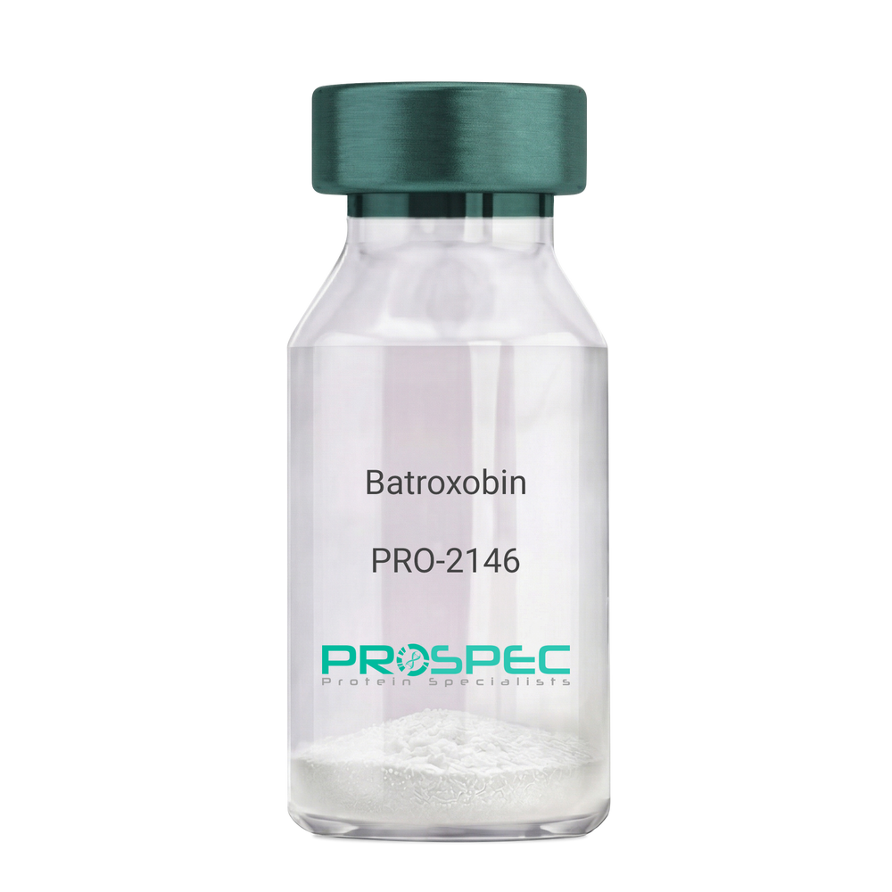
Name :
BatroxobinDescription:
Batroxobin
Thrombin-like enzyme batroxobin, EC 3.4.21.74, BX, Bothrops atrox serine proteinase, Venombin-A, Batroxobin.
Product # :
PRO-2146Price :
Quantity :
Shipping Method :
Shipped at Room temp
More Info
- description
- formulation
- More Info
Description
Batroxobin, isolated from Bothrops atrox snake venom, has an Mw of approximately 43kDa.
Formulation
The Batroxobin protein was lyophilized from a concentrated (1mg/ml) solution with no additives.
More Info
-
Introduction
Batroxobin is a serin protease that reduces fibronogen levels and is originally extracted from snake venom of Bothrops Atrox. Batroxobin is used in defibrinogenation and thrombolysis and also has an effect on c-fos gene and growth factor.
Batroxobin can efficiently restrain proliferation of VSMCs, by blocking the release and uptake of Ca2+, thus influencing [Ca2+]i.
Batroxobin converts fibrinogen to fibrin through the restricted release of fibrinopeptide-A from fibrinogen to promote blood to clot. Unlike thrombin, it is not affected by heparin and hirudin. -
Synonyms
Thrombin-like enzyme batroxobin, EC 3.4.21.74, BX, Bothrops atrox serine proteinase, Venombin-A, Batroxobin.
-
Physical Appearance
Sterile Filtered white lyophilized powder.
-
Stability
Store the lyophilized Batroxobin between 2-8°C. Do not freeze!
-
Solubility
It is recommended to reconstitute the lyophilized Batroxobin in sterile 18MΩ-cm H2O not less than 100µg/ml or more than 10mg/ml solutions.
-
Unit Definition
100BU [Batroxobin Units]=1mg.
ProSpec's products are furnished for LABORATORY RESEARCH USE ONLY. The product may not be used as drugs, agricultural or pesticidal products, food additives or household chemicals.