Skip to content

High-quality research proteins.

  • 1-800-805-6531

  • Mon - Sat 8.00-18.00, Sun - Closed

  • info@prospecbio.com

  • PO Box 6591 East Brunswick NJ 08816

  • Products
    • Cytokines
    • Growth factors
    • Chemokines
    • Cd antigens
    • Neurotrophins
    • Hormones
    • Enzymes
    • Viral antigens
    • Recombinant proteins
    • Natural proteins
    • Monoclonal antibodies
    • Polyclonal antibodies
    • Thermal Packaging
  • About
    • Company
    • Founder
    • Contribution
  • Customers
    • Eastern Asia
    • Eastern Europe
    • North America
    • South America
    • South East Asia
    • Southern Asia
    • Western Europe
  • Ordering
    • Ordering Method
    • Ordering - Credit Card
    • Ordering - PO
    • Shipping Fees
    • FAQs
  • Services
    • DNA Cloning
    • Protein Expression
    • Monoclonal Antibody
    • Fermentation
    • Technology License
  • Contact us
Log in

Country/region

  • USD $ | Albania
  • USD $ | Andorra
  • USD $ | Argentina
  • USD $ | Armenia
  • USD $ | Australia
  • USD $ | Austria
  • USD $ | Azerbaijan
  • USD $ | Belarus
  • USD $ | Belgium
  • USD $ | Bolivia
  • USD $ | Brazil
  • USD $ | Bulgaria
  • USD $ | Cambodia
  • USD $ | Canada
  • USD $ | Chile
  • USD $ | China
  • USD $ | Colombia
  • USD $ | Costa Rica
  • USD $ | Croatia
  • USD $ | Cyprus
  • USD $ | Czechia
  • USD $ | Denmark
  • USD $ | Estonia
  • USD $ | Finland
  • USD $ | France
  • USD $ | Georgia
  • USD $ | Germany
  • USD $ | Greece
  • USD $ | Hong Kong SAR
  • USD $ | Hungary
  • USD $ | Iceland
  • USD $ | India
  • USD $ | Ireland
  • USD $ | Israel
  • USD $ | Italy
  • USD $ | Japan
  • USD $ | Kazakhstan
  • USD $ | Latvia
  • USD $ | Liechtenstein
  • USD $ | Lithuania
  • USD $ | Madagascar
  • USD $ | Malta
  • USD $ | Mexico
  • USD $ | Moldova
  • USD $ | Monaco
  • USD $ | Netherlands
  • USD $ | New Zealand
  • USD $ | North Macedonia
  • USD $ | Norway
  • USD $ | Panama
  • USD $ | Paraguay
  • USD $ | Peru
  • USD $ | Philippines
  • USD $ | Poland
  • USD $ | Portugal
  • USD $ | Romania
  • USD $ | Singapore
  • USD $ | Slovakia
  • USD $ | Slovenia
  • USD $ | South Africa
  • USD $ | South Korea
  • USD $ | Spain
  • USD $ | Sri Lanka
  • USD $ | Sweden
  • USD $ | Switzerland
  • USD $ | Taiwan
  • USD $ | Thailand
  • USD $ | Türkiye
  • USD $ | Ukraine
  • USD $ | United Kingdom
  • USD $ | United States
  • USD $ | Uruguay
  • USD $ | Vatican City
  • USD $ | Venezuela
  • USD $ | Vietnam
  • Facebook
  • Instagram
logoProSpec - Recombinant Protein Manufacturer for academic and R&D industry
Become a distributor
Account Log in
Wishlist
Cart Cart(0)
  • Products
    +
    • Cytokines
    • Growth factors
    • Chemokines
    • Cd antigens
    • Neurotrophins
    • Hormones
    • Enzymes
    • Viral antigens
    • Recombinant proteins
    • Natural proteins
    • Monoclonal antibodies
    • Polyclonal antibodies
    • Thermal Packaging
  • About
    +
    • Company
    • Founder
    • Contribution
  • Customers
    +
    • Eastern Asia
    • Eastern Europe
    • North America
    • South America
    • South East Asia
    • Southern Asia
    • Western Europe
  • Ordering
    +
    • Ordering Method
    • Ordering - Credit Card
    • Ordering - PO
    • Shipping Fees
    • FAQs
  • Services
    +
    • DNA Cloning
    • Protein Expression
    • Monoclonal Antibody
    • Fermentation
    • Technology License
  • Contact us

Item added to your cart

View cart
View All
  • Cytokines
  • Growth factors
  • Chemokines
  • Cd antigens
  • Neurotrophins
  • Hormones
  • Enzymes
  • Viral antigens
  • Recombinant proteins
  • Natural proteins
  • Monoclonal antibodies
  • Polyclonal antibodies
  • Thermal Packaging
  • Tumor Necrosis Factor

    Tumor Necrosis Factor

    View All
  • 4-1BB

    4-1BB

    View All
  • Adiponectin

    Adiponectin

    View All
  • AIF1

    AIF1

    View All
  • Other Cytokines

    Other Cytokines

    View All
  • AITRL

    AITRL

    View All
  • Angiopoietin

    Angiopoietin

    View All
  • Apolipoprotein

    Apolipoprotein

    View All
  • B-Cell Activating Factor

    B-Cell Activating Factor

    View All
  • Beta Defensin

    Beta Defensin

    View All
  • Bone Morphogenetic Protein

    Bone Morphogenetic Protein

    View All
  • B type Natriuretic Peptide

    B type Natriuretic Peptide

    View All
  • BST

    BST

    View All
  • Betacellulin

    Betacellulin

    View All
  • Cardiotrophin

    Cardiotrophin

    View All
  • Activin

    Activin

    View All
  • Fibroblast Growth Factor

    Fibroblast Growth Factor

    View All
  • Other Growth Factors

    Other Growth Factors

    View All
  • CSF

    CSF

    View All
  • CTGF

    CTGF

    View All
  • IGFBP

    IGFBP

    View All
  • VEGF Protein

    VEGF Protein

    View All
  • EGF

    EGF

    View All
  • Epigen

    Epigen

    View All
  • Erythropoietin

    Erythropoietin

    View All
  • Insulin-Like Growth Factor

    Insulin-Like Growth Factor

    View All
  • Growth Hormone

    Growth Hormone

    View All
  • HDGF

    HDGF

    View All
  • Hepatocyte Growth Factor

    Hepatocyte Growth Factor

    View All
  • Insulin

    Insulin

    View All
  • BCA-1/ BLC (CXCL13)

    BCA-1/ BLC (CXCL13)

    View All
  • BRAK (CXCL14)

    BRAK (CXCL14)

    View All
  • C-10 (CCL6)

    C-10 (CCL6)

    View All
  • Other Chemokines

    Other Chemokines

    View All
  • MEC (CCL28)

    MEC (CCL28)

    View All
  • LD78-beta (CCL3L1)

    LD78-beta (CCL3L1)

    View All
  • CTACK (CCL27)

    CTACK (CCL27)

    View All
  • CXCL16

    CXCL16

    View All
  • CXCL17

    CXCL17

    View All
  • Platelet Factor-4 (CXCL4)

    Platelet Factor-4 (CXCL4)

    View All
  • ENA-78 (CXCL5)

    ENA-78 (CXCL5)

    View All
  • Eotaxin (CCL11,24,26)

    Eotaxin (CCL11,24,26)

    View All
  • Exodus-2 (CCL21)

    Exodus-2 (CCL21)

    View All
  • Fractalkine (CX3CL1)

    Fractalkine (CX3CL1)

    View All
  • CXCL6

    CXCL6

    View All
  • Other CD Antigens

    Other CD Antigens

    View All
  • CD14

    CD14

    View All
  • CD164

    CD164

    View All
  • CD1

    CD1

    View All
  • CD2

    CD2

    View All
  • CD200

    CD200

    View All
  • CD207

    CD207

    View All
  • CD226

    CD226

    View All
  • CD23

    CD23

    View All
  • CD244

    CD244

    View All
  • CD247

    CD247

    View All
  • CD27

    CD27

    View All
  • CD274

    CD274

    View All
  • CD300

    CD300

    View All
  • CD33

    CD33

    View All
  • Other Neurotrophins

    Other Neurotrophins

    View All
  • Beta-NGF

    Beta-NGF

    View All
  • BDNF

    BDNF

    View All
  • CDNF

    CDNF

    View All
  • CNTF

    CNTF

    View All
  • GDNF

    GDNF

    View All
  • Glia Maturation Factor

    Glia Maturation Factor

    View All
  • MANF

    MANF

    View All
  • Midkine

    Midkine

    View All
  • Neuroglobin

    Neuroglobin

    View All
  • Neurotrophic factors

    Neurotrophic factors

    View All
  • Neuregulin

    Neuregulin

    View All
  • Neuropilin

    Neuropilin

    View All
  • Pigment Epithelium-Derived Factor

    Pigment Epithelium-Derived Factor

    View All
  • Pleiotrophin

    Pleiotrophin

    View All
  • Peptide Hormones

    Peptide Hormones

    View All
  • Other Hormones

    Other Hormones

    View All
  • HCG

    HCG

    View All
  • Endothelin

    Endothelin

    View All
  • Exendin

    Exendin

    View All
  • FSH

    FSH

    View All
  • Glucagon

    Glucagon

    View All
  • GHRP

    GHRP

    View All
  • GLP

    GLP

    View All
  • Thyrostimulin

    Thyrostimulin

    View All
  • Inhibin A

    Inhibin A

    View All
  • LHRH

    LHRH

    View All
  • Melanotan

    Melanotan

    View All
  • Procalcitonin

    Procalcitonin

    View All
  • PTH

    PTH

    View All
  • Peroxiredoxin

    Peroxiredoxin

    View All
  • Peptidase

    Peptidase

    View All
  • Synthetase

    Synthetase

    View All
  • Transferase

    Transferase

    View All
  • Hydrolase

    Hydrolase

    View All
  • Dehydrogenase

    Dehydrogenase

    View All
  • ACE2

    ACE2

    View All
  • Esterase

    Esterase

    View All
  • Protein Kinases

    Protein Kinases

    View All
  • Other Enzymes

    Other Enzymes

    View All
  • Phosphatase

    Phosphatase

    View All
  • Deaminase

    Deaminase

    View All
  • Oxygenase

    Oxygenase

    View All
  • Adenylate Kinase

    Adenylate Kinase

    View All
  • Lyase

    Lyase

    View All
  • Chagas

    Chagas

    View All
  • SARS

    SARS

    View All
  • Influenza

    Influenza

    View All
  • ASFV

    ASFV

    View All
  • Astrovirus

    Astrovirus

    View All
  • Borrelia

    Borrelia

    View All
  • Helicobacter Pylori

    Helicobacter Pylori

    View All
  • Chikungunya

    Chikungunya

    View All
  • Chlamydia

    Chlamydia

    View All
  • Cytomegalo

    Cytomegalo

    View All
  • Dengue

    Dengue

    View All
  • EBV

    EBV

    View All
  • Ebola

    Ebola

    View All
  • Feline Leukemia Virus

    Feline Leukemia Virus

    View All
  • Hepatitis A

    Hepatitis A

    View All
  • Other

    Other

    View All
  • Actin

    Actin

    View All
  • ADAM

    ADAM

    View All
  • Complement Component

    Complement Component

    View All
  • Ag85

    Ag85

    View All
  • Eukaryotic Translation Initiation Factor

    Eukaryotic Translation Initiation Factor

    View All
  • Anterior Gradient Protein

    Anterior Gradient Protein

    View All
  • Heat Shock Protein

    Heat Shock Protein

    View All
  • Alpha-2-HS-Glycoprotein

    Alpha-2-HS-Glycoprotein

    View All
  • Hemoglobin

    Hemoglobin

    View All
  • Allergy

    Allergy

    View All
  • Anaplasma

    Anaplasma

    View All
  • Angiogenin

    Angiogenin

    View All
  • Ankyrin Repeat Domain

    Ankyrin Repeat Domain

    View All
  • Annexin

    Annexin

    View All
  • Other Natural Proteins

    Other Natural Proteins

    View All
  • Aprotinin

    Aprotinin

    View All
  • Transferrin

    Transferrin

    View All
  • Avidin

    Avidin

    View All
  • Anti Coagulation Factors

    Anti Coagulation Factors

    View All
  • Natural Albumin

    Natural Albumin

    View All
  • Natural Coagulation Factors

    Natural Coagulation Factors

    View All
  • Fibronectin

    Fibronectin

    View All
  • Thrombin

    Thrombin

    View All
  • Anti Human Enzyme

    Anti Human Enzyme

    View All
  • Other Monoclonal Antibodies

    Other Monoclonal Antibodies

    View All
  • Anti Human Cytokine

    Anti Human Cytokine

    View All
  • Anti Human Heat Shock Protein

    Anti Human Heat Shock Protein

    View All
  • Anti Mouse Lymphocyte

    Anti Mouse Lymphocyte

    View All
  • Anti Human Chemokine

    Anti Human Chemokine

    View All
  • Anti Human Lymphocyte

    Anti Human Lymphocyte

    View All
  • Anti Viral Monoclonal

    Anti Viral Monoclonal

    View All
  • Anti Mouse Cytokine

    Anti Mouse Cytokine

    View All
  • Anti-GST Monoclonal

    Anti-GST Monoclonal

    View All
  • Other Polyclonal Antibodies

    Other Polyclonal Antibodies

    View All
  • Anti Viral

    Anti Viral

    View All
  • Anti-GST Monoclonal

    Anti-GST Monoclonal

    View All
Back

Search results

897 results found for “Density Lipoprotein”

Name

Description

Product #

Price

Quantity

Shipping Method

  • View Data Sheet

    Name :

    Dengue Envelope-1 22kDa

    Description:

    Dengue Virus Subtype-1 Envelope 22kDa Recombinant

    Product # :

    DEN-005

    Price :

    Quantity :

    Shipping Method :

    Ice Icon

    Shipped with Ice Packs

    Add To Cart

    More Info

    • description
    • formulation
    • purity
    • More Info

    Description

    The E.coli derived recombinant 22 kDa protein is genetically engineered peptide which is derived from Dengue Type-1 N-terminus Envelope immunodeterminant regions and fused with a 6 aa His Tag. This region also contains a common antigen for Dengue type 1, 2 and 3.

    Formulation

    Phosphate buffered saline, pH-7.4.

    Purity

    Protein is >95% pure as determined by 10% PAGE (coomassie staining).

    More Info

    • Introduction

      Caused by one of four closely related virus serotypes of the genus Flavivirus, family Flaviviridae, each serotype is sufficiently different that there is no cross-protection and epidemics caused by multiple serotypes (hyperendemicity) can occur. In cell culture experiments and mice Morpholino antisense oligos have shown specific activity against Dengue virus.

    • Stability

      Dengue Envelope ST1 although stable at 4°C for 1 week, should be stored below -18°C. Please prevent freeze thaw cycles.

    • Applications

      Each laboratory should determine an optimum working titer for use in its particular application.

    • Purification Method

      Purified by proprietary chromatographic technique.

    ProSpec's products are furnished for LABORATORY RESEARCH USE ONLY. The product may not be used as drugs, agricultural or pesticidal products, food additives or household chemicals.

    Dengue Envelope St1
  • View Data Sheet

    Name :

    Der F1

    Description:

    Der F1 Mosaic Protein Recombinant

    Product # :

    ALR-004

    Price :

    Quantity :

    Shipping Method :

    Ice Icon

    Shipped with Ice Packs

    Add To Cart

    More Info

    • description
    • source
    • formulation
    • purity
    • More Info

    Description

    The E.Coli derived recombinant protein contains the Dermatophagoides farina Der F1 full length protein (a.a. 1-320) and fused to a 6 His Tag at C-terminus, having a total Mw of 36kDa, pI 5.88.

    Source

    E.Coli.

    Formulation

    60mM NaCl, 50mM Tris-HCl pH 8.0 and 1.2M Urea.

    Purity

    Protein is >95% pure as determined by 10% PAGE (coomassie staining) and RP-HPLC.

    More Info

    • Introduction

      DERF1 is a thiol protease that hydrolyzes proteins, with a preference for Phe or basic residues. DERF1 is a C1 peptidase family member. DERF1 has extensive endopeptidase specificity. DERF1 causes an allergic reaction in humans. Common symptoms of mite allergy are bronchial asthma, allergic rhinitis and conjunctivitis.

    • Physical Appearance

      Sterile filtered colorless solution.

    • Stability

      Der F1 although stable at 4°C for 1 week, should be stored below -18°C. Please prevent freeze thaw cycles.

    • Purification Method

      Purified by proprietary chromatographic technique.

    ProSpec's products are furnished for LABORATORY RESEARCH USE ONLY. The product may not be used as drugs, agricultural or pesticidal products, food additives or household chemicals.

    Der F1
  • View Data Sheet

    Name :

    Borrelia Bavariensis DbpA

    Description:

    Borrelia Bavariensis DbpA Recombinant

    Decorin binding protein A, dbpA.

    Product # :

    BOR-023

    Price :

    Quantity :

    Shipping Method :

    Ice Icon

    Shipped with Ice Packs

    Add To Cart

    More Info

    • description
    • source
    • formulation
    • purity
    • More Info

    Description

    Recombinant Borrelia Bavariensis DbpA produced in E.coli is a non-glycosylated, polypeptide chain having a calculated molecular mass of 18kDa. Borrelia Bavariensis DbpA is expressed with a 10x His tag at N-terminus and purified by proprietary chromatographic techniques.

    Source

    Escherichia Coli.

    Formulation

    Borrelia Bavariensis DbpA is supplied in 20mM HEPES buffer pH-7.6, 250mM NaCl and 20% glycerol.

    Purity

    Greater than 80.0% as determined by SDS-PAGE.

    More Info

    • Introduction

      Borrelia is a part of a genus of bacteria of the spirochete phylum. Borrelia causes borreliosis, which is a zoonotic, vector-borne disease transmitted mainly by ticks and some by lice, depending on the species. Borreliella bavariensis is one of the pathogens of the Borreliella burgdorferi sensu lato complex which causes Lyme disease. Decorin binding protein A (DbpA), also known as Osp17 or p17 is expressed during the mammalian phase of infection. DbpA is binds the pathogen to host collagen-associated proteoglycan decorin.

    • Synonyms

      Decorin binding protein A, dbpA.

    • Physical Appearance

      Sterile Filtered clear solution.

    • Stability

      Store at 4°C if entire vial will be used within 2-4 weeks. Store, frozen at -20°C for longer periods of time. Avoid multiple freeze-thaw cycles.

    • Immunological Functions

      1. Binds IgG- and IgM-type human antibodies.2. Immunodot test with Lyme disease positive/negative patient samples.

    • Applications

      Western blot with Patient sample.

    ProSpec's products are furnished for LABORATORY RESEARCH USE ONLY. The product may not be used as drugs, agricultural or pesticidal products, food additives or household chemicals.

    Borrelia Bavarriensis Dbpa
  • View Data Sheet

    Name :

    TNNI3 Human Chimeric

    Description:

    Cardiac Troponin-I Chimeric Human Recombinant

    Troponin I cardiac muscle, Cardiac troponin I, TNNI3, TNNC1, CMH7, RCM1, cTnI, CMD2A, MGC116817.

    Product # :

    PRO-2790

    Price :

    Quantity :

    Shipping Method :

    Room Temp Icon

    Shipped at Room temp

    Add To Cart

    More Info

    • description
    • source
    • formulation
    • purity
    • More Info

    Description

    TNNI3 Human Chimeric produced in E.Coli is a single, non-glycosylated polypeptide chain (28-110 a.a.) and having a molecular mass of 29072 Dalton.

    Source

    Escherichia Coli.

    Formulation

    TNNI3 was lyophilized in 50mM Tris-HCl, 5mM Calcium chloride, 0.7M KCl and 0.1% 2-mercaptoethanol, pH 7.5

    Purity

    Greater than 95.0% as determined by SDS-PAGE.

    More Info

    • Synonyms

      Troponin I cardiac muscle, Cardiac troponin I, TNNI3, TNNC1, CMH7, RCM1, cTnI, CMD2A, MGC116817.

    • Physical Appearance

      Sterile Filtered White lyophilized (freeze-dried) powder.

    • Stability

      Lyophilized Cardiac Troponin-I Chimeric although stable at room temperature for 3 weeks, should be stored desiccated below -18°C. Upon reconstitution TNNI3 should be stored at 4°C between 2-7 days and for future use below -18°C. For long term storage it is recommended to add a carrier protein (0.1% HSA or BSA). Please prevent freeze-thaw cycles.

    • Solubility

      It is recommended to reconstitute the lyophilized TNNI3 in buffer containing BSA not less than 100µg/ml, which can then be further diluted to other aqueous solutions.

    • Background

      Troponin I (TNNI3) is a crucial regulatory protein in cardiac muscle, playing a central role in the regulation of muscle contraction. Understanding the structure and function of TNNI3 is essential for unraveling the complexities of cardiac muscle physiology and exploring therapeutic interventions for cardiac diseases. Chimeric TNNI3 proteins, which combine segments from different isoforms or species, offer a unique opportunity to investigate the role of specific regions in TNNI3 function and to potentially develop novel therapies. This research aims to provide a comprehensive exploration of chimeric TNNI3 proteins, elucidating their functions, structural significance, and potential applications in cardiology and biomedical research.

      The primary objective of this research is to elucidate the functional significance of chimeric TNNI3 proteins in cardiac muscle. In vitro and ex vivo experiments, utilizing engineered chimeric TNNI3 constructs and cardiac tissue models, will be conducted to investigate how these proteins influence muscle contractility, calcium sensitivity, and response to pathological conditions. Understanding these mechanisms is fundamental for deciphering the roles of specific TNNI3 regions in cardiac muscle function.

      The second objective is to assess the therapeutic potential of chimeric TNNI3 proteins in cardiac diseases. Experimental studies involving animal models and cellular systems will explore the use of chimeric TNNI3 proteins as potential therapeutic agents for heart conditions. These investigations may provide valuable insights into novel treatment strategies targeting cardiac muscle function.

      The third objective is to explore the broader applications of chimeric TNNI3 proteins in biotechnology and drug development. Research will investigate the use of chimeric TNNI3-expressing cells and tissues as models for studying cardiac disorders and for developing innovative approaches in regenerative medicine and pharmacology.

      By delving into the functions and roles of chimeric TNNI3 proteins, this research aims to expand our knowledge of cardiac muscle physiology, its implications for cardiac diseases, and its potential applications in cardiology, biotechnology, and drug development

    ProSpec's products are furnished for LABORATORY RESEARCH USE ONLY. The product may not be used as drugs, agricultural or pesticidal products, food additives or household chemicals.

    Tnni3 Chimeric
  • View Data Sheet

    Name :

    GHRL Protein

    Description:

    Ghrelin Human

    Appetite-regulating hormone precursor, Growth hormone secretagogue, Growth hormone-releasing peptide, GHRP, Motilin-related peptide, M46 protein, Ghrelin, Obestatin, MTLRP.

    Product # :

    HOR-297

    Price :

    Quantity :

    Shipping Method :

    Room Temp Icon

    Shipped at Room temp

    Add To Cart

    More Info

    • description
    • formulation
    • purity
    • More Info

    Description

    Ghrelin Human contains 28 amino acids and a total molecular mass of 3370.9 Dalton and a molecular formula of C149H249N47O42.The GHRL is purified by proprietary chromatographic techniques.

    Formulation

    GHRL was lyophilized without additives.

    Purity

    Greater than 97% as determined by:
    (a) Analysis by RP-HPLC.
    (b) Analysis by SDS-PAGE.

    More Info

    • Introduction

      Obestatin is a hormone that is produced in the cells lining the stomach and small intestine of several mammals including humans; it drastically reduces appetite in mice and is expected to do the same in humans. Obestatin is a peptide hormone - a relatively small protein. It is encoded by the same gene that also encodes ghrelin, a peptide hormone that increases appetite. The protein produced by that gene breaks into two smaller peptides, ghrelin and obestatin. Ghrelin is an endogenous ligand for the growth hormone secretagogue receptor and is involved in regulating growth hormone release. Ghrelin is derived from a preprohormone called preproghrelin, which also generates a second peptide called obestatin. Ghrelin is an endogenous ligand for the orphan G protein-coupled receptor GPR39 and is involved in satiety and decreased food intake.

    • Synonyms

      Appetite-regulating hormone precursor, Growth hormone secretagogue, Growth hormone-releasing peptide, GHRP, Motilin-related peptide, M46 protein, Ghrelin, Obestatin, MTLRP.

    • Physical Appearance

      Sterile Filtered Yellowish lyophilized (freeze-dried) powder that may appear as a gel form.

    • Stability

      Store the lyophilized Ghrelin at -20°C. Aliquot the product after reconstitution to avoid repeated freezing/thawing cycles. Reconstituted GHRL can be stored at 4°C for a limited period of time; it does not show any change after two weeks at 4°C.

    • Solubility

      Add deionized water to a working concentration approximately 0.5mg/1ml and let the lyophilized pellet dissolve completely.

    • Amino Acid Sequence

      Gly-Ser-Ser(n-octanoyl)-Phe-Leu-Ser-Pro-Glu-His-Gln-Arg-Val-Gln-Gln-Arg-Lys-Glu-Ser-Lys-Lys-Pro-Pro-Ala-Lys-Leu-Gln-Pro-Arg-OH.

    ProSpec's products are furnished for LABORATORY RESEARCH USE ONLY. The product may not be used as drugs, agricultural or pesticidal products, food additives or household chemicals.

    Ghrelin Human
  • View Data Sheet

    Name :

    SHBG Protein

    Description:

    Sex Hormone-Binding Globulin Human

    Sex hormone-binding globulin, SHBG, Sex steroid-binding protein, SBP, Testis-specific androgen-binding protein, ABP, Testosterone-estradiol-binding globulin, TeBG, Testosterone-estrogen-binding globulin, SHBG.

    Product # :

    PRO-2757

    Price :

    Quantity :

    Shipping Method :

    Ice Icon

    Shipped with Ice Packs

    Add To Cart

    More Info

    • description
    • source
    • formulation
    • More Info

    Description

    SHBG is a protein of approximately 45kD.

    Source

    Human serum.

    Formulation

    The protein is supplied in 0.01M HEPES, PH 7.4 and 0.15M NaCl.

    More Info

    • Introduction

      Sex-hormone-binding globulin (SHBG) is a beta-globulin which specifically binds steroid hormones; it is involved in the transport of sex steroids in plasma. The main site of SHBG synthesis is assumed to be the hepatocytes. The production of SHBG is regulated by androgen/estrogen balance, thyroid hormones, insulin and dietary factors, among others. The concentration of SHBG is a key factor regulating their distribution between protein-bound and free states. SHBG concentration determination is primarily significant in the evaluation of mild disorders of androgen metabolism and it allows detection of women with hirsutism who are likely to react to estrogen therapy. Testosterone/SHBG-ratios correlate well with both measured and calculated values for free testosterone thus aid to distinguish between subjects with excessive androgen activity and normal individuals. SHBG gene polymorphisms are linked with polycystic ovary syndrome and type 2 diabetes mellitus.

    • Synonyms

      Sex hormone-binding globulin, SHBG, Sex steroid-binding protein, SBP, Testis-specific androgen-binding protein, ABP, Testosterone-estradiol-binding globulin, TeBG, Testosterone-estrogen-binding globulin, SHBG.

    • Physical Appearance

      Streile filtered colorless solution.

    • Stability

      Upon arrival, Store at -20°C. Please prevent freeze-thaw cycles.

    • Human Virus Test

      FDA approved Plasma from each donor has been tested and found negative for antibodies to HIV-1 & 2, HCV, HBsAG, Parvovirus B19, HBc, HBV, HIV and Syphilis.

    ProSpec's products are furnished for LABORATORY RESEARCH USE ONLY. The product may not be used as drugs, agricultural or pesticidal products, food additives or household chemicals.

    Shbg Protein
  • View Data Sheet

    Name :

    Thromboplastin Bovine

    Description:

    Thromboplastin Bovine

    Tissue factor, Coagulation factor III, Thromboplastin, CD142, TF, F3, TFA.

    Product # :

    PRO-2760

    Price :

    Quantity :

    Shipping Method :

    Room Temp Icon

    Shipped at Room temp

    Add To Cart

    More Info

    • description
    • source
    • formulation
    • More Info

    Description

    Thromboplastin bovine native

    Source

    Bovine Lung.

    Formulation

    The bovine thromboplastin was lyophilized with no additives.

    More Info

    • Introduction

      Tissue factor is well-known as the main cellular initiator of blood coagulation. The Tissue factor gene encodes coagulation factor III which is a cell surface glycoprotein that enables cells to initiate the blood coagulation cascades, and functions as the high-affinity receptor for the coagulation factor VII. Following vessel injury, the Tissue Factor and Factor VIIa complex activates the coagulation protease cascade, which leads to fibrin deposition and activation of platelets. The ensuing complex presents a catalytic event, which is responsible for initiation of the coagulation protease cascades by specific limited proteolysis. Therefore, Tissue factor has a role in normal hemostasis by initiating the cell-surface assembly and propagation of the coagulation protease cascade. Tissue Factor can also be stimulated by the inflammatory mediators interleukin 1 and TNF, as well as by endotoxin, to appear on monocytes and vascular endothelial cells as a component of cellular immune response.
      Tissue factor is the only one in the coagulation pathway for which a congenital deficiency has not been described. Certain levels of Tissue Factor are essential for the maintained viability and growth of endothelium and Tissue Factor-expressing tumor cells. Additionally, abnormal Tissue Factor expression inside the vasculature initiates life threatening thrombosis in various diseases, for example sepsis, atherosclerosis, and cancer. Alternative spliced Tissue Factor expression advances tumor growth, and is linked to increased tumor cell proliferation and angiogenesis in pancreatic cancer.

    • Synonyms

      Tissue factor, Coagulation factor III, Thromboplastin, CD142, TF, F3, TFA.

    • Physical Appearance

      Sterile Filtered White lyophilized (freeze-dried) powder.

    • Stability

      Lyophilized Bovine Thromboplastin although stable at room temperature for 3 weeks, should be stored desiccated below -18C. Upon reconstitution Prothrombin should be stored at 4C between 2-7 days and for future use below -18C.For long term storage it is recommended to add a carrier protein (0.1% HSA or BSA). Please prevent freeze-thaw cycles

    • Solubility

      It is recommended to reconstitute the lyophilized Bovine Thromboplastin in sterile 0.9% NaCl

    ProSpec's products are furnished for LABORATORY RESEARCH USE ONLY. The product may not be used as drugs, agricultural or pesticidal products, food additives or household chemicals.

    Thromboplastin Bovine
  • View Data Sheet

    Name :

    AMBP

    Description:

    Alpha-1 Microglobulin Human Recombinant

    Alpha-1-microglobulin/bikunin precursor, HCP, ITIL, ITI, EDC1, HI30, IATIL, ITILC, UTI, A1M, bikunin, complex-forming glycoprotein heterogeneous in charge, growth-inhibiting protein 19, inter-alpha-trypsin inhibitor light chain, protein AMBP, protein HC, trypstatin, uristatin, uronic-acid-rich protein.

    Product # :

    PRO-957

    Price :

    Quantity :

    Shipping Method :

    Ice Icon

    Shipped with Ice Packs

    Add To Cart

    More Info

    • description
    • source
    • formulation
    • purity
    • More Info

    Description

    AMBP Human Recombinant produced in E.coli is a single, non-glycosylated polypeptide chain containing 205 amino acids (20-203) and having a molecular mass of 23.1 kDa.AMBP is fused to a 21 amino acid His-tag at N-terminus & purified by proprietary chromatographic techniques.

    Source

    E.coli.

    Formulation

    The AMBP solution (0.5mg/ml) contains 20mM Tris-HCl buffer (pH 8.0), 1mM DTT, 0.1M NaCl and 10% glycerol.

    Purity

    Greater than 90% as determined by SDS-PAGE.

    More Info

    • Introduction

      Alpha 1-microglobulin (A1M) is a lipocalin superfamily member (kernal lipocalins). A1M is a low molecular weight protein component of plasma. A1M is distributed in plasma and extravascular compartments of all organs. Alpha-1 Microglobulin is found in mammals, birds, amphibians and fish. The primary sites of A1M synthesis are the liver and the kidney. Around the opening of the lipocalin pocket three lysyl residues are situated; those residues carry yellow-brown modification derived from the binding and degradation of heme and kynurenin (a tryptophan metabolite). A1-Microglobulin’s reductase and dehydrogenase have broad biological substrate specificity properties due to its’ free cysteine side-chain which is located in a flexible loop. Alpha-1-microglobulin is glycosylated by three separate carbohydrate chains: two complex carbohydrates which are N-linked to asparagines at residues 17 and 96, and the other simple carbohydrate which is O-linked to threonine at position 5. The carbohydrates comprise 22% of the total molecular mass of the protein. The glycosylation varies between species.
      A1M exists in two forms- a free form and complexed to other macromolecules: in humans- complexed to immunoglobulin A (IgA), in rat- complexed to alpha-1-inhibitor-3. Free A1M is exceptionally heterogeneous in charge (therefore also known as protein HC), and is found tightly linked to a chromophore. The free Alpha-1-microglobulin is a monomeric protein composed of one 188 residue polypeptide and contains three cysteines, two of which (residues 75 and 173) form a conserved intra-molecular disulphide link. The chromophoric group is covalently bound to the free cysteine residue at position 34. A1M binds retinol as a major ligand, but this is probably distinct from its covalent chromophore.
      Half of all human plasma A1M (approximately 0.03mg/ml) forms a 1:1 complex with about 5% of plasma immunoglobulin A. The resulting macromolecular complexes’ molecular weight is 200000, and a plasma concentration of 0.3mg/ml. The complex can exhibit both antibody activity and affect many of the biological actions of free Alpha-1-microglobulin.
      Alpha-1-microglobulin was first discovered in pathological human urine.
      It was suggested that A1M might be involved in tissue defense against reactive oxygen species, oxidation by heme and kynurenin. Evidence also suggests that A1M functions in the regulation of the immune system. Other functions include inhibition of stimulation of cultured lymphocytes by protein antigens; induction of cell division of lymphocytes, a mitogenic effect that can either be enhanced or inhibited by the action of other plasma components; inhibition of neutrophil granulocyte migration in vitro; and inhibition of chemotaxis.

    • Synonyms

      Alpha-1-microglobulin/bikunin precursor, HCP, ITIL, ITI, EDC1, HI30, IATIL, ITILC, UTI, A1M, bikunin, complex-forming glycoprotein heterogeneous in charge, growth-inhibiting protein 19, inter-alpha-trypsin inhibitor light chain, protein AMBP, protein HC, trypstatin, uristatin,
      uronic-acid-rich protein.

    • Physical Appearance

      Sterile Filtered colorless solution.

    • Stability

      Store at 4°C if entire vial will be used within 2-4 weeks. Store, frozen at -20°C for longer periods of time. For long term storage it is recommended to add a carrier protein (0.1% HSA or BSA).Avoid multiple freeze-thaw cycles.

    • Amino Acid Sequence

      MGSSHHHHHH SSGLVPRGSH MGPVPTPPDN IQVQENFNIS RIYGKWYNLA IGSTCPWLKK IMDRMTVSTL VLGEGATEAE ISMTSTRWRK GVCEETSGAY EKTDTDGKFL YHKSKWNITM ESYVVHTNYD EYAIFLTKKF SRHHGPTITA KLYGRAPQLR ETLLQDFRVV AQGVGIPEDS IFTMADRGEC VPGEQEPEPI LIPRV.

    ProSpec's products are furnished for LABORATORY RESEARCH USE ONLY. The product may not be used as drugs, agricultural or pesticidal products, food additives or household chemicals.

    Ambp Human
  • View Data Sheet

    Name :

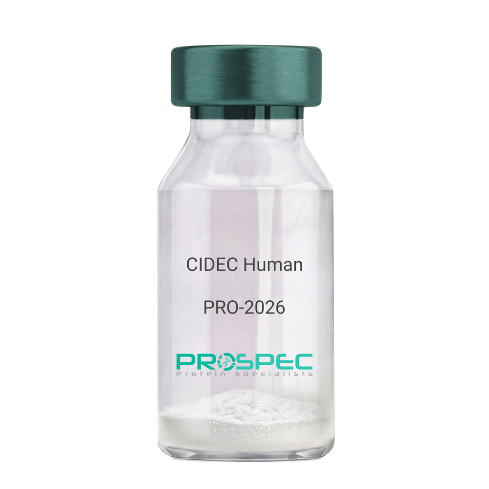
    CIDEC Human

    Description:

    Cell Death-Inducing DFFA-Like Effector C Human Recombinant

    Cell Death-Inducing DFFA-Like Effector C, FSP27, CIDE3, FPLD5, Cell Death-Inducing DFFA-Like Effector Protein C, Fat-Specific Protein FSP27 Homolog, Cell Death Activator CIDE-3, Fat Specific Protein 27, CIDE-3, CIDEC.

    Product # :

    PRO-2026

    Price :

    Quantity :

    Shipping Method :

    Ice Icon

    Shipped with Ice Packs

    Add To Cart

    More Info

    • description
    • source
    • formulation
    • purity
    • More Info

    Description

    CIDEC Human Recombinant produced in E.Coli is a single, non-glycosylated, polypeptide chain (Glu2-Gln238) containing 247 amino acids including a 10 aa His tag at N-terminus. The total calculated molecular mass is 28kDa.

    Source

    Escherichia Coli.

    Formulation

    CIDEC filtered (0.4µm) solution at a concentration of 0.4mg/ml in 30mM acetate buffer and 10mM dithiothreitol, pH 4.

    Purity

    Greater than 95.0% as determined by SDS-PAGE.

    More Info

    • Introduction

      Cell Death-Inducing DFFA-Like Effector C (CIDEC) belongs to the cell death-inducing DNA fragmentation factor-like effector family, whose members have significant roles in apoptosis. CIDEC is expressed mainly in adipocytes, intestine, heart, stomach, and weakly in the brain, kidney and liver. CIDEC overexpression in preadipocytes induces apoptosis. CIDEC regulates enlargement of lipid droplets.

    • Synonyms

      Cell Death-Inducing DFFA-Like Effector C, FSP27, CIDE3, FPLD5, Cell Death-Inducing DFFA-Like Effector Protein C, Fat-Specific Protein FSP27 Homolog, Cell Death Activator CIDE-3, Fat Specific Protein 27, CIDE-3, CIDEC.

    • Physical Appearance

      Filtered colorless solution.

    • Stability

      Store at 4°C if entire vial will be used within 2-4 weeks. Store, frozen at -20°C for longer periods of time. For long term storage it is recommended to add a carrier protein (0.1% HSA or BSA).Avoid multiple freeze-thaw cycles.

    • Amino Acid Sequence

      MKHHHHHHASEYAMKSLSLL YPKSLSRHVS VRTSVVTQQL LSEPSPKAPR ARPCRVSTAD RSVRKGIMAY SLEDLLLKVR DTLMLADKPF FLVLEEDGTT VETEEYFQAL AGDTVFMVLQ KGQKWQPPSE QGTRHPLSLS HKPAKKIDVA RVTFDLYKLN PQDFIGCLNV KATFYDTYSL SYDLHCCGAK RIMKEAFRWA LFSMQATGHV LLGTSCYLQQ LLDATEEGQP PKGKASSLIP TCLKILQ.

    ProSpec's products are furnished for LABORATORY RESEARCH USE ONLY. The product may not be used as drugs, agricultural or pesticidal products, food additives or household chemicals.

    Cidec Human
  • View Data Sheet

    Name :

    RPL5 Human

    Description:

    Ribosomal Protein L5 Human Recombinant

    60S ribosomal protein L5, RPL5, MSTP030, Ribosomal protein L5, DBA6, L5, MSTP030.

    Product # :

    PRO-2058

    Price :

    Quantity :

    Shipping Method :

    Ice Icon

    Shipped with Ice Packs

    Add To Cart

    More Info

    • description
    • source
    • formulation
    • purity
    • More Info

    Description

    RPL5 Human Recombinant produced in E.Coli is a single, non-glycosylated polypeptide chain containing 320 amino acids (1-297 a.a.) and having a molecular mass of 36.8kDa.RPL5 is fused to a 23 amino acid His-tag at N-terminus & purified by proprietary chromatographic techniques.

    Source

    Escherichia Coli.

    Formulation

    RPL5 protein solution (0.25mg/ml) containing 20mM Tris-HCl buffer (pH8.0), 0.15M NaCl, 10% glycerol and 1mM DTT.

    Purity

    Greater than 85% as determined by SDS-PAGE.

    More Info

    • Introduction

      Ribosomal Protein L5, also known as RPL5 is a part of the ribosomal protein L18P family. RPL5 which binds 5S RNA is necessary for rRNA maturation and structure of the 60S ribosomal subunits.

    • Synonyms

      60S ribosomal protein L5, RPL5, MSTP030, Ribosomal protein L5, DBA6, L5, MSTP030.

    • Physical Appearance

      Sterile Filtered clear solution.

    • Stability

      Store at 4°C if entire vial will be used within 2-4 weeks. Store, frozen at -20°C for longer periods of time.For long term storage it is recommended to add a carrier protein (0.1% HSA or BSA).Please avoid freeze thaw cycles.

    • Amino Acid Sequence

      MGSSHHHHHH SSGLVPRGSH MGSMGFVKVV KNKAYFKRYQ VKFRRRREGK TDYYARKRLV IQDKNKYNTP KYRMIVRVTN RDIICQIAYA RIEGDMIVCA AYAHELPKYG VKVGLTNYAA AYCTGLLLAR RLLNRFGMDK IYEGQVEVTG DEYNVESIDG QPGAFTCYLD AGLARTTTGN KVFGALKGAV DGGLSIPHST KRFPGYDSES KEFNAEVHRK HIMGQNVADY MRYLMEEDED AYKKQFSQYI KNSVTPDMME EMYKKAHAAI RENPVYEKKP KKEVKKKRWN RPKMSLAQKK DRVAQKKASF LRAQERAAES.

    ProSpec's products are furnished for LABORATORY RESEARCH USE ONLY. The product may not be used as drugs, agricultural or pesticidal products, food additives or household chemicals.

    Rpl5 Human
  • View Data Sheet

    Name :

    ANGPT1 Human

    Description:

    Angiopoietin-1 Human Recombinant

    Angiopoietin 1, KIAA0003, ANG-1, AGP1, AGPT.

    Product # :

    CYT-074

    Price :

    Quantity :

    Shipping Method :

    Ice Icon

    Shipped with Ice Packs

    Add To Cart

    More Info

    • description
    • source
    • formulation
    • purity
    • More Info
    • sds-page

    Description

    ANGPT1 Human Recombinant produced in E.coli is a single, non-glycosylated polypeptide chain containing 480 amino acids (20-498) and having a molecular mass of 55.6 kDa.The ANGPT1 is purified by proprietary chromatographic techniques.

    Source

    E.coli.

    Formulation

    The ANGPT1 solution (1mg/ml) contains 20mM Tris-HCl buffer (pH 8.0), 1M Urea and 5% glycerol.

    Purity

    Greater than 85% as determined by SDS-PAGE.

    sds-page

    ANGPT1-sds-page - Product image 1

    More Info

    • Introduction

      ANGPT1 is an angiogenic factor which intervenes in blood vessel maturation and takes part in endothelial development. ANGPT1 is a secreted ligand for Tie-2, a cell surface receptor tyrosine kinase expressed in endothelial and hemopoietic cells. The glycosylated ANGPT1 protein has a coiled-coil region in the amino terminus and a fibrinogen-like domain at the carboxy terminus.

    • Synonyms

      Angiopoietin 1, KIAA0003, ANG-1, AGP1, AGPT.

    • Physical Appearance

      Sterile Filtered colorless solution.

    • Stability

      Store at 4°C if entire vial will be used within 2-4 weeks. Store, frozen at -20°C for longer periods of time. For long term storage it is recommended to add a carrier protein (0.1% HSA or BSA).Avoid multiple freeze-thaw cycles.

    • Amino Acid Sequence

      MSNQRRSPEN SGRRYNRIQH GQCAYTFILP EHDGNCREST TDQYNTNALQ RDAPHVEPDF SSQKLQHLEH VMENYTQWLQ KLENYIVENM KSEMAQIQQN AVQNHTATML EIGTSLLSQT AEQTRKLTDV ETQVLNQTSR LEIQLLENSL STYKLEKQLL QQTNEILKIH EKNSLLEHKI LEMEGKHKEE LDTLKEEKEN LQGLVTRQTY IIQELEKQLN RATTNNSVLQ KQQLELMDTV HNLVNLCTKE GVLLKGGKRE EEKPFRDCAD VYQAGFNKSG IYTIYINNMP EPKKVFCNMD VNGGGWTVIQ HREDGSLDFQ RGWKEYKMGF GNPSGEYWLG NEFIFAITSQ RQYMLRIELM DWEGNRAYSQ YDRFHIGNEK QNYRLYLKGH TGTAGKQSSL ILHGADFSTK DADNDNCMCK CALMLTGGWW FDACGPSNLN GMFYTAGQNH GKLNGIKWHY FKGPSYSLRS TTMMIRPLDF

    • Background

      This factor was found in the conditioned medium of the human neuroepithelioma cell line SHEP1 and the mouse myoblast cell line C2C12ras. The cDNA encoding a protein of 498 amino acids was isolated by using a secretion-trap expression cloning procedure exploiting the presence of a signal sequence present in growth factors that are secreted by producer cells (Davis et al, 1996). Murine and human factors show 97 % identity at the amino acid level. For a related factor see: CDT6. The human gene has been mapped to chromosome 8q22.3-q23 (Cheung et al, 1998).

      The expression of angiopoietin-1 mRNA is downregulated by PDGF, EGF, IL1-beta, and TGF-beta (Enholm et al, 1997).

      Angiopoietin-1 is a ligand for the receptor-like tyrosine kinase designated TIE-2 (Davis et al, 1996). Binding of Angiopoietin-1 to its receptor induces tyrosine phosphorylation of the cytoplasmic receptor domain. A naturally occuring antagonist of Angiopoietin-1 binding to TIE-2 is Angiopoietin-2. Angiopoietin-1 also binds to the TIE-1 receptor and this interaction appears to be critical for the development of the right-hand side venous system. It is dispensable for the left-hand side venous system, suggesting that right-hand and left-hand side vascular networks are established early before asymmetrical features of the network become morphologically discernible Loughna and Sato, 2001).

      Angiopoietin-1 does not directly promote the growth of cultured endothelial cells. Angiopoietin-1 is chemotactic for endothelial cells (Witzenbichler et al, 1998). Excess soluble TIE-2 receptors abolish the chemotactic response of endothelial cells toward angiopoietin-1. Angiopoietin-1 has been shown to counteract cell death by apoptosis in cultured endothelial cells (Holash et al, 1999). Angiopoietin-1 also acts as an apoptosis survival factor for endothelial cells and this effect is augmented by the presence of VEGF (Kwak et al, 1999).

      Angiopoietin-2 dose-dependently blocks directed migration toward Angiopoietin-1. Carlson et al (2001) have shown that Ang-1 binds rather selectively to vitronectin and that Ang-1 can directly support adhesion of human umbilical vein endothelial cells and fibroblasts in a process mediated by integrins.

      The physiologic roles of Angiopoietin-1 and its receptor are limited to angiogenic processes that occur subsequent to the earlier vasculogenic and angiogenic actions of the VEGF family and their receptors. However, it is unlike most of the known angiogenesis factors such as VEGF and other classical endothelial cell growth factors in that addition of the factor to cultures of endothelial cells does not directly promote cell growth even though the TIE-2 receptor becomes activated. Angiopoietin-1 also appears to be incapable of inducing the formation of tubules by endothelial cells.

      Angiopoietins can potentiate the effects of other angiogenic cytokines. An investigation of the impact of angiopoietins on neovascularization in vivo in the cornea micropocket assay of neovascularization demonstrates that neither Angiopoietin-1 nor Angiopoietin-2 alone promote neovascularization. Holash et al (1999) have shown that a subset of tumors initially grows by coopting existing host vessels. Regression of these vessels via a process that involves disruption of interactions between endothelial cells and smooth muscle cells as well as cell death by apoptosis of endothelial cells first causes loss of tumour cells before angiogenesis begins at the tumor margin and the tumor is rescued under the influence of VEGF, Angiopoietin-1, and probably other angiogenic stimuli.

      The embryonic expression pattern of Angiopoietin-1 suggests that it plays a particularly important role in the developing heart. Angiopoietin-1 is expressed highly in the myocardial wall surrounding the endocardium expressing TIE-2. Expression of Angiopoietin-1 becomes much more widespread later in development.

      ANGPT1 Protein has a total Mw of 55.6kDa.

      What is the source or expression system of ANGPT1 Protein?
      Escherichia Coli.

      What is the Purity of ANGPT1 Protein?
      ANGPT1 Protein is >85% pure as determined by SDS-PAGE.

      What is the Biological Activity of ANGPT1 Protein?
      The biological functionality of ANGPT1 Protein will be determined in the future.

      What is the amino acid sequence of ANGPT1 Protein?
      MSNQRRSPEN SGRRYNRIQH GQCAYTFILP EHDGNCREST TDQYNTNALQ RDAPHVEPDF SSQKLQHLEH VMENYTQWLQ KLENYIVENM KSEMAQIQQN AVQNHTATML EIGTSLLSQT AEQTRKLTDV ETQVLNQTSR LEIQLLENSL STYKLEKQLL QQTNEILKIH EKNSLLEHKI LEMEGKHKEE LDTLKEEKEN LQGLVTRQTY IIQELEKQLN RATTNNSVLQ KQQLELMDTV HNLVNLCTKE GVLLKGGKRE EEKPFRDCAD VYQAGFNKSG IYTIYINNMP EPKKVFCNMD VNGGGWTVIQ HREDGSLDFQ RGWKEYKMGF GNPSGEYWLG NEFIFAITSQ RQYMLRIELM DWEGNRAYSQ YDRFHIGNEK QNYRLYLKGH TGTAGKQSSL ILHGADFSTK DADNDNCMCK CALMLTGGWW FDACGPSNLN GMFYTAGQNH GKLNGIKWHY FKGPSYSLRS TTMMIRPLDF

      What applications can ANGPT1 Protein be used in?
      ANGPT1 Protein can probably be used in western blot, ELISA and Lateral Flow.

      What is the endotoxin level for ANGPT1 Protein?
      The endotoxin level is minimal, ANGPT1 Protein was purified using conventional chromatography techniques.

    ProSpec's products are furnished for LABORATORY RESEARCH USE ONLY. The product may not be used as drugs, agricultural or pesticidal products, food additives or household chemicals.

    Angpt1 Human
  • View Data Sheet

    Name :

    DNAL4 Human

    Description:

    Dynein Axonemal Light Chain 4 Human Recombinant

    Dynein light chain 4 axonemal, DNAL4, PIG27.

    Product # :

    PRO-227

    Price :

    Quantity :

    Shipping Method :

    Ice Icon

    Shipped with Ice Packs

    Add To Cart

    More Info

    • description
    • source
    • formulation
    • purity
    • More Info

    Description

    DNAL4 Human Recombinant produced in E.Coli is a single, non-glycosylated polypeptide chain containing 125 amino acids (1-105 a.a) and having a molecular mass of 14.1kDa.DNAL4 is fused to a 20 amino acid His-tag at N-terminus & purified by proprietary chromatographic techniques.

    Source

    Escherichia Coli.

    Formulation

    DNAL4 protein solution (1mg/ml) containing 20mM Tris-HCl buffer (pH8.0), 20% glycerol, 0.1M NaCl and 1mM DTT.

    Purity

    Greater than 95% as determined by SDS-PAGE.

    More Info

    • Introduction

      DNAL4 is a member of the dynein light chain family. DNAL4 is a component of the dynein motor complex in cells that converts the chemical energy contained in ATP into the mechanical energy of movement. Dynein transports various cellular cargos by moving the length of cytoskeletal microtubules towards the minus-end of the microtubule, which is typically oriented towards the cell center.

    • Synonyms

      Dynein light chain 4 axonemal, DNAL4, PIG27.

    • Physical Appearance

      Sterile Filtered clear solution.

    • Stability

      Store at 4°C if entire vial will be used within 2-4 weeks. Store, frozen at -20°C for longer periods of time. For long term storage it is recommended to add a carrier protein (0.1% HSA or BSA).Avoid multiple freeze-thaw cycles.

    • Amino Acid Sequence

      MGSSHHHHHH SSGLVPRGSH MGETEGKKDE ADYKRLQTFP LVRHSDMPEE MRVETMELCV TACEKFSNNN ESAAKMIKET MDKKFGSSWH VVIGEGFGFE ITHEVKNLLY LYFGGTLAVC VWKCS.

    ProSpec's products are furnished for LABORATORY RESEARCH USE ONLY. The product may not be used as drugs, agricultural or pesticidal products, food additives or household chemicals.

    Dnal4 Human
  • View Data Sheet

    Name :

    DYNLT1 Human

    Description:

    Dynein, Light Chain, Tctex-Type 1 Human Recombinant

    Dynein light chain Tctex-type 1, Protein CW-1, T-complex testis-specific protein 1 homolog, DYNLT1, TCTEL1, TCTEX-1, TCTEX1, CW-1.

    Product # :

    PRO-757

    Price :

    Quantity :

    Shipping Method :

    Ice Icon

    Shipped with Ice Packs

    Add To Cart

    More Info

    • description
    • source
    • formulation
    • purity
    • More Info

    Description

    DYNLT1 Human Recombinant produced in E.Coli is a single, non-glycosylated polypeptide chain containing 133 amino acids (1-113 a.a.) and having a molecular mass of 14.6kDa.DYNLT1 is fused to a 20 amino acid His-tag at N-terminus & purified by proprietary chromatographic techniques.

    Source

    Escherichia Coli.

    Formulation

    DYNLT1 protein solution (1mg/ml) containing 20mM Tris-HCl buffer (pH 8.0), 1mM DTT, 30% glycerol and 0.1M NaCl.

    Purity

    Greater than 95.0% as determined by SDS-PAGE.

    More Info

    • Introduction

      Dynein light chain Tctex-type 1 (DYNLT1) is a member of the dynein light chain Tctex-type family. DYNLT1 is a dynein light chain involved in cargo binding. DYNLT1 acts as one of several non-catalytic accessory components of the cytoplasmic dynein 1 complex which are believed to be involved in linking dynein to cargos and to adapter proteins that regulate dynein function. Cytoplasmic dynein is a key motor protein complex responsible for minus-end, microtubule-based motile processes. Each dynein complex consists of two heavy chains which have ATPase and motor activities, as well as a group of accessory polypeptides.

    • Synonyms

      Dynein light chain Tctex-type 1, Protein CW-1, T-complex testis-specific protein 1 homolog, DYNLT1, TCTEL1, TCTEX-1, TCTEX1, CW-1.

    • Physical Appearance

      Sterile Filtered colorless solution.

    • Stability

      Store at 4°C if entire vial will be used within 2-4 weeks. Store, frozen at -20°C for longer periods of time. For long term storage it is recommended to add a carrier protein (0.1% HSA or BSA).Avoid multiple freeze-thaw cycles.

    • Amino Acid Sequence

      MGSSHHHHHH SSGLVPRGSH MEDYQAAEET AFVVDEVSNI VKEAIESAIG GNAYQHSKVN QWTTNVVEQT LSQLTKLGKP FKYIVTCVIM QKNGAGLHTA SSCFWDSSTD GSCTVRWENK TMYCIVSAFG LSI.

    ProSpec's products are furnished for LABORATORY RESEARCH USE ONLY. The product may not be used as drugs, agricultural or pesticidal products, food additives or household chemicals.

    Dynlt1 Human
  • View Data Sheet

    Name :

    BEST1 Human

    Description:

    Bestrophin 1 Human Recombinant

    Bestrophin-1, TU15B, Vitelliform macular dystrophy protein 2, BEST1, VMD2, ARB, BEST, BMD, RP50, TU15B, Bestrophin-1 isoform 1, Bestrophin 1.

    Product # :

    PRO-1900

    Price :

    Quantity :

    Shipping Method :

    Ice Icon

    Shipped with Ice Packs

    Add To Cart

    More Info

    • description
    • source
    • formulation
    • purity
    • More Info

    Description

    BEST1 Human Recombinant produced in E.coli is a single, non-glycosylated polypeptide chain containing 317 amino acids (292-585) and having a molecular mass of 36 kDa.BEST1 is fused to a 23 amino acid His-Tag at N-terminus and purified by proprietary chromatographic techniques.

    Source

    Escherichia Coli.

    Formulation

    The BEST1 solution (1mg/ml) contains 20mM Tris-HCl buffer (pH 8.0), 0.4M Urea and 10% glycerol.

    Purity

    Greater than 90.0% as determined by SDS-PAGE.

    More Info

    • Introduction

      Bestrophin 1 (BEST1) is a part of the bestrophin gene family and is also forms calcium-activated chloride-ion channels in epithelial. This small gene family is characterized by proteins with a highly conserved N-terminus with 4-6 transmembrane domains. BEST1 is extremely permeable to bicarbonate.

    • Synonyms

      Bestrophin-1, TU15B, Vitelliform macular dystrophy protein 2, BEST1, VMD2, ARB, BEST, BMD, RP50, TU15B, Bestrophin-1 isoform 1, Bestrophin 1.

    • Physical Appearance

      Sterile Filtered colorless solution.

    • Stability

      Store at 4°C if entire vial will be used within 2-4 weeks. Store, frozen at -20°C for longer periods of time. For long term storage it is recommended to add a carrier protein (0.1% HSA or BSA).Avoid multiple freeze-thaw cycles.

    • Amino Acid Sequence

      MGSSHHHHHH SSGLVPRGSH MGSEQLINPF GEDDDDFETN WIVDRNLQVS LLAVDEMHQD LPRMEPDMYW NKPEPQPPYT AASAQFRRAS FMGSTFNISL NKEEMEFQPN QEDEEDAHAG IIGRFLGLQS HDHHPPRANS RTKLLWPKRE SLLHEGLPKN HKAAKQNVRG QEDNKAWKLK AVDAFKSAPL YQRPGYYSAP QTPLSPTPMF FPLEPSAPSK LHSVTGIDTK DKSLKTVSSG AKKSFELLSE SDGALMEHPE VSQVRRKTVE FNLTDMPEIP ENHLKEPLEQ SPTNIHTTLK DHMDPYWALE NRDEAHS.

    ProSpec's products are furnished for LABORATORY RESEARCH USE ONLY. The product may not be used as drugs, agricultural or pesticidal products, food additives or household chemicals.

    Best1 Human
  • View Data Sheet

    Name :

    PDCD1 Human, Active

    Description:

    Programmed Cell Death 1 Human Recombinant, Active

    Programmed cell death protein 1, CD279, hPD-1, SLEB2, hPD-l, hSLE1, PD-1, PD1, PDCD1.

    Product # :

    PRO-2724

    Price :

    Quantity :

    Shipping Method :

    Ice Icon

    Shipped with Ice Packs

    Add To Cart

    More Info

    • description
    • source
    • formulation
    • purity
    • biological activity
    • More Info

    Description

    PDCD1 Human Recombinant produced in HEK293 Cells is a single, glycosylated polypeptide chain containing 383 amino acids (21-170 a.a) and having a molecular mass of 42.9kDa.PDCD1 is fused to a 233 amino acid hIgG-His-Tag at C-terminus & purified by proprietary chromatographic techniques.

    Source

    HEK293 Cells.

    Formulation

    The PDCD1 solution (0.5mg/1ml) contains phosphate buffered saline (pH7.4) and 10% glycerol.

    Purity

    Greater than 95.0% as determined by SDS-PAGE.

    Biological Activity

    Measured by its binding ability in a functional ELISA with Human PD-L1/B7-H1

    More Info

    • Introduction

      PDCD1 is a cell surface membrane protein of the immunoglobulin superfamily. PDCD1 is expressed in pro-B-cells and is thought to have a role in their differentiation. In mice, expression of PDCD1 gene is induced in the thymus when anti-CD3 antibodies are injected and large numbers of thymocytes undergo apoptosis. Mice deficient for the PDCD1 gene bred on a BALB/c background developed dilated cardiomyopathy and died from congestive heart failure. These studies suggest that the PDCD1 gene product may also be important in T cell function and contribute to the prevention of autoimmune diseases.

    • Synonyms

      Programmed cell death protein 1, CD279, hPD-1, SLEB2, hPD-l, hSLE1, PD-1, PD1, PDCD1.

    • Physical Appearance

      Sterile filtered colorless solution.

    • Stability

      Store at 4°C if entire vial will be used within 2-4 weeks.Store, frozen at -20°C for longer periods of time.For long term storage it is recommended to add a carrier protein (0.1% HSA or BSA).Avoid multiple freeze-thaw cycles.

    • Amino Acid Sequence

      PGWFLDSPDR PWNPPTFSPA LLVVTEGDNA TFTCSFSNTS ESFVLNWYRM SPSNQTDKLA AFPEDRSQPG QDCRFRVTQL PNGRDFHMSV VRARRNDSGT YLCGAISLAP KAQIKESLRA ELRVTERRAE VPTAHPSPSP RPAGQFQTLV LEPKSCDKTH TCPPCPAPEL LGGPSVFLFP PKPKDTLMIS RTPEVTCVVV DVSHEDPEVK FNWYVDGVEV HNAKTKPREE QYNSTYRVVS VLTVLHQDWL NGKEYKCKVS NKALPAPIEK TISKAKGQPR EPQVYTLPPS RDELTKNQVS LTCLVKGFYP SDIAVEWESN GQPENNYKTT PPVLDSDGSF FLYSKLTVDK SRWQQGNVFS CSVMHEALHN HYTQKSLSLS PGK

    ProSpec's products are furnished for LABORATORY RESEARCH USE ONLY. The product may not be used as drugs, agricultural or pesticidal products, food additives or household chemicals.

    Pdcd1 Protein
  • View Data Sheet

    Name :

    PDCD5 Human

    Description:

    Programmed Cell Death-5 Human Recombinant

    Programmed cell death protein 5, TF-1 cell apoptosis-related protein 19, Protein TFAR19, PDCD5, TFAR19, MGC9294.

    Product # :

    PRO-624

    Price :

    Quantity :

    Shipping Method :

    Ice Icon

    Shipped with Ice Packs

    Add To Cart

    More Info

    • description
    • source
    • formulation
    • purity
    • More Info

    Description

    PDCD5 Recombinant produced in E.Coli is a single, non-glycosylated polypeptide chain containing 125 amino acids and having a molecular mass of 14 kDa.

    Source

    Escherichia Coli.

    Formulation

    PDCD5 protein solution contains 1x PBS pH-7.4.

    Purity

    Greater than 95.0% as determined by SDS-PAGE.

    More Info

    • Introduction

      PDCD5 is expressed in tumor cells during apoptosis independent of the apoptosis-inducing stimuli. Prior to apoptosis induction, PDCD5 is distributed in both the nucleus and cytoplasm. Once apoptosis is induced, the amount of PDCD5 increases and by relocation from the cytoplasm, it accumulates in the nucleus.PDCD5 protein has a stable helical core conformation consisting of a triple-helix bundle and two dissociated terminal regions. PDCD5 is an important novel protein that regulates both apoptotic and non-apoptotic programmed cell death. PDCD5 functions in the process of apoptosis.
      PDCD5 plays an important role in regulation of apoptotic processes in gastric cancer cells and gastric tumors.
      PDCD5 plays a role in the pathogenesis of rheumatoid arthritis.
      -27G/-11A SNP is associated with reduced PDCD5 promoter activity and increased susceptibility to chronic myelogenous leukemia.
      PDCD5 gene may be a target gene under the control of some important apoptosis-related transcriptional factors during the cell apoptosis.

    • Synonyms

      Programmed cell death protein 5, TF-1 cell apoptosis-related protein 19, Protein TFAR19, PDCD5, TFAR19, MGC9294.

    • Physical Appearance

      Sterile Filtered colorless solution.

    • Stability

      Store at 4°C if entire vial will be used within 2-4 weeks. Store, frozen at -20°C for longer periods of time. For long term storage it is recommended to add a carrier protein (0.1% HSA or BSA).Avoid multiple freeze-thaw cycles.

    • Amino Acid Sequence

      MADEELEALR RQRLAELQAK HGDPGDAAQQ EAKHRGAEMR NSILAQVLDQ SARARLSNLA LVKPEKTKAV ENYLIQMARY GQLSEKVSEQ GLIEILKKVS QQTEKTTTVK LNRRKVMDSD EDDDY.

    ProSpec's products are furnished for LABORATORY RESEARCH USE ONLY. The product may not be used as drugs, agricultural or pesticidal products, food additives or household chemicals.

    Pdcd5 Human
  • View Data Sheet

    Name :

    QDPR Human

    Description:

    Quinoid Dihydropteridine Reductase Human Recombinant

    Dihydropteridine reductase, HDHPR, Quinoid dihydropteridine reductase, QDPR, DHPR, PKU2, SDR33C1.

    Product # :

    ENZ-163

    Price :

    Quantity :

    Shipping Method :

    Ice Icon

    Shipped with Ice Packs

    Add To Cart

    More Info

    • description
    • source
    • formulation
    • purity
    • More Info

    Description

    QDPR Human Recombinant produced in E.Coli is a single, non-glycosylated polypeptide chain containing 267 amino acids (1-244 a.a.) and having a molecular mass of 28.2kDa.QDPR is fused to a 23 amino acid His-tag at N-terminus & purified by proprietary chromatographic techniques.

    Source

    Escherichia Coli.

    Formulation

    QDPR protein solution (1mg/ml) containing 20mM Tris-HCl buffer (pH 8.0), 10% glycerol and 2mM DTT.

    Purity

    Greater than 90.0% as determined by SDS-PAGE.

    More Info

    • Introduction

      QDPR belongs to the short-chain dehydrogenases/reductase (SDR) family of enzymes. Operating as a homodimer, QDPR has an imperative role in the recycling of tetrahydrobiopterin (BH4), a vital cofactor for the hydroxylation of the aromatic amino acids (tryptophan, tyrosine and phenylalanine). More precisely, QDPR catalyzes the regeneration of BH4 from quinonoid dihydrobiopterin (qBH2), the product generated from the hydroxylation reactions. Mutations in the QDPR gene may lead to phenylketonuria II.

    • Synonyms

      Dihydropteridine reductase, HDHPR, Quinoid dihydropteridine reductase, QDPR, DHPR, PKU2, SDR33C1.

    • Physical Appearance

      Sterile Filtered colorless solution.

    • Stability

      Store at 4°C if entire vial will be used within 2-4 weeks.Store, frozen at -20°C for longer periods of time. For long term storage it is recommended to add a carrier protein (0.1% HSA or BSA).Avoid multiple freeze-thaw cycles.

    • Amino Acid Sequence

      MGSSHHHHHH SSGLVPRGSH MGSMAAAAAA GEARRVLVYG GRGALGSRCV QAFRARNWWV ASVDVVENEE ASASIIVKMT DSFTEQADQV TAEVGKLLGE EKVDAILCVA GGWAGGNAKS KSLFKNCDLM WKQSIWTSTI SSHLATKHLK EGGLLTLAGA KAALDGTPGM IGYGMAKGAV HQLCQSLAGK NSGMPPGAAA IAVLPVTLDT PMNRKSMPEA DFSSWTPLEF LVETFHDWIT GKNRPSSGSL IQVVTTEGRT ELTPAYF.

    ProSpec's products are furnished for LABORATORY RESEARCH USE ONLY. The product may not be used as drugs, agricultural or pesticidal products, food additives or household chemicals.

    Qdpr Human
  • View Data Sheet

    Name :

    MBIP Human

    Description:

    MAP3K12 Binding Inhibitory Protein 1 Human Recombinant

    MAP3K12 binding inhibitory protein 1, MAPK upstream kinase-binding inhibitory protein, MUK-binding inhibitory protein, MBIP.

    Product # :

    PRO-1803

    Price :

    Quantity :

    Shipping Method :

    Ice Icon

    Shipped with Ice Packs

    Add To Cart

    More Info

    • description
    • source
    • formulation
    • purity
    • More Info

    Description

    MBIP Human Recombinant produced in E. coli is a single polypeptide chain containing 367 amino acids (1-344) and having a molecular mass of 41.7kDa. MBIP is fused to a 23 amino acid His-tag at N-terminus & purified by proprietary chromatographic techniques.

    Source

    Escherichia Coli.

    Formulation

    The MBIP solution (1mg/ml) contains 20mM Tris-HCl buffer (pH 8.0), 10% glycerol and 0.4M Urea.

    Purity

    Greater than 85% as determined by SDS-PAGE.

    More Info

    • Introduction

      MAP3K12 Binding Inhibitory Protein 1 (MBIP) is a part of the ATAC complex which consists of histones H3 and H4 with a histone acetyltransferase activity. MBIP inhibits the MAP3K12 activity in order to induce the activation of the JNK/SAPK pathway.

    • Synonyms

      MAP3K12 binding inhibitory protein 1, MAPK upstream kinase-binding inhibitory protein, MUK-binding inhibitory protein, MBIP.

    • Physical Appearance

      Sterile Filtered clear solution.

    • Stability

      Store at 4°C if entire vial will be used within 2-4 weeks. Store frozen at -20°C for longer periods of time. For long term storage it is recommended to add a carrier protein (0.1% HSA or BSA).Avoid multiple freeze-thaw cycles.

    • Amino Acid Sequence

      MGSSHHHHHH SSGLVPRGSH MGSMAAATEL NRPSSGDRNL ERRCRPNLSR EVLYEIFRSL HTLVGQLDLR DDVVKITIDW NKLQSLSAFQ PALLFSALEQ HILYLQPFLA KLQSPIKEEN TTAVEEIGRT EMGNKNEVND KFSIGDLQEE EKHKESDLRD VKKTQIHFDP EVVQIKAGKA EIDRRISAFI ERKQAEINEN NVREFCNVID CNQENSCART DAIFTPYPGF KSHVKVSRVV NTYGPQTRPE GIPGSGHKPN SMLRDCGNQA VEERLQNIEA HLRLQTGGPV PRDIYQRIKK LEDKILELEG ISPEYFQSVS FSGKRRKVQP PQQNYSLAEL DEKISALKQA LLRKSREAES MATHHLP.

    ProSpec's products are furnished for LABORATORY RESEARCH USE ONLY. The product may not be used as drugs, agricultural or pesticidal products, food additives or household chemicals.

    Mbip Human
  • View Data Sheet

    Name :

    DRG1 Human

    Description:

    Developmentally Regulated GTP Binding Protein 1 Human Recombinant

    Developmentally-regulated GTP-binding protein 1, DRG-1, Neural precursor cell expressed developmentally down-regulated protein 3, NEDD-3, DRG1, NEDD3.

    Product # :

    PRO-885

    Price :

    Quantity :

    Shipping Method :

    Ice Icon

    Shipped with Ice Packs

    Add To Cart

    More Info

    • description
    • source
    • formulation
    • purity
    • More Info

    Description

    DRG1 Human Recombinant produced in E.Coli is a single, non-glycosylated polypeptide chain containing 387 amino acids (1-367 a.a.) and having a molecular mass of 42.7kDa.DRG1 is fused to a 20 amino acid His-tag at N-terminus & purified by proprietary chromatographic techniques.

    Source

    Escherichia Coli.

    Formulation

    DRG1 protein solution (0.5mg/ml) containing 20mM Tris-HCl buffer (pH 8.0), 1mM DTT, 30% glycerol and 1mM EDTA.

    Purity

    Greater than 85.0% as determined by SDS-PAGE.

    More Info

    • Introduction

      Developmentally-regulated GTP-binding protein 1 (DRG1) is a member of the GTP1/OBG family. DRG1 has a role in cell proliferation and differentiation, as well as in apoptosis, proposing a role in tumor formation and metastasis. Expression of the DRG1 was considerably reduced in breast tumor cells, particularly in patients with lymph node or bone metastasis as compared to those with localized breast cancer. The DRG1 protein is expressed at high levels in the heart, kidney and skeletal muscle and at lower levels in the brain, liver, placenta, lung, colon and spleen.

    • Synonyms

      Developmentally-regulated GTP-binding protein 1, DRG-1, Neural precursor cell expressed developmentally down-regulated protein 3, NEDD-3, DRG1, NEDD3.

    • Physical Appearance

      Sterile Filtered clear solution.

    • Stability

      Store at 4°C if entire vial will be used within 2-4 weeks. Store, frozen at -20°C for longer periods of time. For long term storage it is recommended to add a carrier protein (0.1% HSA or BSA).Avoid multiple freeze-thaw cycles.

    • Amino Acid Sequence

      MGSSHHHHHH SSGLVPRGSH MSSTLAKIAE IEAEMARTQK NKATAHHLGL LKARLAKLRR ELITPKGGGG GGPGEGFDVA KTGDARIGFV GFPSVGKSTL LSNLAGVYSE VAAYEFTTLT TVPGVIRYKG AKIQLLDLPG IIEGAKDGKG RGRQVIAVAR TCNLILIVLD VLKPLGHKKI IENELEGFGI RLNSKPPNIG FKKKDKGGIN LTATCPQSEL DAETVKSILA EYKIHNADVT LRSDATADDL IDVVEGNRVY IPCIYVLNKI DQISIEELDI IYKVPHCVPI SAHHRWNFDD LLEKIWDYLK LVRIYTKPKG QLPDYTSPVV LPYSRTTVED FCMKIHKNLI KEFKYALVWG LSVKHNPQKV GKDHTLEDED VIQIVKK.

    ProSpec's products are furnished for LABORATORY RESEARCH USE ONLY. The product may not be used as drugs, agricultural or pesticidal products, food additives or household chemicals.

    Drg1 Human
  • View Data Sheet

    Name :

    Borrelia Afzelii OspC

    Description:

    Borrelia Afzelii Outer Surface Protein C Recombinant

    Product # :

    BOR-016

    Price :

    Quantity :

    Shipping Method :

    Ice Icon

    Shipped with Ice Packs

    Add To Cart

    More Info

    • description
    • source
    • formulation
    • purity
    • More Info

    Description

    Recombinant Borrelia Afzelii Outer Surface Protein C produced in E.coli is a non-glycosylated, polypeptide chain having a calculated molecular mass of 22,151 Dalton. Borrelia Afzelii OspC is expressed with a 10xHis tag at N-terminus and purified by proprietary chromatographic techniques.

    Source

    Escherichia Coli.

    Formulation

    Borrelia Afzelii OspC is supplied in 20mM HEPES buffer pH-7.6, 250mM NaCl and 20% glycerol.

    Purity

    Greater than 80.0% as determined by SDS-PAGE.

    More Info

    • Introduction

      Borrelia belongs to a genus of bacteria of the spirochete phylum. Borrelia causes borreliosis, which is a zoonotic, vector-borne disease transmitted mainly by ticks and some by lice, depending on the species. Of the 36 known species of Borrelia, 12 are distinguished to cause Lyme disease or borreliosis and are transmitted by ticks. The main Borrelia species causing Lyme disease are Borrelia burgdorferi, Borrelia afzelii, and Borrelia garinii. The Borrelia genus members have a linear chromosome which is about 900 kbp in length as well as an excess of both linear and circular plasmids in the 5-220 kbp size range. The plasmids are atypical, as compared to most bacterial plasmids, since they contain many paralogous sequences, a large number of pseudogenes and, in some cases, essential genes. Moreover, a number of the plasmids have features suggesting that they are prophages.

    • Physical Appearance

      Sterile Filtered clear solution.

    • Stability

      Store at 4°C if entire vial will be used within 2-4 weeks. Store, frozen at -20°C for longer periods of time. Avoid multiple freeze-thaw cycles.

    • Immunological Functions

      1. Binds IgG- and IgM-type human antibodies.2. Immunodot test with Lyme disease positive/negative plasma.

    ProSpec's products are furnished for LABORATORY RESEARCH USE ONLY. The product may not be used as drugs, agricultural or pesticidal products, food additives or household chemicals.

    Borrelia Afzelii Ospc
  • View Data Sheet

    Name :

    PLCXD3 Human

    Description:

    Phosphatidylinositol Specific Phospholipase C X Domain Human Recombinant

    PI-PLC X domain-containing protein 3, phosphatidylinositol-specific phospholipase C X domain containing 3, phosphatidylinositol-specific phospholipase C, X domain containing 3

    Product # :

    PRO-2651

    Price :

    Quantity :

    Shipping Method :

    Ice Icon

    Shipped with Ice Packs

    Add To Cart

    More Info

    • description
    • source
    • formulation
    • purity
    • More Info

    Description

    PLCXD3 Human Recombinant produced in E.Coli is a single, non-glycosylated polypeptide chain containing 344 amino acids (1-321 a.a.) and having a molecular mass of 38.7 kDa. PLCXD3 is fused to a 23 amino acid His tag at N-Terminus and purified by proprietary chromatographic techniques.

    Source

    Escherichia Coli.

    Formulation

    The PLCXD3 solution (0.25mg/ml) contains 20mM Tris-HCl buffer (pH 8.0), 20% glycerol, 0.15M NaCl and 1mM DTT.

    Purity

    Greater than 85.0% as determined by SDS-PAGE.

    More Info

    • Introduction

      PLCXD3, also referred to asphosphatidylinositol Specific Phospholipase C X Domain Containing 3, contains 1 PI-PLC X-box domain. Creutzfeldt-Jakob Disease, is one of the diseases linked with PLCXD3. It is related to Ectoderm Differentiation, obsolete signal transducer activity and phosphoric diester hydrolase activity.

    • Synonyms

      PI-PLC X domain-containing protein 3, phosphatidylinositol-specific phospholipase C X domain containing 3, phosphatidylinositol-specific phospholipase C, X domain containing 3

    • Physical Appearance

      Sterile Filtered colorless solution.

    • Stability

      Store at 4°C if entire vial will be used within 2-4 weeks. Store, frozen at -20°C for longer periods of time. For long term storage it is recommended to add a carrier protein (0.1% HSA or BSA).Avoid multiple freeze-thaw cycles.

    • Amino Acid Sequence

      MGSSHHHHHH SSGLVPRGSH MGSMASSQGK NELKLADWMA TLPESMHSIP LTNLAIPGSH DSFSFYIDEA SPVGPEQPET VQNFVSVFGT VAKKLMRKWL ATQTMNFTGQ LGAGIRYFDL RISTKPRDPD NELYFAHGLF SAKVNEGLEE INAFLTDHHK EVVFLDFNHF YGMQKYHHEK LVQMLKDIYG NKMCPAIFAQ EVSLKYLWEK DYQVLVFYHS PVALEVPFLW PGQMMPAPWA NTTDPEKLIQ FLQASITERR KKGSFFISQV VLTPKASTVV KGVASGLRET ITERALPAMM QWVRTQKPGE SGINIVTADF VELGDFISTV IKLNYVFDEG EANT

    ProSpec's products are furnished for LABORATORY RESEARCH USE ONLY. The product may not be used as drugs, agricultural or pesticidal products, food additives or household chemicals.

    Plcxd3 Human
  • View Data Sheet

    Name :

    DDT Human

    Description:

    D-Dopachrome Tautomerase Human Recombinant

    EC 4.1.1.84, DDCT, D-dopachtome decarboxylase, D-Dopachrome Tautomerase.

    Product # :

    ENZ-527

    Price :

    Quantity :

    Shipping Method :

    Ice Icon

    Shipped with Ice Packs

    Add To Cart

    More Info

    • description
    • source
    • formulation
    • purity
    • More Info

    Description

    DDT Recombinant E.coli produced in E.Coli is a single, non-glycosylated polypeptide chain containing 138 amino acids (1-118 a.a.) and having a molecular mass of 14.8 kDa. The DDT is fused to a 20 amino acids His-Tag at N-terminus and purified by proprietary chromatographic techniques.

    Source

    Escherichia Coli.

    Formulation

    DDT Human solution containing 20mM Tris-HCl pH-8, & 10% glycerol.

    Purity

    Greater than 95.0% as determined by SDS-PAGE.

    More Info

    • Introduction

      DDT is an enzyme that catayzes the tautomerization of D-dopachrome to give 5,6-dihydroxyindole (DHI). DDT is part of the family of lyases, specifically the carboxy-lyases, which cleave carbon-carbon bonds. DDT shares a homologous amino acid sequence (33% identical) with MIF and has similar tautomerase activity. DDT functions a proinflammatory cytokine.

    • Synonyms

      EC 4.1.1.84, DDCT, D-dopachtome decarboxylase, D-Dopachrome Tautomerase.

    • Physical Appearance

      Sterile filtered colorless solution.

    • Stability

      Store at 4°C if entire vial will be used within 2-4 weeks. Store, frozen at -20°C for longer periods of time. For long term storage it is recommended to add a carrier protein (0.1% HSA or BSA).Avoid multiple freeze-thaw cycles.

    • Amino Acid Sequence

      MGSSHHHHHH SSGLVPRGSH MPFLELDTNL PANRVPAGLE KRLCAAAASI LGKPADRVNV TVRPGLAMAL SGSTEPCAQL SISSIGVVGT AEDNRSHSAH FFEFLTKELA LGQDRILIRF FPLESWQIGK IGTVMTFL.

    ProSpec's products are furnished for LABORATORY RESEARCH USE ONLY. The product may not be used as drugs, agricultural or pesticidal products, food additives or household chemicals.

    Ddt Human
  • View Data Sheet

    Name :

    AZGP1

    Description:

    Alpha-2-Glycoprotein 1 Zinc-Binding Human Recombinant

    Zinc-alpha-2-glycoprotein, ZA2G, ZAG, Zn-alpha-2-GP, Zn-alpha-2-glycoprotein.

    Product # :

    PRO-2169

    Price :

    Quantity :

    Shipping Method :

    Ice Icon

    Shipped with Ice Packs

    Add To Cart

    More Info

    • description
    • source
    • formulation
    • purity
    • More Info

    Description

    AZGP1 Human Recombinant produced in E.Coli is a single, non-glycosylated polypeptide chain containing 301 amino acids (21-298 a.a) and having a molecular mass of 34.5kDa.AZGP1 is fused to a 23 amino acid His-tag at N-terminus & purified by proprietary chromatographic techniques.

    Source

    Escherichia Coli.

    Formulation

    AZGP1 protein solution (0.5mg/ml) containing 20mM Tris-HCl (pH8.0) and 10% glycerol.

    Purity

    Greater than 85.0% as determined by SDS-PAGE.

    More Info

    • Introduction

      Zinc-alpha-2-glycoprotein (AZGP1) protein stimulates lipid degradation in adipocytes and causes the massive fat losses linked with various advanced cancers. AZGP1 may bind polyunsaturated fatty acids.

    • Synonyms

      Zinc-alpha-2-glycoprotein, ZA2G, ZAG, Zn-alpha-2-GP, Zn-alpha-2-glycoprotein.

    • Physical Appearance

      Sterile Filtered clear solution.

    • Stability

      Store at 4°C if entire vial will be used within 2-4 weeks. Store, frozen at -20°C for longer periods of time. For long term storage it is recommended to add a carrier protein (0.1% HSA or BSA).Avoid multiple freeze-thaw cycles.

    • Amino Acid Sequence

      MGSSHHHHHH SSGLVPRGSH MGSQENQDGR YSLTYIYTGL SKHVEDVPAF QALGSLNDLQ FFRYNSKDRK SQPMGLWRQV EGMEDWKQDS QLQKAREDIF METLKDIVEY YNDSNGSHVL QGRFGCEIEN NRSSGAFWKY YYDGKDYIEF NKEIPAWVPF DPAAQITKQK WEAEPVYVQR AKAYLEEECP ATLRKYLKYS KNILDRQDPP SVVVTSHQAP GEKKKLKCLA YDFYPGKIDV HWTRAGEVQE PELRGDVLHN GNGTYQSWVV VAVPPQDTAP YSCHVQHSSL AQPLVVPWEA S.

    ProSpec's products are furnished for LABORATORY RESEARCH USE ONLY. The product may not be used as drugs, agricultural or pesticidal products, food additives or household chemicals.

    Azgp1 Human
  • View Data Sheet

    Name :

    Clusterin Rat

    Description:

    Clusterin Rat Recombinant

    CLI, AAG4, KUB1, SGP2, SGP-2, SP-40, TRPM2, MGC24903, Complement-associated protein SP-40,40, Complement cytolysis inhibitor, NA1/NA2, Apolipoprotein J, Apo-J, Testosterone-repressed prostate message 2, TRPM-2.

    Product # :

    CYT-437

    Price :

    Quantity :

    Shipping Method :

    Room Temp Icon

    Shipped at Room temp

    Add To Cart

    More Info

    • description
    • source
    • formulation
    • purity
    • More Info

    Description

    The Clusterin Rat His-Tagged Fusion Protein, produced in E.coli, is 26.5kDa protein containing 215 amino acid residues of the APO-J Rat and 25 additional amino acid residues: N-terminal fusion of T7-Tag (16AA) and C-terminal fusion of His-Tag (9AA). (Underlined).

    Source

    Escherichia Coli.

    Formulation

    Filtered (0.4µm) and lyophilized from 0.5mg/ml in 0.02M Tris buffer and 0.05M NaCl, pH 7.5.

    Purity

    Greater than 90% as determined by SDS PAGE.

    More Info

    • Introduction

      Clusterin also named Apolipoprotein J (APO-J) is a 75-80 kD disulfide-linked heterodimeric protein containing about 30% of N-linked carbohydrate rich in sialic acid but truncated forms targeted to the nucleus have also been identified.
      The precursor polypeptide chain is cleaved proteolytically to remove the 22-mer secretory signal peptide and subsequently between residues 227/228 to generate the a and b chains. These are assembled in anti-parallel to give a heterodimeric molecule in which the cysteine-rich centers are linked by five disulfide bridges and are flanked by two predicted coiled-coil a-helices and three predicted amphipathic a-helices.
      Across a broad range of species clusterin shows a high degree of sequence homology ranging from 70% to 80%. It is nearly ubiquitously expressed in most mammalian tissues and can be found in plasma, milk, urine, cerebrospinal fluid and semen.
      It is able to bind and form complexes with numerous partners such as immunoglobulins, lipids, heparin, bacteria, complement components, paraoxonase, beta amyloid, leptin and others. Clusterin has been ascribed a plethora of functions such as phagocyte recruitment, aggregation induction, complement attack prevention, apoptosis inhibition, membrane remodeling, lipid transport, hormone transport and/or scavenging, matrix metalloproteinase inhibition.
      A genuine function of clusterin has not been defined. One tempting hypothesis says that clusterin is an extracellular chaperone protecting cells from stress induced insults caused by degraded and misfolded protein precipitates.
      Clusterin is up- or down regulated on the mRNA or protein level in many pathological and clinically relevant situations including cancer, organ regeneration, infection, Alzheimer disease, retinitis pigmentosa, myocardial infarction, renal tubular damage, autoimmunity and others.

    • Synonyms

      CLI, AAG4, KUB1, SGP2, SGP-2, SP-40, TRPM2, MGC24903, Complement-associated protein SP-40,40, Complement cytolysis inhibitor, NA1/NA2, Apolipoprotein J, Apo-J, Testosterone-repressed prostate message 2, TRPM-2.

    • Physical Appearance

      White lyophilized (freeze-dried) powder.

    • Stability

      Store lyophilized protein at -20°C. Aliquot the product after reconstitution to avoid repeated freezing/thawing cycles. Reconstituted protein can be stored at 4°C for a limited period of time; it does not show any change after two weeks at 4°C.

    • Solubility

      It is recommended to add deionized water to a working concentration of 0.5mg/ml and let the lyophilized pellet dissolve completely. Product is not sterile! Please filter the product by an appropriate sterile filter before using it in the cell culture.

    • Amino Acid Sequence

      MASMTGGQQM GRDPNSSSPF YFWMNGDRID SLLESDRQQS QVLDAMQDSF TRASGIIDTL FQDRFFTHEPQDIHHFSPMG FPHKRPHLLY PKSRLVRSLM PLSHYGPLSF HNMFQPFFDM IHQAQQAMDV QLHSPALQFPDVDFLKEGED DRTVCKEIRH NSTGCLKMKG QCEKCQEILS VDCSTNNPAQ ANLRQELNDS LQVAERLTQQYNELLHSLQS KMLNTSSLLE QALEHHHHHH.

    • Background

      What is the molecular weight/Mw of CLUSTERIN Protein?
      CLUSTERIN Protein has a total Mw of 26.5kDa.

      What is the source or expression system of CLUSTERIN Protein?
      Escherichia Coli.

      What is the Purity of CLUSTERIN Protein?
      CLUSTERIN Protein is >90% pure as determined by SDS-PAGE.

      What is the Biological Activity of CLUSTERIN Protein?
      The biological functionality of CLUSTERIN Protein will be determined in the future.

      What is the amino acid sequence of CLUSTERIN Protein?
      MASMTGGQQM GRDPNSSSPF YFWMNGDRID SLLESDRQQS QVLDAMQDSF TRASGIIDTL FQDRFFTHEPQDIHHFSPMG FPHKRPHLLY PKSRLVRSLM PLSHYGPLSF HNMFQPFFDM IHQAQQAMDV QLHSPALQFPDVDFLKEGED DRTVCKEIRH NSTGCLKMKG QCEKCQEILS VDCSTNNPAQ ANLRQELNDS LQVAERLTQQYNELLHSLQS KMLNTSSLLE QALEHHHHHH.

      What applications can CLUSTERIN Protein be used in?
      CLUSTERIN Protein can probably be used in western blot, ELISA and Lateral Flow.

      What is the endotoxin level for CLUSTERIN Protein?
      The endotoxin level is minimal, CLUSTERIN Protein was purified using conventional chromatography techniques.

    ProSpec's products are furnished for LABORATORY RESEARCH USE ONLY. The product may not be used as drugs, agricultural or pesticidal products, food additives or household chemicals.

    Clusterin Rat
  • 1
  • …
  • 10
  • 11
  • 12
  • 13
  • 14
  • …
  • 38
Your browser does not support the video tag.

Products

  • Cytokines
  • Growth Factors
  • Chemokines
  • CD Antigens
  • Neurotrophins
  • Hormones
  • Enzymes
  • Viral Antigens
  • Recombinant Proteins
  • Natural Proteins
  • Monoclonal Antibodies
  • Polyclonal Antibodies
  • Thermal Packaging

About

  • Company
  • Founder
  • Contribution
  • Contact Us
  • My Account

Ordering

  • Ordering Method
  • Ordering - Credit Card
  • Ordering PO
  • Shipping Fees
  • FAQ's

Legal

  • Terms & Conditions
  • Privacy
  • Site Map
  • info@prospecbio.com
  • Facebook
  • Instagram
  • Linkedin Profile

© 2026 ProSpec-Tany TechnoGene Ltd. All Rights Reserved.

  • Choosing a selection results in a full page refresh.
  • Opens in a new window.