Search results
338 results found for “neuronal calcium sensor”
Name
Description
Product #
Price
Quantity
Shipping Method
- View Data Sheet
Name :
CLCF1 HumanDescription:
Neurotrophin-1 Human Recombinant
Cardiotrophin-Like Cytokine Factor 1, Novel Neurotrophin-1 , BSF3, CLC, CRLF1 Associated Cytokine-Like Factor 1, B-Cell Stimulating Factor 3, B-Cell-Stimulating Factor 3, BSF-3, NNT-1, NNT1, Neurotrophin-1/B-Cell Stimulating Factor-3, Cold-Induced Sweating Syndrome 2, B-Cell Stimulatory Factor 3, CISS2, NR6, CLCF1.
Product # :
CYT-869Price :
Quantity :
Shipping Method :
Shipped at Room temp
More Info
- description
- source
- formulation
- purity
- biological activity
- More Info
Description
Neurotrophin-1 Human Recombinant (28-225) produced in E.Coli is a single, non-glycosylated, polypeptide chain containing 199 amino acids and having a molecular mass of 22kDa.The NNT-1 is purified by proprietary chromatographic techniques.
Source
Escherichia Coli.
Formulation
NNT-1 protein was lyophilized from a 0.2µm filtered solution in Acetonitrile and TFA.
Purity
Greater than 95.0% as determined by SDS-PAGE.
Biological Activity
The ED50, as measured in a cell proliferation assay using human TF?1 cells transfected with human CNTF R?, is less than 15ng/ml.More Info
-
Introduction
Cardiotrophin-like cytokine (CLC/ NNT-1) belongs to the IL-6 family of cytokines. All family members share the receptor subunit gp130, which belongs to the type I cytokine receptor superfamily. NNT1 is a trophic factor for motor neurons, a stimulator of ACTH release from corticotrophs, and an inducer of IgE synthesis and B cell proliferation. Cells expressing NNT-1 include embryonic muscle, lung epithelium, and mesenchyme. NNT1 binds to and activates the ILST/gp130 receptor.
-
Synonyms
Cardiotrophin-Like Cytokine Factor 1, Novel Neurotrophin-1 , BSF3, CLC, CRLF1 Associated Cytokine-Like Factor 1, B-Cell Stimulating Factor 3, B-Cell-Stimulating Factor 3, BSF-3, NNT-1, NNT1, Neurotrophin-1/B-Cell Stimulating Factor-3, Cold-Induced Sweating Syndrome 2, B-Cell Stimulatory Factor 3, CISS2, NR6, CLCF1.
-
Physical Appearance
Sterile Filtered White lyophilized (freeze-dried) powder.
-
Stability
Lyophilized Neurotrophin-1 although stable at room temperature for 3 weeks, should be stored desiccated below -18°C. Upon reconstitution NNT1 should be stored at 4°C between 2-7 days and for future use below -18°C. For long term storage it is recommended to add a carrier protein (0.1% HSA or BSA). Please prevent freeze-thaw cycles.
-
Solubility
It is recommended to reconstitute the lyophilized NNT1 in sterile 18M-cm H2O not less than 0.5mg/ml, which can then be further diluted to other aqueous solutions.
-
Amino Acid Sequence
MLNRTGDPGP GPSIQKTYDL TRYLEHQLRS LAGTYLNYLG PPFNEPDFNP PRLGAETLPR ATVDLEVWRS LNDKLRLTQN YEAYSHLLCY LRGLNRQAAT AELRRSLAHF CTSLQGLLGS IAGVMAALGY PLPQPLPGTE PTWTPGPAHS DFLQKMDDFW LLKELQTWLW RSAKDFNRLK KKMQPPAAAV TLHLGAHGF.
-
Background
What is the molecular weight/Mw of CLCF Protein?
CLCF Protein has a total Mw of 22kDa.
What is the source or expression system of CLCF Protein?
Escherichia Coli.
What is the Purity of CLCF Protein?
CLCF Protein is >95% pure as determined by SDS-PAGE.
What is the Biological Activity of CLCF Protein?
The ED50, as measured in a cell proliferation assay using human TF?1 cells transfected with human CNTF R?, is less than 15ng/ml.
What is the amino acid sequence of CLCF Protein?
MLNRTGDPGP GPSIQKTYDL TRYLEHQLRS LAGTYLNYLG PPFNEPDFNP PRLGAETLPR ATVDLEVWRS LNDKLRLTQN YEAYSHLLCY LRGLNRQAAT AELRRSLAHF CTSLQGLLGS IAGVMAALGY PLPQPLPGTE PTWTPGPAHS DFLQKMDDFW LLKELQTWLW RSAKDFNRLK KKMQPPAAAV TLHLGAHGF.
What applications can CLCF Protein be used in?
CLCF Protein can probably be used in western blot, ELISA and Lateral Flow.
What is the endotoxin level for CLCF Protein?
The endotoxin level is minimal, CLCF Protein was purified using conventional chromatography techniques.
ProSpec's products are furnished for LABORATORY RESEARCH USE ONLY. The product may not be used as drugs, agricultural or pesticidal products, food additives or household chemicals.
- View Data Sheet
Name :
LCAT Human, HEKDescription:
Lecithin-Cholesterol Acyltransferase Human Recombinant, HEK
Phosphatidylcholine-sterol acyltransferase, Lecithin-cholesterol acyltransferase, Phospholipid-cholesterol acyltransferase, LCAT.
Product # :
ENZ-254Price :
Quantity :
Shipping Method :
Shipped at Room temp
More Info
- description
- source
- formulation
- purity
- More Info
Description
LCAT Human Recombinant produced in HEK is a single, glycosylated, polypeptide chain containing 429 amino acids (25-440) which includes a 13 amino acid Flag Tag fused at N-terminus and having a total molecular mass of 48.5 kDa. LCAT Human Recombinant is purified by proprietary chromatographic techniques.
Source
Human Embryonic Kidney 293 cells
Formulation
The LCAT protein was lyophilized from 0.4um filtered solution at a concentration of 0.5mg/ml containing 20mM Tris buffer, and 50mM NaCl, pH 7.5.
Purity
Greater than 95.0% as determined by SDS-PAGE.
More Info
-
Introduction
LCAT is an extracellular cholesterol esterifying enzyme, lecithin-cholesterol acyltransferase. The esterification of cholesterol is required for cholesterol transport. LCAT is a essential enzyme in the extracellular metabolism of plasma lipoproteins.
-
Synonyms
Phosphatidylcholine-sterol acyltransferase, Lecithin-cholesterol acyltransferase, Phospholipid-cholesterol acyltransferase, LCAT.
-
Physical Appearance
Filtered White lyophilized (freeze-dried) powder.
-
Stability
Lyophilized LCAT although stable at room temperature for 3 weeks, should be stored desiccated below -18°C. Upon reconstitution LCAT should be stored at 4°C between 2-7 days and for future use below -18°C.For long term storage it is recommended to add a carrier protein (0.1% HSA or BSA).Please prevent freeze-thaw cycles.
-
Solubility
Add deionized water to prepare a working stock solution of approximately 0.5 mg/mL and let the lyophilized pellet dissolve completely. LCAT HEK is not sterile! Please filter the product by an appropriate sterile filter before using it in the cell culture.
-
Amino Acid Sequence
HVDYKDDDDK PAGFWLLNVL FPPHTTPKAE LSNHTRPVIL VPGCLGNQLE AKLDKPDVVN WMCYRKTEDF FTIWLDLNMF LPLGVDCWID NTRVVYNRSS GLVSNAPGVQ IRVPGFGKTY SVEYLDSSKL AGYLHTLVQN LVNNGYVRDE TVRAAPYDWR LEPGQQEEYY RKLAGLVEEM HAAYGKPVFL IGHSLGCLHL LYFLLRQPQA WKDRFIDGFI SLGAPWGGSI KPMLVLASGD NQGIPIMSSI KLKEEQRITT TSPWMFPSRM AWPEDHVFIS TPSFNYTGRD FQRFFADLHF EEGWYMWLQS RDLLAGLPAP GVEVYCLYGV GLPTPRTYIY DHGFPYTDPV GVLYEDGDDT VATRSTELCG LWQGRQPQPV HLLPLHGIQH LNMVFSNLTL EHINAILLGA YRQGPPASPT ASPEPPPPE
ProSpec's products are furnished for LABORATORY RESEARCH USE ONLY. The product may not be used as drugs, agricultural or pesticidal products, food additives or household chemicals.
- View Data Sheet
Name :
UMOD CanineDescription:
Uromodulin Canine
Tamm-Horsfall urinary glycoprotein, THP, FJHN, HNFJ, THGP, MCKD2, ADMCKD2, UMOD, Uromodulin.
Product # :
ENZ-334Price :
Quantity :
Shipping Method :
Shipped at Room temp
More Info
- description
- source
- formulation
- More Info
Description
UMOD is an 85-kDa glycoprotein which is produced in the thick ascending limb of Henle´s loop and early distal convoluted tubules of the nephron.
Source
Canine Urine.
Formulation
The UMOD protein was lyophilized from 0.4µm filtered solution at a concentration of 0.1mg/ml containing deionized water.
More Info
-
Introduction
Uromodulin is the most abundant protein in normal urine. Its secretion in urine follows proteolytic cleavage of the ectodomain of its glycosyl phosphatidylinosital-anchored counterpart that is situated on the luminal cell surface of the loop of Henle. Uromodulin plays a role as a constitutive inhibitor of calcium crystallization in renal fluids. Secretion of uromodulin in urine provides protection against urinary tract infections caused by uropathogenic bacteria. Defects in Uromodulin expression are associated with the autosomal dominant renal disorders medullary cystic kidney disease-2 (MCKD2) and familial juvenile hyperuricemic nephropathy (FJHN). These disorders are characterized by juvenile onset of hyperuricemia, gout, and progressive renal failure. While several transcript variants may exist for this gene, the full-length natures of only two have been described to date. UMOD is involved in regulating the circulating activity of cytokines as it binds to il-1, il-2 and tnf with high affinity.
-
Synonyms
Tamm-Horsfall urinary glycoprotein, THP, FJHN, HNFJ, THGP, MCKD2, ADMCKD2, UMOD, Uromodulin.
-
Physical Appearance
Filtered White lyophilized (freeze-dried) powder.
-
Stability
Lyophilized UMOD although stable at room temperature for 3 weeks, should be stored desiccated below -18°C. Upon reconstitution UMOD should be stored at 4°C between 2-7 days and for future use below -18°C.For long term storage it is recommended to add a carrier protein (0.1% HSA or BSA).Please prevent freeze-thaw cycles.
-
Solubility
Add deionized water to prepare a working stock solution of approximately 0.5mg/mL and let the lyophilized pellet dissolve completely. Product is not sterile! Please filter the product by an appropriate sterile filter before using it in the cell culture.
-
Amino Acid Sequence
RSCSECHSNA TCMEDGMVTT CSCLVGFTGS GFECVDLDEC AIPGAHNCSE GSSCMNTLGS YLCTCPDGFR LTPGLGCIDV DECSEPGLSR CHALATCINN KGNYSCVCPA GYRGDGQHCE CSPGSCGPGL DCVPVGDALV CADPCQEHRI LDEYWRSTEY GAGYTCDVGL NGWYRFTGPG GVRLAETCVP VLHCNTAAPM WLNGTHPTRD QGIVNRTACA HWRGHCCLWD ASIQVKACAG GYYVYNLTET PECYLAYCTD PTSVLGTCEE CSVEEDCKSH DGMWSCQCKQ DFNVTDLFLL DRLECRPNDI KVSLSKCQLK SLGFEKVFMY LRDSQCSGFN ERGDRDWVSV VTPARDGPCG TVMVRNETHA TYSNTLYLAD EIVIRDRNIK INFECSYPLD MKVSLETSLQ PIVSSLNISV GGTGMFTVRM ALFQTPDYTQ PYQGSSVTLT TEAFLYVGTM LDGGDLSRFA LLMTNCYATP SSNATDPLKY FIIQDRCPRT TDSTIQVVEN GESPQGRFSV QMFRFAGNYD LVYLHCEVYL CDIINEKCKP TCSGTRFRSG GIIDQSRVLN LGPITRKNVQ AVVSRAASS.
ProSpec's products are furnished for LABORATORY RESEARCH USE ONLY. The product may not be used as drugs, agricultural or pesticidal products, food additives or household chemicals.
- View Data Sheet
Name :
Resistin Rat, HisDescription:
Resistin Rat Recombinant, His Tag
Cysteine-rich secreted protein FIZZ3, Adipose tissue-specific secretory factor, ADSF, C/EBP-epsilon-regulated myeloid-specific secreted cysteine-rich protein, Cysteine-rich secreted protein A12-alpha-like 2, RSTN, XCP1, RETN1, MGC126603, MGC126609.
Product # :
CYT-458Price :
Quantity :
Shipping Method :
Shipped at Room temp
More Info
- description
- source
- formulation
- purity
- More Info
Description
Resistin Rat Recombinant is manufactured with N-terminal fusion of His tag. Resistin Rat Recombinant His-Tagged Fusion Protein is an 11.9 kDa protein containing 94 amino acid residues of the Resistin Rat and 16 additional amino acid residues – His Tag (underlined).
Source
Escherichia Coli.
Formulation
Filtered (0.4µm) and lyophilized from 0.5mg/ml in 20mM Tris pH 8.0.
Purity
Greater than 95% as determined by SDS-PAGE.
More Info
-
Introduction
Resistin, a product of the RSTN gene, is a peptide hormone belonging to the class of cysteine-rich secreted proteins (monomeric peptide contains 11 cysteine residues) referred to as the RELM family, and is also described as ADSF (Adipose Tissue-Specific Secretory Factor) or FIZZ3 (Found in Inflammatory Zone 3). Mouse resistin is expressed as a 114 amino acid prepeptide; its hydrofobic Nterminal 20 amino acid signal peptide is cleaved before its secretion. Mouse resistin circulates in blood as a homodimeric protein consisting of two 94 amino acid polypeptides, which are disulfide-linked via Cys26.
Resistin may be an important link between obesity. Mouse resistin, specifically produced and secreted by adipocyte, acts on skeletal muscle myocytes, hepatocytes and adipocytes themselves so that it reduces their sensitivity. Steppan et al. have suggested that resistin suppressed the ability to stimulate glucose uptake. They have also suggested that resistin was present at elevated levels in blood of obese mice, and was down regulated by fasting and by antidiabetic drugs. Way et al., on the other hand, have found that resistin expression is severely suppressed in obesity.
Other studies have shown that mouse resistin increases during the differentiation of adipocytes, but it also seems to inhibit adipogenesis. In contrast, the human adipogenic differentiation is likely to be associated with a down regulation of resistin gene expression. -
Synonyms
Cysteine-rich secreted protein FIZZ3, Adipose tissue-specific secretory factor, ADSF, C/EBP-epsilon-regulated myeloid-specific secreted cysteine-rich protein, Cysteine-rich secreted protein A12-alpha-like 2, RSTN, XCP1, RETN1, MGC126603, MGC126609.
-
Physical Appearance
Filtered White lyophilized (freeze-dried) powder.
-
Stability
Store lyophilized protein at -20°C. Aliquot the product after reconstitution to avoid repeated freezing/thawing cycles. Reconstituted protein can be stored at 4°C for a limited period of time; it does not show any change after two weeks at 4°C.
-
Solubility
It is recommended to add deionized water to prepare a working stock solution of approximately 0.5mg/ml and let the lyophilized pellet dissolve completely. Product is not sterile! Please filter the product by an appropriate sterile filter before using it on cell culture.
-
Amino Acid Sequence
MRGSHHHHHH GMASHMPSMS LCPMDEAISK KINQDFSSLL PAAMKNTVLH CWSVSSRGRL ASCPEGTTVT SCSCGSGCGS WDVREDTMCH CQCGSIDWTA ARCCTLRVGS.
ProSpec's products are furnished for LABORATORY RESEARCH USE ONLY. The product may not be used as drugs, agricultural or pesticidal products, food additives or household chemicals.
- View Data Sheet
Name :
NENF HumanDescription:
Neudesin Neurotrophic Factor Human Recombinant
Neudesin, Cell immortalization-related protein 2, Neuron-derived neurotrophic factor, Secreted protein of unknown function, SPUF protein, NENF, CIR2, SPUF, SCIRP10.
Product # :
CYT-778Price :
Quantity :
Shipping Method :
Shipped at Room temp
More Info
- description
- source
- formulation
- purity
- More Info
Description
NENF Human Recombinant produced in E.Coli is a single, non-glycosylated, polypeptide chain (aa 32-172) containing 151 amino acids including a 10 a.a N-terminal His tag and having a molecular mass of 16.9kDa.
Source
Escherichia Coli.
Formulation
NENF was filtered (0.4µm) and lyophilized from 0.5mg/ml in 0.05M phosphate buffer and 0.075M NaCl, pH 7.4.
Purity
Greater than 95.0% as determined by SDS-PAGE.
More Info
-
Introduction
Neudesin Neurotrophic Factor (NENF) is a member of the cytochrome b5 family, MAPR subfamily. NENF contains 1 cytochrome b5 heme-binding domain. NENF exhibits neurotrophic activity and activates phosphorylation of MAPK1/ERK2, MAPK3/ERK1 and AKT1/AKT in primary cultured neurons. NENF doesn’t have mitogenic activity in primary cultured astrocytes. NENF may play a part in neuronal differentiation and may have a transient influence on neural cell proliferation in neural precursor cells. NENF neurotrophic activity is increased by binding to heme. NENF is up-regulated in immortal cells and induced in estrogen receptor positive breast cancer expressing progesterone receptor.
-
Synonyms
Neudesin, Cell immortalization-related protein 2, Neuron-derived neurotrophic factor, Secreted protein of unknown function, SPUF protein, NENF, CIR2, SPUF, SCIRP10.
-
Physical Appearance
Filtered White lyophilized (freeze-dried) powder.
-
Stability
Store lyophilized protein at -20°C. Aliquot the product after reconstitution to avoid repeated freezing/thawing cycles. Reconstituted protein can be stored at 4°C for a limited period of time; it does not show any change after two weeks at 4°C.
-
Solubility
It is recommended to add deionized water to prepare a working stock solution of approximately 0.5 mg/ml and let the lyophilized pellet dissolve completely. NENF is not sterile! Please filter the product by an appropriate sterile filter before using it in the cell culture.
-
Amino Acid Sequence
MKHHHHHHASGQTPRPAERG PPVRLFTEEE LARYGGEEED QPIYLAVKGV VFDVTSGKEF YGRGAPYNAL TGKDSTRGVA KMSLDPADLT HDTTGLTAKE LEALDEVFTK VYKAKYPIVG YTARRILNED GSPNLDFKPE DQPHFDIKDE F.
ProSpec's products are furnished for LABORATORY RESEARCH USE ONLY. The product may not be used as drugs, agricultural or pesticidal products, food additives or household chemicals.
- View Data Sheet
Name :
Midkine HumanDescription:
Midkine Human Recombinant
NEGF-2, Neurite Growth-Promoting Factor 2, MK, Neurite outgrowth-promoting protein, Midgestation and kidney protein, Amphiregulin-associated protein, ARAP, Neurite outgrowth-promoting factor 2, FLJ27379, Midkine, MK1, NEGF2.
Product # :
CYT-192Price :
Quantity :
Shipping Method :
Shipped at Room temp
More Info
- description
- source
- formulation
- purity
- biological activity
- More Info
Description
Midkine Human Recombinant produced in E.Coli is a single, non-glycosylated polypeptide chain containing 123 amino acids and having a molecular mass of 13.4kDa. The Midkine is purified by proprietary chromatographic techniques.
Source
Escherichia Coli.
Formulation
Lyophilized from a 0.2µm filtered concentrated solution in 1×PBS, pH 7.4.
Purity
Greater than 95.0% as determined by:
(a) Analysis by RP-HPLC.
(b) Analysis by SDS-PAGE.Biological Activity
Determined by its ability to chemoattract human neutrophils using a concentration range of 0.1-10 ng/ml corresponding to a specific activity of 100,000-10,000,000IU/mg.More Info
-
Introduction
Midkine (MK) is the product of a retinoic acid responsive gene. It contains 121 amino acid residues including 10 conserved cysteine residues, all of which appear to be disulphide linked.
Midkine is expressed during embryogenesis, showing an expression pattern that suggests functions in neurogenesis, cell migration, secondary organogenetic induction, and mesoderm-epithelial interaction.
The widespread downregulation of MK in the adult human is reverted in a number of cancers, in which polypeptides are able to act as both transforming growth factors and promoters of angiogenesis.
Midkine (MK), induces chemotaxis of human neutrophils and was found to trigger mobilization of intracellular calcium of these cells.
Midkine induces histamine release from rat peritoneal mast cells with a rapid response in a dose dependent manner.
Midkine is also a potent stimulator of collagen and glycosaminoglycan synthesis. -
Synonyms
NEGF-2, Neurite Growth-Promoting Factor 2, MK, Neurite outgrowth-promoting protein, Midgestation and kidney protein, Amphiregulin-associated protein, ARAP, Neurite outgrowth-promoting factor 2, FLJ27379, Midkine, MK1, NEGF2.
-
Physical Appearance
Sterile Filtered White lyophilized (freeze-dried) powder.
-
Stability
Lyophilized Midkine although stable at room temperature for 3 weeks, should be stored desiccated below -18°C. Upon reconstitution Midkine should be stored at 4°C between 2-7 days and for future use below -18°C. For long term storage it is recommended to add a carrier protein (0.1% HSA or BSA).Please prevent freeze-thaw cycles.
-
Solubility
It is recommended to reconstitute the lyophilized Midkine in sterile 18M-cm H2O not less than 100µg/ml, which can then be further diluted to other aqueous solutions.
-
Amino Acid Sequence
VAKKKDKVKK GGPGSECAEW AWGPCTPSSK DCGVGFREGT CGAQTQRIRC RVPCNWKKEF GADCKYKFEN WGACDGGTGT KVRQGTLKKA RYNAQCQETI RVTKPCTPKT KAKAKAKKGK GKD.
ProSpec's products are furnished for LABORATORY RESEARCH USE ONLY. The product may not be used as drugs, agricultural or pesticidal products, food additives or household chemicals.
- View Data Sheet
Name :
DBI MouseDescription:
Diazepam Binding Inhibitor Mouse Recombinant
Acyl-CoA-binding protein, ACBP, Diazepam-binding inhibitor, DBI, Endozepine, EP, Dbi.
Product # :
PRO-2527Price :
Quantity :
Shipping Method :
Shipped with Ice Packs
More Info
- description
- source
- formulation
- purity
- More Info
Description
DBI Mouse Recombinant produced in E.Coli is a single, non-glycosylated polypeptide chain containing 110 amino acids (1-87 a.a) and having a molecular mass of 12.4kDa. DBI is fused to a 23 amino acid His-tag at N-terminus & purified by proprietary chromatographic techniques.
Source
Escherichia Coli.
Formulation
DBI protein solution (1mg/ml) contains Phosphate buffer saline (pH7.4).
Purity
Greater than 95.0% as determined by SDS-PAGE.
More Info
-
Introduction
DBI, also known as Acyl-CoA-binding protein isoform 2, is a diazepam binding inhibitor, which is regulated by hormones. DBI participates in lipid metabolism and in the displacement of beta-carbolines & benzodiazepines, which modulate signal transduction at type A gamma-aminobutyric acid receptors located in brain synapses. Moreover, during adipocyte differentiation the expression of DBI is significantly induced. DBI preforms as an acyl-CoA pool former and regulates LCFA (long-chain fatty acids) metabolism in peripheral tissues.
-
Synonyms
Acyl-CoA-binding protein, ACBP, Diazepam-binding inhibitor, DBI, Endozepine, EP, Dbi.
-
Physical Appearance
Sterile Filtered clear solution.
-
Stability
Store at 4°C if entire vial will be used within 2-4 weeks. Store, frozen at -20°C for longer periods of time. For long term storage it is recommended to add a carrier protein (0.1% HSA or BSA).Avoid multiple freeze-thaw cycles.
-
Amino Acid Sequence
MGSSHHHHHH SSGLVPRGSH MGSMSQAEFD KAAEEVKRLK TQPTDEEMLF IYSHFKQATV GDVNTDRPGL LDLKGKAKWD SWNKLKGTSK ESAMKTYVEK VDELKKKYGI
ProSpec's products are furnished for LABORATORY RESEARCH USE ONLY. The product may not be used as drugs, agricultural or pesticidal products, food additives or household chemicals.
- View Data Sheet
Name :
SNCA NACP112 HumanDescription:
Alpha Synuclein NACP112 Human Recombinant
Alpha-synuclein, Non-A beta component of AD amyloid, Non-A4 component of amyloid precursor, NACP, PD1, PARK1, PARK4, MGC110988, a-Synuclein, SNCA.
Product # :
PRO-162Price :
Quantity :
Shipping Method :
Shipped with Ice Packs
More Info
- description
- source
- formulation
- purity
- More Info
Description
A-Synuclein NACP112 Human Recombinant which is an alternatively spliced (103-129) form of a-Synuclein, produced in E.Coli is a single, non-glycosylated polypeptide chain of 112 amino acids having a molecular mass of 11.3kDa. The Recombinant Human a-Synuclein NACP112 is purified by proprietary chromatographic techniques.
Source
Escherichia Coli.
Formulation
The SNCA NACP112 protein solution (1mg/ml) contains 20mM Tris-HCl buffer pH 7.5 and 100mM NaCl.
Purity
Greater than 95.0% as determined by SDS-PAGE.
More Info
-
Introduction
a-Synuclein (amino acids 1-140), an acidic neuronal protein of 140 amino acids, is extremely heat-resistant and is natively unfolded with an extended structure primarily composed of random coils. a-synuclein has been suggested to be implicated in the pathogenesis of Parkinson’s disease and related neurodegenerative disorders, and more recently, to be an important regulatory component of vesicular transport in neuronal cells. Moreover, recent studies have shown that a-synuclein has chaperone activity and that this activity is lost upon removing its C-terminal acidic tail (amino acids 96-140).
-
Synonyms
Alpha-synuclein, Non-A beta component of AD amyloid, Non-A4 component of amyloid precursor, NACP, PD1, PARK1, PARK4, MGC110988, a-Synuclein, SNCA.
-
Physical Appearance
Sterile filtered colorless solution.
-
Stability
Store at 4°C if entire vial will be used within 2-4 weeks. Store, frozen at -20°C for longer periods of time. For long term storage it is recommended to add a carrier protein (0.1% HSA or BSA). Avoid multiple freeze-thaw cycles.
-
Amino Acid Sequence
MDVFMKGLSK AKEGVVAAAE KTKQGVAEAA GKTKEGVLYV GSKTKEGVVH GVATVAEKTK EQVTNVGGAV VTGVTAVAQK TVEGAGSIAA ATGFVKKDQL GKEGYQDYEP EA.
ProSpec's products are furnished for LABORATORY RESEARCH USE ONLY. The product may not be used as drugs, agricultural or pesticidal products, food additives or household chemicals.
- View Data Sheet
Name :
TECK MouseDescription:
Thymus Expressed Chemokine Mouse Recombinant (CCL25)
C-C motif chemokine 25, Small-inducible cytokine A25, Thymus-expressed chemokine, Chemokine TECK, CCL25, SCYA25, TECK, Ckb15, MGC150327.
Product # :
CHM-259Price :
Quantity :
Shipping Method :
Shipped at Room temp
More Info
- description
- source
- formulation
- purity
- biological activity
- More Info
Description
TECK Mouse Recombinant produced in E.Coli is a single, non-glycosylated, polypeptide chain containing 121 amino acids and having a molecular mass of 14.1kDa. The TECK is purified by proprietary chromatographic techniques.
Source
Escherichia Coli.
Formulation
Filtered (0.2µm) and lyophilized from a concentrated (1mg/ml) solution in in 1×PBS, pH7.4.
Purity
Greater than 97.0% as determined by
(a) Analysis by RP-HPLC.
(b) Analysis by SDS-PAGE.Biological Activity
Determined by its ability to chemoattract human CCR9 transfected BaF3 mouse pro-B cells using a concentration range of 0.1-0.5 ug/ml.More Info
-
Introduction
CCL25 (Teck) is a novel CC chemokine, which is distantly related (about 20% amino acid sequence identity) to other CC chemokines. The mouse CCL25 cDNA has also been cloned and shown to encode a 144 a.a. protein, which exhibits 49% a.a. sequence identity to the human CCL25. Human and mouse CCL25 expression was shown to be greatly restricted to the thymus and small intestine. While dendritic cells are identified as the source of CCL25 production in the thymus, dendritic cells derived from bone marrow do not express CCL25. CCL25 signals through the CCR9 receptor. Teck is possibly involved in T-cell development.
Recombinant human and mouse Teck were shown to be chemotactic for activated macrophages, dendritic cells and thymocytes. The recombinant protein demonstrates chemotactic activity on thymocytes, macrophages, THP-1 cells, and dendritic cells but is inactive on peripheral blood lymphocytes and neutrophils. -
Synonyms
C-C motif chemokine 25, Small-inducible cytokine A25, Thymus-expressed chemokine, Chemokine TECK, CCL25, SCYA25, TECK, Ckb15, MGC150327.
-
Physical Appearance
Sterile Filtered White lyophilized (freeze-dried) powder.
-
Stability
Lyophilized TECK although stable at room temperature for 3 weeks, should be stored desiccated below -18°C. Upon reconstitution TECK should be stored at 4°C between 2-7 days and for future use below -18°C.For long term storage it is recommended to add a carrier protein (0.1% HSA or BSA).Please prevent freeze-thaw cycles.
-
Solubility
It is recommended to reconstitute the lyophilized TECK in sterile 18M-cm H2O not less than 100µg/ml, which can then be further diluted to other aqueous solutions.
-
Amino Acid Sequence
QGAFEDCCLG YQHRIKWNVL RHARNYHQQE VSGSCNLRAV RFYFRQKVVC GNPEDMNVKR AIRILTARKR LVHWKSASDS QTERKKSNHM KSKVENPNST SVRSATLGHP RMVMMPRKTN N
ProSpec's products are furnished for LABORATORY RESEARCH USE ONLY. The product may not be used as drugs, agricultural or pesticidal products, food additives or household chemicals.
- View Data Sheet
Name :
Leptin Super Antagonist RatDescription:
Leptin Super Antagonist Rat Recombinant
Product # :
CYT-1240Price :
Quantity :
Shipping Method :
Shipped at Room temp
More Info
- description
- source
- formulation
- purity
- biological activity
- More Info
Description
Super Leptin Antagonist Rat Recombinant is a single polypeptide chain containing 146 amino acids. Super Rat Leptin Antagonist was mutated, resulting in D23L/L39A/D40A/F41A super Rat leptin antagonist that was purified by proprietary chromatographic techniques.
Source
Escherichia coli.
Formulation
The protein was lyophilized from a concentrated (1mg/ml) solution with 0.003mM NaHCO3.
Purity
Greater than 98.0% as determined by:
(a) Gel filtration analysis.
(b) Analysis by SDS-PAGE.
Biological Activity
ProSpec’s super Rat leptin antagonist is capable of inhibiting leptin-induced proliferation of BAF/3 cells stably transfected with the long form of human leptin receptor. Super Rat leptin antagonist also inhibits various leptin effects in several in vitro bioassays.
More Info
-
Physical Appearance
White lyophilized (freeze-dried) powder.
-
Stability
Lyophilized Super Leptin Antagonist although stable at room temperature for several weeks, should be stored desiccated below -18°C. Upon reconstitution of super Rat leptin antagonist at > 0.1 mg/ml and up to 2 mg/ml and filter sterilization super Rat leptin antagonist can be stored at 4C or even room temperature for several weeks making it suitable for long term infusion studies using osmotic pumps. At lower concentration addition of a carrier protein (0.1% HSA or BSA) is suggested. Please prevent freeze-thaw cycles.
-
Solubility
It is recommended to reconstitute the lyophilized Super Leptin Antagonist in sterile 0.4% NaHCO3 adjusted to pH 8-9, not less than 100µg/ml, which can then be further diluted to other aqueous solutions.
-
Amino Acid Sequence
The sequence of the first five N-terminal amino acids was determined and was found to be Ala-Val-Pro-Ile-His.
-
Background
Leptin is a hormone which takes part in regulating body weight, metabolism and reproductive function and is encoded by the obese gene. Leptin is expressed predominantly by adipocytes, which fits with the idea that body weight is sensed as the total mass of fat in the body. Smaller amounts of leptin are also secreted by cellsin the epithelium of the stomach and in the placenta. Leptin receptors are expressed mainly in areas of the hypothalamus which regulates body weight, as well as in T lymphocytes and vascular endothelial cells.
ProSpec's products are furnished for LABORATORY RESEARCH USE ONLY. The product may not be used as drugs, agricultural or pesticidal products, food additives or household chemicals.
- View Data Sheet
Name :
RARRES2 Human, HisDescription:
Retinoic Acid Receptor Responder 2 Human Recombinant, His Tag
Chemerin, TIG2, Tazarotene-induced gene 2 protein, Retinoic acid receptor responder protein 2, RAR-responsive protein TIG2, RARRES2, HP10433.
Product # :
PRO-1458Price :
Quantity :
Shipping Method :
Shipped with Ice Packs
More Info
- description
- source
- formulation
- purity
- More Info
Description
RARRES2 Human Recombinant produced in E.Coli is a single, non-glycosylated, polypeptide chain containing 158 amino acids (21-157a.a) and having a total molecular mass of 18 kDa. RARRES2 is fused to a 21 amino acid His-tag at N-terminus & purified by proprietary chromatographic techniques.
Source
Escherichia Coli.
Formulation
RARRES2 protein solution (1mg/ml) containing 20mM Tris-HCl buffer (pH 8.0), 0.4M Urea and 10% glycerol.
Purity
Greater than 85.0% as determined by SDS-PAGE.
More Info
-
Introduction
RARRES2 is a secreted chemotactic protein that initiates chemotaxis through the ChemR23 G protein-coupled seven-transmembrane domain ligand. RARRES2 is upregulated by the synthetic retinoid tazarotene and found in a vast variety of tissues. RARRES2 acts as an adipokine, and is truncated on both termini from the proprotein. RARRES2 is structurally related to the cathelicidin precursors, cystatin C and kininogens. RARRES2 promotes calcium mobilization and chemotaxis of immature dendritic cells and macrophages. RARRES2 is secreted as a precursor of little biological activity, which requires proteolytic cleavage of its COOH-terminal domain to be exchangeed into a potent and highly specific agonist of ChemR23. RARRES2 signals via its receptor, ChemR23 (CMKLR1), as a positive regulator of adipocyte differentiation and metabolic function. The Chemerin receptor acts as a coreceptor for SIV and some primary HIV-1 strains. The Chemerin receptor has another ligand, called tazarotene-induced gene.
-
Synonyms
Chemerin, TIG2, Tazarotene-induced gene 2 protein, Retinoic acid receptor responder protein 2, RAR-responsive protein TIG2, RARRES2, HP10433.
-
Physical Appearance
Sterile Filtered colorless solution.
-
Stability
Store at 4°C if entire vial will be used within 2-4 weeks. Store, frozen at -20°C for longer periods of time. For long term storage it is recommended to add a carrier protein (0.1% HSA or BSA).Avoid multiple freeze-thaw cycles.
-
Amino Acid Sequence
MGSSHHHHHH SSGLVPRGSH MELTEAQRRG LQVALEEFHK HPPVQWAFQE TSVESAVDTP FPAGIFVRLE FKLQQTSCRK RDWKKPECKV RPNGRKRKCL ACIKLGSEDK VLGRLVHCPI ETQVLREAEE HQETQCLRVQ RAGEDPHSFY FPGQFAFS.
ProSpec's products are furnished for LABORATORY RESEARCH USE ONLY. The product may not be used as drugs, agricultural or pesticidal products, food additives or household chemicals.
- View Data Sheet
Name :
BD14 MouseDescription:
Beta Defensin-14 Mouse Recombinant
Beta-defensin 14, BD-14, mBD-14, Defensin, beta 14, Defb14.
Product # :
CYT-945Price :
Quantity :
Shipping Method :
Shipped at Room temp
More Info
- description
- source
- formulation
- purity
- More Info
Description
Beta Defensin-14 Mouse Recombinant produced in E.coli is a single, non-glycosylated, polypeptide chain containing 45 amino acids and having a molecular mass of 5.2kDa.The BD14 is purified by proprietary chromatographic techniques.
Source
Escherichia Coli.
Formulation
BD-14 protein was lyophilized from a 0.2µm filtered concentrated solution in PBS, pH 7.4.
Purity
Greater than 96.0% as determined by:
(a) Analysis by RP-HPLC.
(b) Analysis by SDS-PAGE.More Info
-
Introduction
Alpha and Beta Defensins are cationic peptides with antimicrobial activity against Gram-negative and Gram-positive bacteria, fungi and enveloped viruses. These 2-6kDa proteins have vital roles in innate immune system. Mammalian Defensins are classified into alpha, beta and theta categories, based on their size and pattern of disulfide bonding. Beta-Defensins contain a six-cysteine motif which forms 3 intra-molecular disulfide bonds. Since beta-defensins are cationic peptides, they can therefore interact with the membrane of invading microbes, which are negative due to lipopolysaccharides (LPS) and lipoteichoic acid (LTA) found in the cell membrane. In addition, they can affect the stability of the membrane.
-
Synonyms
Beta-defensin 14, BD-14, mBD-14, Defensin, beta 14, Defb14.
-
Physical Appearance
Sterile Filtered White lyophilized (freeze-dried) powder.
-
Stability
Lyophilized Mouse BD14 although stable at room temperature for 3 weeks, should be stored desiccated below -18°C. Upon reconstitution BD-14 should be stored at 4°C between 2-7 days and for future use below -18°C.For long term storage it is recommended to add a carrier protein (0.1% HSA or BSA).Please prevent freeze-thaw cycles.
-
Solubility
It is recommended to reconstitute the lyophilized BD14 in sterile 18M-cm H2O not less than 100µg/ml, which can then be further diluted to other aqueous solutions.
-
Amino Acid Sequence
FLPKTLRKFF CRIRGGRCAV LNCLGKEEQI GRCSNSGRKC CRKKK.
-
Background
What is the molecular weight/Mw of BD14 Protein?
BD14 Protein has a total Mw of 5.2kDa.
What is the source or expression system of BD14 Protein?
Escherichia Coli.
What is the Purity of BD14 Protein?
BD14 Protein is >96% pure as determined by SDS-PAGE.
What is the Biological Activity of BD14 Protein?
The biological functionality of BD14 Protein will be determined in the future.
What is the amino acid sequence of BD14 Protein?
FLPKTLRKFF CRIRGGRCAV LNCLGKEEQI GRCSNSGRKC CRKKK.
What applications can BD14 Protein be used in?
BD14 Protein can probably be used in western blot, ELISA and Lateral Flow.
What is the endotoxin level for BD14 Protein?
The endotoxin level is minimal, BD14 Protein was purified using conventional chromatography techniques.
ProSpec's products are furnished for LABORATORY RESEARCH USE ONLY. The product may not be used as drugs, agricultural or pesticidal products, food additives or household chemicals.
- View Data Sheet
Name :
CDH1 Human, Sf9Description:
E-Cadherin Human Recombinant, Sf9
Cadherin 1, Type 1, E-Cadherin, Cadherin 1, Type 1, E-Cadherin (Epithelial), Epithelial Cadherin, CAM 120/80, Uvomorulin, CDHE, UVO, Calcium-Dependent Adhesion Protein, Epithelial, Cadherin 1, E-Cadherin (Epithelial), Cell-CAM 120/80, CD324 Antigen, Arc-1, CD324, ECAD, LCAM, Cadherin-1.
Product # :
PRO-2265Price :
Quantity :
Shipping Method :
Shipped with Ice Packs
More Info
- description
- source
- formulation
- purity
- More Info
Description
CDH1 produced in Sf9 Baculovirus cells is a single, glycosylated polypeptide chain containing 694 amino acids (24-709a.a.) and having a molecular mass of 76.6kDa. (Molecular size on SDS-PAGE will appear at approximately 70kDa). CDH1 is expressed with an 8 amino acid His tag at C-Terminus and purified by proprietary chromatographic techniques.
Source
Sf9, Baculovirus cells.
Formulation
CDH1 protein solution (0.5mg/ml) contains Phosphate Buffered Saline (pH 7.4) and 10% glycerol.
Purity
Greater than 90% as determined by SDS-PAGE.
More Info
-
Introduction
E-cadherin (uvomorulin, cell-CAM120/80) is a calcium dependent cell adhesion molecule expressed predominately in epithelial tissues. It plays an important role in the growth and development of cells via the mechanisms of control of tissue architecture and the maintenance of tissue integrity. Numerous studies have demonstrated that reduction and/or loss of Ecadherin expression in carcinomas correlates positively with the potential of these tumors for invasion and metastasis.
-
Synonyms
Cadherin 1, Type 1, E-Cadherin, Cadherin 1, Type 1, E-Cadherin (Epithelial), Epithelial Cadherin, CAM 120/80, Uvomorulin, CDHE, UVO, Calcium-Dependent Adhesion Protein, Epithelial, Cadherin 1, E-Cadherin (Epithelial), Cell-CAM 120/80, CD324 Antigen, Arc-1, CD324, ECAD, LCAM, Cadherin-1.
-
Physical Appearance
Sterile Filtered colorless solution.
-
Stability
Store at 4°C if entire vial will be used within 2-4 weeks. Store, frozen at -20°C for longer periods of time. For long term storage it is recommended to add a carrier protein (0.1% HSA or BSA).Avoid multiple freeze-thaw cycles.
-
Amino Acid Sequence
EPEPCHPGFD AESYTFTVPR RHLERGRVLG RVNFEDCTGR QRTAYFSLDT RFKVGTDGVI TVKRPLRFHN PQIHFLVYAW DSTYRKFSTK VTLNTVGHHH RPPPHQASVS GIQAELLTFP NSSPGLRRQK RDWVIPPISC PENEKGPFPK NLVQIKSNKD KEGKVFYSIT GQGADTPPVG VFIIERETGW LKVTEPLDRE RIATYTLFSH AVSSNGNAVE DPMEILITVT DQNDNKPEFT QEVFKGSVME GALPGTSVME VTATDADDDV NTYNAAIAYT ILSQDPELPD KNMFTINRNT GVISVVTTGL DRESFPTYTL VVQAADLQGE GLSTTATAVI TVTDTNDNPP IFNPTTYKGQ VPENEANVVI TTLKVTDADA PNTPAWEAVY TILNDDGGQF VVTTNPVNND GILKTAKGLD FEAKQQYILH VAVTNVVPFE VSLTTSTATV TVDVLDVNEA PIFVPPEKRV EVSEDFGVGQ EITSYTAQEP DTFMEQKITY RIWRDTANWL EINPDTGAIS TRAELDREDF EHVKNSTYTA LIIATDNGSP VATGTGTLLL ILSDVNDNAP IPEPRTIFFC ERNPKPQVIN IIDADLPPNT SPFTAELTHG ASANWTIQYN DPTQESIILK PKMALEVGDY KINLKLMDNQ NKDQVTTLEV SVCDCEGAAG VCRKAQPVEA GLQIPALEHH HHHH.
ProSpec's products are furnished for LABORATORY RESEARCH USE ONLY. The product may not be used as drugs, agricultural or pesticidal products, food additives or household chemicals.
- View Data Sheet
Name :
Ganirelix peptideDescription:
Ganirelix
Product # :
HOR-276Price :
Quantity :
Shipping Method :
Shipped with Ice Packs
More Info
- description
- formulation
- purity
- More Info
Description
Ganirelix acetate is a synthetic decapeptide with high antagonistic activity against naturally occurring gonadotropin-releasing hormone (GnRH). Ganirelix acetate is derived from native GnRH with substitutions of amino acids at positions 1, 2, 3, 6, 8, and 10 to form the following molecular formula of the peptide: N-acetyl-3-(2-napthyl)-D-alanyl-4-chloro-D-phenylalanyl-3-(3-pyridyl)-D-alanyl-L-seryl-L-tyrosyl-N 9 ,N 10 -diethyl- D-homoarginyl-L-leucyl-N 9 ,N 10 -diethyl-L-homoarginyl-L-prolyl-D-alanylamide acetate. The molecular weight for Ganirelix acetate is 1570.4 Dalton as an anhydrous free base.
Formulation
The Ganirelix hormone (0.5mg/ml) contains 0.1mg acetic acid and 23.5mg mannitol pH-5.
Purity
Greater than 98.0% as determined by:
(a) Analysis by RP-HPLC.
(b) Analysis by SDS-PAGE.More Info
-
Physical Appearance
Sterile Filtered colorless liquid formulation.
-
Stability
Ganirelix should be stored between 2°C- 8°C at all time. DO NOT FREEZE.
-
Background
What is the molecular weight/Mw of GANIRELIX PEPTIDE Protein?
GANIRELIX PEPTIDE Protein has a total Mw of 1.57kDa.
What is the Purity of GANIRELIX PEPTIDE Protein?
GANIRELIX PEPTIDE Protein is >98% pure as determined by SDS-PAGE.
What is the Biological Activity of GANIRELIX PEPTIDE Protein?
The biological functionality of GANIRELIX PEPTIDE Protein will be determined in the future.
What applications can GANIRELIX PEPTIDE Protein be used in?
GANIRELIX PEPTIDE Protein can probably be used in western blot, ELISA and Lateral Flow.
What is the endotoxin level for GANIRELIX PEPTIDE Protein?
The endotoxin level is minimal, GANIRELIX PEPTIDE Protein was purified using conventional chromatography techniques.
ProSpec's products are furnished for LABORATORY RESEARCH USE ONLY. The product may not be used as drugs, agricultural or pesticidal products, food additives or household chemicals.
- View Data Sheet
Name :
SNCA A53T HumanDescription:
Alpha Synuclein A53T Human Recombinant
Alpha-synuclein, Non-A beta component of AD amyloid, Non-A4 component of amyloid precursor, NACP, PD1, PARK1, PARK4, MGC110988, a-Synuclein, SNCA.
Product # :
PRO-159Price :
Quantity :
Shipping Method :
Shipped with Ice Packs
More Info
- description
- source
- formulation
- purity
- More Info
Description
A-Synuclein A53T Human Recombinant which is a Parkinson’s disease-related point mutant, produced in E.Coli is a single, non-glycosylated polypeptide chain of 140 amino acids having a molecular mass of 14.4kDa (molecular size on SDS-PAGE will appear higher). The Recombinant Human a-Synuclein A53T is purified by proprietary chromatographic techniques.
Source
Escherichia Coli.
Formulation
The protein (1mg/ml) contains 20mM Tris-HCl buffer (pH 7.5) and 0.1M NaCl.
Purity
Greater than 95.0% as determined by SDS-PAGE.
More Info
-
Introduction
a-Synuclein (amino acids 1-140), an acidic neuronal protein of 140 amino acids, is extremely heat-resistant and is natively unfolded with an extended structure primarily composed of random coils. a-synuclein has been suggested to be implicated in the pathogenesis of Parkinson’s disease and related neurodegenerative disorders, and more recently, to be an important regulatory component of vesicular transport in neuronal cells. Moreover, recent studies have shown that a-synuclein has chaperone activity and that this activity is lost upon removing its C-terminal acidic tail (amino acids 96-140).
-
Synonyms
Alpha-synuclein, Non-A beta component of AD amyloid, Non-A4 component of amyloid precursor, NACP, PD1, PARK1, PARK4, MGC110988, a-Synuclein, SNCA.
-
Physical Appearance
Sterile filtered colorless solution.
-
Stability
Store at 4°C if entire vial will be used within 2-4 weeks. Store, frozen at -20°C for longer periods of time. For long term storage it is recommended to add a carrier protein (0.1% HSA or BSA). Avoid multiple freeze-thaw cycles.
-
Amino Acid Sequence
MDVFMKGLSK AKEGVVAAAE KTKQGVAEAA GKTKEGVLYV GSKTKEGVVH GVTTVAEKTK EQVTNVGGAV VTGVTAVAQK TVEGAGSIAA ATGFVKKDQL GKNEEGAPQE GILEDMPVDP DNEAYEMPSE EGYQDYEPEA.
ProSpec's products are furnished for LABORATORY RESEARCH USE ONLY. The product may not be used as drugs, agricultural or pesticidal products, food additives or household chemicals.
- View Data Sheet
Name :
UCHL1 HumanDescription:
Ubiquitin Carboxyl-Terminal Hydrolase L1 Human Recombinant
Ubiquitin carboxyl-terminal hydrolase isozyme L1, UCH-L1, EC 3.4.19.12, Ubiquitin thioesterase L1, Neuron cytoplasmic protein 9.5, PGP 9.5, UCHL1, PGP9.5, PARK5.
Product # :
PRO-576Price :
Quantity :
Shipping Method :
Shipped with Ice Packs
More Info
- description
- source
- formulation
- purity
- biological activity
- More Info
Description
UCHL1 Human Recombinant produced in E.Coli is a single, non-glycosylated, polypeptide chain containing 223 amino acids (1-223 a.a.) and having a molecular mass of 24.8 kDa (molecular weight on SDS-PAGE will appear higher).
Source
Escherichia Coli.
Formulation
The UCHL1 protein solution (1mg/ml) contains 20mM Tris-HCl buffer (pH 8.0) and 2mM EDTA.
Purity
Greater than 95.0% as determined by SDS-PAGE.
Biological Activity
Specific activity: > 150 pmole/min/ug. Measured by the hydrolysis of Ubiquitin-AMC at pH 8.0, at 37C.
More Info
-
Introduction
UCHL1 (PGP9.5) belongs to a gene family whose products hydrolyze small C-terminal adducts of ubiquitin to produce the ubiquitin monomer. Protein Gene Product (PGP9.5) is a neuron specific protein, structurally and immunologically distinct from neuron specific enolase. Human UCHL1 and UCHL3 have an extremely complicated knot structure for a protein, with five knot crossings. It is considered that the knot structure may increase a protein's resistance to degradation in the proteasome.
The protein, which has a molecular weight of 27 kDa was first defined by high resolution two dimetional PAGE. Standard immunohistochemical techniques have demonstrated the presence of PGP9.5 in neurons and nerve fibers at all levels of the central and peripheral nervous system, in many neurodcrine cells, in segments of the renal tubules, in spermatogonia and leydig cells of the testis, in ova and in some cells of both the pregnant and non pregnant corpus luteum. A point mutation (I93M) in UCHL1 is implicated as the cause of Parkinson's disease in one kindred. On the other hand, a polymorphism (S18Y) in UCHL1 has been found to be associated with a reduced risk for Parkinson's disease. Furthermore, UCHL1 is associated with the Alzheimer's disease, and required for normal synaptic and cognitive function. -
Synonyms
Ubiquitin carboxyl-terminal hydrolase isozyme L1, UCH-L1, EC 3.4.19.12, Ubiquitin thioesterase L1, Neuron cytoplasmic protein 9.5, PGP 9.5, UCHL1, PGP9.5, PARK5.
-
Physical Appearance
Sterile Filtered clear solution.
-
Stability
Store at 4°C if entire vial will be used within 2-4 weeks. Store, frozen at -20°C for longer periods of time. For long term storage it is recommended to add a carrier protein (0.1% HSA or BSA).Avoid multiple freeze-thaw cycles.
-
Amino Acid Sequence
MQLKPMEINP EMLNKVLSRL GVAGQWRFVD VLGLEEESLG SVPAPACALL LLFPLTAQHE NFRKKQIEEL KGQEVSPKVY FMKQTIGNSC GTIGLIHAVA NNQDKLGFED GSVLKQFLSE TEKMSPEDRA KCFEKNEAIQ AAHDAVAQEG QCRVDDKVNF HFILFNNVDG HLYELDGRMP FPVNHGASSE DTLLKDAAKV CREFTEREQG EVRFSAVALC KAA.
ProSpec's products are furnished for LABORATORY RESEARCH USE ONLY. The product may not be used as drugs, agricultural or pesticidal products, food additives or household chemicals.
- View Data Sheet
Name :
SCGN HumanDescription:
Secretagogin Human Recombinant
SCGN, EF-hand calcium binding protein, Setagin, SEGN, CALBL, SECRET, DJ501N12.8, Secretagogin.
Product # :
PRO-599Price :
Quantity :
Shipping Method :
Shipped with Ice Packs
More Info
- description
- source
- formulation
- purity
- More Info
Description
SCGN Human Recombinant produced in E.Coli is a single, non-glycosylated polypeptide chain containing 296 amino acids (1-276 a.a.) and having a molecular mass of 34.2kDa. SCGN is fused to a 20 amino acid His tag at N-terminus and purified by standard chromatography techniques.
Source
Escherichia Coli.
Formulation
SCGN His-Tag (1mg/ml) protein is supplied in 20mM Tris HCl pH-8, 1mM DTT, 0.1M NaCl and 10% glycerol.
Purity
Greater than 90% as determined by SDS-PAGE.
More Info
-
Introduction
SCGN is a secreted calcium-binding protein which is found in the cytoplasm. It is related to calbindin D-28K and calretinin. Secretagogin is involved in KCL-stimulated calcium flux and cell proliferation.
Secretagogin plays a role in human non-functional pituitary adenomas. -
Synonyms
SCGN, EF-hand calcium binding protein, Setagin, SEGN, CALBL, SECRET, DJ501N12.8, Secretagogin.
-
Physical Appearance
Sterile Filtered colorless liquid formulation.
-
Stability
Store at 4°C if entire vial will be used within 2-4 weeks. Store, frozen at -20°C for longer periods of time.For long term storage it is recommended to add a carrier protein (0.1% HSA or BSA).Please avoid freeze thaw cycles.
-
Amino Acid Sequence
MGSSHHHHHH SSGLVPRGSH MDSSREPTLG RLDAAGFWQV WQRFDADEKG YIEEKELDAF FLHMLMKLGT DDTVMKANLH KVKQQFMTTQ DASKDGRIRM KELAGMFLSE DENFLLLFRR ENPLDSSVEF MQIWRKYDAD SSGFISAAEL RNFLRDLFLH HKKAISEAKL EEYTGTMMKI FDRNKDGRLD LNDLARILAL QENFLLQFKM DACSTEERKR DFEKIFAYYD VSKTGALEGP EVDGFVKDMM ELVQPSISGV DLDKFREILL RHCDVNKDGK IQKSELALCL GLKINP.
ProSpec's products are furnished for LABORATORY RESEARCH USE ONLY. The product may not be used as drugs, agricultural or pesticidal products, food additives or household chemicals.
- View Data Sheet
Name :
SPP1 Human, HEKDescription:
Osteopontin Human Recombinant, HEK
Secreted Phosphoprotein-1, OPN, BNSP, BSPI, ETA-1, MGC110940, SPP-1, Osteopontin, Bone sialoprotein 1, Urinary stone protein, Nephropontin, Uropontin, SPP1.
Product # :
CYT-047Price :
Quantity :
Shipping Method :
Shipped at Room temp
More Info
- description
- source
- formulation
- purity
- More Info
Description
Osteopontin Human Recombinant is a single, glycosylated, polypeptide chain produced in HEK293 cells, is a full length protein (amino acids 17-314) fused with a polyhistidine tag at the C-terminus, having a total calculated molecular mass of 34.5kDa (The actual molecular mass may be approximately 60-65kDa in SDS-PAGE under reducing conditions due to glycosylation).Osteopontin is purified by proprietary chromatographic techniques.
Source
HEK293 cells.
Formulation
Osteopontin was lyophilized from a 0.2µM filtered solution of 20mM PBS and 150mM NaCl, pH 7.2.
Purity
Greater than 95.0% as determined by SDS-PAGE.
More Info
-
Introduction
Osteopontin is a glycoprotein that was first identified in osteoblasts and is involved in bone remodeling, immune functions in fibroblasts, macrophages, and lymphocytes during inflammation and wound healing. SPP1 binds tightly to hydroxyapatite. SPP1 forms an integral part of the mineralized matrix. SPP1 is vital to cell-matrix interaction.
Secreted Phosphoprotein-1 protects against cardiac ischemia-reperfusion injury via late preconditioning. Expression of both Ostepontin and CD44 in hepatocellular carcinoma is linked with advanced tumor stage and contributes to prognosis information. SPP1 is the most over-expressed gene in intrahepatic cholangiocarcinoma. Secreted Phosphoprotein-1 overexpression is related with interstitial lung diseases. -
Synonyms
Secreted Phosphoprotein-1, OPN, BNSP, BSPI, ETA-1, MGC110940, SPP-1, Osteopontin, Bone sialoprotein 1, Urinary stone protein, Nephropontin, Uropontin, SPP1.
-
Physical Appearance
Sterile Filtered White lyophilized (freeze-dried) powder.
-
Stability
Store at 4°C if entire vial will be used within 2-4 weeks. Store, frozen at -20°C for longer periods of time.Please avoid freeze thaw cycles.
-
Solubility
It is recommended to reconstitute the lyophilized SPP1 in 1xPBS to a concentration no less than 100 µg/ml, which can then be further diluted to other aqueous solutions.
-
Amino Acid Sequence
IPVKQADSGSSEEKQLYNKYPDAVATWLNPDPSQKQNLLAPQNAVSSEETNDFKQETL
PSKSNESHDHMDDMDDEDDDDHVDSQDSIDSNDSDDVDDTDDSHQSDESHHSDESDEL
VTDFPTDLPATEVFTPVVPTVDTYDGRGDSVVYGLRSKSKKFRRPDIQYPDATDEDIT
SHMESEELNGAYKAIPVAQDLNAPSDWDSRGKDSYETSQLDDQSAETHSHKQSRLYKRK
ANDESNEHSDVIDSQELSKVSREFHSHEFHSHEDMLVVDPKSKEEDKHLKFRISHELDS
ASSEVNVDHHHHHH.
ProSpec's products are furnished for LABORATORY RESEARCH USE ONLY. The product may not be used as drugs, agricultural or pesticidal products, food additives or household chemicals.
- View Data Sheet
Name :
PARK7 AntibodyDescription:
Parkinson Disease Protein 7, Mouse Anti Human
Protein DJ-1, Oncogene DJ1, Parkinson disease protein 7, PARK7, DJ1, DJ-1, FLJ27376.
Product # :
ANT-317Price :
Quantity :
Shipping Method :
Shipped with Ice Packs
More Info
- formulation
- More Info
Formulation
1mg/ml containing PBS, pH-7.4, 0.02% Sodium Azide and 10% Glycerol.
More Info
-
Introduction
The PARK7 is a ubiquitously expressed protein involved in various cellular processes including spermatogenesis and fertilization, cancer, RNA-binding, androgen-receptor signaling and oxidative stress. Mutations in the PARK7 are the cause of autosomal recessive early-onset Parkinson’s disease 7 (Park7).
-
Synonyms
Protein DJ-1, Oncogene DJ1, Parkinson disease protein 7, PARK7, DJ1, DJ-1, FLJ27376.
-
Immunogen
Anti-human PARK7 mAb is derived from hybridization of mouse SP2/O myeloma cells with spleen cells from BALB/c mice immunized with recombinant human PARK7 amino acids 1-189 purified from E. coli.
-
Ig Subclass
Mouse IgG1 heavy chain and κ light chain.
-
Clone
P1B11AT.
-
Applications
PARK7 antibody has been tested by ELISA and Western blot analysis to assure specificity and reactivity. Since application varies, however, each investigation should be titrated by the reagent to obtain optimal results. Recommended dilution range for Western blot analysis is 1:500 ~ 2,000. Recommended starting dilution is 1:1,000.
-
Type
Mouse Anti Human Monoclonal.
-
Storage Procedures
For periods up to 1 month store at 4°C, for longer periods of time, store at -20°C. Prevent freeze thaw cycles.
-
Purification Method
PARK7 antibody was purified from mouse ascitic fluids by protein-G affinity chromatography.
ProSpec's products are furnished for LABORATORY RESEARCH USE ONLY. The product may not be used as drugs, agricultural or pesticidal products, food additives or household chemicals.
- View Data Sheet
Name :
Procalcitonin Human, HisDescription:
Procalcitonin Human Recombinant, His Tag
Procalcitonin, PCT.
Product # :
HOR-295Price :
Quantity :
Shipping Method :
Shipped with Ice Packs
More Info
- description
- source
- formulation
- purity
- More Info
Description
Procalcitonin Human Recombinant produced in E.Coli is a single, non-glycosylated, polypeptide chain containing 114 amino acids fragment (3-116) having a molecular mass of 17.13 kDa and an amino-terminal hexahistidine tag. The PCT is purified by standard chromatographic techniques.
Source
Escherichia Coli.
Formulation
PCT is supplied in 20mM Tris-HCl pH 8.0 and 50% glycerol.
Purity
Greater than 95.0% as determined by SDS-PAGE.
More Info
-
Introduction
Procalcitonin is a peptide hormone mainly produced by the C cells of the thyroid and certain endocrine cells of the lung. Under normal expression conditions, procalcitonin is immediately cleaved into three specific fragments, an N terminal residue, calcitonin and katacalcin. Levels of unprocessed procalcitonin rise significantly after bacterial infection, trauma or shock.
-
Synonyms
Procalcitonin, PCT.
-
Physical Appearance
Sterile Filtered clear solution.
-
Stability
Store at 4°C if entire vial will be used within 2-4 weeks. Store, frozen at -20°C for longer periods of time.Please prevent freeze-thaw cycles.
ProSpec's products are furnished for LABORATORY RESEARCH USE ONLY. The product may not be used as drugs, agricultural or pesticidal products, food additives or household chemicals.
- View Data Sheet
Name :
SARS MERS RBD, ActiveDescription:
SARS MERS Spike Receptor Binding Domain Recombinant, Active
Middle East respiratory syndrome coronavirus, Human betacoronavirus 2c EMC/2012, MERS-CoV, MERS, MERSCoV RBD, MERS RBD, receptor binding domain, RBD, Spike RBD protein, Spike glycoprotein, S glycoprotein, E2, Peplomer protein
Product # :
SARS-060Price :
Quantity :
Shipping Method :
Shipped with Ice Packs
More Info
- description
- source
- formulation
- purity
- biological activity
- More Info
Description
SARS MERS RBD Recombinant produced in Sf9 Baculovirus cells is a single, glycosylated polypeptide chain containing 258 amino acids (358-606 aa) and having a molecular mass of 28.2kDa. SARS MERS RBD is fused to a 6 amino acid His-tag at C-terminus & purified by proprietary chromatographic techniques.
Source
Sf9, Baculovirus cells.
Formulation
The SARS MERS RBD solution (0.5mg/ml) contains 10% glycerol and Phosphate-Buffered Saline (pH 7.4).
Purity
Greater than 90.0% as determined by SDS-PAGE.
Biological Activity
Measured by its binding ability in a functional ELISA with Human DPPIV/CD26 (CAT# enz-1187).
More Info
-
Synonyms
Middle East respiratory syndrome coronavirus, Human betacoronavirus 2c EMC/2012, MERS-CoV, MERS, MERSCoV RBD, MERS RBD, receptor binding domain, RBD, Spike RBD protein, Spike glycoprotein, S glycoprotein, E2, Peplomer protein
-
Physical Appearance
Sterile filtered colorless solution.
-
Stability
Store at 4°C if entire vial will be used within 2-4 weeks.Store, frozen at -20°C for longer periods of time.For long term storage it is recommended to add a carrier protein (0.1% HSA or BSA).Avoid multiple freeze-thaw cycles.
-
Amino Acid Sequence
ADPSGVYSVS SFEAKPSGSV VEQAEGVECD FSPLLSGTPP QVYNFKRLVF TNCNYNLTKL LSLFSVNDFT CSQISPAAIA SNCYSSLILD YFSYPLSMKS DLSVSSAGPI SQFNYKQSFS NPTCLILATV PHNLTTITKP LKYSYINKCS RLLSDDRTEV PQLVNANQYS PCVSIVPSTV WEDGDYYRKQ LSPLEGGGWL VASGSTVAMT EQLQMGFGIT VQYGTDTNSV CPKLEFANDT KIASQLGNCV EYHHHHHH.
-
Background
The severe acute respiratory syndrome coronavirus (SARS-CoV) and Middle East respiratory syndrome coronavirus (MERS-CoV) have posed significant global health threats in recent years. Central to their pathogenesis is the interaction between the viral spike proteins and host cell receptors. This research aims to investigate the receptor binding domain (RBD) of SARS and MERS spike proteins and its implications for viral entry and the development of therapeutic interventions. Understanding the molecular mechanisms underlying viral-host interactions can pave the way for targeted therapeutic strategies against these deadly coronaviruses.
Structure and Function of SARS and MERS Spike RBD:
The spike proteins of SARS-CoV and MERS-CoV are critical for viral entry into host cells. These proteins consist of two subunits: S1, responsible for receptor binding, and S2, involved in membrane fusion. The receptor binding domain (RBD) within the S1 subunit specifically interacts with host cell receptors, enabling viral attachment and entry. The RBDs of SARS and MERS spike proteins exhibit unique structural features and binding affinities for their respective receptors.
Interaction with ACE2 and DPP4 Receptors:
The SARS-CoV spike protein RBD interacts with the angiotensin-converting enzyme 2 (ACE2) receptor, which is abundantly expressed in the respiratory tract. The binding of SARS-CoV RBD to ACE2 facilitates viral entry into host cells. On the other hand, the MERS-CoV spike protein RBD interacts with the dipeptidyl peptidase 4 (DPP4) receptor, predominantly expressed in the lungs and other tissues. The ACE2 and DPP4 receptors play crucial roles in determining the host range and tissue tropism of SARS and MERS coronaviruses.
Implications for Viral Pathogenesis:
The binding of SARS and MERS spike RBDs to their respective receptors triggers conformational changes in the spike protein, leading to membrane fusion and subsequent viral entry. This process is crucial for viral replication and the spread of infection within the host. The specificity and affinity of the RBD-receptor interaction influence viral tropism, tissue damage, and disease severity. Understanding the determinants of RBD-receptor binding can provide insights into viral pathogenesis and potential therapeutic targets.
Development of Therapeutic Interventions:
The RBD of SARS and MERS spike proteins represents a promising target for the development of antiviral therapeutics. Several strategies have been explored, including monoclonal antibodies and small molecule inhibitors, to disrupt the RBD-receptor interaction and inhibit viral entry. These approaches aim to block the binding interface between the spike RBD and the host receptor, thereby preventing viral attachment and entry. Additionally, vaccine development efforts have focused on generating neutralizing antibodies against the RBD to elicit protective immune responses.
Conclusion:
The investigation of the receptor binding domain (RBD) of SARS and MERS spike proteins sheds light on the molecular mechanisms underlying viral entry and pathogenesis. The specific interactions between the spike RBD and host cell receptors play a crucial role in determining viral tropism and tissue damage. Targeting the RBD-receptor interaction holds promise for the development of effective therapeutics against SARS-CoV, MERS-CoV, and potentially other related coronaviruses. Further research and development efforts are needed to exploit the potential of the spike RBD as a therapeutic target and to combat future coronavirus outbreaks.
ProSpec's products are furnished for LABORATORY RESEARCH USE ONLY. The product may not be used as drugs, agricultural or pesticidal products, food additives or household chemicals.
- View Data Sheet
Name :
OrnipressinDescription:
Ornipressin
Product # :
HOR-036Price :
Quantity :
Shipping Method :
Shipped at Room temp
More Info
- description
- formulation
- purity
- More Info
Description
Ornipressin Synthetic is a single, non-glycosylated polypeptide chain containing 9 amino acids, having a molecular mass of 1042.19 Dalton and a Molecular formula of C45H63N13O12 S2.
Formulation
The protein was lyophilized with no additives.
Purity
Greater than 97.0% as determined by analysis by RP-HPLC.
More Info
-
Physical Appearance
Sterile Filtered White lyophilized (freeze-dried) powder.
-
Stability
Lyophilized Ornipressin although stable at room temperature for 3 weeks, should be stored desiccated below -18°C. Upon reconstitution Ornipressin should be stored at 4°C between 2-7 days and for future use below -18°C. For long term storage it is recommended to add a carrier protein (0.1% HSA or BSA). Please prevent freeze-thaw cycles.
-
Solubility
It is recommended to reconstitute the lyophilized Ornipressin in sterile 18MΩ-cm H2O not less than 100 µg/ml, which can then be further diluted to other aqueous solutions.
-
Amino Acid Sequence
H-Cys-Tyr-Phe-Gln-Asn-Cys-Pro-Orn-Gly-NH2.
-
Background
Ornipressin, a naturally occurring non-mammalian vasopressin analogue, has been recognized for its role in vasoconstriction and antidiuresis, making it an invaluable agent in certain clinical settings (Holmes et al., 2003). This paper aims to comprehensively review ornipressin's biochemical properties, its clinical applications, and potential future directions for therapeutic use.
Ornipressin is characterized by its strong vasopressin V1 receptor agonist activity, resulting in intense vasoconstriction. It also exhibits antidiuretic properties through the activation of V2 receptors in the renal collecting ducts, albeit to a lesser extent compared to vasopressin (Evans & Davidson, 1964).
Clinical applications of ornipressin are mainly driven by its potent vasoconstrictor activity. It has shown promise in managing esophageal variceal bleeding, where its vasoconstrictive properties help achieve hemostasis (Bosch et al., 2004).
Furthermore, it has been used as a rescue therapy in vasodilatory shock, where traditional vasoconstrictors may have limited effect (Morelli et al., 2005).
As our understanding of ornipressin continues to expand, future research is directed toward fine-tuning its application in various clinical scenarios and exploring potential new therapeutic roles. Current research is investigating the possible neuroprotective role of ornipressin in conditions such as cerebral ischemia (Uchida et al., 2010).Ornipressin, with its robust vasoconstrictor and antidiuretic properties, plays a crucial role in managing certain clinical situations. As we continue to explore this intriguing compound, its therapeutic potential appears to be far-reaching, warranting continued research in this field
ProSpec's products are furnished for LABORATORY RESEARCH USE ONLY. The product may not be used as drugs, agricultural or pesticidal products, food additives or household chemicals.
- View Data Sheet
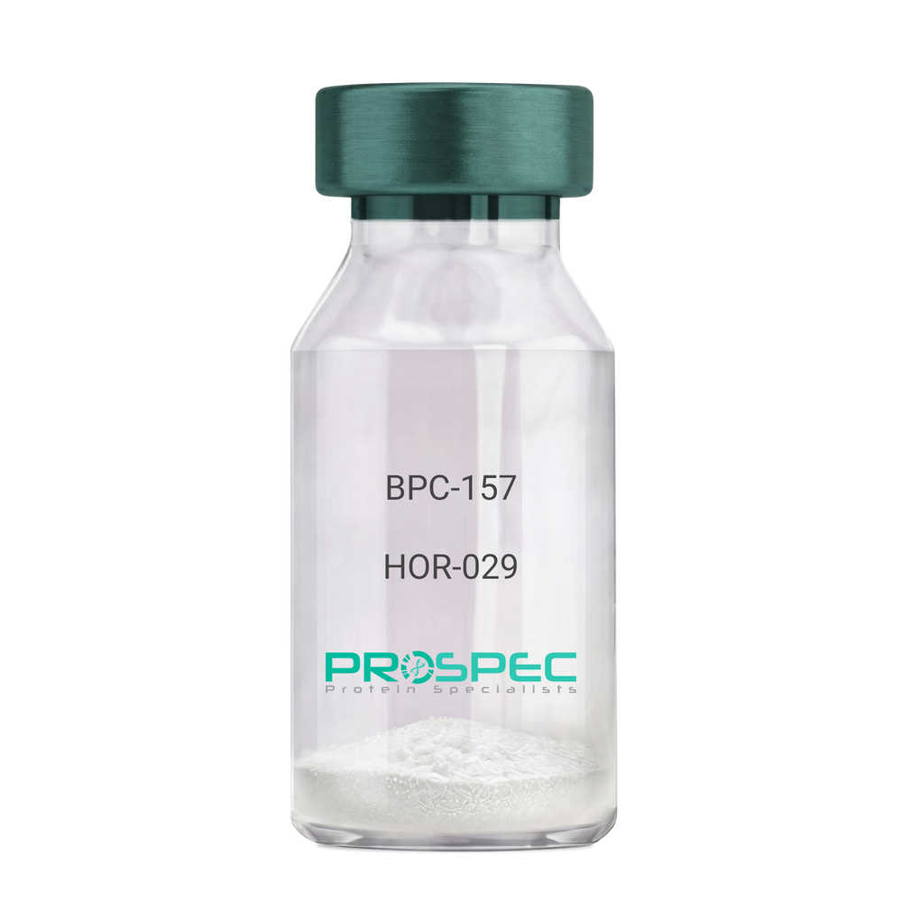
Name :
BPC-157Description:
BPC-157 Pentadecapeptide
Product # :
HOR-029Price :
Quantity :
Shipping Method :
Shipped at Room temp
More Info
- description
- formulation
- purity
- More Info
Description
BPC-157 Synthetic is a single, non-glycosylated polypeptide chain containing 15 amino acids, having a molecular mass of 1419.55 Dalton and a Molecular formula of C62H98N16O22.
Formulation
The protein was lyophilized with no additives.
Purity
Greater than 97.0% as determined by analysis by RP-HPLC.
More Info
-
Physical Appearance
Sterile Filtered White lyophilized (freeze-dried) powder.
-
Stability
Lyophilized BPC-157 although stable at room temperature for 3 weeks, should be stored desiccated below -18°C. Upon reconstitution bpc-157 should be stored at 4°C between 2-7 days and for future use below -18°C. For long term storage it is recommended to add a carrier protein (0.1% HSA or BSA). Please prevent freeze-thaw cycles.
-
Solubility
It is recommended to reconstitute the lyophilized BPC-157 in sterile 18MΩ-cm H2O not less than 100 µg/ml, which can then be further diluted to other aqueous solutions.
-
Amino Acid Sequence
H-Gly-Glu-Pro-Pro-Pro-Gly-Lys-Pro-Ala-Asp-Asp-Ala-Gly-Leu-Val-OH
-
Background
BPC-157, short for Body Protection Compound-157, is a synthetic peptide that has garnered significant attention in the field of regenerative medicine and sports science. This peptide, derived from a portion of the human gastric juice protein known as BPC, exhibits remarkable healing and tissue regeneration properties. BPC-157 has shown promise in various preclinical and clinical studies, demonstrating its potential for the treatment of a wide range of injuries and disorders.
The research on BPC-157 encompasses investigations into its mechanisms of action, efficacy, safety, and potential therapeutic applications. Studies have elucidated the peptide's ability to enhance angiogenesis, promote collagen synthesis, modulate inflammatory responses, and protect against oxidative stress. These properties make BPC-157 an intriguing candidate for accelerating tissue healing, reducing inflammation, and improving overall recovery outcomes.
Preclinical studies have revealed the beneficial effects of BPC-157 in several injury models. For instance, BPC-157 has demonstrated its potential in accelerating tendon and ligament healing, mitigating muscle damage, and promoting bone regeneration. These findings suggest that BPC-157 could be a valuable therapeutic tool in orthopedic medicine and sports-related injuries.
Furthermore, BPC-157 has exhibited promising effects on gastrointestinal health. Studies have highlighted its ability to protect and heal the gut lining, reduce ulcer formation, and alleviate symptoms associated with inflammatory bowel disease. These observations open up avenues for BPC-157 as a potential treatment for gastrointestinal disorders.
In addition to its regenerative properties, BPC-157 has shown potential in neurological and psychiatric conditions. Research has indicated its neuroprotective effects, with implications for the treatment of traumatic brain injury, stroke, and neurodegenerative disorders. Preliminary studies also suggest BPC-157's potential as an antidepressant and anxiolytic agent.
Despite the promising findings, further research is needed to fully understand the mechanisms underlying BPC-157's actions and to assess its long-term safety and efficacy. Clinical trials are underway to explore its potential therapeutic applications in humans, including its use in tendon and ligament repair, inflammatory bowel disease, and neurodegenerative disorders.
This comprehensive review aims to summarize the current state of research on BPC-157, providing an overview of its mechanisms of action and therapeutic potential. By examining relevant studies and findings, we aim to shed light on the diverse applications of BPC-157 and its implications for regenerative medicine, sports science, and various disease
ProSpec's products are furnished for LABORATORY RESEARCH USE ONLY. The product may not be used as drugs, agricultural or pesticidal products, food additives or household chemicals.
- View Data Sheet
Name :
Leptin tA RatDescription:
Leptin Antagonist Triple Mutant Rat Recombinant
Product # :
CYT-355Price :
Quantity :
Shipping Method :
Shipped at Room temp
More Info
- description
- source
- formulation
- purity
- biological activity
- More Info
Description
Leptin Antagonist Triple Mutant Rat Recombinant is a singly non-glycosilated polypeptide chain containing 146 amino and additional Ala at N-terminus acids and having a molecular mass of ~ 16 kDa, Leptin was mutated, resulting in L39A/D40A/F41A mutant. Leptin Antagonist Triple Mutant Rat Recombinant was purified by proprietary chromatographic techniques.
Source
Escherichia coli.
Formulation
The protein was lyophilized from a concentrated (0.85mg/ml) solution with 0.003mM NaHCO3.
Purity
Greater than 99.0% as determined by:
(a) Gel filtration analysis.
(b) Analysis by SDS-PAGE.Biological Activity
ProSpec’s Leptin Antagonist Triple Mutant Rat Recombinant is capable of inhibiting leptin-induced proliferation of BAF/3 cells stably transfected with the long form of human leptin receptor. It also inhibits various leptin effects in several in vitro bioassays.More Info
-
Physical Appearance
White lyophilized (freeze-dried) powder.
-
Stability
Lyophilized Leptin-Antagonist Triple Mutant Rat Recombinant although stable at room temperature for several weeks, should be stored desiccated below -18°C. Upon reconstitution at > 0.1 Leptin mutant mg/ml and up to 2 mM and filter sterilization LEP-tA mutant can be stored at 4°C or even room temperature for several weeks making it suitable for long term infusion studies using osmotic pumps. At lower concentration addition of a carrier protein (0.1% HSA or BSA) is suggested. Please prevent freeze-thaw cycles.
-
Solubility
It is recommended to reconstitute the lyophilized Leptin Antagonist Triple Mutant Rat Recombinant in sterile water or sterile 0.4% NaHCO3 adjusted to pH 8-9, not less than 100µg/ml, which can then be further diluted with other aqueous solutions.
-
Amino Acid Sequence
The sequence of the first five N-terminal amino acids was determined and was found to be Ala-Val-Pro-Ile-Gln.
-
Protein content
Protein quantization was carried out by UV spectroscopy at 280 nm using the absorbency value of 0.21 as the extinction coefficient for a 0.1% (1mg/ml) solution at pH 8.0. This value is calculated by the PC GENE computer analysis program of protein sequences (IntelliGenetics).
ProSpec's products are furnished for LABORATORY RESEARCH USE ONLY. The product may not be used as drugs, agricultural or pesticidal products, food additives or household chemicals.