Search results
960 results found for “insulin-like growth factor”
Name
Description
Product #
Price
Quantity
Shipping Method
- View Data Sheet
Name :
Activin A Human PlantDescription:
Activin A Human Recombinant, Plant
Inhba, Inhibin beta A, FSH releasing protein.
Product # :
CYT-052Price :
Quantity :
Shipping Method :
Shipped at Room temp
More Info
- description
- source
- formulation
- purity
- More Info
Description
Activin A human Recombinant produced in Nicotiana benthamiana plant is a disulfide-linked homodimers of two betaA chains, each containing 116 amino residues (molecular formula C600H911N173O174S13) and 6-His-tag at the N-terminal having the total molecular mass of 27.4kDa.
Source
Nicotiana benthamiana plant
Formulation
Lyophilized from 1mg/ml solution in Tris HCl 0.05M buffer at pH 7.4.
Purity
Greater than 97.0% as determined by Analysis by SDS-PAGE.
More Info
-
Introduction
Activins are homodimers or heterodimers of the different ? subunit isoforms, part of the TGF? family. Mature Activin A has two 116 amino acids residues betaA subunits (bA-bA). Activin displays an extensive variety of biological activities, including mesoderm induction, neural cell differentiation, bone remodelling, haematopoiesis, and reproductive physiology. Activins takes part in the production and regulation of hormones such as FSH, LH, GnRH and ACTH. Cells that are identified to express Activin A include fibroblasts, endothelial cells, hepatocytes, vascular smooth muscle cells, macrophages, keratinocytes, osteoclasts, bone marrow monocytes, prostatic epithelium, neurons, chondrocytes, osteoblasts, Leydig cells, Sertoli cells, and ovarian granulosa cells.
-
Synonyms
Inhba, Inhibin beta A, FSH releasing protein.
-
Physical Appearance
Sterile Filtered White lyophilized (freeze-dried) powder.
-
Stability
Lyophilized Activin A although stable at room temperature for 3 weeks, should be stored desiccated below -18°C. Upon reconstitution Activin A should be stored at 4°C between 2-7 days and for future use below -18°C. For long term storage it is recommended to add a carrier protein (0.1% HSA or BSA). Please prevent freeze-thaw cycles.
-
Solubility
It is recommended to reconstitute the lyophilized Activin A in sterile 18MΩ-cm H2O not less than 100µg/ml, which can then be further diluted to other aqueous solutions.
-
Amino Acid Sequence
HHHHHHGLEC DGKVNICCKK QFFVSFKDIG WNDWIIAPSG YHANYCEGEC PSHIAGTSGS SLSFHSTVIN HYRMRGHSPF ANLKSCCVPT KLRPMSMLYY DDGQNIIKKD IQNMIVEECG CS
-
Background
What is the molecular weight / Mw of Activin A Protein?
Activin A Protein has a total Mw of 27.4 kDa.
What is the source or expression system of Activin A Protein?
Nicotinia
What is the Purity of Activin A Protein?
Activin A Protein is >97% pure as determined by SDS-PAGE.
What is the Biological Activity of Activin A Protein?
The biological functionality of Activin-A Protein will be determined in the future.
What is the endotoxin level for Activin A Protein?
The endotoxin level is minimal, ACTIVIN A Protein was purified using conventional chromatography techniques.
What is the amino acid sequence of ACTIVIN A Protein?
HHHHHHGLEC DGKVNICCKK QFFVSFKDIG WNDWIIAPSG YHANYCEGEC PSHIAGTSGS SLSFHSTVIN HYRMRGHSPF ANLKSCCVPT KLRPMSMLYY DDGQNIIKKD IQNMIVEECG CS
What applications can ACTIVIN A Protein be used in?
ACTIVIN A Protein can probably be used in western blot, ELISA and Lateral Flow.
-
Serological Identification
The protein was electrophoresed under reducing condition on a 15% SDS-polyacrylamide gel, transferred by electroblotting to a NC membrane and visualized by immune-detection with specific antibody Activin A.
ProSpec's products are furnished for LABORATORY RESEARCH USE ONLY. The product may not be used as drugs, agricultural or pesticidal products, food additives or household chemicals.
- View Data Sheet
Name :
FGF 21 Mouse, HisDescription:
Fibroblast Growth Factor-21 Mouse Recombinant, His Tag
Fibroblast growth factor 21, FGF-21.
Product # :
CYT-516Price :
Quantity :
Shipping Method :
Shipped at Room temp
More Info
- description
- source
- formulation
- purity
- More Info
Description
Fibroblast Growth Factor -21 Mouse Recombinant produced in E.Coli is a single, non-glycosylated, polypeptide chain containing 192 amino acids and having a molecular mass of 21.2 kDa. The amino acid sequence of the recombinant human FGF21 is 100% homologous to the amino acid sequence of the Mouse FGF21 without signal sequence and contains 10 a.a. His tag at N-terminal.The FGF-21 is purified by proprietary chromatographic techniques.
Source
Escherichia Coli.
Formulation
Filtered (0.4 µm) and lyophilized from 0.5 mg/ml in 20mM TRIS, 20mM NaCl, pH 7.5.
Purity
Greater than 95.0% as determined by:
(a) Analysis by RP-HPLC.
(b) Analysis by SDS-PAGE.More Info
-
Introduction
The FGFs are a family of more than 20 small (~17–26 kDa) secreted peptides. The initial characterization of these proteins focused on their ability to stimulate fibroblast proliferation. This mitogenic activity was mediated through FGF receptors (FGFRs) 1, 2, or 3. A fourth closely related tyrosine kinase receptor (FGFR4) was able to bind the FGFs but did not lead to a mitogenic response.
FGFs modulate cellular activity via at least 5 distinct subfamilies of high-affinity FGF receptors (FGFRs): FGFR-1, -2, -3, and -4, all with intrinsic tyrosine kinase activity and, except for FGFR-4, multiple splice isoforms, and FGFR-5, which lacks an intracellular kinase domain. There is growing evidence that FGFRs can be important for regulation of glucose and lipid homeostasis. The overexpression of a dominant negative form of FGFR-1 in ? cells leads to diabetes in mice, which thus implies that proper FGF signaling is required for normal ? cell function and glycemia maintenance. FGFR-2 appears to be a key molecule during pancreatic development. Moreover, FGFR-4 has been implicated in cholesterol metabolism and bile acid synthesis.
FGF-19, has been shown to cause resistance to diet-induced obesity and insulin desensitization and to improve insulin, glucose, and lipid profiles in diabetic rodents. Since these effects, at least in part, are mediated through the observed changes in metabolic rates, FGF-19 can be considered as a regulator of energy expenditure.
FGF-21 is preferentially expressed in liver, but an exact knowledge of FGF-21 bioactivity and its mode of action have been lacking to date. FGF-21 is a potent activator of glucose uptake on adipocytes, protects animals from diet-induced obesity when overexpressed in transgenic mice, and lowers blood glucose and triglyceride levels when therapeutically administered to diabetic rodents. -
Synonyms
Fibroblast growth factor 21, FGF-21.
-
Physical Appearance
Filtered white lyophilized powder.
-
Stability
Lyophilized FGF-21 Mouse Recombinant although stable at room temperature for 3 weeks, should be stored desiccated below -18°C. Upon reconstitution Fibroblast Growth Factor 21 should be stored at 4°C between 2-7 days and for future use below -18°C. For long term storage it is recommended to add a carrier protein (0.1% HSA or BSA).Please prevent freeze-thaw cycles.
-
Solubility
It is recommended to add deionized water to prepare a working stock solution of approximately 0.5 mg/ml and let the lyophilized pellet dissolve completely. Product is not sterile! Please filter the product by an appropriate sterile filter before using it in the cell culture. Add DTT (0.2mM) and NaCl (0.1-0.15M) before freezing to prevent potential aggregation.
-
Amino Acid Sequence
MKHHHHHHAS AYPIPDSSPL LQFGGQVRQR YLYTDDDQDT EAHLEIREDG TVVGAAHRSP ESLLELKALKPGVIQILGVK ASRFLCQQPD GALYGSPHFD PEACSFRELL LEDGYNVYQS EAHGLPLRLP QKDSPNQDATSWGPVRFLPM PGLLHEPQDQ AGFLPPEPPD VGSSDPLSMV EPLQGRSPSY AS.
-
Background
What is the molecular weight/Mw of FGF21 MOUSE,HIS Protein?
FGF21 MOUSE,HIS Protein has a total Mw of 21.2kDa.
What is the source or expression system of FGF21 MOUSE,HIS Protein?
Escherichia Coli.
What is the Purity of FGF21 MOUSE,HIS Protein?
FGF21 MOUSE,HIS Protein is >95% pure as determined by SDS-PAGE.
What is the Biological Activity of FGF21 MOUSE,HIS Protein?
The biological functionality of FGF21 MOUSE,HIS Protein will be determined in the future.
What is the amino acid sequence of FGF21 MOUSE,HIS Protein?
MKHHHHHHAS AYPIPDSSPL LQFGGQVRQR YLYTDDDQDT EAHLEIREDG TVVGAAHRSP ESLLELKALKPGVIQILGVK ASRFLCQQPD GALYGSPHFD PEACSFRELL LEDGYNVYQS EAHGLPLRLP QKDSPNQDATSWGPVRFLPM PGLLHEPQDQ AGFLPPEPPD VGSSDPLSMV EPLQGRSPSY AS.
What applications can FGF21 MOUSE,HIS Protein be used in?
FGF21 MOUSE,HIS Protein can probably be used in western blot, ELISA and Lateral Flow.
What is the endotoxin level for FGF21 MOUSE,HIS Protein?
The endotoxin level is minimal, FGF21 MOUSE,HIS Protein was purified using conventional chromatography techniques.
ProSpec's products are furnished for LABORATORY RESEARCH USE ONLY. The product may not be used as drugs, agricultural or pesticidal products, food additives or household chemicals.
- View Data Sheet
Name :
VEGFC Human, Sf9Description:
Vascular Endothelial Growth Factor C Human Recombinant, Sf9
Vascular Endothelial Growth Factor C, Vascular Endothelial Growth Factor-Related Protein, Flt4-L, VRP, FLT4 Ligand DHM, Flt4 Ligand, LMPH1D, VEGF-C, Vascular endothelial growth factor C, VEGF-C, Flt4 ligand, Flt4-L, Vascular endothelial growth factor-related protein.
Product # :
CYT-948Price :
Quantity :
Shipping Method :
Shipped with Ice Packs
More Info
- description
- source
- formulation
- purity
- More Info
Description
VEGFC Human Recombinant produced in Sf9 Baculovirus cells is a single, glycosylated polypeptide chain containing 125 amino acids (112-227a.a.) and having a molecular mass of 14.2kDa (Molecular size on SDS-PAGE will appear at approximately 18-28kDa). VEGFC is expressed with a 6 amino acid His tag at C-Terminus and purified by proprietary chromatographic techniques.
Source
Sf9, Baculovirus cells.
Formulation
VEGFC protein solution (0.5mg/ml) contains Phosphate Buffered Saline (pH 7.4) and 20% glycerol.
Purity
Greater than 90.0% as determined by SDS-PAGE.
More Info
-
Introduction
VEGF-C, also known as Vascular Endothelial Growth Factor Related Protein (VRP), is a recently discovered VEGF growth factor family member that is most closely related to VEGF-D. Human VEGF-C cDNA encodes a pre-pro-protein of 416 amino acids residues. It is almost identical to the mouse VEGF-C protein. Similar to VEGF-D, VEGF-C has a VEGF homology domain spanning the middle third of the precursor molecule and long N- and C-terminal extensions. In adults, VEGF-C is highly expressed in heart, placenta, ovary and small intestine. Recombinant human VEGF-C, lacking the N- and C-terminal extensions and containing only the middle VEGF homology domain, forms primarily non-covalently linked dimers. This protein is a ligand for both VEGFR-2/KDR and VEGFR-3/FLT-4. Since VEGFR-3 is strongly expressed in lymphatic endothelial cells, it has been postulated that VEGF-C is involved in the regulation of the growth and/or differentiation of lymphatic endothelium. Although recombinant human VEGF-C is also a mitogen for vascular endothelial cells, it is much less potent than VEGF-A.
-
Synonyms
Vascular Endothelial Growth Factor C, Vascular Endothelial Growth Factor-Related Protein, Flt4-L, VRP, FLT4 Ligand DHM, Flt4 Ligand, LMPH1D, VEGF-C, Vascular endothelial growth factor C, VEGF-C, Flt4 ligand, Flt4-L, Vascular endothelial growth factor-related protein.
-
Physical Appearance
Sterile filtered colorless solution.
-
Stability
Store at 4°C if entire vial will be used within 2-4 weeks. Store, frozen at -20°C for longer periods of time. For long term storage it is recommended to add a carrier protein (0.1% HSA or BSA). Avoid multiple freeze-thaw cycles.
-
Amino Acid Sequence
ADPAHYNTEI LKSIDNEWRK TQCMPREVCI DVGKEFGVAT NTFFKPPCVS VYRCGGCCNS EGLQCMNTST SYLSKTLFEI TVPLSQGPKP VTISFANHTS CRCMSKLDVY RQVHSIIRRH HHHHH.
ProSpec's products are furnished for LABORATORY RESEARCH USE ONLY. The product may not be used as drugs, agricultural or pesticidal products, food additives or household chemicals.
- View Data Sheet
Name :
GHBP Human, Sf9Description:
Growth Hormone Binding Protein Human Recombinant, Sf9
GHR, GHBP, GHIP.
Product # :
CYT-1152Price :
Quantity :
Shipping Method :
Shipped with Ice Packs
More Info
- description
- source
- formulation
- purity
- biological activity
- More Info
Description
GHBP Human produced in Sf9 Baculovirus cells is a single, glycosylated polypeptide chain containing 254 amino acids (19-264aa) and having a molecular mass of 29.4kDa.GHBP is fused to a 8 amino acid His tag at C-terminus and purified by proprietary chromatographic techniques.
Source
Sf9, Baculovirus cells.
Formulation
The GHBP solution (1mg/ml) contains 10% Glycerol and Phosphate-Buffered Saline (pH 7.4).
Purity
Greater than 95.0% as determined by SDS-PAGE.
Biological Activity
Measured by ability to inhibit GH-induced proliferation assay using Nb2-11 Rat lymphoma cells in the presence of 1.25ng/ml of human growth hormone. The ED50 for this effect is equal or less than 10ng/ml.
More Info
-
Introduction
Growth Hormone Binding Protein or GHR, is a protein, part of the cytokine receptor superfamily. GHR binds to 2 receptors, therefore it enhances signal transduction via dimerization of receptors. In elevated levels, growth hormone operates as an antagonist due to high variance in the binding sites affinities. The antagonist operation can be embellished even more when the binding site is reduced its affinity.
-
Synonyms
GHR, GHBP, GHIP.
-
Physical Appearance
Sterile Filtered colorless solution.
-
Stability
Store at 4°C if entire vial will be used within 2-4 weeks. Store, frozen at -20°C for longer periods of time. For long term storage it is recommended to add a carrier protein (0.1% HSA or BSA).Avoid multiple freeze-thaw cycles.
-
Amino Acid Sequence
FSGSEATAAI LSRAPWSLQS VNPGLKTNSS KEPKFTKCRS PERETFSCHW TDEVHHGTKN LGPIQLFYTR RNTQEWTQEW KECPDYVSAG ENSCYFNSSF TSIWIPYCIK LTSNGGTVDE KCFSVDEIVQ PDPPIALNWT LLNVSLTGIH ADIQVRWEAP RNADIQKGWM VLEYELQYKE VNETKWKMMD PILTTSVPVY SLKVDKEYEV RVRSKQRNSG NYGEFSEVLY VTLPQMSQFT
CEEDFYLEHH HHHH
ProSpec's products are furnished for LABORATORY RESEARCH USE ONLY. The product may not be used as drugs, agricultural or pesticidal products, food additives or household chemicals.
- View Data Sheet
Name :
SCF Human, Sf9Description:
Stem Cell Factor Human Recombinant, Sf9
Kit ligand Precursor, C-kit ligand, SCF, Mast cell growth factor, MGF, SF, KL-1, Kitl, DKFZp686F2250.
Product # :
CYT-421Price :
Quantity :
Shipping Method :
Shipped at Room temp
More Info
- description
- source
- formulation
- purity
- biological activity
- More Info
Description
Stem Cell Factor Human Recombinant produced in insect cells is a single, glycosylated polypeptide chain containing 165 amino acids and having a molecular mass of 18409 Dalton. The SCF is fused to a C-terminal His-tag (6xHis) and purified by proprietary chromatographic techniques.
Source
Sf9, Insect Cells.
Formulation
The protein is supplied in 1xPBS, pH 7.4.
Purity
Greater than 98.0% as determined by analysis by SDS-PAGE.
Biological Activity
The ED50 as determined by the dose-dependent stimulation of Human TF-1 cells is typically 1-5 ng/ml.More Info
-
Introduction
Stem cell factor / KIT ligand (SCF) is a cytokine which binds CD117(c-Kit). SCF is also known as "steel factor" or "c-kit ligand". SCF exists in two forms, cell surface bound SCF and soluble (or free) SCF. Soluble SCF is produced by the cleavage of surface bound SCF by metalloproteases.
SCF is a growth factor important for the survival, proliferation, and differentiation of hematopoietic stem cells and other hematopoietic progenitor cells. One of its roles is to change the BFU-E (burst-forming unit-erythroid) cells, which are the earliest erythrocyte precursors in the erythrocytic series, into the CFU-E (colony-forming unit-erythroid). -
Synonyms
Kit ligand Precursor, C-kit ligand, SCF, Mast cell growth factor, MGF, SF, KL-1, Kitl, DKFZp686F2250.
-
Physical Appearance
Sterile Filtered White lyophilized (freeze-dried) powder.
-
Stability
Lyophilized KIT ligand although stable at room temperature for 3 weeks, should be stored desiccated below -18°C. Upon reconstitution SCF should be stored at 4°C between 2-7 days and for future use below -18°C. For long term storage it is recommended to add a carrier protein (0.1% HSA or BSA).Please prevent freeze-thaw cycles.
-
Solubility
It is recommended to reconstitute the lyophilized Stem Cell Factor in 10mM acetic acid not less than 100µg/ml, which can then be further diluted to other aqueous solutions.
-
Protein content
Protein quantitation was carried out by two independent methods:1. UV spectroscopy at 280 nm using the absorbency value of 0.52 as the extinction coefficient for a 0.1% (1mg/ml) solution. This value is calculated by the PC GENE computer analysis program of protein sequences (IntelliGenetics). 2. Analysis by RP-HPLC, using a standard solution of Stem Cell Factor as a Reference Standard.
ProSpec's products are furnished for LABORATORY RESEARCH USE ONLY. The product may not be used as drugs, agricultural or pesticidal products, food additives or household chemicals.
- View Data Sheet
Name :
FGFR3 HumanDescription:
Fibroblast Growth Factor Receptor 3 Fc Chimera Human Recombinant
Achondroplasia, Thanatophoric Dwarfism, CD333, ACH, CEK2, JTK4, HSFGFR3EX, FGFR3.
Product # :
PKA-232Price :
Quantity :
Shipping Method :
Shipped at Room temp
More Info
- description
- source
- formulation
- purity
- More Info
Description
Soluble FGFR3 Human recombinant fused using a Xa cleavage site with the Fc part of human IgG1 produced in Fc Chimera is a heterodimeric, glycosylated, Polypeptide chain containing 593aa and having a molecular mass of 170kDa.
Source
Insect Cells.
Formulation
FGFR3 was lyophilized from a concentrated (1 mg/ml) sterile solution containing PBS.
Purity
Greater than 90.0% as determined by:(a) Analysis by silver stain.(b) Analysis by SDS-PAGE.
More Info
-
Introduction
Fibroblast Growth Factors (FGFs) comprise a family of at least eighteen structurally realted proteins that are involved in a multitude of physiological and pathological cellular processes, including cell growth, differentation, angiogenesis, wound healing and tumorgenesis. The biological activities of the FGFs are mediated by a family if type I transmembrane tyrosine kinases which undergo dimerization and autophosphorylation after ligand binding. Four distinct genes encoding closely related FGF receptors, FGFR-1to -4 are known. Multiple forms of FGFR-1 to -3 are generated by alternative splicing of the mRNAs. A frequent splicing event involving FGFR-1 and -2 results in receptors containing all three Ig domains, referred to as the alpha isoform, or only IgII and IgIII, referred to as the ? isoform. Only the alpha isoform has been identified for FGFR-3 and FGFR-4. Additional splicing events for FGFR-1 to -3, involving the C-terminal half of the IgIII domain encoded by two mutually exclusive alternative exons, generate FGF receptors with alternative IgIII domains (IIIb and IIIc). A IIIa isoform which is a secreted FGF binding protein containing only the N-terminal half of the IgIII domain plus some intron sequences has also been reported for FGFR-1. Mutations in FGFR-1 to -3 have been found in patients with birth defects involving craniosynostosis.
-
Synonyms
Achondroplasia, Thanatophoric Dwarfism, CD333, ACH, CEK2, JTK4, HSFGFR3EX, FGFR3.
-
Physical Appearance
Sterile Filtered White lyophilized (freeze-dried) powder.
-
Stability
Lyophilized FGFR3 although stable at room temperature for 3 weeks, should be stored desiccated below -18oC. Upon reconstitution FGFR3 should be stored at 4oC between 2-7 days and for future use below -18oC.For long term storage it is recommended to add a carrier protein (0.1% HSA or BSA).Please prevent freeze-thaw cycles.
-
Solubility
It is recommended to reconstitute the lyophilized FGFR-3 in sterile PBS not less than 50µg/ml, which can then be further diluted to other aqueous solutions.
-
Amino Acid Sequence
ESLGTEQRVV GRAAEVPGPE PGQQEQLVFG SGDAVELSCP PPGGGPMGPT VWVKDGTGLV PSERVLVGPQ RLQVLNASHE DSGAYSCRQR LTQRVLCHFS VRVTDAPSSG DDEDGEDEAE DTGVDTGAPY WTRPERMDKK LLAVPAANTV RFRCPAAGNP TPSISWLKNG REFRGEHRIG GIKLRHQQWS LVMESVVPSD RGNYTCVVEN KFGSIRQTYT LDVLERSPHR PILQAGLPAN QTAVLGSDVE FHCKVYSDAQ PHIQWLKHVE VNGSKVGPDG TPYVTVLKTA GANTTDKELE VLSLHNVTFE DAGEYTCLAG NSIGFSHHSA WLVVLPAEEE LVEADEAGDP RRASIEGRGD PEEPKSCDKT HTCPPCPAPE LLGGPSVFLF PPKPKDTLMI SRTPEVTCVV VDVSHEDPEV KFNWYVDGVE VHNAKTKPRE EQYNSTYRVV SVTVLHQDWL NGKEYKCKVS NKALPAPIEK TISKAKGQPR EPQVYTLPPS RDELTKNQVS LTCLVKGFYP SDIAVEWESN GQPENNYKTT PPVLDSDGSF FLYSKLTVDK SRWQQGNVFS CSVMHEALHN HYTQKSLSLS PGK.
ProSpec's products are furnished for LABORATORY RESEARCH USE ONLY. The product may not be used as drugs, agricultural or pesticidal products, food additives or household chemicals.
- View Data Sheet
Name :
FIBP HumanDescription:
FGF-1 Intracellular-Binding Protein Human Recombinant
FGFIBP, FIBP-1, Acidic fibroblast growth factor intracellular-binding protein, aFGF intracellular-binding protein, FGF-1 intracellular-binding protein, FIBP.
Product # :
CYT-758Price :
Quantity :
Shipping Method :
Shipped with Ice Packs
More Info
- description
- source
- formulation
- purity
- More Info
- sds-page
Description
FIBP Human Recombinant produced in E.coli is a single, non-glycosylated polypeptide chain containing 387amino acids (1-364) and having a molecular mass of 44.3kDa. The FIBP is fused to a 23 amino acid His-Tag at N-terminus and purified by proprietary chromatographic techniques.
Source
E.coli.
Formulation
FIBP protein (0.5mg/ml) is supplied in 20mM Tris-HCl buffer (pH 8.0) and 10% glycerol.
Purity
Greater than 95% as determined by SDS-PAGE.
sds-page
More Info
-
Introduction
FGF-1 Intracellular-Binding Protein (FIBP) which is a member of Fibroblast growth factors (FGFs) binds to internalized FGF-1 and is thought to be involved in mitogenic function of FGF-1. FGF activity effects development, adult tissue homeostasis, angiogenesis and cancer progression. FIBP localizes to the nucleus and is greatly expressed in heart, skeletal muscle and pancreas and at lower levels in brain, placenta, liver and kidney.
-
Synonyms
FGFIBP, FIBP-1, Acidic fibroblast growth factor intracellular-binding protein, aFGF intracellular-binding protein, FGF-1 intracellular-binding protein, FIBP.
-
Physical Appearance
Sterile Filtered colorless solution.
-
Stability
Store at 4°C if entire vial will be used within 2-4 weeks. Store, frozen at -20°C for longer periods of time. For long term storage it is recommended to add a carrier protein (0.1% HSA or BSA).Avoid multiple freeze-thaw cycles.
-
Amino Acid Sequence
MGSSHHHHHH SSGLVPRGSH MGSMTSELDI FVGNTTLIDE DVYRLWLDGY SVTDAVALRV RSGILEQTGA TAAVLQSDTM DHYRTFHMLE RLLHAPPKLL HQLIFQIPPS RQALLIERYY AFDEAFVREV LGKKLSKGTK KDLDDISTKT GITLKSCRRQ FDNFKRVFKV VEEMRGSLVD NIQQHFLLSD RLARDYAAIV FFANNRFETG KKKLQYLSFG DFAFCAELMI QNWTLGAVGE APTDPDSQMD DMDMDLDKEF LQDLKELKVL VADKDLLDLH KSLVCTALRG KLGVFSEMEA NFKNLSRGLV NVAAKLTHNK DVRDLFVDLV EKFVEPCRSD HWPLSDVRFF LNQYSASVHS LDGFRHQALW DRYMGTLRGC LLRLYHD.
-
Background
What is the molecular weight/Mw of FIBP Protein?
FIBP Protein has a total Mw of 44.3kkDa.
What is the source or expression system of FIBP Protein?
Escherichia Coli.
What is the Purity of FIBP Protein?
FIBP Protein is >95% pure as determined by SDS-PAGE.
What is the Biological Activity of FIBP Protein?
The biological functionality of FIBP Protein will be determined in the future.
What is the amino acid sequence of FIBP Protein?
MGSSHHHHHH SSGLVPRGSH MGSMTSELDI FVGNTTLIDE DVYRLWLDGY SVTDAVALRV RSGILEQTGA TAAVLQSDTM DHYRTFHMLE RLLHAPPKLL HQLIFQIPPS RQALLIERYY AFDEAFVREV LGKKLSKGTK KDLDDISTKT GITLKSCRRQ FDNFKRVFKV VEEMRGSLVD NIQQHFLLSD RLARDYAAIV FFANNRFETG KKKLQYLSFG DFAFCAELMI QNWTLGAVGE APTDPDSQMD DMDMDLDKEF LQDLKELKVL VADKDLLDLH KSLVCTALRG KLGVFSEMEA NFKNLSRGLV NVAAKLTHNK DVRDLFVDLV EKFVEPCRSD HWPLSDVRFF LNQYSASVHS LDGFRHQALW DRYMGTLRGC LLRLYHD.
What applications can FIBP Protein be used in?
FIBP Protein can probably be used in western blot, ELISA and Lateral Flow.
What is the endotoxin level for FIBP Protein?
The endotoxin level is minimal, FIBP Protein was purified using conventional chromatography techniques.
ProSpec's products are furnished for LABORATORY RESEARCH USE ONLY. The product may not be used as drugs, agricultural or pesticidal products, food additives or household chemicals.
- View Data Sheet
Name :
RANK HumanDescription:
RANK Human Recombinant
TNFRSF11A, ODFR, RANK, Tumor Necrosis Factor Receptor Superfamily, Member 11a, Activator Of NFKB, Receptor Activator Of Nuclear Factor-Kappa B, CD265 Antigen, LOH18CR1, TRANCER, CD265
Product # :
CYT-734Price :
Quantity :
Shipping Method :
Shipped at Room temp
More Info
- description
- source
- formulation
- purity
- biological activity
- More Info
Description
RANK Human Recombinant produced in E.Coli is a single, non-glycosylated polypeptide chain containing 174 amino acids and having a molecular mass of 19.1kDa. The RANK is purified by proprietary chromatographic techniques.
Source
Escherichia Coli.
Formulation
Lyophilized from a 0.2 µm filtered concentrated solution in 20mM Tris-HCl, pH 8.0 and 150mM NaCl.
Purity
Greater than 98.0% as determined by:
(a) Analysis by RP-HPLC.
(b) Analysis by SDS-PAGE.Biological Activity
The ED50 as determined by its ability to inhibit sRANK Ligand induced nuclear factor kappa B (NFkappaB) in RAW 264.7 cells is less than 50 ng/ml, corresponding to a specific activity of
> 2.0 × 10⁴ IU/mg in the presence of 15 ng/ml of recombinant sRANK Ligand.More Info
-
Introduction
sRANK Receptor is a part of of the TNF superfamily of ligands and receptors which participates in the regulation of specific immunity and bone turnover. sRANK Receptor was originally acknowledged as a dendritic-cell-membrane protein, which by interacting with RANKL augments the capacity of dendritic cells to stimulate naive T cell proliferation and to endorse the survival of RANK and T cells. The full length human RANK cDNA encodes a type I transmembrane protein of 616 amino acids with a predicted 183 amino acid extracellular domain and a 383 amino acid cytoplasmic domain. sRANK Receptor is also expressed in a various tissues including skeletal muscle, thymus, liver, colon, small intestine and adrenal gland.
-
Synonyms
TNFRSF11A, ODFR, RANK, Tumor Necrosis Factor Receptor Superfamily, Member 11a, Activator Of NFKB, Receptor Activator Of Nuclear Factor-Kappa B, CD265 Antigen, LOH18CR1, TRANCER, CD265
-
Physical Appearance
Sterile Filtered White lyophilized (freeze-dried) powder.
-
Stability
Lyophilized RANK although stable at room temperature for 3 weeks, should be stored desiccated below -18°C. Upon reconstitution sRANK Receptor should be stored at 4°C between 2-7 days and for future use below -18°C. For long term storage it is recommended to add a carrier protein (0.1% HSA or BSA).Please prevent freeze-thaw cycles.
-
Solubility
It is recommended to reconstitute the lyophilized RANK in sterile 18M-cm H2O not less than 100µg/ml, which can then be further diluted to other aqueous solutions.
-
Amino Acid Sequence
QIAPPCTSEK HYEHLGRCCN KCEPGKYMSS KCTTTSDSVC LPCGPDEYLD SWNEEDKCLL HKVCDTGKAL VAVVAGNSTT PRRCACTAGY HWSQDCECCR RNTECAPGLG AQHPLQLNKD TVCKPCLAGY FSDAFSSTDK CRPWTNCTFL GKRVEHHGTE KSDAVCSSSL PARK.
ProSpec's products are furnished for LABORATORY RESEARCH USE ONLY. The product may not be used as drugs, agricultural or pesticidal products, food additives or household chemicals.
- View Data Sheet
Name :
GCSF RatDescription:
Granulocyte-Colony Stimulating Factor Rat Recombinant
Granulocyte colony stimulating factor, Protein Csf3, Csf3.
Product # :
CYT-940Price :
Quantity :
Shipping Method :
Shipped at Room temp
More Info
- description
- source
- formulation
- purity
- biological activity
- More Info
Description
GCSF Rat Recombinant produced in E.Coli is a single, non-glycosylated, polypeptide chain containing 195 amino acids and having a molecular mass of 21.5kDa.The G-CSF is purified by proprietary chromatographic techniques.
Source
Escherichia Coli.
Formulation
Lyophilized from a 0.2µm filtered solution in 5mM Sodium Citrate, pH 4.0.
Purity
Greater than 97.0% as determined by:
(a)Analysis by RP-HPLC.
(b)Analysis by SDS-PAGE.Biological Activity
The ED50 determined by a cell proliferation assay using murine NFS-60 cells is less than 0.05ng/ml, corresponding to a specific activity of > 2.0× 10⁷ IU/mg.More Info
-
Introduction
GCSF is a cytokine that controls the production, differentiation, and function of granulocytes. The active protein is found extracellularly. Three transcript variants encoding three different isoforms have been found for the GCSF gene. Granulocyte/macrophage colony-stimulating factors are cytokines that act in hematopoiesis by controlling the production, differentiation, and function of 2 related white cell populations of the blood, the granulocytes and the monocytes-macrophages. This csf induces granulocytes.
-
Synonyms
Granulocyte colony stimulating factor, Protein Csf3, Csf3.
-
Physical Appearance
Sterile Filtered White lyophilized (freeze-dried) powder.
-
Stability
Lyophilized G-CSF although stable at room temperature for 3 weeks, should be stored desiccated below -18°C. Upon reconstitution GCSF should be stored at 4°C between 2-7 days and for future use below -18°C.For long term storage it is recommended to add a carrier protein (0.1% HSA or BSA).Please prevent freeze-thaw cycles.
-
Solubility
It is recommended to reconstitute the lyophilized GCSF in sterile 18M-cm H2O not less than 100µg/ml, which can then be further diluted to other aqueous solutions.
-
Amino Acid Sequence
KKIPLLTVSS LPPSLPLPRS FLLKSLEQVR KIQARNTELL EQLCATYKLC HPEELVLFGH SLGIPKASLS SCSSQALQQT KCLSQLHSGL FLYQGLLQAL AGISSELAPT LDMLHLDVDN FATTIWQQME SLGVAPTVQP TQSTMPIFTS AFQRRAGGVL VTSYLQSFLE TAHHALHHLP RPAQKHFPES LFISI.
-
Background
What is the molecular weight/Mw of G CSF RAT Protein?
G CSF RAT Protein has a total Mw of 21.5kDa.
What is the source or expression system of G CSF RAT Protein?
Escherichia Coli.
What is the Purity of G CSF RAT Protein?
G CSF RAT Protein is >97% pure as determined by SDS-PAGE.
What is the Biological Activity of G CSF RAT Protein?
The ED50 determined by a cell proliferation assay using murine NFS-60 cells is less than 0.05ng/ml, corresponding to a specific activity of > 2.0× 10⁷ IU/mg.
What is the amino acid sequence of G CSF RAT Protein?
KKIPLLTVSS LPPSLPLPRS FLLKSLEQVR KIQARNTELL EQLCATYKLC HPEELVLFGH SLGIPKASLS SCSSQALQQT KCLSQLHSGL FLYQGLLQAL AGISSELAPT LDMLHLDVDN FATTIWQQME SLGVAPTVQP TQSTMPIFTS AFQRRAGGVL VTSYLQSFLE TAHHALHHLP RPAQKHFPES LFISI.
What applications can G CSF RAT Protein be used in?
G CSF RAT Protein can probably be used in western blot, ELISA and Lateral Flow.
What is the endotoxin level for G CSF RAT Protein?
The endotoxin level is minimal, G CSF RAT Protein was purified using conventional chromatography techniques.
ProSpec's products are furnished for LABORATORY RESEARCH USE ONLY. The product may not be used as drugs, agricultural or pesticidal products, food additives or household chemicals.
- View Data Sheet
Name :
LFA 3 HumanDescription:
Lymphocyte Function Associated Antigen-3 Human Recombinant , Fusion Protein
CD58, LFA-3, Ag3, Surface glycoprotein LFA-3.
Product # :
CYT-423Price :
Quantity :
Shipping Method :
Shipped at Room temp
More Info
- description
- source
- formulation
- purity
- More Info
Description
Lymphocyte Function-Associated Antigen-3 Fusion Protein Recombinant Human is produced by recombinant DNA technology in a Chinese Hamster Ovary (CHO) mammalian cell expression system. The molecular weight is 91.4 kDa. Recombinant LFA3 is purified by proprietary chromatographic techniques.
Source
Chinese Hamster Ovary.
Formulation
Each mg of CD58 contains 0.8mg sucrose, 0.3mg glycine, 0.25mg sodium citrate dihydrate, and 4µg citric acid monohydrate.
Purity
Greater than 98.0% as determined by(a) Analysis by RP-HPLC.
(b) Analysis by SDS-PAGE.More Info
-
Introduction
LFA-3 is ligand of the t-lymphocyte cd2 glycoprotein. This interaction is important in mediating thymocyte interactions with thymic epithelial cells, antigen-independent and dependent interactions of t-lymphocytes with target cells and antigen- presenting cells and the t-lymphocyte rosetting with erythrocytes. In addition, the lfa-3/cd2 interaction may prime response by both the cd2+ and lfa-3+ cells.
-
Synonyms
CD58, LFA-3, Ag3, Surface glycoprotein LFA-3.
-
Physical Appearance
Sterile Filtered White lyophilized (freeze-dried) powder.
-
Stability
Lyophilized LFA3 Human although stable at room temperature for 3 weeks, should be stored desiccated below -18°C. Upon reconstitution Human LFA-3 should be stored at 4°C between 2-7 days and for future use below -18°C. For long term storage it is recommended to add a carrier protein (0.1% HSA or BSA).Please prevent freeze-thaw cycles.
-
Solubility
It is recommended to reconstitute the lyophilized LFA-3 Human in sterile 18MΩ-cm H2O not less than 100µg/ml, which can then be further diluted to other aqueous solutions.
ProSpec's products are furnished for LABORATORY RESEARCH USE ONLY. The product may not be used as drugs, agricultural or pesticidal products, food additives or household chemicals.
- View Data Sheet
Name :
GMFB HumanDescription:
Glia Maturation Factor Beta Human Recombinant
Glia maturation factor beta, GMFB, GMF-B, GMF-beta, GMF.
Product # :
CYT-565Price :
Quantity :
Shipping Method :
Shipped at Room temp
More Info
- description
- source
- formulation
- purity
- More Info
Description
Glia Maturation Factor-Beta (GMF-Beta) Human Recombinant produced in E.Coli is a signle, non-glycosylated, polypeptide chain containing 141 amino acids and having a total molecular mass of 16.5 kDa. Glia Maturation Factor-Beta, GMF-Beta, Human Recombinant is purified by proprietary chromatographic techniques.
Source
Escherichia Coli.
Formulation
The GMF-beta protein was lyophilized after dialysis against 20mM PBS pH=7.4 and 130mM NaCl.
Purity
Greater than 98.0% as determined by(a) Analysis by RP-HPLC.
(b) Analysis by SDS-PAGE.More Info
-
Introduction
Glia Maturation Factor-Beta (GMF-Beta) is a 17 kDa protein nerve gorwth factor identified as a growth and differentiation factor in the vertebrate brain.
Glia Maturation Factor-Beta stimulates differentiation of normal neurons as well as glial cells. GMFB inhibits the proliferation of the N-18 neuroblastoma line and the C6 glioma line while promoting their phenotypic expression.
GMF-beta inhances the phenotypic expression of glia & neurons thus inhibits the proliferation of their respective tumors when added to cell culture. Although astrocytes produce GMF-b and stores it inside the cells, they don’t secrete the GMF-B into the cultured medium. Cell- surface GMFb acts on the target cells at close range when cells are in direct contact. GMF-Beta is produced by thymic epithelial cells and plays an important role in T cell development in favor of CD4+ T cells.
GMF-Beta is a brain-specific protein which belongs to the actin-binding proteins (ADF) family. GMF-beta appears to play a role in the differentiation, maintenance, and regeneration of the nervous system. It also supports the progression of certain auto-immune diseases, possibly through its ability to induce the production and secretion of various pro-inflammatory cytokines. -
Synonyms
Glia maturation factor beta, GMFB, GMF-B, GMF-beta, GMF.
-
Physical Appearance
Sterile Filtered White lyophilized (freeze-dried) powder.
-
Stability
Lyophilized GMF-B although stable at room temperature for 3 weeks, should be stored desiccated below -18°C. Upon reconstitution GMF-beta should be stored at 4°C between 2-7 days and for future use below -18°C.For long term storage it is recommended to add a carrier protein (0.1% HSA or BSA).Please prevent freeze-thaw cycles.
-
Solubility
It is recommended to reconstitute the lyophilized GMFB in sterile 18MΩ-cm H2O not less than 100µg/ml, which can then be further diluted to other aqueous solutions.
-
Amino Acid Sequence
SESLVVCDVAEDLVEKLRKFRFRKETNNAAIIMKIDKDKRLVVLDEELEGISPD
ELKELPERQPRFIVYSYKYQHDDGRVSYPLCFIFSSPVGCKPEQQMMYAGSKN
KLVQT AELTKVFEIRNTEDLTEEWLREKLGFFH. -
Background
What is the molecular weight/Mw of GMFB HUMAN Protein?
GMFB HUMAN Protein has a total Mw of 16.5kDa.
What is the source or expression system of GMFB HUMAN Protein?
Escherichia Coli.
What is the Purity of GMFB HUMAN Protein?
GMFB HUMAN Protein is >98% pure as determined by SDS-PAGE.
What is the Biological Activity of GMFB HUMAN Protein?
The biological functionality of GMFB HUMAN Protein will be determined in the future.
What is the amino acid sequence of GMFB HUMAN Protein?
SESLVVCDVAEDLVEKLRKFRFRKETNNAAIIMKIDKDKRLVVLDEELEGISPD
ELKELPERQPRFIVYSYKYQHDDGRVSYPLCFIFSSPVGCKPEQQMMYAGSKN
KLVQT AELTKVFEIRNTEDLTEEWLREKLGFFH.
What applications can GMFB HUMAN Protein be used in?
GMFB HUMAN Protein can probably be used in western blot, ELISA and Lateral Flow.
What is the endotoxin level for GMFB HUMAN Protein?
The endotoxin level is minimal, GMFB HUMAN Protein was purified using conventional chromatography techniques.
ProSpec's products are furnished for LABORATORY RESEARCH USE ONLY. The product may not be used as drugs, agricultural or pesticidal products, food additives or household chemicals.
- View Data Sheet
Name :
Noggin MouseDescription:
Noggin Mouse Recombinant
Noggin, SYM1, SYNS1, NOG.
Product # :
CYT-600Price :
Quantity :
Shipping Method :
Shipped at Room temp
More Info
- description
- source
- formulation
- purity
- biological activity
- More Info
Description
Noggin Mouse Recombinant produced in E.Coli is a non-glycosylated, disulfide-linked protein consisting of two 206 amino acid polypeptide chains, having a total molecular mass of approximately 46.4 kDa (each chain 23.2 kDa).
Source
Escherichia Coli.
Formulation
Lyophilized from a 0.2μm filtered solution in 30% acetonitrile, 0.1% TFA.
Purity
Greater than 95.0% as determined by SDS-PAGE.
Biological Activity
The ED50 as determined by inhibiting BMP-4-induced alkaline phosphatase production of murine ATDC5 cells is less than 2ng/ml, corresponding to a specific activity of > 5.0 × 10⁵ IU/mg in the presence of 5ng/ml BMP-4.More Info
-
Introduction
The secreted polypeptide noggin, encoded by the NOG gene, binds and inactivates members of the transforming growth factor-beta (TGF-beta) superfamily signaling proteins, such as bone morphogenetic protein-4 (BMP4). By diffusing through extracellular matrices more efficiently than members of the TGF-beta superfamily, noggin may have a principal role in creating morphogenic gradients. Noggin appears to have pleiotropic effect, both early in development as well as in later stages. It was originally isolated from Xenopus based on its ability to restore normal dorsal-ventral body axis in embryos that had been artificially ventralized by UV treatment. The results of the mouse knockout of noggin suggest that it is involved in numerous developmental processes, such as neural tube fusion and joint formation. Recently, several dominant human NOG mutations in unrelated families with proximal symphalangism (SYM1) and multiple synostoses syndrome (SYNS1) were identified; both SYM1 and SYNS1 have multiple joint fusion as their principal feature, and map to the same region (17q22) as NOG. All NOG mutations altered evolutionarily conserved amino acid residues. The amino acid sequence of human noggin is highly homologous to that of Xenopus, rat and mouse.
-
Synonyms
Noggin, SYM1, SYNS1, NOG.
-
Physical Appearance
Sterile Filtered White lyophilized (freeze-dried) powder.
-
Stability
Lyophilized Mouse Noggin although stable at room temperature for 3 weeks, should be stored desiccated below -18°C. Upon reconstitution Mouse Noggin should be stored at 4°C between 2-7 days and for future use below -18°C.For long term storage it is recommended to add a carrier protein (0.1% HSA or BSA).Please prevent freeze-thaw cycles.
-
Solubility
It is recommended to be briefly centrifuged prior to opening to bring the contents to the bottom. Reconstitute in 10mM HAc to a concentration of 0.1-1.0 mg/ml. Further dilutions should be made in appropriate buffered solutions.
-
Amino Acid Sequence
MQHYLHIRPAPSDNLPLVDLIEHPDPIFDPKEKDLNETLLRSLLGGHYD
PGFMATSPPEDRPGGGGGPAGGAEDLAELDQLLRQRPSGAMPSEIKG
LEFSEGLAQGKKQRLSKKLRRKLQMWLWSQTFCPVLYAWNDLGSRF
WPRYVKVGSCFSKRSCSVPEGMVCKPSKSVHLTVLRWRCQRRGQR
CGWIPIQYPIISECKCSC.
ProSpec's products are furnished for LABORATORY RESEARCH USE ONLY. The product may not be used as drugs, agricultural or pesticidal products, food additives or household chemicals.
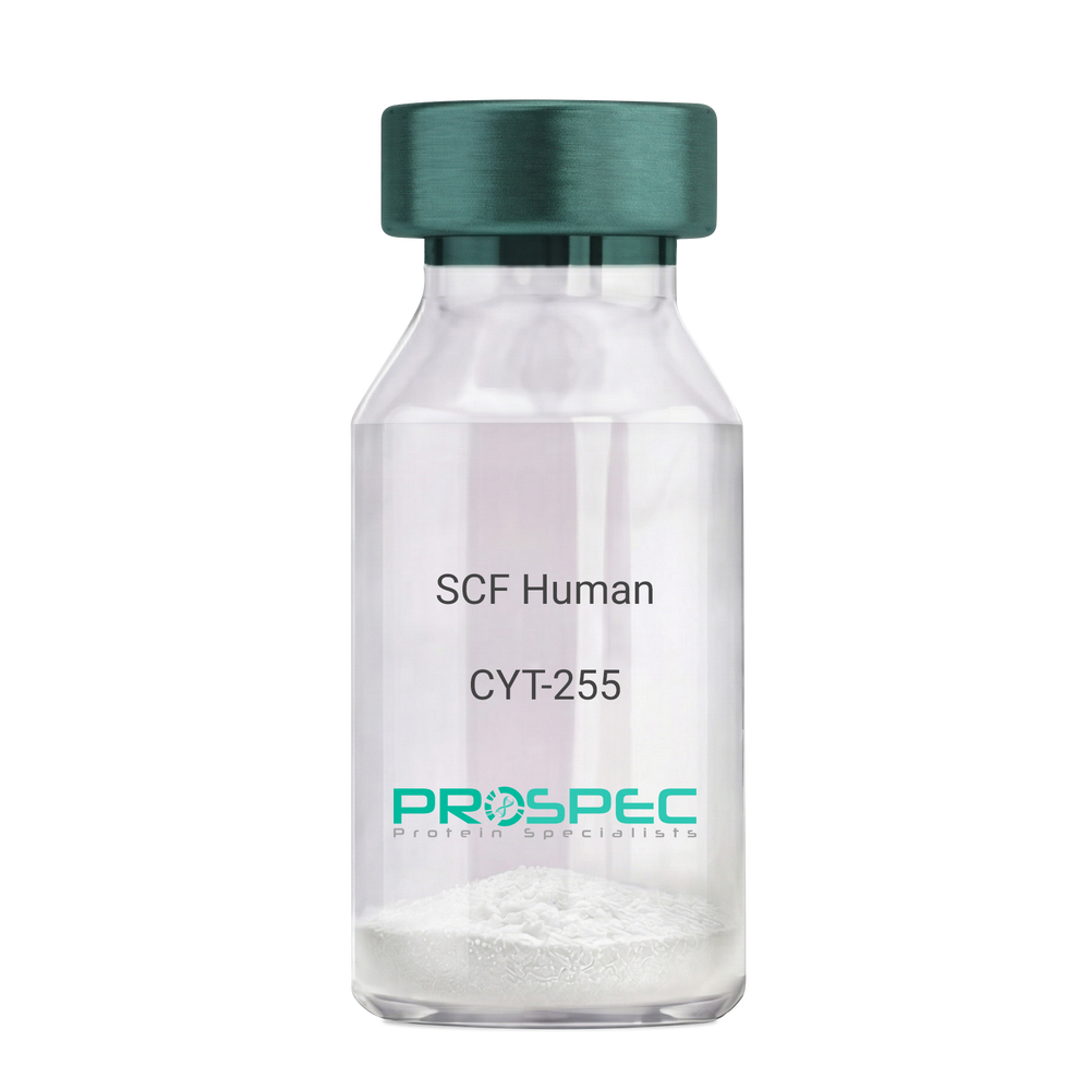
- View Data Sheet
Name :
SCF HumanDescription:
Stem Cell Factor Human Recombinant
Kit ligand Precursor, C-kit ligand, SCF, Mast cell growth factor, MGF, SF, KL-1, Kitl, DKFZp686F2250.
Product # :
CYT-255Price :
Quantity :
Shipping Method :
Shipped at Room temp
More Info
- description
- source
- formulation
- purity
- biological activity
- More Info
Description
Stem Cell Factor Human Recombinant produced in E.Coli is a single, non-glycosylated polypeptide chain containing 165 amino acids and having a molecular mass of 18409 Dalton. The SCF is purified by proprietary chromatographic techniques.
Source
Escherichia Coli.
Formulation
Lyophilized from a concentrated (1mg/ml) solution in water containing 0.02% NaHCO3.
Purity
Greater than 98.0% as determined by:
(a) Analysis by SEC-HPLC.
(b) Analysis by SDS-PAGE.Biological Activity
The ED50 as determined by the dose-dependant stimulation of Human TF-1 cells is < 2 ng/ml, corresponding to a Specific Activity of 500,000 IU/mg.More Info
-
Introduction
Stem cell factor / KIT ligand (SCF) is a cytokine which binds CD117(c-Kit). SCF is also known as "steel factor" or "c-kit ligand". SCF exists in two forms, cell surface bound SCF and soluble (or free) SCF. Soluble SCF is produced by the cleavage of surface bound SCF by metalloproteases. SCF is a growth factor important for the survival, proliferation, and differentiation of hematopoietic stem cells and other hematopoietic progenitor cells. One of its roles is to change the BFU-E (burst-forming unit-erythroid) cells, which are the earliest erythrocyte precursors in the erythrocytic series, into the CFU-E (colony-forming unit-erythroid).
-
Synonyms
Kit ligand Precursor, C-kit ligand, SCF, Mast cell growth factor, MGF, SF, KL-1, Kitl, DKFZp686F2250.
-
Physical Appearance
Sterile Filtered White lyophilized (freeze-dried) powder.
-
Stability
Lyophilized KIT ligand although stable at room temperature for 3 weeks, should be stored desiccated below -18°C. Upon reconstitution SCF should be stored at 4°C between 2-7 days and for future use below -18°C.For long term storage it is recommended to add a carrier protein (0.1% HSA or BSA).Please prevent freeze-thaw cycles.
-
Solubility
It is recommended to reconstitute the lyophilized Stem Cell Factor in sterile 18MΩ-cm H2O not less than 100µg/ml, which can then be further diluted to other aqueous solutions.
-
Amino Acid Sequence
The sequence of the first five N-terminal amino acids was determined and was found to be Met-Glu-Gly-Ile-Cys.
-
Protein content
Protein quantitation was carried out by two independent methods1. UV spectroscopy at 280 nm using the absorbency value of 0.52 as the extinction coefficient for a 0.1% (1mg/ml) solution. This value is calculated by the PC GENE computer analysis program of protein sequences (IntelliGenetics). 2. Analysis by RP-HPLC, using a standard solution of Stem Cell Factor as a Reference Standard.
ProSpec's products are furnished for LABORATORY RESEARCH USE ONLY. The product may not be used as drugs, agricultural or pesticidal products, food additives or household chemicals.
- View Data Sheet
Name :
PDGFD HumanDescription:
Platelet Derived Growth Factor-D Human Recombinant
Platelet Derived Growth Factor D, Spinal Cord-Derived Growth Factor B, Iris-Expressed Growth Factor, SCDGF-B, IEGF, PDGF-D, MSTP036.
Product # :
CYT-155Price :
Quantity :
Shipping Method :
Shipped with Ice Packs
More Info
- description
- source
- formulation
- purity
- More Info
Description
PDGFD Human Recombinant produced in E. coli is a single polypeptide chain containing 146 amino acids (250-370) and having a molecular mass of 16.6 kDa.PDGFD is fused to a 25 amino acid His-tag at N-terminus & purified by proprietary chromatographic techniques.
Source
E.coli.
Formulation
The PDGFD solution (1mg/1ml) contains 20mM Tris-HCl buffer (pH 8.0), 0.4M Urea and 10% glycerol.
Purity
Greater than 90% as determined by SDS-PAGE.
More Info
-
Introduction
Platelet-derived growth factor D (PDGFD) belongs to the platelet-derived growth factor family. PDGFD gene product only forms homodimers and, thus, does not dimerize with the other 3 family members. PDGFD has an imperative role in wound healing. PDGFD induces macrophage recruitment, increased interstitial pressure, and blood vessel maturation during angiogenesis. PDGFD initiates events which lead to a mesangial proliferative glomerulonephritis, including influx of monocytes and macrophages and production of extracellular matrix. The 4 members of the PDGF family are mitogenic factors for cells of mesenchymal origin and are distinguished by a core motif of eight cysteines, 7 of which are found in this factor. PDGFD differs from alpha and beta members of this family by having an odd N-terminal domain, the CUB domain.
-
Synonyms
Platelet Derived Growth Factor D, Spinal Cord-Derived Growth Factor B, Iris-Expressed Growth Factor, SCDGF-B, IEGF, PDGF-D, MSTP036.
-
Physical Appearance
Sterile Filtered colorless solution.
-
Stability
Store at 4°C if entire vial will be used within 2-4 weeks. Store, frozen at -20°C for longer periods of time. For long term storage it is recommended to add a carrier protein (0.1% HSA or BSA).Avoid multiple freeze-thaw cycles.
-
Amino Acid Sequence
MGSSHHHHHH SSGLVPRGSH MGSHMSYHDR KSKVDLDRLN DDAKRYSCTP RNYSVNIREE LKLANVVFFP RCLLVQRCGG NCGCGTVNWR SCTCNSGKTV KKYHEVLQFE PGHIKRRGRA KTMALVDIQL DHHERCDCIC SSRPPR.
ProSpec's products are furnished for LABORATORY RESEARCH USE ONLY. The product may not be used as drugs, agricultural or pesticidal products, food additives or household chemicals.
- View Data Sheet
Name :
VEGF C Rat (152 a.a.)Description:
Vascular Endothelial Growth Factor-C (152 a.a) Rat Recombinant
VEGF-C152, Vascular endothelial growth factor C 152, VRP, Flt4 ligand, Flt4-L.
Product # :
CYT-284Price :
Quantity :
Shipping Method :
Shipped at Room temp
More Info
- description
- source
- formulation
- purity
- biological activity
- More Info
Description
Vascular Endothelial Growth Factor -C 152 Rat Recombinant contains 152 amino acids residues and was fused to a His-tag (6x His) at the C-terminal end. As a result of glycosylation VEGF-C migrates as an 18-24 kDa protein in SDS-PAGE under reducing conditions.
Source
Sf9, Insect Cells.
Formulation
The protein was lyophilized from a concentrated (1mg/ml) solution with BSA.
Purity
Greater than 90.0% as determined by:
(a) Analysis by RP-HPLC.
(b) Analysis by SDS-PAGE.Biological Activity
Measured by its ability to stimulate phosphorylation of the VEGFR-3/FLT-4 receptor in porcine aortic endothelial cells (PAE/FLT -4 cells). The ED50 for this effect is typically 150-300 ng/ml, corresponding to a specific activity of 3.3-6.6KUnits/mg.More Info
-
Introduction
VEGF-C152S is a point mutant generated by the replacement of the second conserved Cys residue of the recombinant processed VEGF-C by a Ser residue. VEGF-C 152S is analog to the human VEGF-C 156S mutant and only active toward VEGFR-3/FLT-4 but, unlike wild type VEGF-C, is unable to bind to and to activate signaling through VEGFR-2/KDR. VEGF-C152S was inactive in the vascular permeability assay and did not increase migration of the capillary endothelial cells, indicating that these VEGF-like effects of VEGF-C require VEGFR-2 binding. VEGF-C, also known as Vascular Endothelial Growth Factor Related Protein (VRP), is a recently discovered VEGF growth factor family member that is most closely related to VEGF-D. The rat VEGF-C cDNA encodes a pre-pro-protein of 416 amino acids residues. It is almost identical to the mouse VEGF-C protein. Similar to VEGF-D, VEGF-C has a VEGF homology domain spanning the middle third of the precursor molecule and long N- and C-terminal extensions. In adults, VEGF-C is highly expressed in heart, placenta, ovary and small intestine. Recombinant rat VEGF-C, lacking the N- and C-terminal extensions and containing only the middle VEGF homology domain, forms primarily non-covalently linked dimers. This protein is a ligand for both VEGFR-2/KDR and VEGFR-3/FLT -4. Since VEGFR-3 is strongly expressed in lymphatic endothelial cells, it has been postulated that VEGF-C is involved in the regulation of the growth and/or differentiation of lymphatic endothelium. Although recombinant rat VEGF-C is also a mitogen for vascular endothelial cells, it is much less potent than VEGF-A.
-
Synonyms
VEGF-C152, Vascular endothelial growth factor C 152, VRP, Flt4 ligand, Flt4-L.
-
Physical Appearance
Sterile Filtered White lyophilized (freeze-dried) powder.
-
Stability
Lyophilized Vascular Endothelial Growth Factor-C152 although stable at room temperature for 3 weeks, should be stored desiccated below -18°C. Upon reconstitution VEGF-C 152 should be stored at 4°C between 2-7 days and for future use below -18°C.Please prevent freeze-thaw cycles.
-
Solubility
It is recommended to reconstitute the lyophilized Vascular Endothelial Growth Factor C 152 in sterile 18MΩ-cm H2O not less than 100µg/ml, which can then be further diluted to other aqueous solutions.
ProSpec's products are furnished for LABORATORY RESEARCH USE ONLY. The product may not be used as drugs, agricultural or pesticidal products, food additives or household chemicals.
- View Data Sheet
Name :
FLT4 HumanDescription:
Vascular Endothelial Growth Factor Receptor-3 Human Recombinant
Tyrosine-protein kinase receptor FLT4, PCL, FLT41, FMS-LIKE TYROSINE KINASE 4, VEGFR-3, VEGFR3.
Product # :
PKA-244Price :
Quantity :
Shipping Method :
Shipped at Room temp
More Info
- description
- source
- formulation
- purity
- biological activity
- More Info
Description
Soluble FLT4 Human Recombinant fused with a carboxy-terminal 6X histidine-tag produced in baculovirus is a monomeric, glycosylated, polypeptide containing the extracellular part, 25-774 amino acids and having a total molecular mass of 120 kDa. The soluble receptor protein contains only the first 7 extracellular domains, which contain all the information necessary for ligand binding. The FLT4 is purified by proprietary chromatographic techniques.
Source
Insect Cells.
Formulation
FLT4 was lyophilized from a concentrated (1mg/ml) sterile solution containing 1xPBS.
Purity
Greater than 90.0% as determined by SDS-PAGE.
Biological Activity
Measured by its ability to bind recombinant rat VEGF-C in a functional solid phase binding assay. Immobilised recombinant human VEGFR-3/FLT-4 at 5 µg/ml can bind recombinant rat VEGF-C in a linear range of 8-500 ng/ml.More Info
-
Introduction
All three VEGF receptors belong to the class III subfamily of receptor tyrosine kinases (RTKs) characterised by the seven immunoglobulin-like loops in the extracellular domain. The expression of VEGFR-1 to -3 is almost exclusively restricted to hematopoietic precursor cells, vascular and lymphatic endothelial cells and to the monocyte/macrophage lineage. They play key roles in vasculogenesis, hematopoiesis, angiogenesis and lymphangiogenesis. The FLT-4 cDNA encodes a 1298 amino acid (aa) residue precursor protein with a 23 aa residue signal peptide. Mature VEGFR-3/FLT-4 is composed of a 751 aa residue extracellular domain, a 22 aa transmembrane domain and a 482 aa residue cytoplasmic domain. Both VEGF family members VEGF-C and VEGF-D have been shown to bind and activate VEGFR-3/FLT-4. The Flt-4 gene is widely expressed in the early embryo but becomes restricted to the lymphatic endothelial a latter stages of development. It is important for lymphangiogenesis.
-
Synonyms
Tyrosine-protein kinase receptor FLT4, PCL, FLT41, FMS-LIKE TYROSINE KINASE 4, VEGFR-3, VEGFR3.
-
Physical Appearance
Sterile Filtered White lyophilized (freeze-dried) powder.
-
Stability
Lyophilized FLT4 although stable at room temperature for 3 weeks, should be stored desiccated below -18°C. Upon reconstitution FLT4 should be stored at 4°C between 2-7 days and for future use below -18°C.For long term storage it is recommended to add a carrier protein (0.1% HSA or BSA).Please prevent freeze-thaw cycles.
-
Solubility
It is recommended to reconstitute the lyophilized FLT4 in sterile water not less than 100 µg/ml, which can then be further diluted to other aqueous solutions.
ProSpec's products are furnished for LABORATORY RESEARCH USE ONLY. The product may not be used as drugs, agricultural or pesticidal products, food additives or household chemicals.
- View Data Sheet
Name :
GDF5 Mouse, HisDescription:
Growth differentiation factor 5 Mouse Recombinant, His Tag
Bmp-14, Bp, GDF-5, Bone morphogenetic protein 14, GDF5.
Product # :
CYT-852Price :
Quantity :
Shipping Method :
Shipped with Ice Packs
More Info
- description
- source
- formulation
- purity
- More Info
- sds-page
Description
GDF5 Mouse Recombinant produced in E.Coli is a single, non-glycosylated polypeptide chain containing 143 amino acids (376-495 a.a) and having a molecular mass of 16kDa.GDF5 is fused to a 23 amino acid His-tag at N-terminus & purified by proprietary chromatographic techniques.
Source
Escherichia Coli.
Formulation
GDF5 protein solution (0.5mg/ml) containing 20mM Tris-HCl buffer (pH 8.0) and 10% glycerol.
Purity
Greater than 90.0% as determined by SDS-PAGE.
sds-page
More Info
-
Introduction
GDF-5 is a member of the bone morphogenetic protein (BMP) family and the TGF-beta superfamily. This group of proteins is characterized by a polybasic proteolytic processing site which is cleaved to produce a mature protein containing seven conserved cysteine residues. The members of this family are regulators of cell growth and differentiation in both embryonic and adult tissues. Mutations in this gene are associated with acromesomelic dysplasia, Hunter-Thompson type; brachydactyly, type C; and chondrodysplasia, Grebe type. These associations confirm that the gene product plays a role in skeletal development.
-
Synonyms
Bmp-14, Bp, GDF-5, Bone morphogenetic protein 14, GDF5.
-
Physical Appearance
Sterile Filtered colorless solution.
-
Stability
Store at 4°C if entire vial will be used within 2-4 weeks. Store, frozen at -20°C for longer periods of time. For long term storage it is recommended to add a carrier protein (0.1% HSA or BSA).Avoid multiple freeze-thaw cycles.
-
Amino Acid Sequence
MGSSHHHHHH SSGLVPRGSH MGSAPLANRQ GKRPSKNLKA RCSRKALHVN FKDMGWDDWI IAPLEYEAFH CEGLCEFPLR SHLEPTNHAV IQTLMNSMDP ESTPPTCCVP TRLSPISILF IDSANNVVYK QYEDMVVESC GCR.
-
Background
What is the molecular weight/Mw of GDF5 MOUSE Protein?
GDF5 MOUSE Protein has a total Mw of 16kDa.
What is the source or expression system of GDF5 MOUSE Protein?
Escherichia Coli.
What is the Purity of GDF5 MOUSE Protein?
GDF5 MOUSE Protein is >90% pure as determined by SDS-PAGE.
What is the Biological Activity of GDF5 MOUSE Protein?
The biological functionality of GDF5 MOUSE Protein will be determined in the future.
What is the amino acid sequence of GDF5 MOUSE Protein?
MGSSHHHHHH SSGLVPRGSH MGSAPLANRQ GKRPSKNLKA RCSRKALHVN FKDMGWDDWI IAPLEYEAFH CEGLCEFPLR SHLEPTNHAV IQTLMNSMDP ESTPPTCCVP TRLSPISILF IDSANNVVYK QYEDMVVESC GCR.
What applications can GDF5 MOUSE Protein be used in?
GDF5 MOUSE Protein can probably be used in western blot, ELISA and Lateral Flow.
What is the endotoxin level for GDF5 MOUSE Protein?
The endotoxin level is minimal, GDF5 MOUSE Protein was purified using conventional chromatography techniques.
ProSpec's products are furnished for LABORATORY RESEARCH USE ONLY. The product may not be used as drugs, agricultural or pesticidal products, food additives or household chemicals.
- View Data Sheet
Name :
TGFB1 Human, HEKDescription:
Transforming Growth Factor-Beta 1 Human Recombinant, HEK
Transforming growth factor beta-1, TGF-beta-1, CED, DPD1, TGFB, TGF-b 1, LAP, TGFB1.
Product # :
CYT-1260Price :
Quantity :
Shipping Method :
Shipped at Room temp
More Info
- description
- source
- formulation
- purity
- biological activity
- More Info
Description
TGFB1 Human Recombinant produced in 293 cells is a glycosylated homodimer polypeptide chain containing 2 x 112 amino acids and having a total molecular mass of 25.0kDa. The TGFB1 is purified by proprietary chromatographic techniques.
Source
HEK 293 cells.
Formulation
Lyophilized from a sterile filtered solution containing TFA (0.1%).
Purity
Greater than 98.0% as determined by: (a) Analysis by HPLC. (b) Analysis by SDS-PAGE.
Biological Activity
The ED50 as determined by the dose-dependent inhibition of IL-4-induced proliferation of HT-2 cells is ≤ 0.05 ng/ml, corresponding to a specific activity of ≥ 2 x 10⁷ units/mg.
More Info
-
Synonyms
Transforming growth factor beta-1, TGF-beta-1, CED, DPD1, TGFB, TGF-b 1, LAP, TGFB1.
-
Physical Appearance
Sterile Filtered White lyophilized (freeze-dried) powder.
-
Stability
Lyophilized TGFB1 although stable at room temperature for 3 weeks, should be stored desiccated below -18°C. Upon reconstitution TGFB1 Human should be stored at 4°C between 2-7 days and for future use below -18°C. For long term storage it is recommended to add a carrier protein (0.1% HSA or BSA). Please prevent freeze-thaw cycles.
-
Solubility
It is recommended to reconstitute the lyophilized TGFB1 in sterile in 18MΩ-cm H2O at a concentration of 0.1 mg/ml, which can then be further diluted to other aqueous solutions.
-
Amino Acid Sequence
ALDTNYCFSS TEKNCCVRQL YIDFRKDLGW KWIHEPKGYH ANFCLGPCPY IWSLDTQYSK VLALYNQHNP GASAAPCCVP QALEPLPIVY YVGRKPKVEQ LSNMIVRSCK CS.
-
Background
Transforming growth factor betas (TGF Betas) mediate many cell-cell interactions that occur during embryonic development. 3 TGF Betas have been identified in mammals: TGF Beta 1, TGF Beta 2 and TGF Beta 3. each are synthesized as precursor proteins that are very similar in that each is cleaved to yield a 112 amino acid polypeptide that remains associated with the latent portion of the molecule.
ProSpec's products are furnished for LABORATORY RESEARCH USE ONLY. The product may not be used as drugs, agricultural or pesticidal products, food additives or household chemicals.
- View Data Sheet
Name :
EGF Mouse, BiotinDescription:
Epidermal Growth Factor Mouse Recombinant, Biotin
Urogastrone, URG, EGF.
Product # :
CYT-841Price :
Quantity :
Shipping Method :
Shipped with Ice Packs
More Info
- description
- source
- formulation
- purity
- biological activity
- More Info
Description
EGF Mouse Recombinant, Biotin produced in E.Coli is a non-glycosylated polypeptide chain containing 61 amino acids and having a total molecular mass of 7.0kDa. This version of EGF has a N terminal leader sequence hosting a biotin conjugation. There are 0.5 biotins for each EGF protein.
Source
Escherichia Coli.
Formulation
The protein (0.5mg/ml) solution contains sterile PBS.
Purity
Greater than 95.0% as determined by analysis by SDS-PAGE.
Biological Activity
The ED50 as determined by the dose-dependent proliferation of mouse BALB/c 3T3 cells is 0.14-0.2ng/ml, corresponding to a specific activity of 7.1x10⁶units/mg.More Info
-
Introduction
Epidermal growth factor has a profound effect on the differentiation of specific cells in vivo and is a potent mitogenic factor for a variety of cultured cells of both ectodermal and mesodermal origin. The EGF precursor is believed to exist as a membrane-bound molecule which is proteolytically cleaved to generate the 53-amino acid peptide hormone that stimulates cells to divide. EGF stimulates the growth of various epidermal and epithelial tissues in vivo and in vitro and of some fibroblasts in cell culture.
-
Synonyms
Urogastrone, URG, EGF.
-
Physical Appearance
Sterile Filtered clear solution.
-
Stability
Should be stored at 4°C.Please do not freeze.
-
Amino Acid Sequence
MKKIDDDKNS YPGCPSSYDG YCLNGGVCMH IESLDSYTCN CVIGYSGDRC QTRDLEWWEL R.
-
Background
Synergistic Explorations: Epidermal Growth Factor Mouse Recombinant and Biotin Conjugation for Enhanced Therapeutic Potential
Abstract:
This research paper delves into the innovative convergence of Epidermal Growth Factor Mouse Recombinant (EGF-MR) and biotin conjugation, unraveling their intricate interplay, molecular attributes, and therapeutic implications. By employing cutting-edge methodologies involving protein engineering, conjugation chemistry, and cellular assays, this study uncovers the augmented cellular responses driven by EGF-MR-biotin complex. The findings highlight a novel avenue for tailored regenerative medicine and targeted therapy.
Introduction:
Epidermal Growth Factor (EGF) governs pivotal cellular processes. This paper navigates the unexplored realm of Epidermal Growth Factor Mouse Recombinant (EGF-MR) in synergy with biotin conjugation, elucidating their combined molecular attributes and therapeutic potential.
Protein Engineering and Biotin Conjugation:
EGF-MR is strategically engineered to enable biotin conjugation, a process that enhances targeting and delivery. This paper delves into site-specific modification approaches, ensuring precise and controlled conjugation of biotin moieties to EGF-MR.
Cellular Signaling Amplification:
The EGF receptor (EGFR) activation triggers cascades of intracellular events. Structural studies and binding kinetics illuminate how the biotin-conjugated EGF-MR modulates EGFR interactions, amplifying downstream signaling pathways like the MAPK and PI3K/Akt cascades.
Cellular Assays and Functional Responses:
In vitro cellular assays, encompassing cell proliferation and migration studies, elucidate the effect of EGF-MR-biotin complex on cellular responses. Live-cell imaging techniques reveal enhanced cell motility and survival, underpinning the potential therapeutic impact.
Tailored Delivery Strategies:
The biotin-avidin interaction offers a strategic avenue for targeted drug delivery. Employing this interaction, EGF-MR-biotin complex can be directed to specific cell types, revolutionizing precision medicine and enabling tailored therapeutic interventions.
Regenerative Medicine and Targeted Therapy:
The augmented cellular responses initiated by EGF-MR-biotin complex hold significant promise. In regenerative medicine, the complex's potential to accelerate tissue regeneration becomes evident. Furthermore, in targeted therapy, the complex's enhanced cellular uptake offers a novel approach to modulate tumor microenvironments.
Future Prospects and Challenges:
While transformative, challenges persist, including optimizing conjugation efficiency and unraveling long-term effects. Future research should focus on refining delivery strategies and conducting comprehensive long-term studies to harness the full therapeutic potential.
Conclusion:
In a convergence of ingenious methodologies and visionary therapeutic approaches, the synergy between Epidermal Growth Factor Mouse Recombinant and biotin emerges as a captivating frontier. The molecular marriage between EGF-MR and biotin not only amplifies cellular responses but also opens doors for targeted interventions and precision therapies, revolutionizing the landscape of medical advancements.
What is the molecular weight/Mw of MEGF, BIOTIN Protein?
MEGF, BIOTIN Protein has a total Mw of 7kDa.
What is the source or expression system of MEGF, BIOTIN Protein?
Escherichia Coli.
What is the Purity of MEGF, BIOTIN Protein?
MEGF, BIOTIN Protein is >95% pure as determined by SDS-PAGE.
What is the Biological Activity of MEGF, BIOTIN Protein?
The ED50 as determined by the dose-dependent proliferation of mouse BALB/c 3T3 cells is 0.14-0.2ng/ml, corresponding to a specific activity of 7.1x10⁶units/mg.
What is the amino acid sequence of MEGF, BIOTIN Protein?
MKKIDDDKNS YPGCPSSYDG YCLNGGVCMH IESLDSYTCN CVIGYSGDRC QTRDLEWWEL R.
What applications can MEGF, BIOTIN Protein be used in?
MEGF, BIOTIN Protein can probably be used in western blot, ELISA and Lateral Flow.
What is the endotoxin level for MEGF, BIOTIN Protein?
The endotoxin level is minimal, MEGF, BIOTIN Protein was purified using conventional chromatography techniques.
ProSpec's products are furnished for LABORATORY RESEARCH USE ONLY. The product may not be used as drugs, agricultural or pesticidal products, food additives or household chemicals.
- View Data Sheet
Name :
FGFR4 HumanDescription:
Fibroblast Growth Factor Receptor 4 Fc Chimera Human Recombinant
Fibroblast Growth Factor Receptor 4, EC 2.7.10.1, JTK2, TKF, Tyrosine Kinase Related To Fibroblast Growth Factor Receptor, Hydroxyaryl-Protein Kinase, Protein-Tyrosine Kinase, Tyrosylprotein Kinase, CD334 Antigen, EC 2.7.10, FGFR-4, CD334, FGFR4.
Product # :
PKA-233Price :
Quantity :
Shipping Method :
Shipped at Room temp
More Info
- description
- source
- formulation
- purity
- biological activity
- More Info
Description
Soluble FGFR-4a (IIIc) Fc Chimera Human Recombinant fused with Xa cleavage site with the Fc part of human IgG1 produced in baculovirus is a heterodimeric, glycosylated, Polypeptide chain and having a molecular mass of 170 kDa. The FGFR4 is purified by proprietary chromatographic techniques.
Source
Insect Cells.
Formulation
CD334 was lyophilized from a concentrated (1mg/ml) sterile solution containing no additives.
Purity
Greater than 90.0% as determined by:
(a) Analysis by RP-HPLC.
(b) Analysis by SDS-PAGE.Biological Activity
Determined by its ability to inhibit human FGF acidic-dependent proliferation on R1 cells. The ED50 for this effect is typically at 15.0-30.0 ng/ml.More Info
-
Introduction
Fibroblast growth factors (FGFs) comprise a family of at least eighteen structurally related proteins that are involved in a multitude of physiological and pathological cellular processes, including cell growth, differentiation, angiogenesis, wound healing and tumorgenesis. The biological activities of the FGFs are mediated by a family of type I transmembrane tyrosine kinases which undergo dimerization and autophosphorylation after ligand binding. Four distinct genes encoding closely related FGF receptors, FGF R1 - 4, are known. All four genes for FGF Rs encode proteins with an N-terminal signal peptide, three immunoglobulin (Ig)-like domains, an acid-box region containing a run of acidic residues between the IgI and IgII domains, a transmembrane domain and the split tyrosine-kinase domain. Multiple forms of FGF R1 - 3 are generated by alternative splicing of the mRNAs. A frequent splicing event involving FGF R1 and 2 results in receptors containing all three Ig domains, referred to as the a isoform, or only IgII and IgIII, referred to as the b isoform. Only the a isoform has been identified for FGF R3 and FGF R4. Additional splicing events for FGF R1 - 3, involving the C-terminal half of the IgIII domain encoded by two mutually exclusive alternative exons, generate FGF receptors with alternative IgIII domains (IIIb and IIIc). A IIIa isoform which is a secreted FGF binding protein containing only the N-terminal half of the IgIII domain plus some intron sequences has also been reported for FGF R1. Mutations in FGF R1 - 3 have been found in patients with birth defects involving craniosynostosis. The complex patterns of expression of these receptors as well as the specificity of their interactions with the various FGF ligand family members are under investigation.
-
Synonyms
Fibroblast Growth Factor Receptor 4, EC 2.7.10.1, JTK2, TKF, Tyrosine Kinase Related To Fibroblast Growth Factor Receptor, Hydroxyaryl-Protein Kinase, Protein-Tyrosine Kinase, Tyrosylprotein Kinase, CD334 Antigen, EC 2.7.10, FGFR-4, CD334, FGFR4.
-
Physical Appearance
Sterile Filtered White lyophilized (freeze-dried) powder.
-
Stability
Lyophilized FGFR4 although stable at room temperature for 3 weeks, should be stored desiccated below -18°C. Upon reconstitution FGFR4 should be stored at 4°C between 2-7 days and for future use below -18°C.For long term storage it is recommended to add a carrier protein (0.1% HSA or BSA).Please prevent freeze-thaw cycles.
-
Solubility
It is recommended to reconstitute the lyophilized FGFR-4 in sterile PBS not less than 100 µg/ml, which can then be further diluted to other aqueous solutions.
ProSpec's products are furnished for LABORATORY RESEARCH USE ONLY. The product may not be used as drugs, agricultural or pesticidal products, food additives or household chemicals.
- View Data Sheet
Name :
PDGF BB Human, YeastDescription:
Platelet Derived Growth Factor-BB Human Recombinant, Yeast
Glioma-derived growth factor, GDGF, Osteosarcoma-derived Growth Factor, ODGF, SIS, SSV, PDGF2, c-sis, FLJ12858, PDGF-BB, PDGF B-chain, Platelet-derived growth factor beta polypeptide, Becaplermin.
Product # :
CYT-242Price :
Quantity :
Shipping Method :
Shipped at Room temp
More Info
- description
- source
- formulation
- purity
- biological activity
- More Info
Description
Platelet-Derived Growth Factor BB Human Recombinant is a glycosylated homodimer produced in Saccharomyces cerevisiae, containing 2x10⁹ amino acids and having a molecular mass of 32kDa. PDGF-BB is purified by proprietary chromatographic techniques.
Source
Saccharomyces cerevisiae.
Formulation
The PDGF-BB was lyophilized from 0.2 µm filtered solution in 20mM sodium phosphate in absence of any carrier protein.
Purity
Greater than 98.0% as determined by SDS-PAGE.
Biological Activity
The ED50, as measured by its ability to stimulate proliferation of mouse 3T3 fibroblasts is typically 1 ng/ml, corresponding to a Specific Activity of 1,000,000IU/mg.More Info
-
Introduction
PDGF-BB is a member of the platelet-derived growth factor family. The four members of this family are mitogenic factors for cells of mesenchymal origin and are characterized by a motif of eight cysteines. This gene product can exist either as a homodimer (PDGF-BB) or as a heterodimer with the platelet-derived growth factor alpha polypeptide (PDGF-AB), where the dimers are connected by disulfide bonds. Mutations in this gene are associated with meningioma. Reciprocal translocations between chromosomes 22 and 7, at sites where this gene and that for COL1A1 are located, are associated with a particular type of skin tumor called dermatofibrosarcoma protuberans resulting from unregulated expression of growth factor. Two splice variants have been identified for this gene.
-
Synonyms
Glioma-derived growth factor, GDGF, Osteosarcoma-derived Growth Factor, ODGF, SIS, SSV, PDGF2, c-sis, FLJ12858, PDGF-BB, PDGF B-chain, Platelet-derived growth factor beta polypeptide, Becaplermin.
-
Physical Appearance
Sterile Filtered White lyophilized (freeze-dried) powder.
-
Stability
Lyophilized Platelet-derived Growth Factor BB although stable at room temperature for 3 weeks, should be stored desiccated below -18°C. Upon reconstitution PDGF BB should be stored at 4°C between 2-7 days and for future use below -18°C.Please prevent freeze-thaw cycles.
-
Solubility
It is recommended to reconstitute the lyophilized Platelet-derived Growth Factor-BB in sterile 18M-cm H2O not less than 100µg/ml, which can then be further diluted to other aqueous solutions.
-
Amino Acid Sequence
The sequence of the first five N-terminal amino acids was determined and was found to be Ser-Leu-Gly-Ser-Leu.
ProSpec's products are furnished for LABORATORY RESEARCH USE ONLY. The product may not be used as drugs, agricultural or pesticidal products, food additives or household chemicals.
- View Data Sheet
Name :
FGFR1 Human, (22-285)Description:
Fibroblast Growth Factor Receptor-1 Human Recombinant, (22-285 a.a.)
FGFR-1, bFGF-R, C-FGR, CD331, fms-related tyrosine kinase 2, Pfeiffer syndrome, CEK, FLG, FLT2, KAL2, BFGFR, FGFBR, HBGFR, FGFR1/FGFR1OP2 FUSION GENE, FGFR1/ZNF198 FUSION GENE, FLG FGFR1/BCR FUSION GENE, FLG protein, FMS-LIKE GENE, N-sam tyrosine kinase, basic fibroblast growth factor receptor 1.
Product # :
PKA-114Price :
Quantity :
Shipping Method :
Shipped with Ice Packs
More Info
- description
- source
- formulation
- purity
- More Info
Description
FGFR1 Human Recombinant produced in Sf9 Baculovirus cells is a single, glycosylated polypeptide chain containing 272 amino acids (22-285) and having a molecular mass of 30.4kDa. (Molecular size on SDS-PAGE will appear at approximately 40-57kDa). FGFR1 is fused to a 8 amino acid His-tag at C-terminus & purified by proprietary chromatographic techniques.
Source
Sf9, Baculovirus cells.
Formulation
The FGFR1 solution (0.25mg/1ml) contains phosphate buffered Saline (pH7.4), and 10% glycerol.
Purity
Greater than 90.0% as determined by SDS-PAGE.
More Info
-
Introduction
Fibroblast Growth Factors (FGFs) comprise a family of at least 18 structurally related proteins that are involved in a multitude of physiological and pathological cellular processes, including cell growth, differentiation, angiogenesis, wound healing and tumorigenesis. The biological activities of the FGFs are mediated by a family of type I transmembrane tyrosine kinases which undergo dimerization and autophosphorylation after ligand binding. Multiple forms of FGFR-1 to -3 are generated by alternative splicing of the mRNAs. A frequent splicing event involving FGFR-1 and -2 results in receptors containing all three Ig domains, referred to as the alpha isoform, or only IgII and IgIII, referred to as the ? isoform. Only the alpha isoform has been identified for FGFR-3 and FGFR-4. Additional splicing events for FGFR-1 to -3, involving the C-terminal half of the IgIII domain encoded by two mutually exclusive alternative exons, generate FGF receptors with alternative IgIII domains (IIIb and IIIc). An IIIa isoform which is a secreted FGF binding protein containing only the N-terminal half of the IgIII domain plus some intron sequences has also been reported for FGFR-1. Mutations in FGFR-1 to -3 have been found in patients with birth defects involving craniosynostosis.
-
Synonyms
FGFR-1, bFGF-R, C-FGR, CD331, fms-related tyrosine kinase 2, Pfeiffer syndrome, CEK, FLG, FLT2, KAL2, BFGFR, FGFBR, HBGFR, FGFR1/FGFR1OP2 FUSION GENE, FGFR1/ZNF198 FUSION GENE, FLG FGFR1/BCR FUSION GENE, FLG protein, FMS-LIKE GENE, N-sam tyrosine kinase, basic fibroblast growth factor receptor 1.
-
Physical Appearance
Sterile Filtered colorless solution.
-
Stability
Store at 4°C if entire vial will be used within 2-4 weeks.Store, frozen at -20°C for longer periods of time. For long term storage it is recommended to add a carrier protein (0.1% HSA or BSA).Avoid multiple freeze-thaw cycles.
-
Amino Acid Sequence
RPSPTLPEQD ALPSSEDDDD DDDSSSEEKE TDNTKPNPVA PYWTSPEKME KKLHAVPAAK TVKFKCPSSG TPNPTLRWLK NGKEFKPDHRIGGYKVRYAT WSIIMDSVVP SDKGNYTCIV ENEYGSINHT YQLDVVERSP HRPILQAGLP ANKTVALGSN VEFMCKVYSD PQPHIQWLKH IEVNGSKIGP DNLPYVQILK TAGVNTTDKE MEVLHLRNVS FEDAGEYTCL AGNSIGLSHH SAWLTVLEAL EERPAVMTSP LYLELEHHHH HH.
ProSpec's products are furnished for LABORATORY RESEARCH USE ONLY. The product may not be used as drugs, agricultural or pesticidal products, food additives or household chemicals.
- View Data Sheet
Name :
G CSF Human, HEKDescription:
Granulocyte-Colony Stimulating Factor Human Recombinant, HEK
CSF-3, MGI-1G, GM-CSF beta, Pluripoietin, Filgrastim, Lenograstim, G-CSF, MGC45931, GCSF.
Product # :
CYT-088Price :
Quantity :
Shipping Method :
Shipped at Room temp
More Info
- description
- source
- formulation
- purity
- biological activity
- More Info
Description
G-CSF Human Recombinant produced in HEK cells is a glycosylated monomer, having a molecular weight range of 21-25kDa due to glycosylation.The G-CSF is purified by proprietary chromatographic techniques.
Source
HEK.
Formulation
The G-CSF was lyophilized from 1mg/ml in 1xPBS.
Purity
Greater than 95% as obsereved by SDS-PAGE.
Biological Activity
The specific activity was determined by the dose-dependent stimulation of the proliferation of murine M-NFS-60 cells (Mouse Myeloid Leukemia indicator cell line), the ED50 is <0.01ng/ml.
More Info
-
Introduction
GCSF is a cytokine that controls the production, differentiation, and function of granulocytes. The active protein is found extracellularly. Three transcript variants encoding three different isoforms have been found for the GCSF gene. Granulocyte/macrophage colony-stimulating factors are cytokines that act in hematopoiesis by controlling the production, differentiation, and function of 2 related white cell populations of the blood, the granulocytes and the monocytes-macrophages. This csf induces granulocytes.
-
Synonyms
CSF-3, MGI-1G, GM-CSF beta, Pluripoietin, Filgrastim, Lenograstim, G-CSF, MGC45931, GCSF.
-
Physical Appearance
Sterile Filtered White lyophilized (freeze-dried) powder.
-
Stability
Lyophilized G-CSF although stable at room temperature for 3 weeks, should be stored desiccated below -18°C. Upon reconstitution G-CSF should be stored at 4°C between 2-7 days and for future use below -18°C.For long term storage it is recommended to add a carrier protein (0.1% HSA or BSA).Please prevent freeze-thaw cycles.
-
Solubility
It is recommended to reconstitute the lyophilized G-CSF in sterile 1xPBS containing 0.1% endotoxin-free recombinant HSA.
-
Background
What is the molecular weight/Mw of G CSF HUMAN, HEK Protein?
G CSF HUMAN, HEK Protein has a total Mw of 23kDa.
What is the source or expression system of G CSF HUMAN, HEK Protein?
HEK.
What is the Purity of G CSF HUMAN, HEK Protein?
G CSF HUMAN, HEK Protein is >95% pure as determined by SDS-PAGE.
What is the Biological Activity of G CSF HUMAN, HEK Protein?
The specific activity was determined by the dose-dependent stimulation of the proliferation of murine M-NFS-60 cells (Mouse Myeloid Leukemia indicator cell line), the ED50 is <0.01ng/ml.
What applications can G CSF HUMAN, HEK Protein be used in?
G CSF HUMAN, HEK Protein can probably be used in western blot, ELISA and Lateral Flow.
What is the endotoxin level for G CSF HUMAN, HEK Protein?
The endotoxin level is minimal, G CSF HUMAN, HEK Protein was purified using conventional chromatography techniques.
ProSpec's products are furnished for LABORATORY RESEARCH USE ONLY. The product may not be used as drugs, agricultural or pesticidal products, food additives or household chemicals.
- View Data Sheet
Name :
IRF1 HumanDescription:
IFN Regulatory Factor-1 Human Recombinant
IRF-1, IRF1, MAR.
Product # :
CYT-449Price :
Quantity :
Shipping Method :
Shipped with Ice Packs
More Info
- description
- source
- formulation
- purity
- More Info
Description
IRF1 Human Recombinant produced in E.Coli is a single, non-glycosylated, polypeptide chain containing 134 amino acids (1-114) with a His Tag of 20 aa, and having a molecular mass of 15 kDa.The IRF1 is purified by proprietary chromatographic techniques.
Source
Escherichia Coli.
Formulation
1 mg/ml in 20mM Tris pH-8 and 10% glycerol.
Purity
Greater than 90.0% as determined by(a) Analysis by RP-HPLC.
(b) Analysis by SDS-PAGE.More Info
-
Introduction
IRF1, IFN regulatory factor 1, is a member of the IFN regulatory transcription factor (IRF) family which regulates gene expression critical to immune response, hematopoiesis and proliferation. IRF-1 is a transcriptional activator for IFN-A, IFN-B, and IFN-G stimulated genes. IRF1 is also a tumor suppressor transcription factor inducing apoptosis of tumorigenic cell lines.
-
Synonyms
IRF-1, IRF1, MAR.
-
Physical Appearance
Sterile Filtered colorless solution.
-
Stability
Liquid IRF1 although stable at 10°C for 1 week, should be stored below -18°C. For long term storage it is recommended to add a carrier protein (0.1% HSA or BSA).Please prevent freeze-thaw cycles.
-
Amino Acid Sequence
MGSSHHHHHH SSGLVPRGSH MPITRMRMRP WLEMQINSNQ IPGLIWINKE EMIFQIPWKHAAKHGWDINK DACLFRSWAI HTGRYKAGEK EPDPKTWKAN FRCAMNSLPD IEEVKDQSRN KGSSAVRVYR MLPP.
-
Background
What is the molecular weight/Mw of IRF1 HUMAN Protein?
IRF1 HUMAN Protein has a total Mw of XX15kDa.
What is the source or expression system of IRF1 HUMAN Protein?
Escherichia Coli.
What is the Purity of IRF1 HUMAN Protein?
IRF1 HUMAN Protein is >90% pure as determined by SDS-PAGE.
What is the Biological Activity of IRF1 HUMAN Protein?
The biological functionality of IRF1 HUMAN Protein will be determined in the future.
What is the amino acid sequence of IRF1 HUMAN Protein?
MGSSHHHHHH SSGLVPRGSH MPITRMRMRP WLEMQINSNQ IPGLIWINKE EMIFQIPWKHAAKHGWDINK DACLFRSWAI HTGRYKAGEK EPDPKTWKAN FRCAMNSLPD IEEVKDQSRN KGSSAVRVYR MLPP.
What applications can IRF1 HUMAN Protein be used in?
IRF1 HUMAN Protein can probably be used in western blot, ELISA and Lateral Flow.
What is the endotoxin level for IRF1 HUMAN Protein?
The endotoxin level is minimal, IRF1 HUMAN Protein was purified using conventional chromatography techniques.
ProSpec's products are furnished for LABORATORY RESEARCH USE ONLY. The product may not be used as drugs, agricultural or pesticidal products, food additives or household chemicals.