Search results
1000 results found for “GRO (CXCL1,2,3)”
Name
Description
Product #
Price
Quantity
Shipping Method
- View Data Sheet
Name :
GDI1 HumanDescription:
GDP Dissociation Inhibitor 1 Human Recombinant
Rab GDP dissociation inhibitor alpha, Rab GDI alpha, GDP Dissociation Inhibitor 1, GDI1, Guanosine diphosphate dissociation inhibitor 1, GDI-1, Oligophrenin-2, XAP-4, GDIL, OPHN2, RABGDIA, XAP4, MRX41, MRX48, 1A, RABGD1A.
Product # :
PRO-2023Price :
Quantity :
Shipping Method :
Shipped with Ice Packs
More Info
- description
- source
- formulation
- purity
- More Info
Description
GDI1 Human Recombinant produced in E. coli is a single polypeptide chain containing 470 amino acids (1-447) and having a molecular mass of 53 kDa.GDI1 is fused to 23 amino acid His-tag at N-terminus & purified by proprietary chromatographic techniques.
Source
Escherichia Coli.
Formulation
The GDI1 solution (0.25mg/1ml) contains 20mM Tris-HCl buffer (pH 8.0), 0.15M NaCl, 20% glycerol and 1mM DTT.
Purity
Greater than 90.0% as determined by SDS-PAGE.
More Info
-
Introduction
GDP Dissociation Inhibitor 1 (GDI1) is expressed mainly in neural and sensory tissues. GDI1 regulates the GDP/GTP exchange reaction of nearly all Rab proteins by inhibiting the dissociation of GDP from them, and the subsequent binding of GTP to them. GDI1 is also promoting the dissociation of GDP-bound Rab proteins from the membrane and inhibits their activation. Mutations in GDI1 have been associated with X-linked nonspecific mental retardation.
-
Synonyms
Rab GDP dissociation inhibitor alpha, Rab GDI alpha, GDP Dissociation Inhibitor 1, GDI1, Guanosine diphosphate dissociation inhibitor 1, GDI-1, Oligophrenin-2, XAP-4, GDIL, OPHN2, RABGDIA, XAP4, MRX41, MRX48, 1A, RABGD1A.
-
Physical Appearance
Sterile Filtered clear solution.
-
Stability
Store at 4°C if entire vial will be used within 2-4 weeks. Store, frozen at -20°C for longer periods of time. For long term storage it is recommended to add a carrier protein (0.1% HSA or BSA).Avoid multiple freeze-thaw cycles.
-
Amino Acid Sequence
MGSSHHHHHH SSGLVPRGSH MGSMDEEYDV IVLGTGLTEC ILSGIMSVNG KKVLHMDRNP YYGGESSSIT PLEELYKRFQ LLEGPPESMG RGRDWNVDLI PKFLMANGQL VKMLLYTEVT RYLDFKVVEG SFVYKGGKIY KVPSTETEAL ASNLMGMFEK RRFRKFLVFV ANFDENDPKT FEGVDPQTTS MRDVYRKFDL GQDVIDFTGH ALALYRTDDY LDQPCLETVN RIKLYSESLA RYGKSPYLYP LYGLGELPQG FARLSAIYGG TYMLNKPVDD IIMENGKVVG VKSEGEVARC KQLICDPSYI PDRVRKAGQV IRIICILSHP IKNTNDANSC QIIIPQNQVN RKSDIYVCMI SYAHNVAAQG KYIAIASTTV ETTDPEKEVE PALELLEPID QKFVAISDLY EPIDDGCESQ VFCSCSYDAT THFETTCNDI KDIYKRMAGT AFDFENMKRK QNDVFGEAEQ.
ProSpec's products are furnished for LABORATORY RESEARCH USE ONLY. The product may not be used as drugs, agricultural or pesticidal products, food additives or household chemicals.
- View Data Sheet
Name :
GLP1R HumanDescription:
Glucagon Like Peptide 1 Receptor Human Recombinant
GLP1, GLP2, GRPP
Product # :
HOR-027Price :
Quantity :
Shipping Method :
Shipped at Room temp
More Info
- description
- source
- formulation
- purity
- More Info
Description
GLP1R Human Recombinant produced in E.Coli is a single, non-glycosylated, polypeptide chain (aa 24-145) containing 132 amino acids including a 10 a.a N-terminal His tag. The total molecular mass is 15.55 kDa (calculated).
Source
Escherichia Coli.
Formulation
GLP1R filtered (0.4 µm) and lyophilized from 0.5mg/ml in 50mM Acetate Buffer, pH 4.0.
Purity
Greater than 90.0% as determined by SDS-PAGE.
More Info
-
Introduction
The glucagon-like peptide-1 receptor, also known as GLP1R, is a receptor protein found on brain neurons and beta cells of the pancreas. GLP1R activates a signalling cascade that leads to the initiation of adenylyl cyclase and increased intracellular cAMP levels. In humans, GLP1R is synthesized by GLP1R gene, which is present on chromosome 6.
GLP1R takes part in the control of blood sugar level by enhancing insulin secretion. GLP1R is a part of the glucagon receptor family of G protein-coupled receptors. GLP1R consists of two domains, one transmembrane (TMD) domain, that binds the N-terminal region of GLP-1. And one extracellular (ECD) which binds the C-terminal helix of GLP-1. The TMD domain contains a fulcrum of polar residues that controls the biased signaling of the receptor, while the transmembrane helical boundaries and extracellular surface are a trigger for biased agonism. -
Synonyms
GLP1, GLP2, GRPP
-
Physical Appearance
Filtered White lyophilized (freeze-dried) powder.
-
Stability
Store lyophilized protein at -20°C. Aliquot the product after reconstitution to avoid repeated freezing/thawing cycles. Reconstituted protein can be stored at 4°C for a limited period of time; it does not show any change after two weeks at 4°C.
-
Solubility
It is recommended to reconstitute the lyophilized Hepatocyte Growth Factor in 100mM Acetate buffer to a working concentration approximately 0.5mg/ml. Further dilutions should be made using buffer containing protein.
-
Amino Acid Sequence
MKHHHHHHAS RPQGATVSLW ETVQKWREYR RQCQRSLTED PPPATDLFCN RTFDEYACWP DGEPGSFVNV SCPWYLPWAS SVPQGHVYRF CTAEGLWLQK DNSSLPWRDL SECEESKRGE RSSPEEQLLF LY
ProSpec's products are furnished for LABORATORY RESEARCH USE ONLY. The product may not be used as drugs, agricultural or pesticidal products, food additives or household chemicals.
- View Data Sheet
Name :
TXNRD1 Human 161-649 a.a.Description:
Thioredoxin Reductase 1 161-649 a.a. Human Recombinant
Thioredoxin reductase 1 cytoplasmic, TR, Gene associated with retinoic and interferon-induced mortality 12 protein, GRIM-12, Gene associated with retinoic and IFN-induced mortality 12 protein, KM-102-derived reductase-like factor, Thioredoxin reductase TR1, TXNRD1, GRIM12, KDRF, TR1, TXNR, TRXR1, MGC9145.
Product # :
ENZ-042Price :
Quantity :
Shipping Method :
Shipped with Ice Packs
More Info
- description
- source
- formulation
- purity
- More Info
Description
TXNRD1 Human Recombinant fused with a 21 amino acid His tag at N-terminus produced in E.Coli is a single, non-glycosylated, polypeptide chain containing 510 amino acids (161-649 a.a.) and having a molecular mass of 55.9kDa. The TXNRD1 is purified by proprietary chromatographic techniques.
Source
Escherichia Coli.
Formulation
The TXNRD1 solution (1 mg/ml) contains 20mM Tris-HCl buffer (pH8.0) and 10% Glycerol.
Purity
Greater than 90.0% as determined by SDS-PAGE.
More Info
-
Introduction
TXNRD1 belongs to the selenium-containing pyridine nucleotide-disulphide oxidoreductase family, which has a conserved catalytic site of Cys-Val-Asn-Val-Gly-Cys. TXNRD1 decreases thioredoxins as well as other substrates, and participates in selenium metabolism and protection against oxidative stress. Inhibition of TXNRD1 activity serves as a potential treatment for cancer, AIDS and other autoimmune diseases as well as bacterial infections and parasitic diseases.
-
Synonyms
Thioredoxin reductase 1 cytoplasmic, TR, Gene associated with retinoic and interferon-induced mortality 12 protein, GRIM-12, Gene associated with retinoic and IFN-induced mortality 12 protein, KM-102-derived reductase-like factor, Thioredoxin reductase TR1, TXNRD1, GRIM12, KDRF, TR1, TXNR, TRXR1, MGC9145.
-
Physical Appearance
Sterile Filtered colorless solution.
-
Stability
Store at 4°C if entire vial will be used within 2-4 weeks. Store, frozen at -20°C for longer periods of time. For long term storage it is recommended to add a carrier protein (0.1% HSA or BSA).Avoid multiple freeze-thaw cycles.
-
Amino Acid Sequence
MGSSHHHHHH SSGLVPRGSH MYDYDLIIIG GGSGGLAAAK EAAQYGKKVM VLDFVTPTPL GTRWGLGGTC VNVGCIPKKL MHQAALLGQA LQDSRNYGWK VEETVKHDWD RMIEAVQNHI GSLNWGYRVA LREKKVVYEN AYGQFIGPHR IKATNNKGKE KIYSAERFLI ATGERPRYLG IPGDKEYCIS SDDLFSLPYC PGKTLVVGAS YVALECAGFL AGIGLDVTVM VRSILLRGFD QDMANKIGEH MEEHGIKFIR QFVPIKVEQI EAGTPGRLRV VAQSTNSEEI IEGEYNTVML AIGRDACTRK IGLETVGVKI NEKTGKIPVT DEEQTNVPYI YAIGDILEDK VELTPVAIQA GRLLAQRLYA GSTVKCDYEN VPTTVFTPLE YGACGLSEEK AVEKFGEENI EVYHSYFWPL EWTIPSRDNN KCYAKIICNT KDNERVVGFH VLGPNAGEVT QGFAAALKCG LTKKQLDSTI GIHPVCAEVF TTLSVTKRSG ASILQAGCCG.
ProSpec's products are furnished for LABORATORY RESEARCH USE ONLY. The product may not be used as drugs, agricultural or pesticidal products, food additives or household chemicals.
- View Data Sheet
Name :
MCL1 HumanDescription:
Myeloid Cell Leukemia Sequence 1 Human Recombinant
Myeloid cell leukemia sequence 1 (BCL2-related), induced myeloid leukemia cell differentiation protein Mcl-1, myeloid cell leukemia ES, Bcl-2-related protein EAT/mcl1, Bcl-2-like protein 3, MCL1-ES, MCL1S, mcl1/EAT, BCL2L3, MCL1L.
Product # :
PRO-1202Price :
Quantity :
Shipping Method :
Shipped with Ice Packs
More Info
- description
- source
- formulation
- purity
- More Info
Description
MCL1 Human Recombinant produced in E. coli is a single polypeptide chain containing 347 amino acids (1-327) and having a molecular mass of 37.2 kDa.MCL1 is fused to a 20 amino acid His-tag at N-terminus & purified by proprietary chromatographic techniques.
Source
E.coli.
Formulation
The MCL1 solution (0.25mg/1ml) contains 20mM Tris-HCl buffer (pH 8.0), 0.2M NaCl, 2mM DTT and 20% glycerol.
Purity
Greater than 90% as determined by SDS-PAGE.
More Info
-
Introduction
MCL1 is a member of the Bcl-2 family. MCL1 takes part in the control of apoptosis against cell existence, and in the preservation of viability but not of proliferation. Alternative splicing happens at this locus and two transcript variants encoding different isoforms are known. Isoform 1 is the longer gene product and it increases cell existence by inhibiting apoptosis. Isoform 2 is a shorter gene product which indorses apoptosis and is death-inducing.
-
Synonyms
Myeloid cell leukemia sequence 1 (BCL2-related), induced myeloid leukemia cell differentiation protein Mcl-1, myeloid cell leukemia ES, Bcl-2-related protein EAT/mcl1, Bcl-2-like protein 3, MCL1-ES, MCL1S, mcl1/EAT, BCL2L3, MCL1L.
-
Physical Appearance
Sterile Filtered colorless solution.
-
Stability
Store at 4°C if entire vial will be used within 2-4 weeks. Store, frozen at -20°C for longer periods of time. For long term storage it is recommended to add a carrier protein (0.1% HSA or BSA).Avoid multiple freeze-thaw cycles.
-
Amino Acid Sequence
MGSSHHHHHH SSGLVPRGSH MFGLKRNAVI GLNLYCGGAG LGAGSGGATR PGGRLLATEK EASARREIGG GEAGAVIGGS AGASPPSTLT PDSRRVARPP PIGAEVPDVT ATPARLLFFA PTRRAAPLEE MEAPAADAIM SPEEELDGYE PEPLGKRPAV LPLLELVGES GNNTSTDGSL PSTPPPAEEE EDELYRQSLE IISRYLREQA TGAKDTKPMG RSGATSRKAL ETLRRVGDGV QRNHETAFQG MLRKLDIKNE DDVKSLSRVM IHVFSDGVTN WGRIVTLISF GAFVAKHLKT INQESCIEPL AESITDVLVR TKRDWLVKQR GWDGFVEFFH VEDLEGG.
ProSpec's products are furnished for LABORATORY RESEARCH USE ONLY. The product may not be used as drugs, agricultural or pesticidal products, food additives or household chemicals.
- View Data Sheet
Name :
MCP1 Rat, Sf9Description:
Monocyte Chemotactic Protein-1 Rat Recombinant, Sf9
Small inducible cytokine A2, CCL2, Monocyte chemotactic protein 1, MCP-1, Monocyte chemoattractant protein 1, Monocyte chemotactic and activating factor, MCAF, Monocyte secretory protein JE, HC11, chemokine (C-C motif) ligand 2, MCP1, SCYA2, GDCF-2, SMC-CF, HSMCR30, MGC9434, GDCF-2 HC11
Product # :
CHM-044Price :
Quantity :
Shipping Method :
Shipped with Ice Packs
More Info
- description
- source
- formulation
- purity
- More Info
Description
MCP1 Rat Recombinant produced in Sf9 Baculovirus cells is a single, glycosylated polypeptide chain containing 134 amino acids (24-148 aa) and having a molecular mass of 15.1kDa.MCP1 is fused to a 6 amino acid His tag at C-terminus and purified by proprietary chromatographic techniques.
Source
Sf9, Baculovirus cells
Formulation
The MCP1 Rat protein solution (0.5mg/ml) contains 10% glycerol and Phosphate-Buffered Saline (pH 7.4).
Purity
Greater than 90% as determined by SDS-PAGE.
More Info
-
Introduction
Chemokine (C-C motif) ligand 2 (CCL2) is a small cytokine belongs to the CC chemokine family which is also known as monocyte chemotactic protein-1 (MCP-1). MCP 1 is found at the site of tooth eruption and bone degradation. In the bone, CCL2 is expressed by mature osteoclasts. CCL2 recruits immune cells, such as monocytes, to sites of tissue injury and infection. MCP 1is produced as a protein precursor containing signal peptide of 23 amino acids and a mature peptide of 76 amino acids. As with many other CC chemokines, CCL2 is located on chromosome 17 in humans. The cell surface receptors that bind CCL2 are CCR2 and CCR5.
-
Synonyms
Small inducible cytokine A2, CCL2, Monocyte chemotactic protein 1, MCP-1, Monocyte chemoattractant protein 1, Monocyte chemotactic and activating factor, MCAF, Monocyte secretory protein JE, HC11, chemokine (C-C motif) ligand 2, MCP1, SCYA2, GDCF-2, SMC-CF, HSMCR30, MGC9434, GDCF-2 HC11
-
Physical Appearance
Sterile filtered colorless solution.
-
Stability
Store at 4°C if entire vial will be used within 2-4 weeks.Store, frozen at -20°C for longer periods of time.For long term storage it is recommended to add a carrier protein (0.1% HSA or BSA).Avoid multiple freeze-thaw cycles.
-
Amino Acid Sequence
ADPQPDAVNA PLTCCYSFTG KMIPMSRLEN YKRITSSRCP KEAVVFVTKL KREICADPNK EWVQKYIRKL DQNQVRSETT VFYKIASTLR TSAPLNVNLT HKSEANASTL FSTTTSSTSV EVTSMTENHH HHHH
ProSpec's products are furnished for LABORATORY RESEARCH USE ONLY. The product may not be used as drugs, agricultural or pesticidal products, food additives or household chemicals.
- View Data Sheet
Name :
PLGF1 Human, HEKDescription:
Placental Growth Factor-1 Human Recombinant, HEK
PIGF, PGF, PLGF-1.
Product # :
CYT-1227Price :
Quantity :
Shipping Method :
Shipped with Ice Packs
More Info
- description
- source
- formulation
- purity
- biological activity
- More Info
Description
PLGF1 Human Recombinant produced in HEK293 cells is a single, glycosylated polypeptide chain (a.a 19-149) containing 137 amino acids and having a molecular mass of 15.5kDa. PLGF1 is fused to a 6 amino acid His tag at C-terminus and purified by proprietary chromatographic techniques.
Source
HEK293 cells.
Formulation
PLGF1 protein (0.5mg/ml) contains Phosphate-Buffered Saline (pH 7.4) and 10% glycerol.
Purity
Greater than 90.0% as determined by SDS-PAGE.
Biological Activity
Measured by its binding ability in a functional ELISA with Human VEGFR1/Flt-1.
More Info
-
Synonyms
PIGF, PGF, PLGF-1.
-
Physical Appearance
Sterile Filtered colorless solution.
-
Stability
Store at 4°C if entire vial will be used within 2-4 weeks. Store, frozen at -20°C for longer periods of time. For long term storage it is recommended to add a carrier protein (0.1% HSA or BSA). Avoid multiple freeze-thaw cycles.
-
Amino Acid Sequence
LPAVPPQQWA LSAGNGSSEV EVVPFQEVWG RSYCRALERL VDVVSEYPSE VEHMFSPSCV SLLRCTGCCG DENLHCVPVE TANVTMQLLK IRSGDRPSYV ELTFSQHVRC ECRPLREKMK PERCGDAVPR RHHHHHH.
-
Background
Implications in Pathological Angiogenesis:
While PLGF1 is essential for normal vascular development, dysregulation of its expression is associated with pathological angiogenesis. In conditions such as cancer, PLGF1 can contribute to the formation of abnormal blood vessels that support tumor growth and metastasis. Investigations involving PLGF1 Human Recombinant provide insights into the mechanisms by which PLGF1 contributes to pathological angiogenesis, offering potential targets for anti-angiogenic therapies.
Challenges and Future Directions:
While the potential of PLGF1 Human Recombinant in understanding angiogenesis is evident, challenges persist. Fine-tuning its applications, understanding its interactions with other angiogenic factors, and deciphering the context-dependent nature of its functions are critical considerations for translational success. Additionally, developing strategies to selectively target PLGF1 in pathological conditions without compromising its physiological roles poses a challenge in the pursuit of therapeutic interventions.
PLGF1 Human Recombinant stands at the forefront of angiogenesis research, offering a controlled platform for scientific exploration. Its structural insights, angiogenic signaling functions, and implications in both physiological and pathological contexts position it as a key player in the evolving landscape of vascular biology. As researchers continue to delve into the molecular intricacies of PLGF1, they not only enhance our understanding of angiogenesis but also pave the way for transformative advancements in vascular-targeted therapies, shaping the future of precision medicine and anti-angiogenic interventions.
ProSpec's products are furnished for LABORATORY RESEARCH USE ONLY. The product may not be used as drugs, agricultural or pesticidal products, food additives or household chemicals.
- View Data Sheet
Name :
SCGB1A1 MouseDescription:
Uteroglobin Mouse Recombinant
Uteroglobin, Clara cell 17 kDa protein, Clara cell phospholipid-binding protein, CCPBP, Clara cells 10 kDa secretory protein, CC10, PCB-binding protein, Secretoglobin family 1A member 1, Scgb1a1, Ugb, Utg, UG, CC16, CCSP, PCB-BP.
Product # :
CYT-746Price :
Quantity :
Shipping Method :
Shipped at Room temp
More Info
- description
- source
- formulation
- purity
- biological activity
- More Info
Description
Uteroglobin Mouse Recombinant produced in E.coli is a non-glycosylated disulfide-linked homodimeric protein containing 2x75 amino acids chains and having a molecular mass of 16.7kDa.The SCGB1A1 is purified by proprietary chromatographic techniques.
Source
Escherichia Coli.
Formulation
The Uteroglobin protein was lyophilized from a 0.2µm filtered concentrated solution in PBS, pH 7.4.
Purity
Greater than 98.0% as determined by:
(a) Analysis by RP-HPLC.
(b) Analysis by SDS-PAGE.Biological Activity
The ED50 as determined by the ability of the immobilized protein to support the adhesion of the A549 human lung carcinoma cells is less than 5.0µg/ml, corresponding to a specific activity of > 200 IU/mg.More Info
-
Introduction
Uteroglobin (SCGB1A1) which belongs to the Secretoglobin (SCGBs) superfamily, is a multifunctional protein that exerts anti-inflammatory and anti-tumorigenic effects by binding small hydrophobic molecules such as phospholipids and prostaglandins. Uteroglobin is involved in numerous functions including anti-inflammation, inhibition of phospholipase A2 and the sequestering of hydrophobic ligands. SCGB1A1 is expressed by Clara cells, the non-ciliated, non-mucous secretory cells predominant in lung bronchioles, and by other epithelia which communicate with the external environment. On top of sequestering pro-inflammatory mediators and carcinogens, Uteroglobin is implicated in the inhibition of cell migration and invasion, platelet aggregation, and T cell differentiation. SCGB1A1 gene defects are associated with a susceptibility to asthma.
-
Synonyms
Uteroglobin, Clara cell 17 kDa protein, Clara cell phospholipid-binding protein, CCPBP, Clara cells 10 kDa secretory protein, CC10, PCB-binding protein, Secretoglobin family 1A member 1, Scgb1a1, Ugb, Utg, UG, CC16, CCSP, PCB-BP.
-
Physical Appearance
Sterile Filtered White lyophilized (freeze-dried) powder.
-
Stability
Lyophilized Uteroglobin although stable at room temperature for 3 weeks, should be stored desiccated below -18°C. Upon reconstitution SCGB1A1 should be stored at 4°C between 2-7 days and for future use below -18°C. For long term storage it is recommended to add a carrier protein (0.1% HSA or BSA). Please prevent freeze-thaw cycles.
-
Solubility
It is recommended to reconstitute the lyophilized SCGB1A1 in sterile 18M-cm H2O not less than 100µg/ml, which can then be further diluted to other aqueous solutions.
-
Amino Acid Sequence
DICPGFLQVL EALLMESESG YVASLKPFNP GSDLQNAGTQ LKRLVDTLPQ ETRINIMKLT EKILTSPLCK QDLRF.
ProSpec's products are furnished for LABORATORY RESEARCH USE ONLY. The product may not be used as drugs, agricultural or pesticidal products, food additives or household chemicals.
- View Data Sheet
Name :
BCL10 HumanDescription:
B-cell CLL/Lymphoma 10 Human Recombinant
B-cell CLL/lymphoma 10, B-cell lymphoma/leukemia 10, Mammalian CARD-containing adapter molecule E10, CARD-containing molecule enhancing NF-kappa-B, CED-3/ICH-1 prodomain homologous E10-like regulator, CARD-containing apoptotic signaling protein, caspase-recruiting domain-containing protein, Cellular homolog of vCARMEN, cellular-E10, c-E10, CARD-like apoptotic protein, cCARMEN, mE10, CLAP, CIPER, Bcl-10.
Product # :
PRO-1058Price :
Quantity :
Shipping Method :
Shipped with Ice Packs
More Info
- description
- source
- formulation
- purity
- More Info
Description
BCL10 Human Recombinant produced in E. coli is a single polypeptide chain containing 257 amino acids (1-233) and having a molecular mass of 28.8kDa (molecular weight on SDS-PAGE will appear higher).BCL10 is fused to a 24 amino acid His-tag at N-terminus & purified by proprietary chromatographic techniques.
Source
E.coli.
Formulation
The BCL10 solution (0.5mg/ml) contains 20mM Tris-HCl buffer (pH 8.0), 50mM NaCl, 1mM DTT and 10% glycerol.
Purity
Greater than 85% as determined by SDS-PAGE.
More Info
-
Introduction
BCL10 holds a caspase recruitment domain (CARD), and is known as an inducer of apoptosis and as NF-kappaB activator. BCL10 cooperates with other CARD domain containing proteins such as CARD9, 10, 11 and 14, which operates as upstream regulators in NF-kappaB signaling. BCL10 is known to create a complex with MALT1, a protein encoded by another gene and is translocated in MALT lymphoma. MALT1 and BCL10 protein synergize the activation of NF-kappaB, and the deregulation of either of them facilitates the same pathogenetic process which leads to the malignancy.
-
Synonyms
B-cell CLL/lymphoma 10, B-cell lymphoma/leukemia 10, Mammalian CARD-containing adapter molecule E10, CARD-containing molecule enhancing NF-kappa-B, CED-3/ICH-1 prodomain homologous E10-like regulator, CARD-containing apoptotic signaling protein, caspase-recruiting domain-containing protein, Cellular homolog of vCARMEN, cellular-E10, c-E10, CARD-like apoptotic protein, cCARMEN, mE10, CLAP, CIPER, Bcl-10.
-
Physical Appearance
Sterile Filtered colorless solution.
-
Stability
Store at 4°C if entire vial will be used within 2-4 weeks. Store, frozen at -20°C for longer periods of time. For long term storage it is recommended to add a carrier protein (0.1% HSA or BSA).Avoid multiple freeze-thaw cycles.
-
Amino Acid Sequence
MGSSHHHHHH SSGLVPRGSH MGSHMEPTAP SLTEEDLTEV KKDALENLRV YLCEKIIAER HFDHLRAKKI LSREDTEEIS CRTSSRKRAG KLLDYLQENP KGLDTLVESI RREKTQNFLI QKITDEVLKL RNIKLEHLKG LKCSSCEPFP DGATNNLSRS NSDESNFSEK LRASTVMYHP EGESSTTPFF STNSSLNLPV LEVGRTENTI FSSTTLPRPG DPGAPPLPPD LQLEEEGTCA NSSEMFLPLR SRTVSRQ.
ProSpec's products are furnished for LABORATORY RESEARCH USE ONLY. The product may not be used as drugs, agricultural or pesticidal products, food additives or household chemicals.
- View Data Sheet
Name :
FLT1 Human, HEK ActiveDescription:
Vascular Endothelial Growth Factor receptor-1 Human Recombinant, HEK Active
FLT-1, FLT1, Tyrosine-protein kinase receptor FLT, Flt-1, Tyrosine-protein kinase FRT, Fms-like tyrosine kinase 1, VEGFR-1.
Product # :
PKA-134Price :
Quantity :
Shipping Method :
Shipped with Ice Packs
More Info
- description
- source
- formulation
- purity
- biological activity
- More Info
Description
FLT1 Human Recombinant is a single, glycosylated polypeptide chain containing 535 amino acids (27-328a.a) and having a molecular mass of 60.3kDa (calculated). FLT1 is fused to a 233 amino acid hIgG-Tag at C-terminus and is purified by proprietary chromatographic techniques.
Source
HEK293
Formulation
FLT1 protein solution (0.5mg/ml) contains 10% Glycerol and Phosphate-Buffered Saline (pH 7.4).
Purity
Greater than 90.0% as determined by SDS-PAGE.
Biological Activity
The ED50 range ≤ 60ng/ml and is measured by its ability to inhibit proliferation using HUVEC human umbilical vein endothelial cells in the
presence of Human VEGF165.More Info
-
Synonyms
FLT-1, FLT1, Tyrosine-protein kinase receptor FLT, Flt-1, Tyrosine-protein kinase FRT, Fms-like tyrosine kinase 1, VEGFR-1.
-
Physical Appearance
Filtered clear solution.
-
Stability
Store at 4°C if entire vial will be used within 2-4 weeks.Store, frozen at -20°C for longer periods of time.Avoid multiple freeze-thaw cycles.
-
Amino Acid Sequence
SKLKDPELSL KGTQHIMQAG QTLHLQCRGE AAHKWSLPEM VSKESERLSI TKSACGRNGK QFCSTLTLNT AQANHTGFYS CKYLAVPTSK KKETESAIYI FISDTGRPFV EMYSEIPEII HMTEGRELVI PCRVTSPNIT VTLKKFPLDT LIPDGKRIIW DSRKGFIISN ATYKEIGLLT CEATVNGHLY KTNYLTHRQT NTIIDVQIST PRPVKLLRGH TLVLNCTATT PLNTRVQMTW SYPDEKNKRA SVRRRIDQSN SHANIFYSVL TIDKMQNKDK GLYTCRVRSG PSFKSVNTSV HILEPKSCDK THTCPPCPAP ELLGGPSVFL FPPKPKDTLM ISRTPEVTCV VVDVSHEDPE VKFNWYVDGV EVHNAKTKPR EEQYNSTYRV VSVLTVLHQD WLNGKEYKCK VSNKALPAPI EKTISKAKGQ PREPQVYTLP PSRDELTKNQ VSLTCLVKGF YPSDIAVEWE SNGQPENNYK TTPPVLDSDG SFFLYSKLTV DKSRWQQGNV FSCSVMHEAL HNHYTQKSLS LSPGK.
-
Background
VEGFR-1 (Vascular Endothelial Growth Factor Receptor-1), also known as Flt-1 (Fms-like tyrosine kinase 1), is a critical receptor involved in angiogenesis and vascular development. This research paper delves into the structure, function, and therapeutic implications of VEGFR-1, shedding light on its multifaceted role in various physiological and pathological processes.
VEGFR-1 is a transmembrane receptor tyrosine kinase belonging to the VEGF receptor family. It is primarily expressed on endothelial cells and plays a pivotal role in mediating the cellular responses to VEGF ligands. Upon ligand binding, VEGFR-1 initiates intracellular signaling cascades that regulate endothelial cell proliferation, migration, and survival, ultimately contributing to the formation of new blood vessels.
The structure of VEGFR-1 comprises distinct domains, including an extracellular ligand-binding domain, a transmembrane domain, and an intracellular tyrosine kinase domain. The extracellular domain facilitates the interaction between VEGF ligands and the receptor, while the intracellular domain transduces downstream signals by phosphorylating specific tyrosine residues.
VEGFR-1 exhibits not only ligand-dependent but also ligand-independent functions. In addition to its role as a VEGF receptor, it can act as a decoy receptor, sequestering VEGF and modulating the bioavailability of VEGF ligands. This unique property allows VEGFR-1 to regulate VEGF signaling and influence angiogenic processes.
The signaling pathways activated by VEGFR-1 are diverse and intricate, involving multiple downstream effectors, such as PI3K/AKT, MAPK/ERK, and STAT proteins. These pathways regulate endothelial cell behaviors, including proliferation, migration, and differentiation, which are crucial for angiogenesis. Perturbations in VEGFR-1 signaling have been implicated in various pathological conditions, including cancer, retinopathy, and inflammatory disorders.
The therapeutic targeting of VEGFR-1 has gained considerable attention for its potential in managing angiogenesis-related diseases. Inhibitors specifically designed to block VEGFR-1 have been developed to suppress aberrant angiogenesis and impede tumor growth. Moreover, VEGFR-1-based therapies have been explored for ocular diseases like wet age-related macular degeneration (AMD) and diabetic retinopathy, aiming to alleviate pathological neovascularization.
The availability of VEGFR-1 human recombinant proteins has facilitated in-depth research and the development of potential therapeutic interventions. Recombinant VEGFR-1 proteins serve as valuable tools for investigating VEGF-VEGFR-1 interactions, screening drug candidates, and elucidating the underlying molecular mechanisms of VEGFR-1-mediated signaling pathways.
ProSpec's products are furnished for LABORATORY RESEARCH USE ONLY. The product may not be used as drugs, agricultural or pesticidal products, food additives or household chemicals.
- View Data Sheet
Name :
PLGF1 Human, 132 a.a.Description:
Placental Growth Factor-1, 132 a.a. Human Recombinant
PIGF, PGF, PLGF-1.
Product # :
CYT-1133Price :
Quantity :
Shipping Method :
Shipped at Room temp
More Info
- description
- source
- formulation
- purity
- biological activity
- More Info
Description
Placental Growth Factor-1 Human Recombinant produced in E.Coli is a non-glycosylated, disulfide-linked homodimer consisting of 2x132 amino acid polypeptide chains, having a total molecular mass of approximately 29.7kDa. PLGF1 is purified by proprietary chromatographic techniques.
Source
Escherichia Coli.
Formulation
Lyophilized from a 0.2μm filtered concentrated solution in 10mM Sodium Phosphate pH 7.5.
Purity
Greater than 95.0% as determined by:
(a) Analysis by RP-HPLC.
(b) Analysis by SDS-PAGE.Biological Activity
Determined by its ability to chemoattract human monocytes of using a concentration range of 1.0-10.0 ng/ml.
More Info
-
Introduction
PLGF is a growth factor active in angiogenesis, and endothelial cell growth, stimulating their proliferation and migration. It binds to receptor vegfr-1/flt1.
-
Synonyms
PIGF, PGF, PLGF-1.
-
Physical Appearance
Sterile Filtered White lyophilized (freeze-dried) powder.
-
Stability
Lyophilized PLGF1 although stable at room temperature for 3 weeks, should be stored desiccated below -18°C. Upon reconstitution Placental Growth Factor-1 should be stored at 4°C between 2-7 days and for future use below -18°C. Please prevent freeze-thaw cycles.
-
Solubility
It is recommended to reconstitute the lyophilized Placental Growth Factor-1 in sterile 18MΩ-cm H2O not less than 100µg/ml, which can then be further diluted to other aqueous solutions.
-
Amino Acid Sequence
MLPAVPPQQW ALSAGNGSSE VEVVPFQEVW GRSYCRALER LVDVVSEYPS EVEHMFSPSC VSLLRCTGCC GDENLHCVPV ETANVTMQLL KIRSGDRPSY VELTFSQHVR CECRPLREKM KPERCGDAVP RR.
ProSpec's products are furnished for LABORATORY RESEARCH USE ONLY. The product may not be used as drugs, agricultural or pesticidal products, food additives or household chemicals.
- View Data Sheet
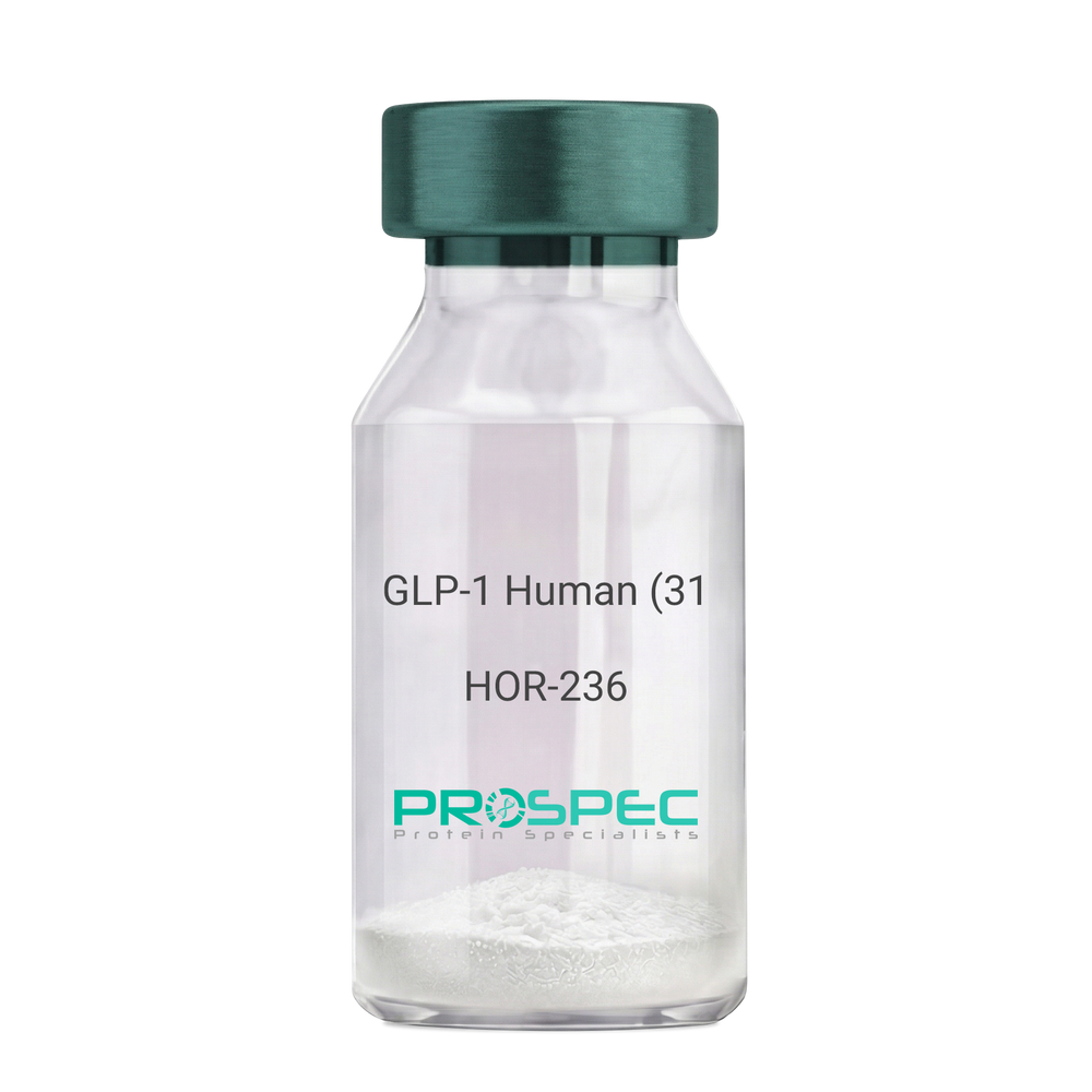
Name :
GLP-1 Human (31 a.a.)Description:
Glucagon Like Peptide-1 (31 a.a.) Human Recombinant
GLP1, GLP2, GRPP.
Product # :
HOR-236Price :
Quantity :
Shipping Method :
Shipped at Room temp
More Info
- description
- source
- formulation
- purity
- More Info
Description
Glucagon Like Peptide-1 Human Recombinant produced in E.Coli is a single, non-glycosylated, polypeptide chain containing 31 amino acids and having a molecular mass of 3.3 kDa. The GLP-1 is purified by proprietary chromatographic techniques.
Source
Escherichia Coli.
Formulation
The Recombinant GLP-1 concentrated solution (1mg/ml) was lyophilized after extensive dialyses against 0.1mg sodium phosphate monobasic & 1.6mg sodium phosphate dibasic.
Purity
Greater than 98.0% as determined by:
(a) Analysis by RP-HPLC.
(b) Analysis by SDS-PAGE.More Info
-
Introduction
Glucagon-like peptide-1 (GLP-1) is derived from the transcription product of the proglucagon gene. The major source of GLP-1 in the body is the intestinal L cell that secretes GLP-1 as a guthormone. The biologically active forms of GLP-1 are: GLP-1-(7-37) and GLP-1-(7-36)NH2.
GLP-1 secretion by L cells is dependent on the presence of nutrients in the lumen of the small intestine. The secretagogues (agents that causes or stimulates secretion) of this hormone include major nutrients like carbohydrate, proteinand lipid. Once in the circulation, GLP-1 has a half life of less than 2 minutes, due to rapid degradation by the enzyme dipeptidyl peptidase-4.
GLP-1 possesses several physiological properties that make it a subject of intensive investigation as a potential treatment of diabetes mellitus. The known physiological functions of GLP-1 include: Increases insulin secretion from the pancreas in a glucose-dependent manner, decreases glucagon secretion from the pancreas, increases beta cells mass and insulin gene expression, inhibits acid secretion and gastric emptying in the stomach, decreases food intake by increasing satiety. -
Synonyms
GLP1, GLP2, GRPP.
-
Physical Appearance
Sterile Filtered White lyophilized (freeze-dried) powder.
-
Stability
Lyophilized Glucagon Like Peptide-1 although stable at room temperature for 3 weeks, should be stored desiccated below -18°C. Upon reconstitution GLP-1 should be stored at 4°C between 2-7 days and for future use below -18°C. For long term storage it is recommended to add a carrier protein (0.1% HSA or BSA).Please prevent freeze-thaw cycles.
-
Solubility
It is recommended to reconstitute the lyophilized Glucagon Like Peptide-1 in sterile 18MΩ-cm H2O not less than 100µg/ml, which can then be further diluted to other aqueous solutions.
-
Amino Acid Sequence
HAEGTFTSDV SSYLEGQAAK EFIAWLVKGR G.
ProSpec's products are furnished for LABORATORY RESEARCH USE ONLY. The product may not be used as drugs, agricultural or pesticidal products, food additives or household chemicals.
- View Data Sheet
Name :
TXN1 E.ColiDescription:
Thioredoxin E.Coli Recombinant
Thioredoxin-1, Trx-1, trxA, fipA, tsnC, b3781, JW5856.
Product # :
PRO-334Price :
Quantity :
Shipping Method :
Shipped at Room temp
More Info
- description
- source
- formulation
- purity
- biological activity
- More Info
Description
Recombinant Thioredoxin was purified from E. coli harboring its gene.
Source
Escherichia Coli.
Formulation
Each mg of protein contains 20mM phosphate buffer pH 7.4.
Purity
Greater than 90.0% as determined by:
(a) Analysis by RP-HPLC.
(b) Analysis by SDS-PAGE.Biological Activity
TRX activity is assayed by measuring the change in absorbance at 650 nm at 25°C using 0.13µM bovine insulin containing 0.33mM DTT (pH 6.5).
The specific activity was found to be 3IU/mg.More Info
-
Introduction
Thioredoxins are small disulphide-containing redox proteins (within the conserved Cys-Gly-Pro-Cys active site) that have been found in all the kingdoms of living organisms. Thioredoxin contains a single disulfide active site and serves as a general protein disulphide oxidoreductase. Thioredoxins are involved in the first unique step in DNA synthesis. It interacts with a broad range of proteins by a redox mechanism based on reversible oxidation of two cysteine thiol groups to a disulphide, accompanied by the transfer of two electrons and two protons. The net result is the covalent interconversion of a disulphide and a dithiol. Trx also provides control over a number of transcription factors affecting cell proliferation and death through a mechanism referred to as redox regulation. It has been suggested that thioredoxin may catalyze the formation of correct disulfides during protein folding because of its ability to act as an efficient oxidoreductant. This could be especially useful in refolding proteins expressed in E. coli. To this end, thioredoxin has been shown to act as a protein disulfide isomerase.Its Molecular Weight is 11.9kDa. and the pI is 4.67.
-
Synonyms
Thioredoxin-1, Trx-1, trxA, fipA, tsnC, b3781, JW5856.
-
Physical Appearance
Sterile Lyophilized Powder.
-
Stability
TRX although stable at 4°C for 3 weeks, should be stored desiccated below -18°C. Please prevent freeze thaw cycles.
-
Solubility
It is recommended to reconstitute the lyophilized TRX in sterile 18MΩ-cm H2O.
-
Amino Acid Sequence
HMSDKIIHL TDDSFDTDVLKADGAIL VDFW AEWCGPCKMIAPILDEI GKLTVAKLNIDQNPGTAPKYGIRGIPTLLLFKNGEVAATKVGAL DANLA.
ProSpec's products are furnished for LABORATORY RESEARCH USE ONLY. The product may not be used as drugs, agricultural or pesticidal products, food additives or household chemicals.
- View Data Sheet
Name :
CGB3 HumanDescription:
CGB3 Human Recombinant
CGB3 CGB5 CGB8, CGB5, CGB7, CGB8, HCGB, CG-beta.
Product # :
PRO-2398Price :
Quantity :
Shipping Method :
Shipped with Ice Packs
More Info
- description
- source
- formulation
- purity
- More Info
Description
CGB3 Human Recombinant produced in Sf9 Baculovirus cells is a single, glycosylated polypeptide chain containing 154 amino acids (21-165.a.) and having a molecular mass of 16.6kDa (Molecular size on SDS-PAGE will appear at approximately 18-28kDa). CGB3 is expressed with a 6 amino acid His tag at C-Terminus and purified by proprietary chromatographic techniques.
Source
Sf9, Baculovirus cells.
Formulation
CGB3 protein solution (0.25mg/ml) contains Phosphate Buffered Saline (pH 7.4) and 10% glycerol.
Purity
Greater than 90.0% as determined by SDS-PAGE.
More Info
-
Introduction
CGB3, belongs to the glycoprotein hormone beta chain family. CGB3 encodes the beta 3 subunit. CGB3 stimulates the expression of GCM1 target genes, as well as the fusogenic protein syncytin-1, to promote placental cell fusion. Furthermore, CGB3 is synthesized in large quantities by the growing placenta, reaching peak concentrations in maternal blood throughout the late first trimester and early mid-trimester of pregnancy. CGB3 is expressed solely by trophoblasts. CGB3 is a significant biomarker in pregnancy and oncology, where it is routinely detected and quantified by specific immunoassays.
-
Synonyms
CGB3 CGB5 CGB8, CGB5, CGB7, CGB8, HCGB, CG-beta.
-
Physical Appearance
Sterile Filtered clear solution.
-
Stability
Store at 4°C if entire vial will be used within 2-4 weeks. Store, frozen at -20°C for longer periods of time. For long term storage it is recommended to add a carrier protein (0.1% HSA or BSA).Avoid multiple freeze-thaw cycles.
-
Amino Acid Sequence
ADPSKEPLRP RCRPINATLA VEKEGCPVCI TVNTTICAGY CPTMTRVLQG VLPALPQVVC NYRDVRFESI RLPGCPRGVN PVVSYAVALS CQCALCRRST TDCGGPKDHP LTCDDPRFQD SSSSKAPPPS LPSPSRLPGP SDTPILPQHH HHHH.
ProSpec's products are furnished for LABORATORY RESEARCH USE ONLY. The product may not be used as drugs, agricultural or pesticidal products, food additives or household chemicals.
- View Data Sheet
Name :
HexarelinDescription:
Hexarelin
Product # :
HOR-288Price :
Quantity :
Shipping Method :
Shipped at Room temp
More Info
- description
- formulation
- purity
- More Info
Description
Hexarelin has 7 amino acids H-His-D-2-Methyl-Trp-Ala-Trp-D-Phe-Lys-NH2 and having a molecular weight of 887 Dalton. The Molecular Formula is C47H58N12O6.
Formulation
The Hexarelin peptide was lyophilized with no additives.
Purity
Greater than 98.0% as determined by:
(a) Analysis by RP-HPLC.
(b) Analysis by SDS-PAGE.More Info
-
Introduction
Hexarelin stimulates GH secretion. Hexarelin is more resistant to proteolytic degradation than GHRP-6.
-
Physical Appearance
Sterile Filtered lyophilized (freeze-dried) powder.
-
Stability
Lyophilized Hexarelin although stable at room temperature for 3 weeks, should be stored desiccated below -18°C. Upon reconstitution Hexarelin should be stored at 4°C between 2-7 days and for future use below -18°C. For long term storage it is recommended to add a carrier protein (0.1% HSA or BSA).Please prevent freeze-thaw cycles.
-
Solubility
It is recommended to reconstitute the lyophilized Hexarelin 18MΩ-cm H2O not less than 100 µg/ml, which can then be further diluted to other aqueous solutions.
ProSpec's products are furnished for LABORATORY RESEARCH USE ONLY. The product may not be used as drugs, agricultural or pesticidal products, food additives or household chemicals.
- View Data Sheet
Name :
CTGF AntibodyDescription:
Mouse Anti Human Connective Tissue Growth Factor
CCN2, NOV2, HCS24, IGFBP8, MGC102839, CTGF, Connective Tissue Growth Factor.
Product # :
ANT-699Price :
Quantity :
Shipping Method :
Shipped with Ice Packs
More Info
- formulation
- More Info
Formulation
1mg/ml containing PBS, pH-7.4, 10% Glycerol and 0.02% Sodium Azide.
More Info
-
Introduction
Connective Tissue Growth Factor belongs to the CCN family of proteins. The CCN family presently consists of six members in human also known as: Cyr61 (Cystein rich 61), CTGF (Connective Tissue Growth Factor), Nov (Nephroblastoma Overexpressed gene), WISP-1, 2 and 3 (Wnt-1 Induced Secreted Proteins). The CCN genes encode secreted proteins associated with the Extracellular Matrix (ECM) and cell membrane.
CCN proteins are matricellular proteins which are involved in the regulation of various cellular functions including: proliferation, differentiation, survival, adhesion and migration. They are expressed in derivatives of the three embryonic sheets and are implicated in the development of kidney, nervous system, muscle, bone marrow, cartilage and bone. During adulthood, they are implicated in wound healing, bone fracture repair, and pathologies such as: fibrosis, vascular ailments and tumorigenesis.
Full length secreted CCN proteins can show an antiproliferative activity, whereas truncated isoforms are likely to stimulate proliferation and behave as oncogenes.
The full length protein consists of four modulesModule I shares partial identity with the N-terminal part of the Insulin-like Growth Factor Binding Proteins (IGFBPs).
Module II includes a stretch of 70amino acid residues – which shares sequence identity with the Von Willebrand Factor Type C repeat (VWC).
Module III contains sequences sharing identity with the Thrombospondin type 1 repeat (TSP1) (WSXCSXXCG), which is thought to be implicated in the binding of sulfated glycoconjugates and to be important for cell adhesion.
Module IV, also designated CT, is encoded by exon5. It is the leasts conserved one of the four domains at the level of nucleotide sequence, but it appears to be critical for several of the biological functions attributed to the CCN proteins. Module IV resembles the CT domain of several extracellular protein including, Von Willebrand's factor and mucins. Sequence similarities to heparin-binding motifs are also found within this domain.
Proteolysis of the secreted full-length CCN proteins that has been reported in the case of CCN2 and CCN3 might result in the production of CCN-derived peptides with high affinity for ligands that full-length CNN proteins bind only poorly. Amino-truncated CCN2 isoforms were biologically active whereas no specific biological activity has been attributed to the truncated CCN3. Although the molecular processes underlying the production of these secreted isoforms is presently unknown, it is important to note that proteolysis occur at the same amino acid residues in both CCN2 and CCN3. An elevated expression of CCN2 has also been detected by Northern blotting in human invasive mammary ductal carcinomas, dermatofibromas, pyogenic granuloma, endothelial cells of angiolipomas and angioleiomyomas, and in pancreatic tumors. A study performed with chondrosarcomas representative of various histological grades established that CCN2 expression was closely correlated with increasing levels of malignancy.
In agreement with CCN2 playing a role in brain tumor angiogenesis, immunocytochemistry studies indicated that both glioblastoma tumor cells and proliferating endothelial cells stained positive for CCN2. In astrocytomas, CCN2 expression was particularly elevated in high grade tumors, with a marked effect of CCN2 on cell proliferation. Downregulation of CCN2 expression in these cells was associated with a growth arrest at the G1/S transition while over-expression of CCN2 induced a two-fold increase of the number of cells in the G1 phase. Gene profiling analysis allowed to identify a set of about 50 genes whose expression might account for the proliferative activity of CCN2 in these cells.
CCN2 was seen in a higher proportion of mononuclear cells of patients with acute lymphoblastic leukemia. -
Synonyms
CCN2, NOV2, HCS24, IGFBP8, MGC102839, CTGF, Connective Tissue Growth Factor.
-
Physical Appearance
Sterile filtered colorless solution.
-
Immunogen
Anti-human CTGF mAb, is derived from hybridization of mouse F0 myeloma cells with spleen cells from BALB/c mice immunized with a recombinant human CTGF protein 27-349 amino acids purified from E. coli.
-
Ig Subclass
Mouse IgG2a heavy chain and κ light chain.
-
Clone
PAT18E7AT.
-
Applications
CTGF antibody has been tested by ELISA, Western blot analysis and ICC/IF to assure specificity and reactivity. Since application varies, however, each investigation should be titrated by the reagent to obtain optimal results.
-
Type
Mouse Anti Human Monoclonal.
-
Storage Procedures
For periods up to 1 month store at 4°C, for longer periods of time, store at -20°C. Prevent freeze thaw cycles.
-
Purification Method
CTGF antibody was purified by protein-A affinity chromatography.
ProSpec's products are furnished for LABORATORY RESEARCH USE ONLY. The product may not be used as drugs, agricultural or pesticidal products, food additives or household chemicals.
- View Data Sheet
Name :
NRG1 (HRG-beta3) AntibodyDescription:
Neuregulin1 isoform HRG-beta3, Mouse Anti Human
Product # :
ANT-458Price :
Quantity :
Shipping Method :
Shipped with Ice Packs
More Info
- formulation
- More Info
Formulation
1mg/ml containing PBS, pH-7.4, & 0.1% Sodium Azide.
More Info
-
Introduction
NRG1 is one of 4 proteins in the neuregulin family which act on the EGFR family of receptors. NRG1 was initially identified as a 44kDa glycoprotein that interacts with the ERBB2 receptor tyrosine kinase to increase its phosphorylation on tyrosine residues. In various brain areas, NRG1 is expressed at synaptic regions and its processing can be regulated by neuronal activity. The HRG-beta3variant of NRG1, along with HRG-alpha, beta1 and beta2 variants, was identified in various normal tissues and cancer cell lines.
-
Immunogen
Anti-human NRG1 mAb is derived from hybridization of mouse F0 myeloma cells with spleen cells from BALB/c mice immunized with recombinant human NRG1 amino acids 20-241 purified from E. coli.
-
Ig Subclass
Mouse IgG1 heavy chain and κ light chain.
-
Clone
Pk1G13AT.
-
Applications
NRG1 antibody has been tested by ELISA and Western blot analysis to assure specificity and reactivity. Since application varies, however, each investigation should be titrated by the reagent to obtain optimal results. Recommended dilution range for Western blot analysis is 1:1,000 ~ 2,000. Recommended starting dilution is 1:1,000.
-
Type
Mouse Anti Human Monoclonal.
-
Storage Procedures
For periods up to 1 month store at 4°C, for longer periods of time, store at -20°C. Prevent freeze thaw cycles.
-
Purification Method
NRG1 antibody was purified from mouse ascitic fluids by protein-G affinity chromatography.
ProSpec's products are furnished for LABORATORY RESEARCH USE ONLY. The product may not be used as drugs, agricultural or pesticidal products, food additives or household chemicals.
- View Data Sheet
Name :
GSTP1 AntibodyDescription:
Glutathione S-Transferase pi 1, Mouse Anti Human
Glutathione S-transferase P 1, Gst P1, GST YF-YF, GST class-pi, GST-piB, Preadipocyte growth factor.
Product # :
ANT-729Price :
Quantity :
Shipping Method :
Shipped with Ice Packs
More Info
- formulation
- More Info
Formulation
1mg/ml containing PBS, pH-7.4, 10% Glycerol and 0.02% Sodium Azide.
More Info
-
Introduction
GSTP1 is a polymorphic gene encoding active, functionally different GSTP1 variant proteins that are believed to function in xenobiotic metabolism and have a part in susceptibility to cancer, and other diseases. GSTP1 is a glutathione S-transferase belonging to the pi class. GST family enzymes play a significant role in detoxification by catalyzing the conjugation of many hydrophobic and electrophilic compounds with reduced glutathione. Based upon the biochemical, immunologic and structural properties of the soluble GSTs they are grouped into 4 main classes: alpha, mu, pi, and theta. The GSTP1 enzyme acts by catalyzing the reaction of glutathione with an acceptor molecule to form a Sulfur-substituted glutathione. The reactions employing glutathione contribute the transformation of a broad range of electrophiles, including reactive products of lipid, protein, carcinogens, therapeutic drugs, environmental toxins, and products of oxidative stress.
The GSTP1 inactivation through CpG hypermethylation is frequent in pituitary adenomas and may be a factor in aggressive pituitary tumor behavior. GSTP1 is may be a transcriptional target of the p53 tumor suppressor gene. Single-nucleotide polymorphism in GSTP1 is linked to modified protein binding, which influence GSTP1's contribution to carcinogen and drug metabolism, and possibly disease pathogenesis and/or drug response. GST-pi might have central roles in proliferation of androgen-independent human prostate cancer cells. -
Synonyms
Glutathione S-transferase P 1, Gst P1, GST YF-YF, GST class-pi, GST-piB, Preadipocyte growth factor.
-
Physical Appearance
Sterile filtered colorless solution.
-
Immunogen
Anti-human GSTP1 mAb, is derived from hybridization of mouse F0 myeloma cells with spleen cells from BALB/c mice immunized with recombinant human GSTP1 amino acids 1-210 purified from E. coli.
-
Ig Subclass
Mouse IgG1 heavy chain and κ light chain.
-
Clone
PAT12C10AT
-
Applications
GSTP1 antibody has been tested by ELISA, Western blot analysis, ICC/IF and Flow cytometry to assure specificity and reactivity. Since application varies, however, each investigation should be titrated by the reagent to obtain optimal results.
-
Type
Mouse Anti Human Monoclonal.
-
Storage Procedures
For periods up to 1 month store at 4°C, for longer periods of time, store at -20°C. Prevent freeze thaw cycles.
-
Purification Method
GSTP1 antibody was purified from mouse ascitic fluids by protein-A affinity chromatography.
ProSpec's products are furnished for LABORATORY RESEARCH USE ONLY. The product may not be used as drugs, agricultural or pesticidal products, food additives or household chemicals.
- View Data Sheet
Name :
Flt3 Ligand HumanDescription:
Flt3 Ligand Human Recombinant
Fms-Related Tyrosine Kinase 3 Ligand, Flt3 Ligand, Flt-3 Ligand, SL Cytokine.
Product # :
CYT-331Price :
Quantity :
Shipping Method :
Shipped at Room temp
More Info
- description
- source
- formulation
- purity
- biological activity
- More Info
- sds-page
Description
Flt3-Ligand Human Recombinant produced in E.Coli is a non-glycosylated, polypeptide chain containing 155 amino acids and having a molecular mass of approximately 17.6kDa. Flt3-Ligand is purified by proprietary chromatographic techniques.
Source
Escherichia Coli.
Formulation
The protein was lyophilized with no additives.
Purity
Greater than 97.0% as determined by:
(a) Analysis by RP-HPLC.
(b) Analysis by SDS-PAGE.Biological Activity
The ED50 as calculated by the dose-dependent stimulation of the proliferation of human AML5 cells is less than 1.0 ng/ml, corresponding to a Specific Activity of 1.0×10⁶ IU/mg.sds-page
More Info
-
Introduction
FLT3 ligand is a receptor for the fl cytokine has a tyrosine-protein kinase activity & a growth factor that regulates proliferation of early hematopoietic cells. Flt3-Ligand synergizes with other CSFs and interleukins to induce growth and differentiation.
-
Synonyms
Fms-Related Tyrosine Kinase 3 Ligand, Flt3 Ligand, Flt-3 Ligand, SL Cytokine.
-
Physical Appearance
Sterile Filtered White lyophilized (freeze-dried) powder.
-
Stability
Lyophilized Flt3-Ligand although stable at room temperature for 3 weeks, should be stored desiccated below -18°C. Upon reconstitution Flt3-Ligand should be stored at 4°C between 2-7 days and for future use below -18°C.For long term storage it is recommended to add a carrier protein (0.1% HSA or BSA).Please prevent freeze-thaw cycles.
-
Solubility
It is recommended to reconstitute the lyophilized Flt3-L in sterile 18MΩ-cm H2O not less than 100µg/ml, which can then be further diluted to other aqueous solutions.
-
Amino Acid Sequence
TQDCSFQHSP ISSDFAVKIR ELSDYLLQDY PVTVASNLQD EELCGGLWRL VLAQRWMERL KTVAGSKMQG LLERVNTEIH FVTKCAFQPP PSCLRFVQTN ISRLLQETSE QLVALKPWIT RQNFSRCLEL QCQPDSSTLP PPWSPRPLEA TAPTA.
-
Background
What is the molecular weight/Mw of FLT3 LIGAND HUMAN Protein?
FLT3 LIGAND HUMAN Protein has a total Mw of 17.6kDa.
What is the source or expression system of FLT3 LIGAND HUMAN Protein?
Escherichia Coli.
What is the Purity of FLT3 LIGAND HUMAN Protein?
FLT3 LIGAND HUMAN Protein is >97% pure as determined by SDS-PAGE.
What is the Biological Activity of FLT3 LIGAND HUMAN Protein?
The ED50 as calculated by the dose-dependent stimulation of the proliferation of human AML5 cells is less than 1.0 ng/ml, corresponding to a Specific Activity of 1.0×10⁶ IU/mg.
What is the amino acid sequence of FLT3 LIGAND HUMAN Protein?
TQDCSFQHSP ISSDFAVKIR ELSDYLLQDY PVTVASNLQD EELCGGLWRL VLAQRWMERL KTVAGSKMQG LLERVNTEIH FVTKCAFQPP PSCLRFVQTN ISRLLQETSE QLVALKPWIT RQNFSRCLEL QCQPDSSTLP PPWSPRPLEA TAPTA.
What applications can FLT3 LIGAND HUMAN Protein be used in?
FLT3 LIGAND HUMAN Protein can probably be used in western blot, ELISA and Lateral Flow.
What is the endotoxin level for FLT3 LIGAND HUMAN Protein?
The endotoxin level is minimal, FLT3 LIGAND HUMAN Protein was purified using conventional chromatography techniques.
ProSpec's products are furnished for LABORATORY RESEARCH USE ONLY. The product may not be used as drugs, agricultural or pesticidal products, food additives or household chemicals.
- View Data Sheet
Name :
CD3G HumanDescription:
CD3G Human Recombinant
T-cell surface glycoprotein CD3 gamma chain, CD3-GAMMA, T3G, CD3G.
Product # :
PRO-1392Price :
Quantity :
Shipping Method :
Shipped with Ice Packs
More Info
- description
- source
- formulation
- purity
- More Info
Description
CD3G Human Recombinant produced in E.Coli is a single, non-glycosylated polypeptide chain containing 117 amino acids (23-116a.a.) and having a molecular mass of 13.1kDa. CD3G is fused to a 23 amino acid His-tag at N-terminus & purified by proprietary chromatographic techniques.
Source
E.coli.
Formulation
CD3G protein solution (1mg/ml) containing 20mM Tris-HCl buffer (pH 8.0), 0.4M urea and 10% glycerol.
Purity
Greater than 90% as determined by SDS-PAGE.
More Info
-
Introduction
CD3-gamma polypeptide is encoded by CD3 Gamma (CD3G) which together with CD3-epsilon, -delta and -zeta, and the T-cell receptor alpha/beta and gamma/delta heterodimers, configures the T-cell receptor-CD3 complex. The T-cell receptor-CD3 complex takes part in coupling antigen recognition to numerous intracellular signal-transduction pathways. The genes encoding the epsilon, gamma and delta polypeptides are located in the same cluster on chromosome 11. Deficiencies in this gene are associated with T cell immunodeficiency.
-
Synonyms
T-cell surface glycoprotein CD3 gamma chain, CD3-GAMMA, T3G, CD3G.
-
Physical Appearance
Sterile Filtered colorless solution.
-
Stability
Store at 4°C if entire vial will be used within 2-4 weeks. Store, frozen at -20°C for longer periods of time. For long term storage it is recommended to add a carrier protein (0.1% HSA or BSA).Avoid multiple freeze-thaw cycles.
-
Amino Acid Sequence
MGSSHHHHHH SSGLVPRGSH MGSQSIKGNH LVKVYDYQED GSVLLTCDAE AKNITWFKDG KMIGFLTEDK KKWNLGSNAK DPRGMYQCKG SQNKSKPLQV YYRMCQNCIE LNAATIS.
ProSpec's products are furnished for LABORATORY RESEARCH USE ONLY. The product may not be used as drugs, agricultural or pesticidal products, food additives or household chemicals.
- View Data Sheet
Name :
GM CSF Human, Sf9Description:
Granulocyte Macrophage-Colony Stimulating Factor Human Recombinant, Sf9
CSF-2, MGI-1GM, GMCSF, Pluripoietin-alpha, Molgramostin, Sargramostim.
Product # :
CYT-416Price :
Quantity :
Shipping Method :
Shipped at Room temp
More Info
- description
- source
- formulation
- purity
- biological activity
- More Info
Description
GM-CSF Human Recombinant produced in insect cells is a single, glycosylated, polypeptide chain containing 127 amino acids (18-144) and having a molecular mass of 14.6kDa. GM-CSF is fused to a C-terminal His -tag (6x His) and purified by proprietary chromatographic techniques.
Source
Insect Cells.
Formulation
The protein was lyophilized with PBS.
Purity
Greater than 98.0% as determined by SDS-PAGE.
Biological Activity
The ED50 as determined by the dose-dependant stimulation of the proliferation of human TF-1 cells (human erythroleukemic indicator cell line) is < 0.1 ng/ml, corresponding to a Specific Activity of 10,000,000IU/mg.More Info
-
Introduction
GMCSF is a cytokine that controls the production, differentiation, and function of granulocytes and macrophages. The active form of the protein is found extracellularly as a homodimer. This gene has been localized to a cluster of related genes at chromosome region 5q31, which is known to be associated with interstitial deletions in the 5q- syndrome and acute myelogenous leukemia. Other genes in the cluster include those encoding interleukins 4, 5, and 13.
GM-CSF stimulates the growth and differentiation of hematopoietic precursor cells from various lineages, including granulocytes, macrophages, eosinophils and erythrocytes. -
Synonyms
CSF-2, MGI-1GM, GMCSF, Pluripoietin-alpha, Molgramostin, Sargramostim.
-
Physical Appearance
Sterile Filtered White lyophilized (freeze-dried) powder.
-
Stability
Lyophilized Granulocyte Macrophage Colony Stimulating Factor although stable at room temperature for 3 weeks, should be stored desiccated below -18°C. Upon reconstitution GMCSF should be stored at 4°C between 2-7 days and for future use below -18°C.For long term storage it is recommended to add a carrier protein (0.1% HSA or BSA).Please prevent freeze-thaw cycles.
-
Solubility
It is recommended to reconstitute the lyophilized Granulocyte Macrophage Colony Stimulating Factor in sterile 18MΩ-cm H2O not less than 100µg/ml, which can then be further diluted to other aqueous solutions.
-
Amino Acid Sequence
The sequence of the first five N-terminal amino acids was determined and was found to be Ala-Pro-Ala-Arg-Ser.
-
Background
Recombinant Granulocyte-Macrophage Colony-Stimulating Factor (GMCSF) is a protein that plays a crucial role in the production and differentiation of white blood cells, including granulocytes and macrophages. It is a potent stimulator of hematopoietic stem cells, which are responsible for the production of all blood cells in the body. Recombinant GMCSF is a synthetic version of the protein that is produced using recombinant DNA technology.
Recombinant GMCSF has been extensively studied for its potential therapeutic applications in a variety of medical conditions, including cancer, autoimmune diseases, and infectious diseases. In cancer, GMCSF is used as an immunostimulatory agent to enhance the immune response against cancer cells. By stimulating the production and differentiation of white blood cells, GM-CSF can increase the number of immune cells that can recognize and attack cancer cells. This approach has been successfully used in the treatment of several types of cancer, including melanoma and leukemia.
In autoimmune diseases, recombinant GMCSF has been investigated as a potential treatment for conditions such as rheumatoid arthritis and multiple sclerosis. These diseases are characterized by an overactive immune response that attacks healthy tissues in the body. By modulating the immune response, GMCSF may be able to reduce inflammation and prevent further damage to affected tissues.
In infectious diseases, recombinant GMCSF has been studied as a potential treatment for conditions such as sepsis and HIV/AIDS. In sepsis, a severe bacterial infection, GMCSF may be able to stimulate the production of white blood cells and improve the immune response against the infection. In HIV/AIDS, GMCSF may be able to enhance the immune response against the virus and reduce the risk of opportunistic infections.
Recombinant GMCSF is typically administered by injection, either directly into the affected tissue or into the bloodstream. It is generally well-tolerated, although some patients may experience side effects such as fever, fatigue, and muscle pain.
In conclusion, recombinant GMCSF is a promising therapeutic agent with potential applications in a variety of medical conditions. Its ability to stimulate the production and differentiation of white blood cells makes it a valuable tool in the treatment of cancer, autoimmune diseases, and infectious diseases. Ongoing research is likely to uncover new uses for this protein and further refine its therapeutic potential.
ProSpec's products are furnished for LABORATORY RESEARCH USE ONLY. The product may not be used as drugs, agricultural or pesticidal products, food additives or household chemicals.
- View Data Sheet
Name :
GOT1 MouseDescription:
Glutamic-Oxaloacetic Transaminase 1 Mouse Recombinant
Aspartate aminotransferase, cytoplasmic, cAspAT, Cysteine aminotransferase, cytoplasmic, Cysteine transaminase, cytoplasmic, cCAT, Glutamate oxaloacetate transaminase 1, Transaminase A.
Product # :
ENZ-872Price :
Quantity :
Shipping Method :
Shipped with Ice Packs
More Info
- description
- source
- formulation
- purity
- More Info
Description
GOT1 Mouse Recombinant produced in E.Coli is a single, non-glycosylated polypeptide chain containing 436 amino acids (1-413a.a) and having a molecular mass of 48.6kDa.GOT1 is fused to a 23 amino acid His-tag at N-terminus & purified by proprietary chromatographic techniques.
Source
Escherichia Coli.
Formulation
GOT1 protein solution (0.5mg/ml) containing Phosphate buffered saline (pH7.4), 10% glycerol and 1mM DTT.
Purity
Greater than 95.0% as determined by SDS-PAGE.
More Info
-
Introduction
GOT1 is a pyridoxal phosphate-dependent enzyme which exists in cytoplasmic and mitochondrial forms, GOT1 and GOT2, which participate in amino acid metabolism and the urea and tricarboxylic acid cycles. Both enzymes are homodimeric and show close homology.
-
Synonyms
Aspartate aminotransferase, cytoplasmic, cAspAT, Cysteine aminotransferase, cytoplasmic, Cysteine transaminase, cytoplasmic, cCAT, Glutamate oxaloacetate transaminase 1, Transaminase A.
-
Physical Appearance
Sterile filtered colorless solution.
-
Stability
Store at 4°C if entire vial will be used within 2-4 weeks.Store, frozen at -20°C for longer periods of time.For long term storage it is recommended to add a carrier protein (0.1% HSA or BSA).Avoid multiple freeze-thaw cycles.
-
Amino Acid Sequence
MGSSHHHHHH SSGLVPRGSH MGSMAPPSVF AQVPQAPPVL VFKLTADFRD DPDPRKVNLG VGAYRTDESQ PWVLPVVRKV EQKIANDNSL NHEYLPILGL AEFRSCASRL VLGDNSPAIR ENRVGGVQSL GGTGALRIGA DFLGRWYNGT DNKNTPIYVS SPTWENHNAV FSAAGFKDIR PYCYWDAEKR GLDLQGFLND LENAPEFSIF VLHACAHNPT GTDPTPEQWK QIAAVMQRRF LFPFFDSAYQ GFASGDLEKD AWAIRYFVSE GFELFCAQSF SKNFGLYNER VGNLTVVGKE SDSVLRVLSQ MEKIVRITWS NPPAQGARIV AATLSDPELF KEWKGNVKTM ADRILTMRSE LRARLEALKT PGTWSHITEQ IGMFSFTGLN PKQVEYLVNE KHIYLLPSGR INMCGLTTKN LDYVATSIHE AVTKIQ.
ProSpec's products are furnished for LABORATORY RESEARCH USE ONLY. The product may not be used as drugs, agricultural or pesticidal products, food additives or household chemicals.
- View Data Sheet
Name :
CoV-2 S1 (16-685), FcDescription:
Coronavirus 2019 Spike Glycoprotein-S1 (16-685 a.a.), Fc Recombinant
Product # :
SARS-027Price :
Quantity :
Shipping Method :
Shipped at Room temp
More Info
- description
- source
- formulation
- purity
- More Info
Description
The HEK293 derived recombinant protein contains the Coronavirus 2019 CoV-2 Spike Glycoprotein S1, Wuhan-Hu-1 strain, amino acids 16-685 fused to Fc tag at C-terminal.
Source
HEK293 Cells.
Formulation
CoV-2 S1 protein is supplied in sodium chloride, glycine, arginine pH-7.5 & 5% trehalose.
Purity
Protein is >95% pure as determined SDS-PAGE.
More Info
-
Introduction
A human infecting coronavirus (viral pneumonia) called 2019 novel coronavirus, 2019-nCoV was found in the fish market at the city of Wuhan, Hubei province of China on December 2019.
The 2019-nCoV shares an 87% identity to the 2 bat-derived severe acute respiratory syndrome 2018 SARS-CoV-2 located in Zhoushan of eastern China. 2019-nCoV has an analogous receptor-BD-structure to that of 2018 SARS-CoV, even though there is a.a. diversity so thus the 2019-nCoV might bind to ACE2 receptor protein (angiotensin-converting enzyme 2) in humans.
While bats are possibly the host of 2019-nCoV, researchers suspect that animal from the ocean sold at the seafood market was an intermediate host. RSCU analysis proposes that the 2019-nCoV is a recombinant within the viral spike glycoprotein between the bat coronavirus and an unknown coronavirus.
-
Physical Appearance
Sterile Filtered White lyophilized (freeze-dried) powder.
-
Stability
Lyophilized Cov-2 Spike S1 Glycoprotein although stable at room temperature for 3 weeks, should be stored desiccated below -18°C. Upon reconstitution CoV2 Spike protein should be stored at 4°C between 2-7 days and for future use below -18°C.For long term storage it is recommended to add a carrier protein (0.1% HSA or BSA).Please prevent freeze-thaw cycles.
-
Purification Method
Purified by Protein-G chromatographic technique.
ProSpec's products are furnished for LABORATORY RESEARCH USE ONLY. The product may not be used as drugs, agricultural or pesticidal products, food additives or household chemicals.
- View Data Sheet
Name :
CTACK HumanDescription:
CTACK Human Recombinant (CCL27)
ALP, CTACK, ESKINE, ILC, PESKY, SCYA27, CCL27, C-C motif chemokine 27, Small-inducible cytokine A27, IL-11 R-alpha-locus chemokine, Skinkine, ESkine, Cutaneous T-cell-attracting chemokine.
Product # :
CHM-373Price :
Quantity :
Shipping Method :
Shipped with Ice Packs
More Info
- description
- source
- formulation
- purity
- More Info
- SDS-PAGE
Description
CTACK Human Recombinant produced in E.Coli is a single, non-glycosylated, polypeptide chain containing 89 amino acids (25-112 a.a.) and having a molecular mass of 10.3kDa. CTACK protein is purified by standard chromatography.
Source
Escherichia Coli.
Formulation
CTACK Human solution containing 10mM sodium citrate pH-3.5 & 10% glycerol.
Purity
Greater than 95% as determined by SDS-PAGE.
SDS-PAGE
More Info
-
Introduction
CTACK is a chemotactic factor that attracts skin-associated memory T-lymphocytes. CTACK is involved in mediating homing of lymphocytes to cutaneous sites. CTACK Binds to CCR10.
-
Synonyms
ALP, CTACK, ESKINE, ILC, PESKY, SCYA27, CCL27, C-C motif chemokine 27, Small-inducible cytokine A27, IL-11 R-alpha-locus chemokine, Skinkine, ESkine, Cutaneous T-cell-attracting chemokine.
-
Physical Appearance
Sterile filtered colorless solution.
-
Stability
Store at 4°C if entire vial will be used within 2-4 weeks. Store, frozen at -20°C for longer periods of time. For long term storage it is recommended to add a carrier protein (0.1% HSA or BSA).Avoid multiple freeze-thaw cycles.
-
Amino Acid Sequence
MFLLPPSTAC CTQLYRKPLS DKLLRKVIQV ELQEADGDCH LQAFVLHLAQ RSICIHPQNP SLSQWFEHQE RKLHGTLPKL NFGMLRKMG.
-
Background
What is the molecular weight/Mw of CTACK HUMAN Protein?
CTACK HUMAN Protein has a total Mw of 10.3kDa.
What is the source or expression system of CTACK HUMAN Protein?
Escherichia Coli.
What is the Purity of CTACK HUMAN Protein?
CTACK HUMAN Protein is >95% pure as determined by SDS-PAGE.
What is the Biological Activity of CTACK HUMAN Protein?
The biological functionality of CTACK HUMAN Protein will be determined in the future.
What is the amino acid sequence of CTACK HUMAN Protein?
MFLLPPSTAC CTQLYRKPLS DKLLRKVIQV ELQEADGDCH LQAFVLHLAQ RSICIHPQNP SLSQWFEHQE RKLHGTLPKL NFGMLRKMG.
What applications can CTACK HUMAN Protein be used in?
CTACK HUMAN Protein can probably be used in western blot, ELISA and Lateral Flow.
What is the endotoxin level for CTACK HUMAN Protein?
The endotoxin level is minimal, CTACK HUMAN Protein was purified using conventional chromatography techniques.
ProSpec's products are furnished for LABORATORY RESEARCH USE ONLY. The product may not be used as drugs, agricultural or pesticidal products, food additives or household chemicals.
- View Data Sheet
Name :
C17ORF103 HumanDescription:
Chromosome 17 Open Reading Frame 103 Human Recombinant
Chromosome 17 Open Reading Frame 103, Transcript Expressed During Hematopoiesis 2, Gene Trap Locus F3b, Gtlf3b, Protein GTLF3B.
Product # :
PRO-1619Price :
Quantity :
Shipping Method :
Shipped with Ice Packs
More Info
- description
- source
- formulation
- purity
- More Info
Description
C17ORF103 Human Recombinant produced in E.coli is a single, non-glycosylated polypeptide chain containing 136 amino acids (1-113) and having a molecular mass of 15.4kDa.C17ORF103 is fused to a 23 amino acid His-tag at N-terminus & purified by proprietary chromatographic techniques.
Source
Escherichia Coli.
Formulation
The C17ORF103 solution contains 20mM Tris-HCl buffer (pH 8.0), 0.1M NaCl, 1mM DTT and 20% glycerol.
Purity
Greater than 95% as determined by SDS-PAGE.
More Info
-
Introduction
C17orf103 is a member of the GTLF3B family and is encoded by a gene which is located in human chromosome 17p11.2. BRCA1 and p53 are 2 main tumor suppressor genes which are linked to chromosome 17. Tumor suppressor p53 is necessary for maintenance of cellular genetic integrity by moderating cell fate through DNA repair versus cell death. C17orf103 is also related to neurofibromatosis, a disorder characterized by neural and epidermal lesions, and dysregulated Schwann cell growth.
-
Synonyms
Chromosome 17 Open Reading Frame 103, Transcript Expressed During Hematopoiesis 2, Gene Trap Locus F3b, Gtlf3b, Protein GTLF3B.
-
Physical Appearance
Sterile filtered colorless solution.
-
Stability
Store at 4°C if entire vial will be used within 2-4 weeks. Store, frozen at -20°C for longer periods of time. For long term storage it is recommended to add a carrier protein (0.1% HSA or BSA).Avoid multiple freeze-thaw cycles.
-
Amino Acid Sequence
MGSSHHHHHH SSGLVPRGSH MGSMAHSAAA VPLGALEQGC PIRVEHDRRR RQFTVRLNGC HDRAVLLYEY VGKRIVDLQH TEVPDAYRGR GIAKHLAKAA LDFVVEEDLK AHLTCWYIQK YVKENPLPQY LERLQP
ProSpec's products are furnished for LABORATORY RESEARCH USE ONLY. The product may not be used as drugs, agricultural or pesticidal products, food additives or household chemicals.