Search results
1000 results found for “Chagas”
Name
Description
Product #
Price
Quantity
Shipping Method
- View Data Sheet
Name :
Dengue Envelope-3 32kDaDescription:
Dengue Virus Subtype 3 Envelope 32kDa Recombinant
Product # :
DEN-019Price :
Quantity :
Shipping Method :
Shipped with Ice Packs
More Info
- description
- source
- formulation
- purity
- More Info
- Citations
Description
Recombinant Dengue Virus Subtype 3 Envelope 32kDa, produced in E.coli and fused with a 6xHis Tag, containing domains I+II of the dengue envelope.
Source
E.coli.
Formulation
Phosphate buffered saline, pH-7.4.
Purity
Protein is >95% pure as determined by 10% PAGE (coomassie staining).
Citations
More Info
-
Introduction
Dengue fever is affected by 1 of 4 closely linked virus serotypes of the genus Flavivirus, family Flaviviridae. One might have dengue fever infected by the different serotype virus after the primary infection. Detection of particular antibodies to dengue viruses is used for the diagnosis in clinic. Recently, lateral flow rapid test products have become a most suitable and known method in the clinical diagnosis. Though, there is difficulty for manufacturers to have the dengue antigens with a whole coverage for Dengue IgG & IgM recognition for all 4 serotype infections as well as with colloid gold binding ability. 8 dengue antigens have been established for the lateral flow products which are well defined for their coverage of dengue IgG & IgM recognition. The researcher can select the products below based on their own specific application.
-
Stability
Dengue Envelope-3 32kDa although stable at 4°C for 1 week, should be stored below -18°C. Please prevent freeze thaw cycles.
-
Applications
Immunoassay.
-
Purification Method
Purified by proprietary chromatographic technique.
ProSpec's products are furnished for LABORATORY RESEARCH USE ONLY. The product may not be used as drugs, agricultural or pesticidal products, food additives or household chemicals.
- View Data Sheet
Name :
HBsAg adw CHODescription:
Hepatitis B Surface Antigen Adw subtype Recombinant, CHO
Product # :
HBS-001Price :
Quantity :
Shipping Method :
Shipped with Ice Packs
More Info
- description
- source
- formulation
- purity
- More Info
- Citations
Description
Recombinant Hepatitis B Surface Antigen Adw subtype, CHO has a Mw of 23kDa, containing 226 amino acids and was purified from CHO cell line.The Recombinant Hepatitis B Surface Antigen Adw subtype, CHO is purified by proprietary chromatographic technique.
Source
Chinese Hamster Ovary Cells (CHO).
Formulation
HBsAg adw CHO solution contains PBS, PH 6.5.
Purity
Protein is >95% pure as determined by 12% SDS-PAGE (coomassie staining).
Citations
More Info
-
Introduction
HBsAg is the surface antigen of the Hepatitis-B-Virus (HBV). The capsid of a virus has different surface proteins from the rest of the virus. The antigen is a protein that binds specifically on one of these surface proteins. It is commonly referred to as the Australian Antigen. HBsAg is an important parameter to establish a diagnosis of hepatitis B disease in the clinic and to monitor antiviral treatment.
-
Physical Appearance
Sterile Filtered clear solution.
-
Stability
Store at 4°C if entire vial will be used within 2-4 weeks.Store, frozen at -20°C for longer periods of time.For long term storage it is recommended to add a carrier protein (0.1% HSA or BSA).Avoid multiple freeze-thaw cycles.
ProSpec's products are furnished for LABORATORY RESEARCH USE ONLY. The product may not be used as drugs, agricultural or pesticidal products, food additives or household chemicals.
- View Data Sheet
Name :
T.gondii p29Description:
Toxoplasma Gondii p29 (GRA7) Recombinant
Product # :
TOX-263Price :
Quantity :
Shipping Method :
Shipped with Ice Packs
More Info
- description
- source
- formulation
- purity
- More Info
- Citations
Description
The E.coli derived recombinant protein contains the p29 (GRA7) immunodominant regions, amino acids 24-100 and fused to a 26kDa GST tag.
Source
Escherichia Coli.
Formulation
50mM Tris-HCl, 1.5M urea and 50% glycerol.
Purity
Toxoplasma protein is >95% pure as determined by 10% PAGE (coomassie staining).
Citations
More Info
-
Introduction
The life cycle of Toxoplasma gondii has two phases. The coccidia like takes place only in members of the Felidae family which makes these animals the parasite's primary host. The asexual part of the life cycle can take place in any warm-blooded animal, like other mammals(including felines) and birds. T. gondii constructing daughter scaffolds within the mother cell. In the intermediate hosts (including felines), the parasite invades cells, forming intracellular so-called parasitophorous vacuoles containing bradyzoites, the slowly replicating form of the parasit. Vacuoles form tissue cysts mainly within the muscles and brain. Since they are within cells, the host's immune system does not detect these cysts. Resistance to antibiotics varies, but the cysts are very difficult to eradicate entirely. Within these vacuoles T. gondii propagates by a series of binary fissions until the infected cell eventually bursts and tachyzoites are released. Tachyzoites are the motile, asexually reproducing form of the parasite. Unlike the bradyzoites, the free tachyzoites are usually efficiently cleared by the host's immune response, although some manage to infect cells and form bradyzoites, thus maintaining the infection.
-
Stability
Toxoplasma protein although stable at 4°C for 1 week, should be stored below -18°C. Please prevent freeze thaw cycles.
-
Applications
Toxoplasma antigen is suitable for ELISA and Western blots, excellent antigen for detection of Toxoplasma gondii- with minimal specificity problems.
-
Specificity
Immunoreactive with sera of Toxoplasma gondii-infected individuals.
-
Purification Method
Toxoplasma protein was purified by proprietary chromatographic technique.
ProSpec's products are furnished for LABORATORY RESEARCH USE ONLY. The product may not be used as drugs, agricultural or pesticidal products, food additives or household chemicals.
- View Data Sheet
Name :
TBEV gEDescription:
Tick-Borne Encephalitis Virus gE Recombinant
Product # :
TBE-281Price :
Quantity :
Shipping Method :
Shipped with Ice Packs
More Info
- description
- source
- formulation
- purity
- More Info
- Citations
Description
The E.coli derived recombinant protein contains the Tick-borne Encephalitis Virus glycoprotein E regions, 95-229 amino acids.
Source
Escherichia Coli.
Formulation
20mM Tris-MES pH 6.5, 8M urea, 200mM NaCl and 0.05% Tween-20.
Purity
Protein is >90% pure as determined by SDS- PAGE.
Citations
More Info
-
Introduction
TBE is caused by tick-borne encephalitis virus (TBEV), a member of the family Flaviviridae.
A closely related virus in Far Eastern Eurasia, Russian spring-summer encephalitis virus (RSSEV).
The family Flaviviridae includes other tick-borne viruses are closely related to TBEV and RSSEV, such as Omsk hemorrhagic fever virus & Kyasanur Forest virus.
Louping ill virus is also a member of this family. -
Stability
Encephalitis protein although stable at 4°C for 1 week, should be stored below -18°C. Please prevent freeze thaw cycles.
-
Applications
Encephalitis antigen in ELISA and Western blots, excellent antigen for detection of Tick-borne encephalitis virus with minimal specificity problems.
-
Specificity
Immunoreactive with sera of encephalitis virus infected individuals.
-
Purification Method
Encephalitis protein was purified by proprietary chromatographic technique.
ProSpec's products are furnished for LABORATORY RESEARCH USE ONLY. The product may not be used as drugs, agricultural or pesticidal products, food additives or household chemicals.
- View Data Sheet
Name :
TBEV NS3Description:
Tick-Borne Encephalitis Virus NS3 Recombinant
Product # :
TBE-286Price :
Quantity :
Shipping Method :
Shipped with Ice Packs
More Info
- description
- source
- formulation
- purity
- More Info
- Citations
Description
The E.coli derived recombinant protein contains the Tick-borne Encephalitis Virus NS3 protein epitopes.
Source
Escherichia Coli.
Formulation
20mM MES pH 6.5, 8M urea, 200mM NaCl and 0.05% Tween-20.
Purity
Encephalitis protein is >95% pure as determined by 10% PAGE (coomassie staining).
Citations
More Info
-
Introduction
TBE is caused by tick-borne encephalitis virus (TBEV), a member of the family Flaviviridae.
A closely related virus in Far Eastern Eurasia, Russian spring-summer encephalitis virus (RSSEV).
The family Flaviviridae includes other tick-borne viruses are closely related to TBEV and RSSEV, such as Omsk hemorrhagic fever virus & Kyasanur Forest virus.
Louping ill virus is also a member of this family. -
Stability
Encephalitis protein although stable at 4°C for 1 week, should be stored below -18°C. Please prevent freeze thaw cycles.
-
Applications
Encephalitis antigen is suitable for ELISA and Western blots, excellent antigen for detection of Tick-borne encephalitis virus with minimal specificity problems.
-
Specificity
Immunoreactive with sera of encephalitis virus infected individuals.
-
Purification Method
Encephalitis protein was purified by proprietary chromatographic technique.
ProSpec's products are furnished for LABORATORY RESEARCH USE ONLY. The product may not be used as drugs, agricultural or pesticidal products, food additives or household chemicals.
- View Data Sheet
Name :
CMV gBDescription:
Cytomegalo Virus gB Recombinant
Product # :
CMV-211Price :
Quantity :
Shipping Method :
Shipped with Ice Packs
More Info
- description
- source
- formulation
- purity
- More Info
- Citations
Description
The E.Coli derived recombinant artificial mosaic protein contains the CMV gB immunodominant regions 11-67 amino acids, fused with a 26 kDa GST Tag, the protein weight is 6.5kDa, having a total weight of 32.5 kDa.
Source
Escherichia Coli.
Formulation
50mM Tris pH 7.2, 1mM EDTA and 50% glycerol.
Purity
CMV gB protein is >95% pure as determined by 10% PAGE (coomassie staining).
Citations
More Info
-
Introduction
MV belongs to the Beta herpesvirinae subfamily of Herpesviridae which includes herpes simplex virustypes 1 and 2, varicella-zoster virus, and Epstein-Barrvirus. The herpesviruses share a characteristic ability to remain latentover long periods.CMV is a double-stranded linear DNA virus with 162 hexagonal protein capsomeres surrounded by a lipid membrane. CMV has the largest genome of the herpes viruses, ranging from 230-240 kilobase pairs. Human CMV is composed of unique and inverted repeats that include the existence of 4 genome isomers caused by inversion of L-S genome components (class E). Replication may be divided into immediate early, delayed early, and late gene expression based on time of synthesis after infection. The DNA is replicated by rolling circles. In vitro, CMV replicates in human fibroblasts.
-
Stability
CMV gB protein although stable at 4°C for 1 week, should be stored below -18°C. Please prevent freeze thaw cycles.
-
Specificity
Immunoreactive with sera of CMV-infected individuals.
-
Purification Method
CMV gB was purified by proprietary chromatographic technique.
ProSpec's products are furnished for LABORATORY RESEARCH USE ONLY. The product may not be used as drugs, agricultural or pesticidal products, food additives or household chemicals.
- View Data Sheet
Name :
Borrelia Afzelii BmpADescription:
Borrelia Afzelii Basic Membrane Protein A Recombinant
Product # :
BOR-012Price :
Quantity :
Shipping Method :
Shipped with Ice Packs
More Info
- description
- source
- formulation
- purity
- More Info
- Citations
Description
Recombinant Borrelia Afzelii Basic Membrane Protein A produced in E.coli is a non-glycosylated, polypeptide chain having a calculated molecular mass of 36,227 Dalton. Borrelia Afzelii BmpA is expressed with a -6x His tag at N-terminus and purified by proprietary chromatographic techniques.
Source
Escherichia Coli.
Formulation
Borrelia Afzelii BmpA is supplied in 20mM HEPES buffer pH-7.6, 250mM NaCl and 20% glycerol.
Purity
Greater than 80.0% as determined by SDS-PAGE.
Citations
More Info
-
Introduction
Borrelia belongs to a genus of bacteria of the spirochete phylum. Borrelia causes borreliosis, which is a zoonotic, vector-borne disease transmitted mainly by ticks and some by lice, depending on the species. Of the 36 known species of Borrelia, 12 are distinguished to cause Lyme disease or borreliosis and are transmitted by ticks. The main Borrelia species causing Lyme disease are Borrelia burgdorferi, Borrelia afzelii, and Borrelia garinii. The Borrelia genus members have a linear chromosome which is about 900 kbp in length as well as an excess of both linear and circular plasmids in the 5-220 kbp size range. The plasmids are atypical, as compared to most bacterial plasmids, since they contain many paralogous sequences, a large number of pseudogenes and, in some cases, essential genes. Moreover, a number of the plasmids have features suggesting that they are prophages.
-
Physical Appearance
Sterile Filtered clear solution.
-
Stability
Store at 4°C if entire vial will be used within 2-4 weeks. Store, frozen at -20°C for longer periods of time. Avoid multiple freeze-thaw cycles.
-
Immunological Functions
1. Binds IgG- and IgM-type human antibodies.2. Immunodot test with Lyme disease positive/negative plasma; suitable for LTT (lymphocyte transformation test).
-
Applications
Western blot with Lyme positive plasma.
ProSpec's products are furnished for LABORATORY RESEARCH USE ONLY. The product may not be used as drugs, agricultural or pesticidal products, food additives or household chemicals.
- View Data Sheet
Name :
Borrelia OspADescription:
Borrelia Burgdorferi Outer Surface Protein A Recombinant
Product # :
BOR-013Price :
Quantity :
Shipping Method :
Shipped with Ice Packs
More Info
- description
- source
- formulation
- purity
- More Info
- Citations
Description
Recombinant Borrelia Burgdorferi (strain B31) OspA produced in E.coli is a non-glycosylated, full-length polypeptide chain having a calculated molecular mass of 27kDa. Borrelia OspA is expressed with a -6x His tag at N-terminus and purified by proprietary chromatographic techniques.
Source
Escherichia Coli.
Formulation
Borrelia OspA is supplied in 20mM HEPES buffer pH-8.0, 200mM NaCl and 20% glycerol.
Purity
Greater than 95.0% as determined by SDS-PAGE.
Citations
More Info
-
Introduction
Borrelia belongs to a genus of bacteria of the spirochete phylum. Borrelia causes borreliosis, which is a zoonotic, vector-borne disease transmitted mainly by ticks and some by lice, depending on the species. Of the 36 known species of Borrelia, 12 are distinguished to cause Lyme disease or borreliosis and are transmitted by ticks. The main Borrelia species causing Lyme disease are Borrelia burgdorferi, Borrelia afzelii, and Borrelia garinii. The Borrelia genus members have a linear chromosome which is about 900 kbp in length as well as an excess of both linear and circular plasmids in the 5-220 kbp size range. The plasmids are atypical, as compared to most bacterial plasmids, since they contain many paralogous sequences, a large number of pseudogenes and, in some cases, essential genes. Moreover, a number of the plasmids have features suggesting that they are prophages.
-
Physical Appearance
Sterile Filtered clear solution.
-
Stability
Store at 4°C if entire vial will be used within 2-4 weeks. Store, frozen at -20°C for longer periods of time. Avoid multiple freeze-thaw cycles.
ProSpec's products are furnished for LABORATORY RESEARCH USE ONLY. The product may not be used as drugs, agricultural or pesticidal products, food additives or household chemicals.
- View Data Sheet
Name :
FCGR3A Human, Sf9Description:
CD16a Human Recombinant, Sf9
Low affinity immunoglobulin gamma Fc region receptor III-A, CD16a antigen, Fc-gamma, RIII-alpha, Fc-gamma RIII, Fc-gamma RIIIa, FcRIII, FcRIIIa, FcR-10, IgG Fc receptor III-2, CD16a, FCGR3A, FCG3, FCGR3, IGFR3, CD16, FCGRIII.
Product # :
PRO-2360Price :
Quantity :
Shipping Method :
Shipped with Ice Packs
More Info
- description
- source
- formulation
- purity
- More Info
- Citations
Description
FCGR3A Human Recombinant produced in Sf9 Baculovirus cells is a single, glycosylated polypeptide chain containing 200 amino acids (18-208 a.a.) and having a molecular mass of 22.8kDa (Molecular size on SDS-PAGE will appear at approximately 28-40kDa).FCGR3A is fused with a 6 amino acids His tag at C-Terminus and purified by proprietary chromatographic techniques.
Source
Sf9, Baculovirus cells.
Formulation
FCGR3A protein solution (0.25mg/ml) contains Phosphate buffered saline (pH7.4) and 10% glycerol.
Purity
Greater than 95.0% as determined by SDS-PAGE.
Citations
More Info
-
Introduction
Low affinity immunoglobulin gamma Fc region receptor III-A (FCGR3A) is a receptor for the Fc portion of immunoglobulin G, and is involved in the elimination of antigen-antibody complexes from the circulation, as well as other antibody-dependent responses. FCGR3A needs to associate with the gamma subunit of Fc epsilon. The FCGR3A receptor is expressed on natural killer (NK) cells as an integral membrane glycoprotein anchored through a transmembrane peptide, while FCGR3B is expressed on polymorphonuclear neutrophils (PMN) where the receptor is anchored through a phosphatidylinositol (PI) linkage. In addition, FCGR3A is expressed on macrophages, subpopulation of T-cells, immature thymocytes and placental trophoblasts. FCGR3A mediates antibody-dependent cellular cytotoxicity (ADCC) and other antibody-dependent responses, such as phagocytosis. FCGR3A gene mutations are linked with susceptibility to recurrent viral infections, susceptibility to systemic lupus erythematosus, and alloimmune neonatal neutropenia.
-
Synonyms
Low affinity immunoglobulin gamma Fc region receptor III-A, CD16a antigen, Fc-gamma, RIII-alpha, Fc-gamma RIII, Fc-gamma RIIIa, FcRIII, FcRIIIa, FcR-10, IgG Fc receptor III-2, CD16a, FCGR3A, FCG3, FCGR3, IGFR3, CD16, FCGRIII.
-
Physical Appearance
Sterile filtered colorless solution.
-
Stability
Store at 4°C if entire vial will be used within 2-4 weeks. Store, frozen at -20°C for longer periods of time. For long term storage it is recommended to add a carrier protein (0.1% HSA or BSA).Avoid multiple freeze-thaw cycles.
-
Amino Acid Sequence
ADPMRTEDLP KAVVFLEPQW YRVLEKDSVT LKCQGAYSPE DNSTQWFHNE SLISSQASSY FIDAATVDDS GEYRCQTNLS TLSDPVQLEV HIGWLLLQAP RWVFKEEDPI HLRCHSWKNT ALHKVTYLQN GKGRKYFHHN SDFYIPKATL KDSGSYFCRG LFGSKNVSSE TVNITITQGL AVSTISSFFP PGYQHHHHHH.
-
Background
CD16A, also known as Fc gamma receptor IIIA (FcγRIIIA), is a key receptor involved in immune responses and antibody-dependent cell-mediated cytotoxicity (ADCC). This research paper aims to explore the structure, function, and therapeutic implications of CD16A, shedding light on its diverse roles in immune regulation and targeted therapies.
CD16A is a transmembrane protein expressed primarily on natural killer (NK) cells, macrophages, and a subset of activated neutrophils. It interacts with the Fc portion of immunoglobulin G (IgG) antibodies, thereby mediating ADCC. Upon binding to IgG-coated target cells, CD16A triggers intracellular signaling cascades that initiate immune effector functions, leading to target cell destruction.
The structure of CD16A consists of an extracellular domain responsible for IgG binding, a transmembrane domain, and an intracellular domain involved in signal transduction. Genetic polymorphisms in CD16A have been identified, leading to variations in its affinity for IgG subclasses and influencing immune responses and disease susceptibility.
The function of CD16A extends beyond its role in ADCC. It also participates in immune cell activation, cytokine production, and regulation of immune responses. CD16A engagement on NK cells can trigger cytotoxicity and cytokine release, contributing to antiviral and antitumor immune responses. Moreover, CD16A-mediated cross-talk between immune cells modulates immune surveillance and inflammation.
Therapeutically, CD16A has emerged as a potential target for immunotherapy. Monoclonal antibodies designed to enhance CD16A-mediated ADCC have shown promising results in the treatment of cancer. By engaging CD16A on immune effector cells, these antibodies facilitate targeted cell killing and tumor eradication. Additionally, CD16A-based immunotherapies have been explored in infectious diseases and autoimmune disorders.
The availability of CD16A human recombinant proteins has facilitated extensive research and drug development efforts. Recombinant CD16A proteins serve as valuable tools for studying CD16A interactions, optimizing immunotherapeutic strategies, and evaluating the efficacy of novel therapeutic agents.
ProSpec's products are furnished for LABORATORY RESEARCH USE ONLY. The product may not be used as drugs, agricultural or pesticidal products, food additives or household chemicals.
- View Data Sheet
Name :
IFNG EquineDescription:
Interferon-Gamma Equine Recombinant
Interferon gamma, IFN-gamma, IFNG.
Product # :
CYT-739Price :
Quantity :
Shipping Method :
Shipped at Room temp
More Info
- description
- source
- formulation
- purity
- biological activity
- More Info
- Citations
Description
Recombinant Equine Interferon-gamma produced in E.Coli is a single, non-glycosylated, polypeptide chain containing 143 amino acids and having a molecular mass of 16.7kDa.The IFN-gamma Equine is purified by proprietary chromatographic techniques.
Source
Escherichia Coli.
Formulation
Lyophilized from a 0.2µm filtered concentrated solution in PBS, pH 7.4 and 5% trehalose.
Purity
Greater than 97.0% as determined by:
(a) Analysis by RP-HPLC.
(b) Analysis by SDS-PAGE.Biological Activity
The ED50 as determined by an anti-viral assay using human HeLa cells infected with encephalomyocarditis (EMC) virus is less than 10.0 ng/ml, corresponding to a specific activity of > 1.0 × 10⁵ IU/mg.Citations
More Info
-
Introduction
IFN-gamma produced by lymphocytes activated by specific antigens or mitogens.
IFN-gamma, in addition to having antiviral activity, has important immunoregulatory functions, it is a potent activator of macrophages, and has antiproliferative effects on transformed cells and it can potentiate the antiviral and antitumor effects of the type I interferons. -
Synonyms
Interferon gamma, IFN-gamma, IFNG.
-
Physical Appearance
Sterile Filtered White lyophilized (freeze-dried) powder.
-
Stability
Lyophilized Interferon-gamma although stable at room temperature for 3 weeks, should be stored desiccated below -18°C. Upon reconstitution IFN-gamma should be stored at 4°C between 2-7 days and for future use below -18°C.For long term storage it is recommended to add a carrier protein (0.1% HSA or BSA).Please prevent freeze-thaw cycles.
-
Solubility
It is recommended to reconstitute the lyophilized Interferon-gamma in sterile 18M-cm H2O not less than 100µg/ml, which can then be further diluted to other aqueous solutions.
-
Amino Acid Sequence
QAAFFKEIEN LKEYFNASNP DVGDGGPLFL DILKNWKEDS DKKIIQSQIV SFYFKLFENL KDNQVIQKSM DTIKEDLFVK FFNSSTSKLE DFQKLIQIPV NDLKVQRKAI SELIKVMNDL SPKANLRKRK RSQNPFRGRR ALQ.
-
Background
What is the molecular weight/Mw of IFNG EQUINE Protein?
IFNG EQUINE Protein has a total Mw of 16.7kDa.
What is the source or expression system of IFNG EQUINE Protein?
Escherichia Coli.
What is the Purity of IFNG EQUINE Protein?
IFNG EQUINE Protein is >97% pure as determined by SDS-PAGE.
What is the Biological Activity of IFNG EQUINE Protein?
The ED50 as determined by an anti-viral assay using human HeLa cells infected with encephalomyocarditis (EMC) virus is less than 10.0 ng/ml, corresponding to a specific activity of > 1.0 × 10⁵ IU/mg.
What is the amino acid sequence of IFNG EQUINE Protein?
QAAFFKEIEN LKEYFNASNP DVGDGGPLFL DILKNWKEDS DKKIIQSQIV SFYFKLFENL KDNQVIQKSM DTIKEDLFVK FFNSSTSKLE DFQKLIQIPV NDLKVQRKAI SELIKVMNDL SPKANLRKRK RSQNPFRGRR ALQ.
What applications can IFNG EQUINE Protein be used in?
IFNG EQUINE Protein can probably be used in western blot, ELISA and Lateral Flow.
What is the endotoxin level for IFNG EQUINE Protein?
The endotoxin level is minimal, IFNG EQUINE Protein was purified using conventional chromatography techniques.
ProSpec's products are furnished for LABORATORY RESEARCH USE ONLY. The product may not be used as drugs, agricultural or pesticidal products, food additives or household chemicals.
- View Data Sheet
Name :
PPARG Human (1-477)Description:
Peroxisome Proliferator Activated Receptor Gamma Human Recombinant, (1-477 a.a)
Peroxisome proliferator-activated receptor gamma, PPAR-gamma, PPARG, NR1C3, PPARG1, PPARG2.
Product # :
PKA-334Price :
Quantity :
Shipping Method :
Shipped with Ice Packs
More Info
- description
- source
- formulation
- More Info
- Citations
Description
PPARG Human Recombinant encoding amino acids 1-477 expressed in E.coli, is fused to a GST tag and it is antibody reactive.The PPARG is purified by proprietary chromatographic techniques.
Source
Escherichia Coli.
Formulation
PPARG at 0.1mg/ml in 50mM Tris-HCl and 10mM L-glutathione (reduced).
Citations
More Info
-
Introduction
Peroxisome proliferators are non-genotoxic carcinogens which are purported to exert their effect on cells through their interaction with members of the nuclear hormone receptor family termed peroxisome proliferators activated receptors (PPARs). Nuclear hormone receptors are ligand-dependent intracellular proteins that stimulate transcription of specific genes by binding to specific DNA sequences following activation by the appropriate ligand. Studies indicate that PPARs are activated by peroxisome proliferators such as clofibric acid, nafenopin, and WY-14,643, as well as by some fatty acids.
-
Synonyms
Peroxisome proliferator-activated receptor gamma, PPAR-gamma, PPARG, NR1C3, PPARG1, PPARG2.
-
Physical Appearance
Sterile Filtered clear solution.
-
Stability
Store vial at -20°C to -80°C. When stored at the recommended temperature, this protein is stable for 12 months. Avoid multiple freeze-thaw cycles.
-
Applications
ELISA.
Inhibition Assays.
Western Blotting.
ProSpec's products are furnished for LABORATORY RESEARCH USE ONLY. The product may not be used as drugs, agricultural or pesticidal products, food additives or household chemicals.
- View Data Sheet
Name :
CCL5 MouseDescription:
Rantes Mouse Recombinant (CCL5)
Small inducible cytokine A5, CCL5, T-cell-specific RANTES protein, SIS-delta, T cell- specific protein P228, TCP228, chemokine (C-C motif) ligand 5, SISd, SCYA5, RANTES, D17S136E, MGC17164.
Product # :
CHM-342Price :
Quantity :
Shipping Method :
Shipped at Room temp
More Info
- description
- source
- formulation
- purity
- biological activity
- More Info
- Citations
Description
Rantes Mouse Recombinant produced in E.Coli is a single, non-glycosylated polypeptide chain containing 68 amino acids and having a molecular mass of 7876 Dalton. The Mouse Rantes is purified by proprietary chromatographic techniques.
Source
Escherichia Coli.
Formulation
Filtered (0.2µm) and lyophilized from a solution in 2×PBS, pH7.4.
Purity
Greater than 97.0% as determined by:
(a) Analysis by RP-HPLC.
(b) Analysis by SDS-PAGE.
Biological Activity
The activity is determined by the ability to chemoattract total human lymphocytes population at a concentration between 1-10ng/ml.Citations
More Info
-
Introduction
Regulated upon Activation, Normal T-cell Expressed, and Secreted or RANTES is an 8 kDa protein classified as a chemotactic cytokine or chemokine. It has recently been renamed CCL5. RANTES is chemotactic for T cells, eosinophils and basophils and plays an active role in recruiting leukocytes into inflammatory sites. With the help of particular cytokines (i.e. IL-2 and IFN-?) that are released by T cells, RANTES also induces the proliferation and activation of certain natural killer (NK) cells to form CHAK (CC-Chemokine-activated killer) cells. It is also a HIV-suppressive factor released from CD8+ T cells. This chemokine has been localized to chromosome 17 in humans.
-
Synonyms
Small inducible cytokine A5, CCL5, T-cell-specific RANTES protein, SIS-delta, T cell- specific protein P228, TCP228, chemokine (C-C motif) ligand 5, SISd, SCYA5, RANTES, D17S136E, MGC17164.
-
Physical Appearance
Sterile Filtered White lyophilized (freeze-dried) powder.
-
Stability
Lyophilized Mouse Rantes although stable at room temperature for 3 weeks, should be stored desiccated below -18°C. Upon reconstitution Mouse CCL5 should be stored at 4°C between 2-7 days and for future use below -18°C.For long term storage it is recommended to add a carrier protein (0.1% HSA or BSA).Please prevent freeze-thaw cycles.
-
Solubility
It is recommended to reconstitute the lyophilized Rantes in sterile 18MΩ-cm H2O not less than 100µg/ml, which can then be further diluted to other aqueous solutions.
-
Amino Acid Sequence
SPYGSDTTPC CFAYLSLALP RAHVKEYFYT SSKCSNLAVV FVTRRNRQVC ANPEKKWVQE YINYLEMS.
-
Background
What is the molecular weight/Mw of CCL5 MOUSE Protein?
CCL5 MOUSE Protein has a total Mw of 7.8kDa.
What is the source or expression system of CCL5 MOUSE Protein?
Escherichia Coli.
What is the Purity of CCL5 MOUSE Protein?
CCL5 MOUSE Protein is >97% pure as determined by SDS-PAGE.
What is the Biological Activity of CCL5 MOUSE Protein?
The activity is determined by the ability to chemoattract total human lymphocytes population at a concentration between 1-10ng/ml.
What is the amino acid sequence of CCL5 MOUSE Protein?
SPYGSDTTPC CFAYLSLALP RAHVKEYFYT SSKCSNLAVV FVTRRNRQVC ANPEKKWVQE YINYLEMS.
What applications can CCL5 MOUSE Protein be used in?
CCL5 MOUSE Protein can probably be used in western blot, ELISA and Lateral Flow.
What is the endotoxin level for CCL5 MOUSE Protein?
The endotoxin level is minimal, CCL5 MOUSE Protein was purified using conventional chromatography techniques.
Who supplies recombinant CCL5 MOUSE that is in stock and available to order?
ProSpec has the protein in stock and available for immediate delivery.
Where can I order bulk mg quantities of CCL5 MOUSE?
ProSpec supplies bulk mg quantities at affordable prices that meets research and academic budgets.
ProSpec's products are furnished for LABORATORY RESEARCH USE ONLY. The product may not be used as drugs, agricultural or pesticidal products, food additives or household chemicals.
- View Data Sheet
Name :
CTSB MouseDescription:
Cathepsin-B Mouse Recombinant
Cathepsin B, Cathepsin B1, CTSB.
Product # :
PRO-2304Price :
Quantity :
Shipping Method :
Shipped with Ice Packs
More Info
- description
- source
- formulation
- purity
- More Info
- Citations
Description
CTSB Mouse Recombinant produced in Sf9 Baculovirus cells is a single, glycosylated polypeptide chain containing 330 amino acids (18-339a.a.) and having a molecular mass of 36.4kDa (Molecular size on SDS-PAGE will appear at approximately 28-40kDa). CTSB is expressed with an 8 amino acid His tag at C-Terminus and purified by proprietary chromatographic techniques.
Source
Sf9, Baculovirus cells.
Formulation
CTSB protein solution (0.5mg/ml) containing Phosphate Buffered Saline (pH 7.4) and 10% glycerol.
Purity
Greater than 90% as determined by SDS-PAGE.
Citations
More Info
-
Introduction
Cathepsin B preproprotein (CTSB) is a papain-family cysteine protease which is normally located in lysosomes. CTSB is able to degrade several extracellular matrix components at both neutral and acidic pH and has been implicated in the progression of some human and rodent tumors progression and arthritis.
-
Synonyms
Cathepsin B, Cathepsin B1, CTSB.
-
Physical Appearance
Sterile Filtered clear solution.
-
Stability
Store at 4°C if entire vial will be used within 2-4 weeks. Store, frozen at -20°C for longer periods of time.For long term storage it is recommended to add a carrier protein (0.1% HSA or BSA).Please avoid freeze thaw cycles.
-
Amino Acid Sequence
HDKPSFHPLS DDLINYINKQ NTTWQAGRNF YNVDISYLKK LCGTVLGGPK LPGRVAFGED IDLPETFDAR EQWSNCPTIG QIRDQGSCGS CWAFGAVEAI SDRTCIHTNG RVNVEVSAED LLTCCGIQCG DGCNGGYPSG AWSFWTKKGL VSGGVYNSHV GCLPYTIPPC EHHVNGSRPP CTGEGDTPRC NKSCEAGYSP SYKEDKHFGY TSYSVSNSVK EIMAEIYKNG PVEGAFTVFS DFLTYKSGVY KHEAGDMMGG HAIRILGWGV ENGVPYWLAA NSWNLDWGDN GFFKILRGEN HCGIESEIVA GIPRTDQYWG RFLEHHHHHH.
ProSpec's products are furnished for LABORATORY RESEARCH USE ONLY. The product may not be used as drugs, agricultural or pesticidal products, food additives or household chemicals.
- View Data Sheet
Name :
AMIGO2 HumanDescription:
Adhesion Molecule with Ig-Like Domain 2 Human Recombinant
Adhesion Molecule with Ig-Like Domain 2, Differentially Expressed in Gastric, Adenocarcinomas, AMIGO-2, ALI1, DEGA, Differentially Expressed in Gastric, Adenocarcinoma, Amphoterin-Induced Gene and Open Reading Frame 2, Transmembrane Protein AMIGO2, Amphoterin Induced Gene, Alivin 1, Alivin-1.
Product # :
PRO-2254Price :
Quantity :
Shipping Method :
Shipped with Ice Packs
More Info
- description
- source
- formulation
- purity
- More Info
- Citations
Description
AMIGO2 produced in Sf9 Insect cells is a single, glycosylated polypeptide chain containing 367 amino acids (40-398a.a.) and having a molecular mass of 41.9kDa (Molecular size on SDS-PAGE will appear at approximately 40-57kDa).AMIGO2 is expressed with an 8 amino acid His tag at C-Terminus and purified by proprietary chromatographic techniques.
Source
Sf9, Insect cells.
Formulation
AMIGO2 protein solution (0.5mg/ml) contains Phosphate Buffered Saline (pH 7.4) and 10% glycerol.
Purity
Greater than 95.0% as determined by SDS-PAGE.
Citations
More Info
-
Introduction
Adhesion Molecule with Ig-Like Domain 2, also known as AMIGO2 is a member of the leucine-rich repeat family. AMIGO2 arbitrates homophilic in addition to heterophilic cell-cell interaction with AMIGO1 or AMIGO3. Furthermore, AMIGO2 contributes to signal transduction via its intracellular domain and is also essential for tumorigenesis of a subset of gastric adenocarcinomas.
-
Synonyms
Adhesion Molecule with Ig-Like Domain 2, Differentially Expressed in Gastric, Adenocarcinomas, AMIGO-2, ALI1, DEGA, Differentially Expressed in Gastric, Adenocarcinoma, Amphoterin-Induced Gene and Open Reading Frame 2, Transmembrane Protein AMIGO2, Amphoterin Induced Gene, Alivin 1, Alivin-1.
-
Physical Appearance
Sterile filtered colorless solution.
-
Stability
Store at 4°C if entire vial will be used within 2-4 weeks.Store, frozen at -20°C for longer periods of time. For long term storage it is recommended to add a carrier protein (0.1% HSA or BSA).Avoid multiple freeze-thaw cycles.
-
Amino Acid Sequence
VCPTACICAT DIVSCTNKNL SKVPGNLFRL IKRLDLSYNR IGLLDSEWIP VSFAKLNTLI
LRHNNITSIS TGSFSTTPNL KCLDLSSNKL KTVKNAVFQE LKVLEVLLLY NNHISYLDPS AFGGLSQLQK LYLSGNFLTQ FPMDLYVGRF KLAELMFLDV SYNRIPSMPM HHINLVPGKQ LRGIYLHGNP FVCDCSLYSL LVFWYRRHFS SVMDFKNDYT CRLWSDSRHS RQVLLLQDSF MNCSDSIING SFRALGFIHE AQVGERLMVH CDSKTGNANT DFIWVGPDNR LLEPDKEMEN FYVFHNGSLV IESPRFEDAG VYSCIAMNKQ RLLNETVDVT INVSNFTVSR SHAHEAFNTL EHHHHHH.
ProSpec's products are furnished for LABORATORY RESEARCH USE ONLY. The product may not be used as drugs, agricultural or pesticidal products, food additives or household chemicals.
- View Data Sheet
Name :
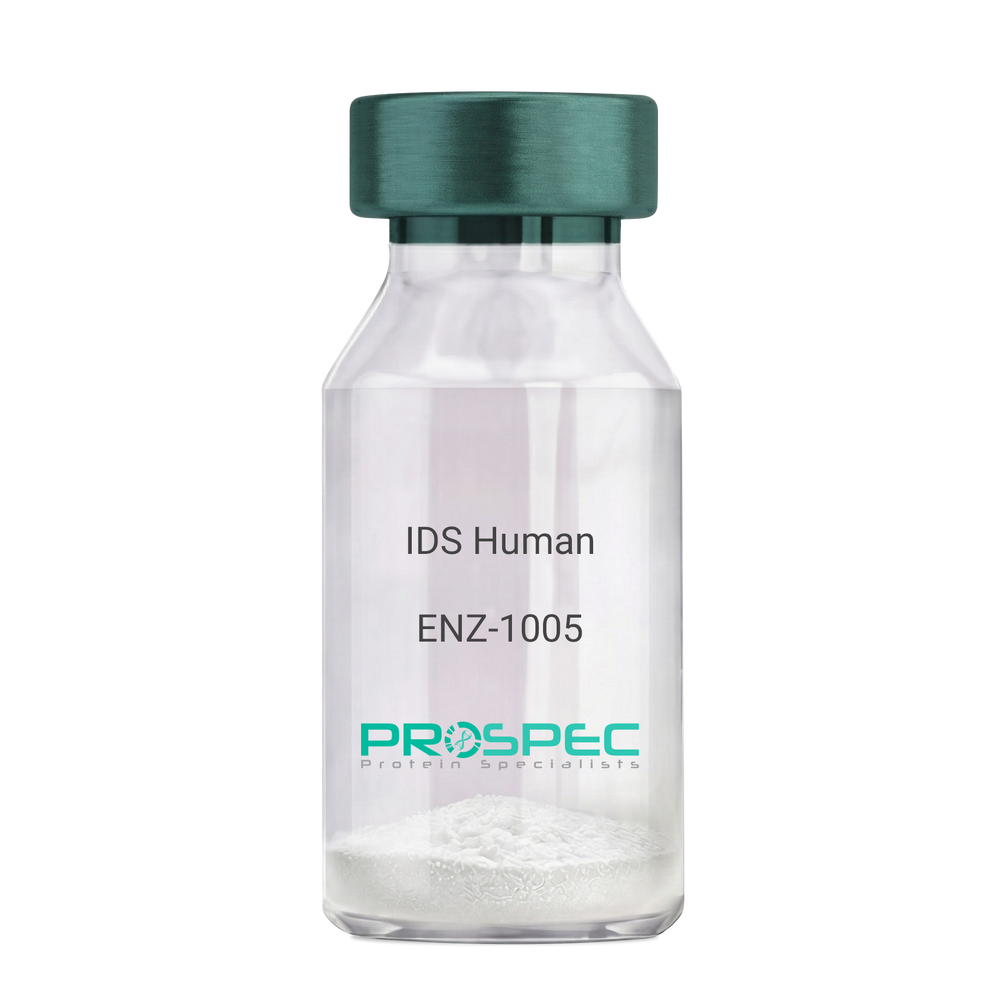
IDS HumanDescription:
Iduronate 2-Sulfatase Human Recombinant
Iduronate 2-Sulfatase, Alpha-L-Iduronate Sulfate Sulfatase, SIDS, Iduronate 2-Sulfatase 14 KDa Chain, Iduronate 2-Sulfatase 42 KDa Chain, Hunter Syndrome, EC 3.1.6.13, MPS2, Iduronate 2-sulfatase, Alpha-L-iduronate sulfate sulfatase.
Product # :
ENZ-1005Price :
Quantity :
Shipping Method :
Shipped with Ice Packs
More Info
- description
- source
- formulation
- purity
- More Info
- Citations
Description
IDS Human Recombinant produced in Sf9 Baculovirus cells is a single, glycosylated polypeptide chain containing 533 amino acids (26-550a.a) and having a molecular mass of 60.3kDa. (Molecular size on SDS-PAGE will appear at approximately 35-70kDa). IDS is fused to an 8 amino acid His-tag at C-terminus & purified by proprietary chromatographic techniques.
Source
Sf9, Baculovirus cells.
Formulation
IDS protein solution (0.25mg/ml) containing Phosphate Buffered Saline (pH 7.4) and 10% glycerol.
Purity
Greater than 90% as determined by SDS-PAGE.
Citations
More Info
-
Introduction
Iduronate 2-Sulfatase also known as IDS, belongs to the highly-conserved sulfatase family of enzymes which catalyze the hydrolysis of O-sulfate and N-salfate esters from a variety of substrates. IDS is essential for the lysosomal degradation of the glycosaminoglycans (GAG) heparan sulfate as well as dermatan sulfate. Furthermore, IDS hydrolyzes the 2-sulfate group of the IDS units of the GAG.
-
Synonyms
Iduronate 2-Sulfatase, Alpha-L-Iduronate Sulfate Sulfatase, SIDS, Iduronate 2-Sulfatase 14 KDa Chain, Iduronate 2-Sulfatase 42 KDa Chain, Hunter Syndrome, EC 3.1.6.13, MPS2, Iduronate 2-sulfatase, Alpha-L-iduronate sulfate sulfatase.
-
Physical Appearance
Sterile Filtered clear solution.
-
Stability
Store at 4°C if entire vial will be used within 2-4 weeks. Store, frozen at -20°C for longer periods of time. For long term storage it is recommended to add a carrier protein (0.1% HSA or BSA).Avoid multiple freeze-thaw cycles.
-
Amino Acid Sequence
SETQANSTTD ALNVLLIIVD DLRPSLGCYG DKLVRSPNID QLASHSLLFQ NAFAQQAVCA PSRVSFLTGR RPDTTRLYDF NSYWRVHAGN FSTIPQYFKE NGYVTMSVGK VFHPGISSNH TDDSPYSWSF PPYHPSSEKY ENTKTCRGPD GELHANLLCP VDVLDVPEGT LPDKQSTEQA IQLLEKMKTS ASPFFLAVGY HKPHIPFRYP KEFQKLYPLE NITLAPDPEV PDGLPPVAYN PWMDIRQRED VQALNISVPY GPIPVDFQRK IRQSYFASVS YLDTQVGRLL SALDDLQLAN STIIAFTSDH GWALGEHGEW AKYSNFDVAT HVPLIFYVPG RTASLPEAGE KLFPYLDPFD SASQLMEPGR QSMDLVELVS LFPTLAGLAG LQVPPRCPVP SFHVELCREG KNLLKHFRFR DLEEDPYLPG NPRELIAYSQ YPRPSDIPQW NSDKPSLKDI KIMGYSIRTI DYRYTVWVGF NPDEFLANFS DIHAGELYFV DSDPLQDHNM YNDSQGGDLF QLLMPLEHHH HHH.
ProSpec's products are furnished for LABORATORY RESEARCH USE ONLY. The product may not be used as drugs, agricultural or pesticidal products, food additives or household chemicals.
- View Data Sheet
Name :
IFNG Human, His ActiveDescription:
Interferon-gamma Human Recombinant, His Tag Active
Immune Interferon, type II interferon, T cell interferon, MAF, IFNG, IFG, IFI, IFN-gamma.
Product # :
CYT-1053Price :
Quantity :
Shipping Method :
Shipped with Ice Packs
More Info
- description
- source
- formulation
- purity
- biological activity
- More Info
- SDS-PAGE
- Citations
Description
IFNG Human Recombinant produced in E.Coli is a single, non-glycosylated polypeptide chain containing 159 amino acids (24-161 a.a) and having a molecular mass of 18.4kDa. IFNG is fused to a 21 amino acid His-tag at N-terminus & purified by proprietary chromatographic techniques.
Source
Escherichia Coli.
Formulation
IFNG protein solution (1mg/ml) containing Phosphate buffered saline (pH 7.4) and 20% glycerol.
Purity
Greater than 90.0% as determined by SDS-PAGE.
Biological Activity
Measured in a cytotoxicity assay using WiDr cells. The ED50 for this effect is ≤ 5 ng/ml.
SDS-PAGE
Citations
More Info
-
Introduction
IFN-gamma produced by lymphocytes activated by specific antigens or mitogens.
IFN-gamma, in addition to having antiviral activity, has important immunoregulatory functions, it is a potent activator of macrophages, and has antiproliferative effects on transformed cells and it can potentiate the antiviral and antitumor effects of the type I interferons. -
Synonyms
Immune Interferon, type II interferon, T cell interferon, MAF, IFNG, IFG, IFI, IFN-gamma.
-
Physical Appearance
Sterile Filtered colorless liquid.
-
Stability
Store at 4°C if entire vial will be used within 2-4 weeks. Store, frozen at -20°C for longer periods of time. For long term storage it is recommended to add a carrier protein (0.1% HSA or BSA).Avoid multiple freeze-thaw cycles.
-
Amino Acid Sequence
MGSSHHHHHH SSGLVPRGSH MQDPYVKEAE NLKKYFNAGH SDVADNGTLF LGILKNWKEE SDRKIMQSQI VSFYFKLFKN FKDDQSIQKS VETIKEDMNV KFFNSNKKKR DDFEKLTNYS VTDLNVQRKA IHELIQVMAE LSPAAKTGKR KRSQMLFRG.
-
Background
What is the molecular weight/Mw of IFNG HUMAN, HIS ACTIVE Protein?
IFNG HUMAN, HIS ACTIVE Protein has a total Mw of 18.4kDa.
What is the source or expression system of IFNG HUMAN, HIS ACTIVE Protein?
Escherichia Coli.
What is the Purity of IFNG HUMAN, HIS ACTIVE Protein?
IFNG HUMAN, HIS ACTIVE Protein is >90% pure as determined by SDS-PAGE.
What is the Biological Activity of IFNG HUMAN, HIS ACTIVE Protein?
Measured in a cytotoxicity assay using WiDr cells. The ED50 for this effect is ≤ 5 ng/ml.
What is the amino acid sequence of IFNG HUMAN, HIS ACTIVE Protein?
MGSSHHHHHH SSGLVPRGSH MQDPYVKEAE NLKKYFNAGH SDVADNGTLF LGILKNWKEE SDRKIMQSQI VSFYFKLFKN FKDDQSIQKS VETIKEDMNV KFFNSNKKKR DDFEKLTNYS VTDLNVQRKA IHELIQVMAE LSPAAKTGKR KRSQMLFRG.
What applications can IFNG HUMAN, HIS ACTIVE Protein be used in?
IFNG HUMAN, HIS ACTIVE Protein can probably be used in western blot, ELISA and Lateral Flow.
What is the endotoxin level for IFNG HUMAN, HIS ACTIVE Protein?
The endotoxin level is minimal, IFNG HUMAN, HIS ACTIVE Protein was purified using conventional chromatography techniques.
ProSpec's products are furnished for LABORATORY RESEARCH USE ONLY. The product may not be used as drugs, agricultural or pesticidal products, food additives or household chemicals.
- View Data Sheet
Name :
Borrelia Afzelii OspCDescription:
Borrelia Afzelii Outer Surface Protein C Recombinant
Product # :
BOR-016Price :
Quantity :
Shipping Method :
Shipped with Ice Packs
More Info
- description
- source
- formulation
- purity
- More Info
- Citations
Description
Recombinant Borrelia Afzelii Outer Surface Protein C produced in E.coli is a non-glycosylated, polypeptide chain having a calculated molecular mass of 22,151 Dalton. Borrelia Afzelii OspC is expressed with a 10xHis tag at N-terminus and purified by proprietary chromatographic techniques.
Source
Escherichia Coli.
Formulation
Borrelia Afzelii OspC is supplied in 20mM HEPES buffer pH-7.6, 250mM NaCl and 20% glycerol.
Purity
Greater than 80.0% as determined by SDS-PAGE.
Citations
More Info
-
Introduction
Borrelia belongs to a genus of bacteria of the spirochete phylum. Borrelia causes borreliosis, which is a zoonotic, vector-borne disease transmitted mainly by ticks and some by lice, depending on the species. Of the 36 known species of Borrelia, 12 are distinguished to cause Lyme disease or borreliosis and are transmitted by ticks. The main Borrelia species causing Lyme disease are Borrelia burgdorferi, Borrelia afzelii, and Borrelia garinii. The Borrelia genus members have a linear chromosome which is about 900 kbp in length as well as an excess of both linear and circular plasmids in the 5-220 kbp size range. The plasmids are atypical, as compared to most bacterial plasmids, since they contain many paralogous sequences, a large number of pseudogenes and, in some cases, essential genes. Moreover, a number of the plasmids have features suggesting that they are prophages.
-
Physical Appearance
Sterile Filtered clear solution.
-
Stability
Store at 4°C if entire vial will be used within 2-4 weeks. Store, frozen at -20°C for longer periods of time. Avoid multiple freeze-thaw cycles.
-
Immunological Functions
1. Binds IgG- and IgM-type human antibodies.2. Immunodot test with Lyme disease positive/negative plasma.
ProSpec's products are furnished for LABORATORY RESEARCH USE ONLY. The product may not be used as drugs, agricultural or pesticidal products, food additives or household chemicals.
- View Data Sheet
Name :
Borrelia DbpADescription:
Borrelia Burgdorferi Decorin Binding Protein A Recombinant
Product # :
BOR-005Price :
Quantity :
Shipping Method :
Shipped with Ice Packs
More Info
- description
- source
- formulation
- purity
- More Info
- Citations
Description
Recombinant Borrelia Burgdorferi Decorin Binding Protein A produced in E.coli is a non-glycosylated, polypeptide chain having a calculated molecular mass of 19,916 Dalton. Borrelia DbpA is expressed with a -6x His tag at N-terminus and purified by proprietary chromatographic techniques.
Source
Escherichia Coli.
Formulation
Borrelia DbpA (0.91mg/1ml) is supplied in 20mM HEPES buffer pH-8.0 and 20% glycerol.
Purity
Greater than 80.0% as determined by SDS-PAGE.
Citations
More Info
-
Introduction
Borrelia belongs to a genus of bacteria of the spirochete phylum. Borrelia causes borreliosis, which is a zoonotic, vector-borne disease transmitted mainly by ticks and some by lice, depending on the species. Of the 36 known species of Borrelia, 12 are distinguished to cause Lyme disease or borreliosis and are transmitted by ticks. The main Borrelia species causing Lyme disease are Borrelia burgdorferi, Borrelia afzelii, and Borrelia garinii. The Borrelia genus members have a linear chromosome which is about 900 kbp in length as well as an excess of both linear and circular plasmids in the 5-220 kbp size range. The plasmids are atypical, as compared to most bacterial plasmids, since they contain many paralogous sequences, a large number of pseudogenes and, in some cases, essential genes. Moreover, a number of the plasmids have features suggesting that they are prophages.
-
Physical Appearance
Sterile Filtered clear solution.
-
Stability
Store at 4°C if entire vial will be used within 2-4 weeks. Store, frozen at -20°C for longer periods of time. Avoid multiple freeze-thaw cycles.
-
Applications
Western blot with Lyme positive plasma.
-
Patent Protected Countries
The Sale and/or commercial use of recombinant Borrelia DbpA by ProSpec is prohibited in the following countries: Canada, USA, Germany, Austria, Belgium, Switzerland, Denmark, Finland, France, United Kingdom, Ireland, Netherlands and Sweden.
ProSpec's products are furnished for LABORATORY RESEARCH USE ONLY. The product may not be used as drugs, agricultural or pesticidal products, food additives or household chemicals.
- View Data Sheet
Name :
CD5L MouseDescription:
CD5 Molecule-Like Mouse Recombinant
CD5 antigen-like, Apoptosis inhibitor expressed by macrophages, Apoptosis inhibitory 6 CT-2, SP-alpha, Cd5l' Aim, Api6.
Product # :
PRO-2243Price :
Quantity :
Shipping Method :
Shipped with Ice Packs
More Info
- description
- source
- formulation
- purity
- More Info
- Citations
Description
CD5L produced in Sf9 Insect cells is a single, glycosylated polypeptide chain containing 339 amino acids (22-352a.a.) and having a molecular mass of 37.6 kDa. (Molecular size on SDS-PAGE will appear at approximately 40-57kDa).CD5L is expressed with an 8 amino acid His tag at C-Terminus and purified by proprietary chromatographic techniques.
Source
Sf9, Insect cells.
Formulation
CD5L protein solution (0.25mg/ml) contains phosphate buffered saline (pH7.4) and 10% glycerol.
Purity
Greater than 95.0% as determined by SDS-PAGE.
Citations
More Info
-
Introduction
CD5 antigen-like (CD5L) participates in the regulation of the immune system. CD5L functions as an inhibitor of apoptosis. CD5L is expressed in the spleen, lymph node, thymus and bone marrow.
-
Synonyms
CD5 antigen-like, Apoptosis inhibitor expressed by macrophages, Apoptosis inhibitory 6 CT-2, SP-alpha, Cd5l' Aim, Api6.
-
Physical Appearance
Sterile filtered colorless solution.
-
Stability
Store at 4°C if entire vial will be used within 2-4 weeks.Store, frozen at -20°C for longer periods of time. For long term storage it is recommended to add a carrier protein (0.1% HSA or BSA).Avoid multiple freeze-thaw cycles.
-
Amino Acid Sequence
ESPTKVQLVG GAHRCEGRVE VEHNGQWGTV CDDGWDRRDV AVVCRELNCG AVIQTPRGAS YQPPASEQRV LIQGVDCNGT EDTLAQCELN YDVFDCSHEE DAGAQCENPD SDLLFIPEDV RLVDGPGHCQ GRVEVLHQSQ WSTVCKAGWN LQVSKVVCRQ LGCGRALLTY GSCNKSTQGK GPIWMGKMSC SGQEANLRSC LLSRLENNCT HGEDTWMECE DPFELKLVGG DTPCSGRLEV LHKGSWGSVC DDNWGEKEDQ VVCKQLGCGK SLHPSPKTRK IYGPGAGRIW LDDVNCSGKE QSLEFCRHRL WGYHDCTHKE DVEVICTDFD VLEHHHHHH.
ProSpec's products are furnished for LABORATORY RESEARCH USE ONLY. The product may not be used as drugs, agricultural or pesticidal products, food additives or household chemicals.
- View Data Sheet
Name :
CENPA HumanDescription:
Centromere Protein A Human Recombinant
Histone H3-like centromeric protein A, Centromere protein A, CENP-A, Centromere autoantigen A, CENPA.
Product # :
PRO-389Price :
Quantity :
Shipping Method :
Shipped with Ice Packs
More Info
- description
- source
- formulation
- purity
- More Info
- Citations
Description
CENPA Human Recombinant produced in SF9 is a glycosylated, polypeptide chain having a molecular mass of 17,015 Dalton. CENPA is expressed with a -6x His tag and purified by proprietary chromatographic techniques.
Source
Sf9 insect cells.
Formulation
CENPA is supplied in 20mM HEPES buffer pH-8, 100mM sodium chloride, and 6M Urea.
Purity
Greater than 90% as determined by SDS-PAGE.
Citations
More Info
-
Introduction
Centromere proteins are a group of proteins which form and/or mediate the function of centromeres, the central structures of chromosomes to which spindle fibers/microtubuli attach and pull the chromosomes apart in cell division. Currently, 9 centromere proteins are known and designated CENPA to CENP-I. Most of the centromere proteins are targets of autoantibodies, the anti-centromere antibodies.
CENPA is another important centromeric autoantigen in addition to CENPB: it has a molecular weight of approx. 20 kDa and is incorporated into centromeric chromatin due to its histone-like properties. CENPA antibodies are an important marker for correct diagnosis of Scleroderma / CREST syndrome in CENPB-autoantibody negative patients. -
Synonyms
Histone H3-like centromeric protein A, Centromere protein A, CENP-A, Centromere autoantigen A, CENPA.
-
Physical Appearance
Sterile Filtered liquid formulation.
-
Stability
Store at 4°C if entire vial will be used within 2-4 weeks. Store, frozen at -20°C for longer periods of time. Avoid multiple freeze-thaw cycles.
-
Immunological Functions
1. Binds IgG-type human auto-antibodies.2. Standard ELISA test (checker-board analysis of positive/negative sera panels including CDC international reference sera).
-
coating concentration
0.15-0.4 µg/ml (depending on the type of ELISA plate and coating buffer).Suitable for biotinylation and iodination.
-
Applications
Western-Blot with monoclonal anti-hexa-His-tag antibody & Scleroderma patient sera.
ProSpec's products are furnished for LABORATORY RESEARCH USE ONLY. The product may not be used as drugs, agricultural or pesticidal products, food additives or household chemicals.
- View Data Sheet
Name :
LGALS3 MouseDescription:
Galectin-3 Mouse Recombinant
CBP35, GAL3, GALBP, GALIG, LGALS2, MAC2, Galectin-3, Lectin, galactose binding, soluble 3, Lectin, galactose binding, soluble 3, isoform CRA_a, Lectin, galactose binding, soluble 3, isoform CRA_d, Lgals3.
Product # :
CYT-188Price :
Quantity :
Shipping Method :
Shipped with Ice Packs
More Info
- description
- source
- formulation
- purity
- More Info
- sds-page
- Citations
Description
LGALS3 Mouse Recombinant produced in E. coli is a single polypeptide chain containing 287 amino acids (1-264) and having a molecular mass of 29.8 kDa. LGALS3 is fused to a 23 amino acid His-tag at N-terminus & purified by proprietary chromatographic techniques.
Source
E.coli.
Formulation
The LGALS3 solution (0.5mg/1ml) contains 20mM Tris-HCl buffer (pH 8.0), 0.15M NaCl, 50% glycerol, 1mM DTT and 2mM EDTA.
Purity
Greater than 95% as determined by SDS-PAGE.
sds-page
Citations
More Info
-
Introduction
Galectin-3 mediates with the alpha-3, beta-1 integrin the stimulation by cspg4 of endothelial cells migration. Galectin-3 plays an necessary part during the acquisition of vasculogenic mimicry and angiogenic properties associated with melanoma progression. LGALS3 overexpression is highly expressed in early stages of papillary carcinoma, and its expression intensity declines during tumor progression. Serum levels of LGALS3 are high in patients with thyroid malignancy but there is considerable overlap in serum LGALS3 concentrations between those with benign and malignant nodular thyroid disease. LGLAS3 takes part as an immune regulator to inhibit T-cell immune responses and promote tumor growth, as a result providing a new mechanism for tumor immune tolerance.
-
Synonyms
CBP35, GAL3, GALBP, GALIG, LGALS2, MAC2, Galectin-3, Lectin, galactose binding, soluble 3, Lectin, galactose binding, soluble 3, isoform CRA_a, Lectin, galactose binding, soluble 3, isoform CRA_d, Lgals3.
-
Physical Appearance
Sterile Filtered colorless solution.
-
Stability
Store at 4°C if entire vial will be used within 2-4 weeks. Store, frozen at -20°C for longer periods of time. For long term storage it is recommended to add a carrier protein (0.1% HSA or BSA).Avoid multiple freeze-thaw cycles.
-
Amino Acid Sequence
MGSSHHHHHH SSGLVPRGSH MGSMADSFSL NDALAGSGNP NPQGYPGAWG NQPGAGGYPG AAYPGAYPGQ APPGAYPGQA PPGAYPGQAP PSAYPGPTAP GAYPGPTAPG AYPGSTAPGA FPGQPGAPGA YPSAPGGYPA AGPYGVPAGP LTVPYDLPLP GGVMPRMLIT IMGTVKPNAN RIVLDFRRGN DVAFHFNPRF NENNRRVIVC NTKQDNNWGK EERQSAFPFE SGKPFKIQVL VEADHFKVAV NDAHLLQYNH RMKNLREISQ LGISGDITLT SANHAMI.
-
Background
What is the molecular weight/Mw of LGALS3 Protein?
LGALS3 Protein has a total Mw of 29.8kDa.
What is the source or expression system of LGALS3 Protein?
Escherichia Coli.
What is the Purity of LGALS3 Protein?
LGALS3 Protein is >95% pure as determined by SDS-PAGE.
What is the Biological Activity of LGALS3 Protein?
The biological functionality of LGALS3 Protein will be determined in the future.
What is the amino acid sequence of LGALS3 Protein?
MGSSHHHHHH SSGLVPRGSH MGSMADSFSL NDALAGSGNP NPQGYPGAWG NQPGAGGYPG AAYPGAYPGQ APPGAYPGQA PPGAYPGQAP PSAYPGPTAP GAYPGPTAPG AYPGSTAPGA FPGQPGAPGA YPSAPGGYPA AGPYGVPAGP LTVPYDLPLP GGVMPRMLIT IMGTVKPNAN RIVLDFRRGN DVAFHFNPRF NENNRRVIVC NTKQDNNWGK EERQSAFPFE SGKPFKIQVL VEADHFKVAV NDAHLLQYNH RMKNLREISQ LGISGDITLT SANHAMI.
What applications can LGALS3 Protein be used in?
LGALS3 Protein can probably be used in western blot, ELISA and Lateral Flow.
What is the endotoxin level for LGALS3 Protein?
The endotoxin level is minimal, LGALS3 Protein was purified using conventional chromatography techniques.
ProSpec's products are furnished for LABORATORY RESEARCH USE ONLY. The product may not be used as drugs, agricultural or pesticidal products, food additives or household chemicals.
- View Data Sheet
Name :
LGMN MouseDescription:
Legumain Mouse Recombinant
Legumain, Asparaginyl endopeptidase, Protease, cysteine 1.
Product # :
ENZ-933Price :
Quantity :
Shipping Method :
Shipped with Ice Packs
More Info
- description
- source
- formulation
- purity
- More Info
- sds-page
- Citations
Description
LGMN produced in Sf9 Baculovirus cells is a single, glycosylated polypeptide chain containing 426 amino acids (18-435a.a.) and having a molecular mass of 48.6kDa. LGMN is expressed with an 8 amino acid His tag at C-Terminus and purified by proprietary chromatographic techniques.
Source
Sf9, Baculovirus cells.
Formulation
LGMN protein solution (0.5mg/ml) contains Phosphate Buffered Saline (pH 7.4) and 10% glycerol.
Purity
Greater than 90.0% as determined by SDS-PAGE.
sds-page
Citations
More Info
-
Introduction
Legumain, also known as LGMN is a lysosomal cysteine protease which is found in all mouse tissues, however it was mainly abundant in the kidney as well as placenta. LGMN plays an essential role in the endosomal/lysosomal degradation system as the Legumain deficiency causes the accumulation of pro cathepsins B, H & L, another group of lysosomal cysteine proteases. Furthermore, over expression of LGMN in tumors is important for invasion/metastasis.
-
Synonyms
Legumain, Asparaginyl endopeptidase, Protease, cysteine 1.
-
Physical Appearance
Sterile filtered colorless solution.
-
Stability
Store at 4°C if entire vial will be used within 2-4 weeks.Store, frozen at -20°C for longer periods of time. For long term storage it is recommended to add a carrier protein (0.1% HSA or BSA).Avoid multiple freeze-thaw cycles.
-
Amino Acid Sequence
VPVGVDDPED GGKHWVVIVA GSNGWYNYRH QADACHAYQI IHRNGIPDEQ IIVMMYDDIA NSEENPTPGV VINRPNGTDV YKGVLKDYTG EDVTPENFLA VLRGDAEAVK GKGSGKVLKS GPRDHVFIYF TDHGATGILV FPNDDLHVKD LNKTIRYMYE HKMYQKMVFY IEACESGSMM NHLPDDINVY ATTAANPKES SYACYYDEER GTYLGDWYSV NWMEDSDVED LTKETLHKQY HLVKSHTNTS HVMQYGNKSI STMKVMQFQG MKHRASSPIS LPPVTHLDLT PSPDVPLTIL KRKLLRTNDV KESQNLIGQI QQFLDARHVI EKSVHKIVSL LAGFGETAER HLSERTMLTA HDCYQEAVTH FRTHCFNWHS VTYEHALRYL YVLANLCEAP YPIDRIEMAM DKVCLSHYLE HHHHHH.
ProSpec's products are furnished for LABORATORY RESEARCH USE ONLY. The product may not be used as drugs, agricultural or pesticidal products, food additives or household chemicals.
- View Data Sheet
Name :
M CSF RatDescription:
Macrophage-Colony Stimulating Factor Rat Recombinant
Macrophage colony-stimulating factor 1, CSF-1, MCSF, Csf1, Csfm.
Product # :
CYT-856Price :
Quantity :
Shipping Method :
Shipped at Room temp
More Info
- description
- source
- formulation
- purity
- biological activity
- More Info
- Citations
Description
Macrophage Colony Stimulating Factor Rat Recombinant produced in E.coli is a non-glycosylated homodimer, containing 2 x 155 amino acids and having a total molecular mass of 36.2 kDa.MCSF is purified by proprietary chromatographic techniques.
Source
Escherichia Coli.
Formulation
Lyophilized from a sterile filtered aqueous solution containing 10mM Na3PO4, pH 7.5.
Purity
Greater than 95.0% as determined by SDS-PAGE.
Biological Activity
The activity as determined by dose-dependent induction of M-NFS-60 cell proliferation is 1.65 ng/ml. This corresponds to an expected specific activity of 6.1x10⁵ units/mg.
Citations
More Info
-
Introduction
Granulocyte/Macrophage Colony-Stimulating Factors are cytokines that act in hematopoiesis by controlling the production, differentiation, and function of 2 related white cell populations of the blood, the granulocytes and the monocytes-macrophages. MCSF induces cells of the monocyte/macrophage lineage. MCSF plays a role in immunological defenses, bone metabolism, lipoproteins clearance, fertility and pregnancy.
-
Synonyms
Macrophage colony-stimulating factor 1, CSF-1, MCSF, Csf1, Csfm.
-
Physical Appearance
Sterile Filtered White lyophilized (freeze-dried) powder.
-
Stability
Lyophilized MCSF although stable at room temperature for 3 weeks, should be stored desiccated below -18°C. Upon reconstitution MCSF should be stored at 4°C between 2-7 days and for future use below -18°C.For long term storage it is recommended to add a carrier protein (0.1% HSA or BSA).Please prevent freeze-thaw cycles.
-
Solubility
It is recommended to reconstitute the lyophilized MCSF in sterile 18M-cm H2O not less than 100µg/ml, which can then be further diluted to other aqueous solutions.
-
Amino Acid Sequence
MEVSEHCSHM IGNGHLQILQ QLIDSQMETA CLIEYKFVDQ EQLDDPVCYL KKAFVLVQVI IEETMRFKDN TPNANATERL QELSMKLNSC FIKDYKEQNE ACVQTYKESP LRLLEKIKNF FNETKNFLEK DWNIFSKNCN DSLAKCSSRD VVTKP.
ProSpec's products are furnished for LABORATORY RESEARCH USE ONLY. The product may not be used as drugs, agricultural or pesticidal products, food additives or household chemicals.
- View Data Sheet
Name :
CHRAC1 HumanDescription:
Chromatin Accessibility Complex 1 Human Recombinant
CHARC1, CHARC15, CHRAC-1, CHRAC-15, CHRAC15, YCL1, Chromatin accessibility complex protein 1, Chromatin accessibility complex 15 kDa protein, DNA polymerase epsilon subunit p15.
Product # :
PRO-1391Price :
Quantity :
Shipping Method :
Shipped with Ice Packs
More Info
- description
- source
- formulation
- purity
- More Info
- Citations
Description
CHRAC1 Human Recombinant produced in E.Coli is a single, non-glycosylated polypeptide chain containing 154 amino acids (1-131 a.a.) and having a molecular mass of 17.1kDa. CHRAC1 is fused to a 23 amino acid His-tag at N-terminus & purified by proprietary chromatographic techniques.
Source
E.coli.
Formulation
CHRAC1 protein solution (1mg/ml) contains 20mM Tris-HCl buffer (pH 8.0), 0.4M urea and 10% glycerol.
Purity
Greater than 90% as determined by SDS-PAGE.
Citations
More Info
-
Introduction
Chromatin Accessibility Complex 1 (CHRAC1) is a histone-fold protein which interacts with further histone-fold proteins to bind DNA in a sequence-independent mode. These histone-fold protein dimers combine within larger enzymatic complexes for DNA replication, transcription, and packaging.
-
Synonyms
CHARC1, CHARC15, CHRAC-1, CHRAC-15, CHRAC15, YCL1, Chromatin accessibility complex protein 1, Chromatin accessibility complex 15 kDa protein, DNA polymerase epsilon subunit p15.
-
Physical Appearance
Sterile Filtered colorless solution.
-
Stability
Store at 4°C if entire vial will be used within 2-4 weeks. Store, frozen at -20°C for longer periods of time. For long term storage it is recommended to add a carrier protein (0.1% HSA or BSA).Avoid multiple freeze-thaw cycles.
-
Amino Acid Sequence
MGSSHHHHHH SSGLVPRGSH MGSMADVVVG KDKGGEQRLI SLPLSRIRVI MKSSPEVSSI NQEALVLTAK ATELFVQCLA TYSYRHGSGK EKKVLTYSDL ANTAQQSETF QFLADILPKK ILASKYLKML KEEKREEDEE NDNDNESDHD EADS.
ProSpec's products are furnished for LABORATORY RESEARCH USE ONLY. The product may not be used as drugs, agricultural or pesticidal products, food additives or household chemicals.