Search results
777 results found for “Basic Transcription Factor”
Name
Description
Product #
Price
Quantity
Shipping Method
- View Data Sheet
Name :
FGF 2 MouseDescription:
Fibroblast Growth Factor-Basic Mouse Recombinant
HBGF-2, Prostatropin, FGF-2, FGB-b.
Product # :
CYT-386Price :
Quantity :
Shipping Method :
Shipped at Room temp
More Info
- description
- source
- formulation
- purity
- biological activity
- More Info
- Citations
Description
Fibroblast Growth Factor-basic Mouse Recombinant (FGF-2) produced in E.Coli is a single, non-glycosylated, polypeptide chain containing 146 amino acids and having a molecular mass of 16.3kDa. The FGF-2 is purified by proprietary chromatographic techniques.
Source
Escherichia Coli.
Formulation
FGF-b was lyophilized from 5mM Na2PO4, pH7.5 and 50mM NaCl.
Purity
Greater than 95.0% as determined by SDS-PAGE.
Biological Activity
The activity as calculated by the dose-dependant proliferation of BALB/3T3 cells was found to be less than 1ng/ml corresponding to a specific activity of 1,000,000 units/mg.Citations
More Info
-
Introduction
FGF-basic is a member of the fibroblast growth factor (FGF) family. FGF family members bind and possess broad mitogenic and angiogenic activities. This protein has been implicated in diverse biological processes, such as limb and nervous system development, wound healing, and tumor growth. The mRNA for this gene contains multiple polyadenylation sites, and is alternatively translated from AUG and non-AUG (CUG) initiation codons resulting in five different isoforms with distinct properties. The CUG-initiated isoforms are localized in the nucleus and are responsible for the intracrine effect, whereas, the AUG-initiated form is mostly cytosolic and is responsible for the paracrine and autocrine effects of this FGF.
The binding growth factors are angiogenic agents in vivo and are potent mitogens for a variety of cell types in vitro. there are differences in the tissue distribution and concentration of these 2 growth factors. -
Synonyms
HBGF-2, Prostatropin, FGF-2, FGB-b.
-
Physical Appearance
Sterile Filtered White lyophilized (freeze-dried) powder.
-
Stability
Lyophilized Fibroblast Growth Factor-2 although stable at room temperature for 3 weeks, should be stored desiccated below -18°C. Upon reconstitution FGF-basic should be stored at 4°C between 2-7 days and for future use below -18°C.For long term storage it is recommended to add a carrier protein (0.1% HSA or BSA).Please prevent freeze-thaw cycles.
-
Solubility
It is recommended to reconstitute the lyophilized Fibroblast Growth Factor b in sterile 18M-cm H2O not less than 100µg/ml, which can then be further diluted to other aqueous solutions.
-
Amino Acid Sequence
MPALPEDGGA AFPPGHFKDP KRLYCKNGGF FLRIHPDGRV DGVREKSDPH VKLQLQAEER GVVSIKGVCA NRYLAMKEDG RLLASKCVTE ECFFFERLES NNYNTYRSRK YSSWYVALKR TGQYKLGSKT GPGQKAILFL PMSAKS.
-
Background
What is the molecular weight/Mw of FGF 2 MOUSE Protein?
FGF 2 MOUSE Protein has a total Mw of 16.3kDa.
What is the source or expression system of FGF 2 MOUSE Protein?
Escherichia Coli.
What is the Purity of FGF 2 MOUSE Protein?
FGF 2 MOUSE Protein is >95% pure as determined by SDS-PAGE.
What is the Biological Activity of FGF 2 MOUSE Protein?
The activity as calculated by the dose-dependant proliferation of BALB/3T3 cells was found to be less than 1ng/ml corresponding to a specific activity of 1,000,000 units/mg.
What is the amino acid sequence of FGF 2 MOUSE Protein?
MPALPEDGGA AFPPGHFKDP KRLYCKNGGF FLRIHPDGRV DGVREKSDPH VKLQLQAEER GVVSIKGVCA NRYLAMKEDG RLLASKCVTE ECFFFERLES NNYNTYRSRK YSSWYVALKR TGQYKLGSKT GPGQKAILFL PMSAKS.
What applications can FGF 2 MOUSE Protein be used in?
FGF 2 MOUSE Protein can probably be used in western blot, ELISA and Lateral Flow.
What is the endotoxin level for FGF 2 MOUSE Protein?
The endotoxin level is minimal, FGF 2 MOUSE Protein was purified using conventional chromatography techniques.
-
Protein content
Protein quantitation was carried out by two independent methods:1. UV spectroscopy at 280 nm using the absorbency value of 0.885 as the extinction coefficient for a 0.1% (1mg/ml) solution. This value is calculated by the PC GENE computer analysis program of protein sequences (IntelliGenetics). 2. Analysis by RP-HPLC, using a calibrated solution of FGF2 as a Reference Standard.
ProSpec's products are furnished for LABORATORY RESEARCH USE ONLY. The product may not be used as drugs, agricultural or pesticidal products, food additives or household chemicals.
- View Data Sheet
Name :
BAFF AntibodyDescription:
B-cell Activating Factor, Mouse Anti Human
BAFF, BLYS, CD257, TALL1, THANK, ZTNF4, TALL-1, TNFSF20, TNFSF13B.
Product # :
ANT-367Price :
Quantity :
Shipping Method :
Shipped with Ice Packs
More Info
- formulation
- More Info
- Citations
Formulation
1mg/ml containing PBS, pH-7.4, & 0.1% Sodium Azide.
Citations
More Info
-
Introduction
Binds to tnfrsf13b/taci and tnfrsf17/bcma. tnfsf13/april binds to the same 2 receptors, together, they form a 2 ligands -2 receptors pathway involved in the stimulation of b- and t-cell function and the regulation of humoral immunity. A third b-cell specific baff-receptor (baffr/br3) promotes the survival of mature b-cells and the b-cell response. B Lymphocyte Stimulator functions as a potent B-cell growth factor in costimulation assays. Administration of BAFF Human recombinant to mice disrupts splenic B-cell and T-cell zones and results in elevated levels of serum immunoglobulin.
-
Synonyms
BAFF, BLYS, CD257, TALL1, THANK, ZTNF4, TALL-1, TNFSF20, TNFSF13B.
-
Physical Appearance
Sterile Filtered colorless solution.
-
Immunogen
Anti-human BAFF mAb, is derived from hybridization of mouse F0 myeloma cells with spleen cells from BALB/c mice immunized with Recombinant human BAFF amino acids 134-285 purified from E. coli.
-
Ig Subclass
Mouse IgG3 heavy chain and κ light chain.
-
Clone
PH4C7AT.
-
Applications
BAFF antibody has been tested by ELISA and Western blot analysis to assure specificity and reactivity. Since application varies, however, each investigation should be titrated by the reagent to obtain optimal results. Recommended dilution range for Western blot analysis is 1:500 ~ 1,000. Recommended starting dilution is 1:500.
-
Type
Mouse Anti Human Monoclonal.
-
Storage Procedures
For periods up to 1 month store at 4°C, for longer periods of time, store at -20°C. Prevent freeze thaw cycles.
-
Purification Method
BAFF antibody was purified from mouse ascitic fluids by protein-G affinity chromatography.
ProSpec's products are furnished for LABORATORY RESEARCH USE ONLY. The product may not be used as drugs, agricultural or pesticidal products, food additives or household chemicals.
- View Data Sheet
Name :
Mouse OX40LDescription:
OX40 Ligand Mouse Recombinant
Tumor necrosis factor ligand superfamily member 4, OX40 ligand, OX40L, OX-40L, CD252, Tnfsf4, Ox40l, Txgp1l, gp3, OX4, Ath-1, Ath1, CD134L, gp34, Tnlg2b, Txgp1l
Product # :
CYT-1191Price :
Quantity :
Shipping Method :
Shipped with Ice Packs
More Info
- description
- source
- formulation
- purity
- biological activity
- More Info
- Citations
Description
OX40L Mouse produced in Sf9 Baculovirus cells is a single, glycosylated polypeptide chain containing 157 amino acids (51-198 aa) and having a molecular mass of 17.7kDa.OX40L is fused to a 6 amino acid His tag at C-terminus and purified by proprietary chromatographic techniques.
Source
Sf9, Baculovirus cells.
Formulation
The OX40L solution (1mg/ml) contains 10% Glycerol and Phosphate-Buffered Saline (pH 7.4).
Purity
Greater than 90.0% as determined by SDS-PAGE.
Biological Activity
Determined by its binding ability in a functional ELISA with Human TNFRSF4 (CAT# cyt-1039).
Citations
More Info
-
Introduction
OX40 Ligandis a part of the tumor necrosis factor (TNF)receptor family. OX40L receptor is OX40 which is expressed predominantly on activated CD4+ T cells.OX40 Ligandis expressed on various antigen-presenting cells such asmacrophages, DC2sand activated Blymphocytes. Expression of OX40 is momentary following engagement of T cell receptors. The ligation of OX40-OX40L stimulates proliferation anddifferentiation of activated B cells,increases immunoglobulin secretion and is also a sourceof survival signal for T cells and enables the development of memory T cells.
-
Synonyms
Tumor necrosis factor ligand superfamily member 4, OX40 ligand, OX40L, OX-40L, CD252, Tnfsf4, Ox40l, Txgp1l, gp3, OX4, Ath-1, Ath1, CD134L, gp34, Tnlg2b, Txgp1l
-
Physical Appearance
Sterile filtered colorless solution.
-
Stability
Store at 4°C if entire vial will be used within 2-4 weeks.Store, frozen at -20°C for longer periods of time.For long term storage it is recommended to add a carrier protein (0.1% HSA or BSA).Avoid multiple freeze-thaw cycles.
-
Amino Acid Sequence
ADPSSSPAKD PPIQRLRGAV TRCEDGQLFI SSYKNEYQTM EVQNNSVVIK CDGLYIIYLK GSFFQEVKID LHFREDHNPI SIPMLNDGRR IVFTVVASLA FKDKVYLTVN APDTLCEHLQ INDGELIVVQ LTPGYCAPEG SYHSTVNQVP LHHHHHH
ProSpec's products are furnished for LABORATORY RESEARCH USE ONLY. The product may not be used as drugs, agricultural or pesticidal products, food additives or household chemicals.
- View Data Sheet
Name :
TRAIL Human (114-281 a.a.)Description:
TRAIL/APO 2 Ligand (114-281 a.a.) Human Recombinant
Tumor necrosis factor ligand superfamily member 10, TNF-related apoptosis-inducing ligand, Protein TRAIL, Apo-2 ligand, Apo-2L, CD253 antigen, TL2, APO2L, TNFSF10.
Product # :
CYT-546Price :
Quantity :
Shipping Method :
Shipped with Ice Packs
More Info
- description
- source
- formulation
- purity
- More Info
- Citations
Description
Soluble TNF-related apoptosis-inducing ligand Human Recombinant produced in E.Coli is a single, non-glycosylated polypeptide chain containing 169 amino acids (114-281) and having a molecular mass of 19.6 kDa. The sTRAIL is purified by proprietary chromatographic techniques.
Source
Escherichia Coli.
Formulation
1mg/ml in 20mM Tris-HCl pH-7.5, 300mM NaCl, 0.1mM DTT & 10% glycerol.
Purity
Greater than 95.0% as determined by SDS-PAGE.
Citations
More Info
-
Introduction
TNF-related apoptosis-inducing ligand (TRAIL) is a ligand molecule which induces apoptosis. It is a type II transmembrane protein with homology to other members of the tumor necrosis factor family.
In humans, the gene that encodes for TRAIL is located at chromosome 3q26.
TRAIL binds to the death receptors, DR4 and DR5. The process of apoptosis is caspase-8-dependent. This protein preferentially induces apoptosis in transformed and tumor cells, but does not appear to kill normal cells although it is expressed at a significant level in most normal tissues. -
Synonyms
Tumor necrosis factor ligand superfamily member 10, TNF-related apoptosis-inducing ligand, Protein TRAIL, Apo-2 ligand, Apo-2L, CD253 antigen, TL2, APO2L, TNFSF10.
-
Physical Appearance
Sterile Filtered colorless liquid.
-
Stability
Store at 4°C if entire vial will be used within 2-4 weeks. Store, frozen at -20°C for longer periods of time. For long term storage it is recommended to add a carrier protein (0.1% HSA or BSA).Avoid multiple freeze-thaw cycles.
-
Amino Acid Sequence
MVRERGPQRV AAHITGTRGR SNTLSSPNSK NEKALGRKIN SWESSRSGHS FLSNLHLRNGELVIHEKGFY YIYSQTYFRF QEEIKENTKN DKQMVQYIYK YTSYPDPILL MKSARNSCWSKDAEYGLYSI YQGGIFELKE NDRIFVSVTN EHLIDMDHEA SFFGAFLVG.
ProSpec's products are furnished for LABORATORY RESEARCH USE ONLY. The product may not be used as drugs, agricultural or pesticidal products, food additives or household chemicals.
- View Data Sheet
Name :
Visfatin HumanDescription:
Visfatin Human Recombinant
PBEF, Pre-B cell colony-enhancing factor, Nicotinamide phosphoribosyltransferase NAmPRTase, Nampt, MGC117256, DKFZP666B131, 1110035O14Rik.
Product # :
CYT-318Price :
Quantity :
Shipping Method :
Shipped at Room temp
More Info
- description
- source
- formulation
- purity
- biological activity
- More Info
- Citations
Description
Visfatin Human Recombinant produced in E.Coli is a single, non-glycosylated, polypeptide chain containing 466 amino acids. The total molecular mass is 52.6kDa (calculated). The Visfatin is purified by Flag-affinity chromatography.
Source
Escherichia Coli.
Formulation
Visfatin was lyophilized with no additives.
Purity
Greater than 90.0% as determined by SDS-PAGE.
Biological Activity
The activity is determined by its ability to induce IL-6, IL-1 beta and TNF alpha production from human PBMCs at 100ng/ml.Citations
More Info
-
Introduction
Excess adiposity is the most important risk in the development of type 2 diabetes mellitus (T2DM). Adipose tissue produces several proteins (adipocytokines) such as leptin, adiponectin, resistin, tumor necrosis factor-a, and IL-6, that modulate sensitivity and appear to play an important role in the pathogenesis, diabetes, dyslipidemia, inflammation, and atherosclerosis. Visfatin, also known as pre-B cell colony-enhancing factor (PBEF), is a cytokine that is highly expressed in visceral fat and was originally isolated as a secreted factor that synergizes with IL-7 and stem cell factors to promote the growth of B cell precursors. Visfatin homologs have been identified in carp, invertebrate mollusks, and bacteria, as well as in vertebrates, including humans and the mouse. It has been postulated to play a role in innate immunity.
Visfatin exerts mimetic effects that are dose-dependent and quantitatively similar to stimulating muscle and adipocyte glucose transport, and in inhibiting hepatocyte glucose production. Intravenous injection of recombinant visfatin in mice decreased plasma glucose in a dose-dependent fashion. In keeping with its mimetic effects, visfatin was as effective in reducing hyperglycemia in deficient diabetic mice. Visfatin was also found to be bound to and activate receptor, causing receptor phosphorylation and the activation of downstream signaling molecules. However, visfatin did not compete for binding to the receptor, indicating that the two proteins were recognized by different regions of the receptor. Thus, visfatin might play a role in glucose homeostasis and dysregulation in biosynthesis or signal transduction, and might contribute to the pathogenesis of diabetes. -
Synonyms
PBEF, Pre-B cell colony-enhancing factor, Nicotinamide phosphoribosyltransferase NAmPRTase, Nampt, MGC117256, DKFZP666B131, 1110035O14Rik.
-
Physical Appearance
Filtered White lyophilized (freeze-dried) powder.
-
Stability
Lyophilized Visfatin although stable at room temperature for 3 weeks, should be stored desiccated below -18°C. Upon reconstitution Visfatin should be stored at 4°C between 2-7 days and for future use below -18°C.For long term storage it is recommended to add a carrier protein (0.1% HSA or BSA).Please prevent freeze-thaw cycles.
-
Solubility
Centrifuge vial before opening. When reconstituting the product, gently pipet and wash down the sides of the vial to ensure full recovery of the protein into solution. It is recommended to reconstitute the lyophilized product with 20 mM HCl at a concentration of 0.1 mg/mL, which can be further diluted into other aqueous solutions. Wait several minutes for full reconstitution and solubility.
-
Amino Acid Sequence
MPPNTSKVYS YFECREKKTE NSKLRKVKYE ETVFYGLQYI LNKYLKGKVV TKEKIQEAKD VYKEHFQDDV FNEKGWNYIL EKYDGHLPIE IKAVPEGFVI PRGNVLFTVE NTDPECYWLT NWIETILVQS WYPITVATNS REQKKILAKY LLETSGNLDG LEYKLHDFGY RGVSSQETAG IGASAHLVNF KGTDTVAGLA LIKKYYGTKD PVPGYSVPAA EHSTITAWGK DHEKDAFEHI VTQFSSVPVS VVSDSYDIYN ACEKIWGEDL RHLIVSRSTQ APLIIRPDSG NPLDTVLKVL EILGKKFPVT ENSKGYKLLP PYLRVIQGDG VDINTLQEIV EGMKQKMWSI ENIAFGSGGG LLQKLTRDLL NCSFKCSYVV TNGLGINVFK DPVADPNKRS KKGRLSLHRT PAGNFVTLEE GKGDLEEYGQ DLLHTVFKNG KVTKSYSFDE IRKNAQLNIE LEAAHH.
-
Background
About Visfatin Human
Visfatin is a cytokine expressed in visceral fat that was originally isolated as a secreted
element that synergized with stem cell factors and IL-7. One of its main functions is to
enhance the development of B cell precursors.The cytokine is also known as the “Pre-B Cell Colony-Enhancing Factor (PBEF).” It has been
identified in vertebrates, including mice and humans, and it’s being studied due to its link
to inflammatory conditions, beta cell function, and cardiovascular disease.
What’s the Function of Visfatin Human Recombinant?Visfatin human recombinant is produced in E. Coli. It’s a single, non-glycosylated,
polypeptide chain that contains 466 amino acids, it’s purified by FLAG-affinity
chromatography, and it contains a total molecular mass of 52.6 kDa.
What Are the Main Applications of Visfatin Human Recombinant?The cytokine is being researched because of its involvement in glucose homeostasis,
dysregulation in biosynthesis and signal transduction, and the pathogenesis of diabetes.
Visfatin human recombinant is tailored exclusively for laboratory research, ensuring
experts can get further answers regarding the cytokine’s involvement in different
processes, including pathogenesis, diabetes, inflammation, dyslipidemia, and
atherosclerosis.Findings can also help during the identification of high-risk people for cardiovascular
disease and diabetes.
ProSpec's products are furnished for LABORATORY RESEARCH USE ONLY. The product may not be used as drugs, agricultural or pesticidal products, food additives or household chemicals.
- View Data Sheet
Name :
TGFBR1 HumanDescription:
Transforming Growth Factor Beta Receptor I Human Recombinant
TGFBR1, AAT5, ACVRLK4, ALK-5, ALK5, ESS1, LDS1, LDS1A, LDS2A, MSSE, SKR4, tbetaR-I, TGFR-1.
Product # :
PKA-111Price :
Quantity :
Shipping Method :
Shipped with Ice Packs
More Info
- description
- source
- formulation
- purity
- More Info
- Citations
Description
TGFBR1produced in Sf9 Baculovirus cells is a single, glycosylated polypeptide chain containing 342 amino acids (27-126a.a.) and having a molecular mass of 38.0kDa. (Molecular size on SDS-PAGE will appear at approximately 40-57kDa).TGFBR1is expressed with a 242 amino acid hIgG-His tag at C-Terminus and purified by proprietary chromatographic techniques.
Source
Sf9, Baculovirus cells.
Formulation
TGFBR1protein solution (0.5mg/ml) Phosphate Buffered Saline (pH 7.4) containing 10% glycerol.
Purity
Greater than 90.0% as determined by SDS-PAGE.
Citations
More Info
-
Introduction
TGFBR1 is a single-pass type 1 membrane protein which is a part of the TGFB receptor subfamily and the protein kinase superfamily. TGFBR1 is a secreted protein that performs many cellular functions such as the control of cell growth, cell differentiation, cell proliferation and apoptosis. TGFBR1 also controls the immune system and shows different activities on various types of cell, or cells at different developmental stages.
-
Synonyms
TGFBR1, AAT5, ACVRLK4, ALK-5, ALK5, ESS1, LDS1, LDS1A, LDS2A, MSSE, SKR4, tbetaR-I, TGFR-1.
-
Physical Appearance
Sterile filtered colorless solution.
-
Stability
Store at 4°C if entire vial will be used within 2-4 weeks. Store, frozen at -20°C for longer periods of time. For long term storage it is recommended to add a carrier protein (0.1% HSA or BSA).Avoid multiple freeze-thaw cycles.
-
Amino Acid Sequence
ADLLLPGATA LQCFCHLCTK DNFTCVTDGL CFVSVTETTD KVIHNSMCIA EIDLIPRDRP FVCAPSSKTG SVTTTYCCNQ DHCNKIELPT TVKSSPGLGP VELVEPKSCD KTHTCPPCPA PELLGGPSVF LFPPKPKDTL MISRTPEVTC VVVDVSHEDP EVKFNWYVDG VEVHNAKTKP REEQYNSTYR VVSVLTVLHQ DWLNGKEYKC KVSNKALPAP IEKTISKAKG QPREPQVYTL PPSRDELTKN QVSLTCLVKG FYPSDIAVEW ESNGQPENNY KTTPPVLDSD GSFFLYSKLT VDKSRWQQGN VFSCSVMHEA LHNHYTQKSL SLSPGKHHHH HH.
ProSpec's products are furnished for LABORATORY RESEARCH USE ONLY. The product may not be used as drugs, agricultural or pesticidal products, food additives or household chemicals.
- View Data Sheet
Name :
NTRK2 MouseDescription:
Neurotrophic Receptor Tyrosine Kinase 2 Mouse Recombinant
TrkB, NTRK2, NTRK-2, Tyrosine Kinase Receptor B, BDNF/NT-3 Growth Factors Receptor
Product # :
CYT-919Price :
Quantity :
Shipping Method :
Shipped with Ice Packs
More Info
- description
- source
- formulation
- purity
- More Info
- Citations
Description
NTRK2 Mouse produced in Sf9 Insect cells is a single, glycosylated polypeptide chain containing 409 amino acids (32-429a.a.) and having a molecular mass of 45.7kDa (Molecular size on SDS-PAGE will appear at approximately 57-70kDa under reducing conditions).NTRK2 Mouse is expressed with an 11 amino acid His tag at C-Terminus and purified by proprietary chromatographic techniques.
Source
Sf9, Insect cells.
Formulation
NTRK2 Mouse protein solution (0.5mg/ml) contains PBS pH 7.4 and 10% glycerol.
Purity
Greater than 95.0% as determined by SDS-PAGE.
Citations
More Info
-
Introduction
NTRK2 plays a role in the growth and the development of the central and the peripheral nervous systems using the regulation of neuron proliferation, migration, survival, differentiation, and synapse production. NTRK2 is a receptor for B-DNF and NT4. NTRK2 binds to neurotrophin-3 which regulates neuron survival through NTRK2. NTRK-2 activates numerous effectors such as SHC-1, SH2B-1, PLCG-1 and SH2B-2 that control overlapping signaling cascades.
-
Synonyms
TrkB, NTRK2, NTRK-2, Tyrosine Kinase Receptor B, BDNF/NT-3 Growth Factors Receptor
-
Physical Appearance
Sterile filtered colorless solution.
-
Stability
Store at 4°C if entire vial will be used within 2-4 weeks.Store, frozen at -20°C for longer periods of time. For long term storage it is recommended to add a carrier protein (0.1% HSA or BSA).Avoid multiple freeze-thaw cycles.
-
Amino Acid Sequence
CPTSCKCSSA RIWCTEPSPG IVAFPRLEPN SVDPENITEI LIANQKRLEI INEDDVEAYV GLRNLTIVDS GLKFVAYKAF LKNSNLRHIN FTRNKLTSLS RRHFRHLDLS DLILTGNPFT CSCDIMWLKT LQETKSSPDT QDLYCLNESS KNMPLANLQI PNCGLPSARL AAPNLTVEEG KSVTLSCSVG GDPLPTLYWD VGNLVSKHMN ETSHTQGSLR ITNISSDDSG KQISCVAENL VGEDQDSVNL TVHFAPTITF LESPTSDHHW CIPFTVRGNP KPALQWFYNG AILNESKYIC TKIHVTNHTE YHGCLQLDNP THMNNGDYTL MAKNEYGKDE RQISAHFMGR PGVDYETNPN YPEVLYEDWT TPTDIGDTTN KSNEIPSTDV ADQSNREHAA ALEHHHHHH.
ProSpec's products are furnished for LABORATORY RESEARCH USE ONLY. The product may not be used as drugs, agricultural or pesticidal products, food additives or household chemicals.
- View Data Sheet
Name :
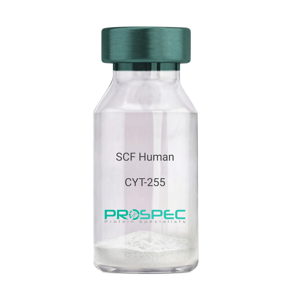
SCF HumanDescription:
Stem Cell Factor Human Recombinant
Kit ligand Precursor, C-kit ligand, SCF, Mast cell growth factor, MGF, SF, KL-1, Kitl, DKFZp686F2250.
Product # :
CYT-255Price :
Quantity :
Shipping Method :
Shipped at Room temp
More Info
- description
- source
- formulation
- purity
- biological activity
- More Info
- Citations
Description
Stem Cell Factor Human Recombinant produced in E.Coli is a single, non-glycosylated polypeptide chain containing 165 amino acids and having a molecular mass of 18409 Dalton. The SCF is purified by proprietary chromatographic techniques.
Source
Escherichia Coli.
Formulation
Lyophilized from a concentrated (1mg/ml) solution in water containing 0.02% NaHCO3.
Purity
Greater than 98.0% as determined by:
(a) Analysis by SEC-HPLC.
(b) Analysis by SDS-PAGE.Biological Activity
The ED50 as determined by the dose-dependant stimulation of Human TF-1 cells is < 2 ng/ml, corresponding to a Specific Activity of 500,000 IU/mg.Citations
More Info
-
Introduction
Stem cell factor / KIT ligand (SCF) is a cytokine which binds CD117(c-Kit). SCF is also known as "steel factor" or "c-kit ligand". SCF exists in two forms, cell surface bound SCF and soluble (or free) SCF. Soluble SCF is produced by the cleavage of surface bound SCF by metalloproteases. SCF is a growth factor important for the survival, proliferation, and differentiation of hematopoietic stem cells and other hematopoietic progenitor cells. One of its roles is to change the BFU-E (burst-forming unit-erythroid) cells, which are the earliest erythrocyte precursors in the erythrocytic series, into the CFU-E (colony-forming unit-erythroid).
-
Synonyms
Kit ligand Precursor, C-kit ligand, SCF, Mast cell growth factor, MGF, SF, KL-1, Kitl, DKFZp686F2250.
-
Physical Appearance
Sterile Filtered White lyophilized (freeze-dried) powder.
-
Stability
Lyophilized KIT ligand although stable at room temperature for 3 weeks, should be stored desiccated below -18°C. Upon reconstitution SCF should be stored at 4°C between 2-7 days and for future use below -18°C.For long term storage it is recommended to add a carrier protein (0.1% HSA or BSA).Please prevent freeze-thaw cycles.
-
Solubility
It is recommended to reconstitute the lyophilized Stem Cell Factor in sterile 18MΩ-cm H2O not less than 100µg/ml, which can then be further diluted to other aqueous solutions.
-
Amino Acid Sequence
The sequence of the first five N-terminal amino acids was determined and was found to be Met-Glu-Gly-Ile-Cys.
-
Protein content
Protein quantitation was carried out by two independent methods1. UV spectroscopy at 280 nm using the absorbency value of 0.52 as the extinction coefficient for a 0.1% (1mg/ml) solution. This value is calculated by the PC GENE computer analysis program of protein sequences (IntelliGenetics). 2. Analysis by RP-HPLC, using a standard solution of Stem Cell Factor as a Reference Standard.
ProSpec's products are furnished for LABORATORY RESEARCH USE ONLY. The product may not be used as drugs, agricultural or pesticidal products, food additives or household chemicals.
- View Data Sheet
Name :
BDNF Human, HisDescription:
Brain-Derived Neurotrophic Factor Human Recombinant, His Tag
Brain-Derived Neurotrophic Factor, Neurotrophin, Abrineurin, ANON2, BULN2, Brain-derived neurotrophic factor.
Product # :
CYT-881Price :
Quantity :
Shipping Method :
Shipped with Ice Packs
More Info
- description
- source
- formulation
- purity
- More Info
- sds-page
- Citations
Description
BDNF Human Recombinant produced in E.Coli is a single, non-glycosylated polypeptide chain containing 140 amino acids (129-247 a.a) and having a molecular mass of 15.8kDa. BDNF is fused to a 21 amino acid His-tag at N-terminus & purified by proprietary chromatographic techniques.
Source
Escherichia Coli.
Formulation
BDNF protein solution (1mg/ml) containing 20mM Tris-HCl buffer (pH 8.0) and 10% glycerol.
Purity
Greater than 85% as determined by SDS-PAGE.
sds-page
Citations
More Info
-
Introduction
BDNF promotes the survival of neuronal populations that are all located either in the central nervous system or directly connected to it. BDNF is a major regulator of synaptic transmission and plasticity at adult synapses in many regions of the cns. The versatility of BDNF is emphasized by its contribution to a range of adaptive neuronal responses including long-term potentiation (ltp), long-term depression (ltd), certain forms of short-term synaptic plasticity, as well as homeostatic regulation of intrinsic neuronal excitability.
-
Synonyms
Brain-Derived Neurotrophic Factor, Neurotrophin, Abrineurin, ANON2, BULN2, Brain-derived neurotrophic factor.
-
Physical Appearance
Sterile filtered colorless solution.
-
Stability
Store at 4°C if entire vial will be used within 2-4 weeks. Store, frozen at -20°C for longer periods of time. For long term storage it is recommended to add a carrier protein (0.1% HSA or BSA).Avoid multiple freeze-thaw cycles.
-
Amino Acid Sequence
MGSSHHHHHH SSGLVPRGSH MHSDPARRGE LSVCDSISEW VTAADKKTAV DMSGGTVTVL EKVPVSKGQL KQYFYETKCN PMGYTKEGCR GIDKRHWNSQ CRTTQSYVRA LTMDSKKRIG WRFIRIDTSC VCTLTIKRGR.
-
Background
What is the molecular weight/Mw of BDNF Protein?
BDNF Protein has a total Mw of 15.8kDa.
What is the source or expression system of BDNF Protein?
Escherichia Coli.
What is the Purity of BDNF Protein?
BDNF Protein is >85% pure as determined by SDS-PAGE.
What is the Biological Activity of BDNF Protein?
The biological functionality of BDNF Protein will be determined in the future.
What is the amino acid sequence of BDNF Protein?
MGSSHHHHHH SSGLVPRGSH MHSDPARRGE LSVCDSISEW VTAADKKTAV DMSGGTVTVL EKVPVSKGQL KQYFYETKCN PMGYTKEGCR GIDKRHWNSQ CRTTQSYVRA LTMDSKKRIG WRFIRIDTSC VCTLTIKRGR.
What applications can BDNF Protein be used in?
BDNF Protein can probably be used in western blot, ELISA and Lateral Flow.
What is the endotoxin level for BDNF Protein?
The endotoxin level is minimal, BDNF Protein was purified using conventional chromatography techniques.
ProSpec's products are furnished for LABORATORY RESEARCH USE ONLY. The product may not be used as drugs, agricultural or pesticidal products, food additives or household chemicals.
- View Data Sheet
Name :
STK16 HumanDescription:
Serine/Threonine Kinase 16 Human Recombinant
Serine/threonine-protein kinase 16, Myristoylated and palmitoylated serine/threonine-protein kinase, MPSK, Protein kinase PKL12, TGF-beta-stimulated factor 1, TSF-1, Tyrosine-protein kinase STK16, hPSK, STK16, MPSK1, PKL12, TSF1, KRCT.
Product # :
PKA-032Price :
Quantity :
Shipping Method :
Shipped with Ice Packs
More Info
- description
- source
- formulation
- purity
- More Info
- Citations
Description
STK16 Human Recombinant produced in E.Coli is a single, non-glycosylated polypeptide chain containing 329 amino acids (1-305 a.a) and having a molecular mass of 37.2kDa.STK16 is fused to a 24 amino acid His-tag at N-terminus & purified by proprietary chromatographic techniques.
Source
Escherichia Coli.
Formulation
STK16 protein solution (1mg/ml) containing 20mM Tris-HCl buffer (pH8.0), 20% glycerol, 0.1M NaCl and 1mM DTT.
Purity
Greater than 90.0% as determined by SDS-PAGE.
Citations
More Info
-
Introduction
Serine/threonine-protein kinase 16 (STK16) is a membrane-associated protein kinase which phosphorylates on serine and threonine residues. STK16 is involved in secretory vesicle trafficking or intracellular signaling. Furthermore, the STK16 protein may have a role in regulating stromal-epithelial interactions which occur during ductal morphogenesis in the mammary gland. STK16 can autophosphorylate on Tyr residue; it is however unclear whether STK16 has tyrosine-protein kinase toward other proteins. STK16 may also be involved in TGF-beta signaling.
-
Synonyms
Serine/threonine-protein kinase 16, Myristoylated and palmitoylated serine/threonine-protein kinase, MPSK, Protein kinase PKL12, TGF-beta-stimulated factor 1, TSF-1, Tyrosine-protein kinase STK16, hPSK, STK16, MPSK1, PKL12, TSF1, KRCT.
-
Physical Appearance
Sterile Filtered colorless solution.
-
Stability
Store at 4°C if entire vial will be used within 2-4 weeks. Store, frozen at -20°C for longer periods of time. For long term storage it is recommended to add a carrier protein (0.1% HSA or BSA).Avoid multiple freeze-thaw cycles.
-
Amino Acid Sequence
MGSSHHHHHH SSGLVPRGSH MGSHMGHALC VCSRGTVIID NKRYLFIQKL GEGGFSYVDL VEGLHDGHFY ALKRILCHEQ QDREEAQREA DMHRLFNHPN ILRLVAYCLR ERGAKHEAWL LLPFFKRGTL WNEIERLKDK GNFLTEDQIL WLLLGICRGL EAIHAKGYAH RDLKPTNILL GDEGQPVLMD LGSMNQACIH VEGSRQALTL QDWAAQRCTI SYRAPELFSV QSHCVIDERT DVWSLGCVLY AMMFGEGPYD MVFQKGDSVA LAVQNQLSIP QSPRHSSALR QLLNSMMTVD PHQRPHIPLL LSQLEALQPP APGQHTTQI.
ProSpec's products are furnished for LABORATORY RESEARCH USE ONLY. The product may not be used as drugs, agricultural or pesticidal products, food additives or household chemicals.
- View Data Sheet
Name :
KGF MouseDescription:
Keratinocye Growth Factor Mouse Recombinant
HBGF-7, FGF7, FGF-7, KGF, Keratinocyte growth factor, Fibroblast growth factor 7, Heparin-binding growth factor 7.
Product # :
CYT-721Price :
Quantity :
Shipping Method :
Shipped at Room temp
More Info
- description
- source
- formulation
- purity
- biological activity
- More Info
- Citations
Description
Keratinocyte Growth Factor-1 Mouse Recombinant produced in E.Coli is a single, non-glycosylated, polypeptide chain containing 164 amino acids and having a molecular mass of 18.9 kDa. The FGF-7 is purified by proprietary chromatographic techniques.
Source
Escherichia Coli.
Formulation
The protein was lyophilized from a concentrated (1mg/ml) solution containing 20mM Phosphate buffer pH-8 and 0.1M NaCl.
Purity
Greater than 95.0% as determined by: (a) Analysis by HPLC. (b) Analysis by SDS-PAGE.
Biological Activity
The ED50, calculated by the dose-dependant stimulation of KGF-responsive BaF3 indicator cells (measured by 3H-thymidine uptake) is < 10ng/ml corresponding to a specific activity of 100,000 Units/mg.Citations
More Info
-
Introduction
KGF is a member of the fibroblast growth factor (FGF) family. FGF family members possess broad mitogenic and cell survival activities, and are involved in a variety of biological processes, including embryonic development, cell growth, morphogenesis, tissue repair, tumor growth and invasion. FGF7 is a potent epithelial cell-specific growth factor, whose mitogenic activity is predominantly exhibited in keratinocytes but not in fibroblasts and endothelial cells. Studies of mouse and rat homologs of this gene implicated roles in morphogenesis of epithelium, reepithelialization of wounds, hair development and early lung organogenesis.
-
Synonyms
HBGF-7, FGF7, FGF-7, KGF, Keratinocyte growth factor, Fibroblast growth factor 7, Heparin-binding growth factor 7.
-
Physical Appearance
Sterile Filtered White lyophilized (freeze-dried) powder.
-
Stability
Lyophilized KGF although stable at room temperature for 3 weeks, should be stored desiccated below -18°C. Upon reconstitution FGF7 should be stored at 4°C between 2-7 days and for future use below -18°C. For long term storage it is recommended to add a carrier protein (0.1% HSA or BSA). Please prevent freeze-thaw cycles.
-
Solubility
It is recommended to reconstitute the lyophilized KGF in sterile 18MΩ-cm H2O not less than 100µg/ml, which can then be further diluted to other aqueous solutions.
-
Amino Acid Sequence
MCNDMSPEQT ATSVNCSSPE RHTRSYDYME GGDIRVRRLF CRTQWYLRID KRGKVKGTQE MKNSYNIMEI RTVAVGIVAI KGVESEYYLA MNKEGKLYAK KECNEDCNFK ELILENHYNT YASAKWTHSG GEMFVALNQK GIPVKGKKTK KEQKTAHFLP MAIT.
ProSpec's products are furnished for LABORATORY RESEARCH USE ONLY. The product may not be used as drugs, agricultural or pesticidal products, food additives or household chemicals.
- View Data Sheet
Name :
GM-CSF RatDescription:
Granulocyte Macrophage-Colony Stimulating Factor Rat Recombinant
CSF-2, MGI-1GM, GM-CSF, Pluripoietin-alpha, Molgramostin, Sargramostim.
Product # :
CYT-395Price :
Quantity :
Shipping Method :
Shipped at Room temp
More Info
- description
- source
- formulation
- purity
- biological activity
- More Info
- Citations
Description
Granulocyte Macrophage Colony Stimulating Factor Rat Recombinant produced in E.Coli is a single, non-glycosylated, polypeptide chain containing 128 amino acids and having a molecular mass of 14590.65 Dalton. GM-CSF Rat Recombinant is purified by proprietary chromatographic techniques.
Source
Escherichia Coli.
Formulation
GM-CSF Rat was lyophilized with no additives.
Purity
Greater than 96.0% as determined by:
(a)Analysis by RP-HPLC.
(b)Analysis by SDS-PAGE.Biological Activity
The ED50 as calculated by the dose-dependent stimulation of the proliferation of murine FDC-P1 cells is less than 0.01ng/ml, corresponding to a specific activity of > 1×10⁸units/mg.
Citations
More Info
-
Introduction
GMCSF is a cytokine that controls the production, differentiation, and function of granulocytes and macrophages. The active form of the protein is found extracellularly as a homodimer. This gene has been localized to a cluster of related genes at chromosome region 5q31, which is known to be associated with interstitial deletions in the 5q- syndrome and acute myelogenous leukemia. Other genes in the cluster include those encoding interleukins 4, 5, and 13.
GM-CSF stimulates the growth and differentiation of hematopoietic precursor cells from various lineages, including granulocytes, macrophages, eosinophils and erythrocytes. -
Synonyms
CSF-2, MGI-1GM, GM-CSF, Pluripoietin-alpha, Molgramostin, Sargramostim.
-
Physical Appearance
Sterile Filtered White lyophilized (freeze-dried) powder.
-
Stability
Lyophilized Granulocyte Macrophage Colony Stimulating Factor although stable at room temperature for 3 weeks, should be stored desiccated below -18°C. Upon reconstitution GMCSF should be stored at 4°C between 2-7 days and for future use below -18°C.For long term storage it is recommended to add a carrier protein (0.1% HSA or BSA).Please prevent freeze-thaw cycles.
-
Solubility
It is recommended to reconstitute the lyophilized Granulocyte Macrophage Colony Stimulating Factor in sterile 18MΩ-cm H2O not less than 100µg/ml, which can then be further diluted to other aqueous solutions.
-
Amino Acid Sequence
MAPTRSPNPV TRPWKHVDAI KEALSLLNDM RALENEKNED VDIISNEFSI
QRPTCVQTRL KLYKQGLRGN LTKLNGALTM IASHYQTNCP PTPETDCEIE
VTTFEDFIKN LKGFLFDIPF DCWKPVQK.
-
Background
What is the molecular weight/Mw of GM-CSF RAT Protein?
GM-CSF RAT Protein has a total Mw of 14.59kDa.
What is the source or expression system of GM-CSF RAT Protein?
Escherichia Coli.
What is the Purity of GM-CSF RAT Protein?
GM-CSF RAT Protein is >96% pure as determined by SDS-PAGE.
What is the Biological Activity of GM-CSF RAT Protein?
The ED50 as calculated by the dose-dependent stimulation of the proliferation of murine FDC-P1 cells is less than 0.01ng/ml, corresponding to a specific activity of > 1×10⁸units/mg.
What is the amino acid sequence of GM-CSF RAT Protein?
MAPTRSPNPV TRPWKHVDAI KEALSLLNDM RALENEKNED VDIISNEFSI
QRPTCVQTRL KLYKQGLRGN LTKLNGALTM IASHYQTNCP PTPETDCEIE
VTTFEDFIKN LKGFLFDIPF DCWKPVQK.
What applications can GM-CSF RAT Protein be used in?
GM-CSF RAT Protein can probably be used in western blot, ELISA and Lateral Flow.
What is the endotoxin level for GM-CSF RAT Protein?
The endotoxin level is minimal, GM-CSF RAT Protein was purified using conventional chromatography techniques.
ProSpec's products are furnished for LABORATORY RESEARCH USE ONLY. The product may not be used as drugs, agricultural or pesticidal products, food additives or household chemicals.
- View Data Sheet
Name :
RAET1E Human, Sf9Description:
Retinoic Acid Early Transcript 1E Human Recombinant, Sf9
Retinoic Acid Early Transcript 1E, Lymphocyte Effector Toxicity Activation Ligand, RAE-1-Like Transcript 4, NKG2DL4, N2DL-4, LETAL, ULBP4, RL-4, NKG2D Ligand 4, BA350J20.7, RAET1E2, N2DL4.
Product # :
PRO-2467Price :
Quantity :
Shipping Method :
Shipped with Ice Packs
More Info
- description
- source
- formulation
- purity
- More Info
- Citations
Description
RAET1E Human Recombinant produced in Sf9 Insect cells is a single, glycosylated polypeptide chain containing 204 amino acids (31-225 a.a.) and having a molecular mass of 23.4kDa (Molecular size on SDS-PAGE will appear at approximately 28-40kDa).RAET1E is expressed with a 6 amino acids His tag at C-Terminus and purified by proprietary chromatographic techniques.
Source
Sf9, Baculovirus cells.
Formulation
RAET1E protein solution (0.5mg/ml) contains Phosphate Buffered Saline (pH 7.4) and 10% glycerol.
Purity
Greater than 90.0% as determined by SDS-PAGE.
Citations
More Info
-
Introduction
RAET1E is a member of the MHC class I family. MHC class I family contains main histocompatibility complex (MHC) class I-related genes positioned in a cluster on chromosome 6q24.2-q25.3. RAET1E and RAET1G protein are different from other RAET1 proteins since they have type I membrane-spanning sequences at their C termini instead glycosylphosphatidylinositol anchor sequences.RAET1E acts as a ligand for NKG2D receptor, expressed on the surface of numerous types of immune cells, involves in innate adaptive immune reactions.RAET1E delivers signals to NK cells and advances tumor immune surveillance by inducing the growth of anti-tumor cytotoxic lymphocyte.
-
Synonyms
Retinoic Acid Early Transcript 1E, Lymphocyte Effector Toxicity Activation Ligand, RAE-1-Like Transcript 4, NKG2DL4, N2DL-4, LETAL, ULBP4, RL-4, NKG2D Ligand 4, BA350J20.7, RAET1E2, N2DL4.
-
Physical Appearance
Sterile Filtered colorless solution.
-
Stability
Store at 4°C if entire vial will be used within 2-4 weeks. Store, frozen at -20°C for longer periods of time. For long term storage it is recommended to add a carrier protein (0.1% HSA or BSA).Avoid multiple freeze-thaw cycles.
-
Amino Acid Sequence
ADPHSLCFNF TIKSLSRPGQ PWCEAQVFLN KNLFLQYNSD NNMVKPLGLL GKKVYATSTW GELTQTLGEV GRDLRMLLCD IKPQIKTSDP STLQVEMFCQ REAERCTGAS WQFATNGEKS LLFDAMNMTW TVINHEASKI KETWKKDRGL EKYFRKLSKG DCDHWLREFL GHWEAMPEPT VSPVNASDIH WSSSSLPDHH HHHH.
ProSpec's products are furnished for LABORATORY RESEARCH USE ONLY. The product may not be used as drugs, agricultural or pesticidal products, food additives or household chemicals.
- View Data Sheet
Name :
SCF Human, HEKDescription:
Stem Cell Factor Human Recombinant, HEK
Kit ligand Precursor, C-kit ligand, SCF, Mast cell growth factor, MGF, SF, KL-1, Kitl, DKFZp686F2250.
Product # :
CYT-111Price :
Quantity :
Shipping Method :
Shipped at Room temp
More Info
- description
- source
- formulation
- purity
- biological activity
- More Info
- Citations
Description
SCF Human Recombinant produced in HEK cells is a glycosylated monomer, having a molecular weight range of 35-45kDa due to glycosylation.The SCF is purified by proprietary chromatographic techniques.
Source
HEK.
Formulation
SCF was lyophilized from a 0.2µm filtered solution (1mg/ml) containing 1xPBS.
Purity
Greater than 95% as obsereved by SDS-PAGE.
Biological Activity
The specific activity was determined by the dose-dependent stimulation of the proliferation of human TF-1 cells (human erythroleukemic indicator cell line).
The EC50 is 15.25ng/ml.Citations
More Info
-
Introduction
Stem cell factor / KIT ligand (SCF) is a cytokine which binds CD117(c-Kit). SCF is also known as "steel factor" or "c-kit ligand". SCF exists in two forms, cell surface bound SCF and soluble (or free) SCF. Soluble SCF is produced by the cleavage of surface bound SCF by metalloproteases. SCF is a growth factor important for the survival, proliferation, and differentiation of hematopoietic stem cells and other hematopoietic progenitor cells. One of its roles is to change the BFU-E (burst-forming unit-erythroid) cells, which are the earliest erythrocyte precursors in the erythrocytic series, into the CFU-E (colony-forming unit-erythroid).
-
Synonyms
Kit ligand Precursor, C-kit ligand, SCF, Mast cell growth factor, MGF, SF, KL-1, Kitl, DKFZp686F2250.
-
Physical Appearance
Sterile Filtered White lyophilized (freeze-dried) powder.
-
Stability
Lyophilized SCF although stable at room temperature for 3 weeks, should be stored desiccated below -18°C. Upon reconstitution SCF should be stored at 4°C between 2-7 days and for future use below -18°C.For long term storage it is recommended to add a carrier protein (0.1% HSA or BSA).Please prevent freeze-thaw cycles.
-
Solubility
It is recommended to reconstitute the lyophilized SCF in sterile PBS containing 0.1% endotoxin-free recombinant HSA.
ProSpec's products are furnished for LABORATORY RESEARCH USE ONLY. The product may not be used as drugs, agricultural or pesticidal products, food additives or household chemicals.
- View Data Sheet
Name :
FGF 2 Human, sf9Description:
Fibroblast Growth Factor-Basic Human Recombinant, Sf9
Prostatropin, HBGH-2, HBGF-2, FGF-2, FGF-b.
Product # :
CYT-365Price :
Quantity :
Shipping Method :
Shipped with Ice Packs
More Info
- description
- source
- formulation
- purity
- biological activity
- More Info
- Citations
Description
Fibroblast Growth Factor-2 Human Recombinant (FGF-2) produced in Sf9 insect cells is a single, glycosylated, polypeptide chain containing 155 amino acids and having a molecular mass of 17353 Dalton.The FGF-basic is purified by proprietary chromatographic techniques.
Source
Baculovirus.
Formulation
The sterile protein solution (0.5mg/ml) contains 20mM Tris pH=7.9, 100mM KCl, 1mM DTT and 20% glycerol.
Purity
Greater than 98.0% as determined by(a) Analysis by RP-HPLC.
(b) Analysis by SDS-PAGE.Biological Activity
The ED50, calculated by the dose-dependant proliferation of BAF3 cells expressing FGF receptors (measured by 3H-thymidine uptake) is <0.5 ng/ml, corresponding to a specific activity of 2MU/mg.Citations
More Info
-
Introduction
Basic fibroblast growth factor is a member of the fibroblast growth factor (FGF) family. FGF family members possess broad mitogenic and cell survival activities, and are involved in a variety of biological processes, including embryonic development, cell growth, morphogenesis, tissue repair, tumor growth and invasion. This protein functions as a modifier of endothelial cell migration and proliferation, as well as an angiogenic factor. It acts as a mitogen for a variety of mesoderm- and neuroectoderm-derived cells in vitro, thus is thought to be involved in organogenesis. Three alternatively spliced variants encoding different isoforms have been described. The heparin-binding growth factors are angiogenic agents in vivo and are potent mitogens for a variety of cell types in vitro. There are differences in the tissue distribution and concentration of these 2 growth factors.
-
Synonyms
Prostatropin, HBGH-2, HBGF-2, FGF-2, FGF-b.
-
Physical Appearance
Sterile Filtered liquid formulation.
-
Stability
Store at 4°C if entire vial will be used within 2-4 weeks. Store, frozen at -20°C for longer periods of time. For long term storage it is recommended to add a carrier protein (0.1% HSA or BSA).Avoid multiple freeze-thaw cycles.
-
Amino Acid Sequence
The sequence of the first five N-terminal amino acids was determined and was found to be Ala-Ala-Gly-Ser-Ile.
-
Background
What is the molecular weight/Mw of FGF 2 Protein?
FGF 2 Protein has a total Mw of 17.3kDa.
What is the source or expression system of FGF 2 Protein?
Baculovirus.
What is the Purity of FGF 2 Protein?
FGF 2 Protein is >98% pure as determined by SDS-PAGE.
What is the Biological Activity of FGF 2 Protein?
The ED50, calculated by the dose-dependant proliferation of BAF3 cells expressing FGF receptors (measured by 3H-thymidine uptake) is <0.5 ng/ml, corresponding to a specific activity of 2MU/mg.
What is the amino acid sequence of FGF 2 Protein?
FGF 2 Protein is composed from155 amino acids.
What applications can FGF 2 Protein be used in?
FGF 2 Protein can probably be used in western blot, ELISA and Lateral Flow.
What is the endotoxin level for FGF 2 Protein?
The endotoxin level is minimal, FGF 2 Protein was purified using conventional chromatography techniques.
ProSpec's products are furnished for LABORATORY RESEARCH USE ONLY. The product may not be used as drugs, agricultural or pesticidal products, food additives or household chemicals.
- View Data Sheet
Name :
GM-CSF Human, PichiaDescription:
Granulocyte Macrophage-Colony Stimulating Factor Human Recombinant, Pichia
CSF-2, MGI-1GM, GM-CSF, Pluripoietin-alpha, Molgramostin, Sargramostim, MGC131935, MGC138897.
Product # :
CYT-324Price :
Quantity :
Shipping Method :
Shipped at Room temp
More Info
- description
- source
- formulation
- purity
- biological activity
- More Info
- Citations
Description
Granulocyte Macrophage Colony Stimulating Factor Human Recombinant produced in Yeast is a single, glycosylated, polypeptide chain containing 127 amino acids and having a molecular mass of 26-32 kDa. rhGMCSF differs from the natural human GM-CSF by a substitution of leucine at position 23 (R to L), and the carbohydrate moiety may be different from the native protein. GM-CSF is purified by proprietary chromatographic techniques.
Source
Pichia Pastoris.
Formulation
The protein was lyophilized from a concentrated (1mg/ml) solution containing 10mM phosphate buffer pH 7.0, 40 mg mannitol and 10 mg sucrose.
Purity
Greater than 97.0% as determined by1. Analysis by RP-HPLC.
2. Analysis by SDS-PAGE.Biological Activity
The ED50 as determined by the dose-dependant stimulation of the proliferation of human TF-1 cells (human erythroleukemic indicator cell line) is < 0.183 ng/ml, corresponding to a Specific Activity of 5,500,000IU/mg.Citations
More Info
-
Introduction
GMCSF is a cytokine that controls the production, differentiation, and function of granulocytes and macrophages. The active form of the protein is found extracellularly as a homodimer. This gene has been localized to a cluster of related genes at chromosome region 5q31, which is known to be associated with interstitial deletions in the 5q- syndrome and acute myelogenous leukemia. Other genes in the cluster include those encoding interleukins 4, 5, and 13.
GM-CSF stimulates the growth and differentiation of hematopoietic precursor cells from various lineages, including granulocytes, macrophages, eosinophils and erythrocytes. -
Synonyms
CSF-2, MGI-1GM, GM-CSF, Pluripoietin-alpha, Molgramostin, Sargramostim, MGC131935, MGC138897.
-
Physical Appearance
Sterile Filtered White lyophilized (freeze-dried) powder.
-
Stability
Lyophilized Granulocyte Macrophage Colony Stimulating Factor although stable at room temperature for 3 weeks, should be stored desiccated below -18°C. Upon reconstitution GMCSF should be stored at 4°C between 2-7 days and for future use below -18°C.For long term storage it is recommended to add a carrier protein (0.1% HSA or BSA).Please prevent freeze-thaw cycles.
-
Solubility
It is recommended to reconstitute the lyophilized Granulocyte Macrophage Colony Stimulating Factor in sterile 18MΩ-cm H2O not less than 100µg/ml, which can then be further diluted to other aqueous solutions.
-
Amino Acid Sequence
The sequence of the first five N-terminal amino acids was determined and was found to be Ala-Pro-Ala-Arg-Ser.
-
Background
What is the molecular weight/Mw of GM-CSF HUMAN, PICHIA Protein?
GM-CSF HUMAN, PICHIA Protein has a total Mw of 29kDa.
What is the source or expression system of GM-CSF HUMAN, PICHIA Protein?
Pichia Pastoris.
What is the Purity of GM-CSF HUMAN, PICHIA Protein?
GM-CSF HUMAN, PICHIA Protein is >97% pure as determined by SDS-PAGE.
What is the Biological Activity of GM-CSF HUMAN, PICHIA Protein?
The ED50 as determined by the dose-dependant stimulation of the proliferation of human TF-1 cells (human erythroleukemic indicator cell line) is < 0.183 ng/ml, corresponding to a Specific Activity of 5,500,000IU/mg.
What is the amino acid sequence of GM-CSF HUMAN, PICHIA Protein?
The sequence of the first five N-terminal amino acids was determined and was found to be Ala-Pro-Ala-Arg-Ser.
What applications can GM-CSF HUMAN, PICHIA Protein be used in?
GM-CSF HUMAN, PICHIA Protein can probably be used in western blot, ELISA and Lateral Flow.
What is the endotoxin level for GM-CSF HUMAN, PICHIA Protein?
The endotoxin level is minimal, GM-CSF HUMAN, PICHIA Protein was purified using conventional chromatography techniques.
ProSpec's products are furnished for LABORATORY RESEARCH USE ONLY. The product may not be used as drugs, agricultural or pesticidal products, food additives or household chemicals.
- View Data Sheet
Name :
Beta NGF MouseDescription:
Beta Nerve Growth Factor Mouse Recombinant
Beta Polypeptide, NGF, NGFB, HSAN5, Beta-NGF, MGC161426, MGC161428.
Product # :
CYT-581Price :
Quantity :
Shipping Method :
Shipped at Room temp
More Info
- description
- source
- formulation
- purity
- biological activity
- More Info
- Citations
Description
Recombinant Mouse b-NGF produced in E.Coli is a noncovalently disulfide-linked homodimer, non-glycosylated, polypeptide chain containing 2 identical chains of 120 amino acids each and having a molecular mass of 13,471 Dalton each.The Recombinant Mouse-beta-NGF is purified by advanced biology purification technology.
Source
Escherichia Coli.
Formulation
The Recombinant Mouse b-NGF was lyophilized without additives.
Purity
Greater than 98% as determined by:
(a) Analysis by RP-HPLC.
(b) Analysis by SDS-PAGE .Biological Activity
The Mouse b-NGF activity was measured in a cell proliferation assay using a factor-dependent human erythroleukemic cell line, TF-1, the ED50 for this effect is 0.2 ng/ml, corresponding to a Specific Activity of 5,000,000 units/mg.Citations
More Info
-
Introduction
NGF-beta has nerve growth stimulating activity and the complex is involved in the regulation of growth and the differentiation of sympathetic and certain sensory neurons. Mutations in this gene have been associated with hereditary sensory and autonomic neuropathy, type 5 (HSAN5), and dysregulation of this gene's expression is associated with allergic rhinitis. Nerve Growth Factor was the 1st protein found from the family of neurotrophic factors that influence the growth and differentiation of sympathetic and sensory neurons. NGF consists of 3 different subunits: alpha, beta, and gamma. The beta subunit is accountable for its growth stimulating activity. The synthesis of NGF in astrocytes is enhanced by a range of cytokines such as IL1, TNF-a, PDGF & TGF-b.
-
Synonyms
Beta Polypeptide, NGF, NGFB, HSAN5, Beta-NGF, MGC161426, MGC161428.
-
Physical Appearance
Sterile Filtered White lyophilized (freeze-dried) powder.
-
Stability
Lyophilized Mouse Beta-NGF although stable at room temperature for 3 weeks, should be stored desiccated below -18°C. Upon reconstitution Murine NGF-Beta should be stored at 4°C between 2-7 days and for future use below -18°C.Please prevent freeze-thaw cycles.
-
Solubility
It is recommended to reconstitute the lyophilized Murine NGF-beta in sterile 18MΩ-cm H2O not less than 100µg/ml, which can then be further diluted to other aqueous solutions.
-
Amino Acid Sequence
MSSTHPVFHM GEFSVCDSVS VWVGDKTTAT DIKGKEVTVL AEVNINNSVF RQYFFETKCR ASNPVESGCR GIDSKHWNSY CTTTHTFVKA LTTDEKQAAW RFIRIDTACV CVLSRKATRR G.
-
Background
What is the molecular weight/Mw of B NGF Protein?
B NGF Protein has a total Mw of 13kDa.
What is the source or expression system of B NGF Protein?
Escherichia Coli.
What is the Purity of B NGF Protein?
B NGF Protein is >98% pure as determined by SDS-PAGE.
What is the Biological Activity of B NGF Protein?
The Mouse b-NGF activity was measured in a cell proliferation assay using a factor-dependent human erythroleukemic cell line, TF-1, the ED50 for this effect is 0.2 ng/ml, corresponding to a Specific Activity of 5,000,000 units/mg.
What is the amino acid sequence of B NGF Protein?
MSSTHPVFHM GEFSVCDSVS VWVGDKTTAT DIKGKEVTVL AEVNINNSVF RQYFFETKCR ASNPVESGCR GIDSKHWNSY CTTTHTFVKA LTTDEKQAAW RFIRIDTACV CVLSRKATRR G.
What applications can B NGF Protein be used in?
B NGF Protein can probably be used in western blot, ELISA and Lateral Flow.
ProSpec's products are furnished for LABORATORY RESEARCH USE ONLY. The product may not be used as drugs, agricultural or pesticidal products, food additives or household chemicals.
- View Data Sheet
Name :
TNFRSF10B Human, Sf9Description:
TNF Ligand Receptor Superfamily Member 10B Human Recombinant, Sf9
TNFRSF10B, CD262, DR5, KILLER, KILLER/DR5, TRAIL-R2, TRAILR2, TRICK2, TRICK2A, TRICK2B, TRICKB, ZTNFR9, Death receptor 5, TNF-related apoptosis-inducing ligand receptor 2.
Product # :
CYT-1055Price :
Quantity :
Shipping Method :
Shipped with Ice Packs
More Info
- description
- source
- formulation
- purity
- biological activity
- More Info
- Citations
Description
TNFRSF10B produced in Sf9 Baculovirus cells is a single, glycosylated polypeptide chain containing 394 amino acids (56-210a.a.) and having a molecular mass of 43.9kDa. (Molecular size on SDS-PAGE will appear at approximately 40-57kDa).TNFRSF10B is expressed with a 239 amino acid hIgG-His tag at C-Terminus and purified by proprietary chromatographic techniques.
Source
Sf9, Insect cells.
Formulation
TNFRSF10B protein solution (1mg/ml) contains Phosphate Buffered Saline (pH 7.4) and 10% glycerol.
Purity
Greater than 90% as determined by SDS-PAGE.
Biological Activity
Measured by its ability to inhibit cytotoxicity assay using Jurkat human T lymphocyte. The ED50 for this effect ≤ to 5ng/ml with TRAIL
.
Citations
More Info
-
Introduction
TRAIL Receptor-1 (DR4) and TRAIL Receptor-2(DR5) are members of the TNFR superfamily of transmembrane proteins and contain a cytoplasmic "death domain", which is capable of activating the cell's apoptotic machinery. These receptors are activated by binding to either membrane anchored or soluble TRAIL/Apo2L.
-
Synonyms
TNFRSF10B, CD262, DR5, KILLER, KILLER/DR5, TRAIL-R2, TRAILR2, TRICK2, TRICK2A, TRICK2B, TRICKB, ZTNFR9, Death receptor 5, TNF-related apoptosis-inducing ligand receptor 2.
-
Physical Appearance
Sterile Filtered colorless solution.
-
Stability
Store at 4°C if entire vial will be used within 2-4 weeks. Store, frozen at -20°C for longer periods of time. For long term storage it is recommended to add a carrier protein (0.1% HSA or BSA).Avoid multiple freeze-thaw cycles.
-
Amino Acid Sequence
ITQQDLAPQQ RAAPQQKRSS PSEGLCPPGH HISEDGRDCI SCKYGQDYST HWNDLLFCLR CTRCDSGEVE LSPCTTTRNT VCQCEEGTFR EEDSPEMCRK CRTGCPRGMV KVGDCTPWSD IECVHKESGT KHSGEVPAVE ETVTSSPGTP ASPCSLEPKS CDKTHTCPPC PAPELLGGPS
VFLFPPKPKD TLMISRTPEV TCVVVDVSHE DPEVKFNWYV DGVEVHNAKT KPREEQYNST YRVVSVLTVL HQDWLNGKEY KCKVSNKALP APIEKTISKA KGQPREPQVY TLPPSRDELT KNQVSLTCLV KGFYPSDIAV EWESNGQPEN NYKTTPPVLD SDGSFFLYSK LTVDKSRWQQ
GNVFSCSVMH EALHNHYTQK SLSLSPGKHH HHHH.
ProSpec's products are furnished for LABORATORY RESEARCH USE ONLY. The product may not be used as drugs, agricultural or pesticidal products, food additives or household chemicals.
- View Data Sheet
Name :
TNFRSF14 Human, Sf9Description:
HVEM-Fc Human Recombinant, Sf9
Tumor necrosis factor receptor superfamily member 14 isoform 1, TNFRSF14, ATAR, CD270, HVEA, HVEM, LIGHTR, TR2, HVEM-Fc, Sf9, Tumor necrosis factor receptor superfamily member 14, Herpes virus entry mediator A, Herpesvirus entry mediator A, HveA.
Product # :
CYT-981Price :
Quantity :
Shipping Method :
Shipped with Ice Packs
More Info
- description
- source
- formulation
- purity
- More Info
- Citations
Description
TNFRSF14 Human Recombinant produced in Sf9 Baculovirus cells is a single, glycosylated polypeptide chain containing 406 amino acids (39-202) and having a molecular mass of 44.6kDa (Molecular size on SDS-PAGE will appear at approximately 40-57kDa).TNFRSF14 is fused to a 239 amino acid IgG His-Tag at C-terminus and purified by proprietary chromatographic techniques.
Source
Sf9, Baculovirus cells.
Formulation
TNFRSF14 protein solution (0.25mg/ml) containing Phosphate Buffered Saline (pH 7.4) and 10% glycerol.
Purity
Greater than 90.0% as determined by analysis by SDS-PAGE.
Citations
More Info
-
Introduction
TNFRSF14, a member of the TNF receptor superfamily, is a type I transmembrane protein. TNFRSF14 is expressed in peripheral blood T cells, B cells, monocytes and in various tissues enriched in lymphoid cells. TNFRSF14 operates as a co-stimulatory factor for the activation of lymphoid cells and as a deterrent to infection by herpesvirus. Additionally, TNFRSF14 encourages the proliferation of T cells, and triggers apoptosis of various tumor cells.
-
Synonyms
Tumor necrosis factor receptor superfamily member 14 isoform 1, TNFRSF14, ATAR, CD270, HVEA, HVEM, LIGHTR, TR2, HVEM-Fc, Sf9, Tumor necrosis factor receptor superfamily member 14, Herpes virus entry mediator A, Herpesvirus entry mediator A, HveA.
-
Physical Appearance
Sterile Filtered clear solution.
-
Stability
Store at 4°C if entire vial will be used within 2-4 weeks. Store, frozen at -20°C for longer periods of time. For long term storage it is recommended to add a carrier protein (0.1% HSA or BSA).Avoid multiple freeze-thaw cycles.
-
Amino Acid Sequence
ADPLPSCKED EYPVGSECCP KCSPGYRVKE ACGELTGTVC EPCPPGTYIA HLNGLSKCLQ CQMCDPAMGL RASRNCSRTE NAVCGCSPGH FCIVQDGDHC AACRAYATSS PGQRVQKGGT ESQDTLCQNC PPGTFSPNGT LEECQHQTKC SWLVTKAGAG TSSSHWVLEP KSCDKTHTCP PCPAPELLGG PSVFLFPPKP KDTLMISRTP EVTCVVVDVS HEDPEVKFNW YVDGVEVHNA KTKPREEQYN STYRVVSVLT VLHQDWLNGK EYKCKVSNKA LPAPIEKTIS KAKGQPREPQ VYTLPPSRDE LTKNQVSLTC LVKGFYPSDI AVEWESNGQP ENNYKTTPPV LDSDGSFFLY SKLTVDKSRW QQGNVFSCSV MHEALHNHYT QKSLSLSPGK HHHHHH.
ProSpec's products are furnished for LABORATORY RESEARCH USE ONLY. The product may not be used as drugs, agricultural or pesticidal products, food additives or household chemicals.
- View Data Sheet
Name :
NT 3 MouseDescription:
Neurotrophin-3 Mouse Recombinant
Neurotrophic factor, Nerve growth factor-2, NGF-2, HDNF, NT-3, Neurotrophin-3, Ntf3, Ntf-3, AI316846, AI835689, Nt3.
Product # :
CYT-688Price :
Quantity :
Shipping Method :
Shipped at Room temp
More Info
- description
- source
- formulation
- purity
- biological activity
- More Info
- Citations
Description
Neurotrophin-3 Mouse Recombinant produced in E.Coli is a single, non-glycosylated polypeptide chain containing 119 amino acids and having a molecular mass of 13.6kDa. The NT-3 is purified by proprietary chromatographic techniques.
Source
Escherichia Coli.
Formulation
Lyophilized from 0.02% TFA.
Purity
Greater than 97.0% as determined by(a) Analysis by RP-HPLC.
(b) Analysis by SDS-PAGE.Biological Activity
The activity, as determined by the dose-dependent proliferation of BaF3 cells transfected with the TrkB receptor, is typically in the range of 1-10 ng/ml, corresponding to a specific activity of 100,000-1,000,000 units/mg.
Citations
More Info
-
Introduction
NT3 a member of the neurotrophin family, that controls survival and differentiation of mammalian neurons. This protein is closely related to both nerve growth factor and brain-derived neurotrophic factor. It may be involved in the maintenance of the adult nervous system, and may affect development of neurons in the embryo when it is expressed in human placenta. NTF3-deficient mice generated by gene targeting display severe movement defects of the limbs. The mature peptide of this protein is identical in all mammals examined including human, pig, rat and mouse.
-
Synonyms
Neurotrophic factor, Nerve growth factor-2, NGF-2, HDNF, NT-3, Neurotrophin-3, Ntf3, Ntf-3, AI316846, AI835689, Nt3.
-
Physical Appearance
Sterile Filtered White lyophilized (freeze-dried) powder.
-
Stability
Lyophilized NGF2 although stable at room temperature for 3 weeks, should be stored desiccated below -18°C. Upon reconstitution NGF-2 should be stored at 4°C between 2-7 days and for future use below -18°C.For long term storage it is recommended to add a carrier protein (0.1% HSA or BSA).Please prevent freeze-thaw cycles.
-
Solubility
It is recommended to reconstitute the lyophilized Neurotrophin-3 in sterile 18MΩ-cm H2O not less than 100µg/ml, which can then be further diluted to other aqueous solutions.
-
Amino Acid Sequence
YAEHKSHRGE YSVCDSESLW VTDKSSAIDI RGHQVTVLGE IKTGNSPVKQ YFYETRCKEA RPVKNGCRGI DDKHWNSQCK TSQTYVRALT SENNKLVGWR WIRIDTSCVC ALSRKIGRT.
ProSpec's products are furnished for LABORATORY RESEARCH USE ONLY. The product may not be used as drugs, agricultural or pesticidal products, food additives or household chemicals.
- View Data Sheet
Name :
CXCL12 Mouse alphaDescription:
Stromal Cell-Derived Factor-1 Alpha Mouse Recombinant (CXCL12)
SDF-1, CXCL12, Pre-B cell growth-stimulating factor, PBSF, hIRH, chemokine (C-X-C motif) ligand 12, SDF1, SDF1A, TPAR1, SCYB12, SDF-1a, TLSF-a, 12-O-tetradecanoylphorbol 13-acetate repressed protein 1, Thymic lymphoma cell-stimulating factor, TLSF.
Product # :
CHM-324Price :
Quantity :
Shipping Method :
Shipped at Room temp
More Info
- description
- source
- formulation
- purity
- biological activity
- More Info
- Citations
Description
Stromal Cell-Derived Factor-1 alpha Mouse Recombinant produced in E.Coli is a non-glycosylated, Polypeptide chain containing 68 amino acids and having a molecular mass of 8 kDa. The SDF-1a is purified by proprietary chromatographic techniques.
Source
Escherichia Coli.
Formulation
The protein was Lyophilized from a sterile filtered aqueous solution containing 0.1% Trifluoroacetic Acid (TFA).
Purity
Greater than 95% as determined by SDS-PAGE.
Biological Activity
The specific activity as determined by its ability to chemoattract human peripheral blood monocytes at 50-100 ng/ml corresponding to a Specific Activity of 10,000-20,000IU/mg.
Citations
More Info
-
Introduction
SDF-1 (stromal cell-derived factor-1) is small cytokine belonging to the chemokine family that is officially designated Chemokine (C-X-C motif) ligand 12 (CXCL12). It is produced in two forms, SDF-1?/CXCL12a and SDF-1?/CXCL12b, by alternate splicing of the same gene. Chemokines are characterized by the presence of four conserved cysteines, which form two disulfide bonds. The CXCL12 proteins belong to the group of CXC chemokines, whose initial pair of cysteines are separated by one intervening amino acid. CXCL12 is strongly chemotactic for lymphocytes and has been implicated as an important cell co-ordinator during development. During embryogenesis it directs the migration of hematopoietic cells from foetal liver to bone marrow. Mice which were knocked-out for CXCL12 gene were lethal before the birth or within just 1 hour of life. As another role, CXCL12a alters also the electrophysiology of neurons. CXCL12 was shown to be expressend in many tissues in mice (including brain, thymus, heart, lung, liver, kidney, spleen and bone marrow).
The receptor for this chemokine is CXCR4, which was previously called fusin. This CXCL12-CXCR4 interaction used to be considered exclusive (unlike for other chemokines and their receptors), but recently it was suggested that CXCL12 is also bound by CXCR7 receptor.
The gene for CXCL12 is located on human chromosome 10. In human and mouse both CXCL12 and CXCR4 show high identity of sequence: 99% and 90%, respectively. -
Synonyms
SDF-1, CXCL12, Pre-B cell growth-stimulating factor, PBSF, hIRH, chemokine (C-X-C motif) ligand 12, SDF1, SDF1A, TPAR1, SCYB12, SDF-1a, TLSF-a, 12-O-tetradecanoylphorbol 13-acetate repressed protein 1, Thymic lymphoma cell-stimulating factor, TLSF.
-
Physical Appearance
Sterile Filtered White lyophilized (freeze-dried) powder.
-
Stability
Lyophilized SDF-1a although stable at room temperature for 3 weeks, should be stored desiccated below -18°C. Upon reconstitution CXCL12 should be stored at 4°C between 2-7 days and for future use below -18°C. For long term storage it is recommended to add a carrier protein (0.1% HSA or BSA).Please prevent freeze-thaw cycles.
-
Solubility
It is recommended to reconstitute the lyophilized Stromal Cell-Derived Factor-1a in sterile 18MΩ-cm H2O not less than 100µg/ml, which can then be further diluted to other aqueous solutions.
-
Amino Acid Sequence
KPVSLSYRCP CRFFESHIAR ANVKHLKILN TPNCALQIVA RLKNNNRQVC IDPKLKWIQE
YLEKALNK.
-
Background
What is the molecular weight/Mw of CXCL12 MOUSE ALPHA Protein?
CXCL12 MOUSE ALPHA Protein has a total Mw of 8kDa.
What is the source or expression system of CXCL12 MOUSE ALPHA Protein?
Escherichia Coli.
What is the Purity of CXCL12 MOUSE ALPHA Protein?
CXCL12 MOUSE ALPHA Protein is > 95% pure as determined by SDS-PAGE.
What is the Biological Activity of CXCL12 MOUSE ALPHA Protein?
The specific activity as determined by its ability to chemoattract human peripheral blood monocytes at 50-100 ng/ml corresponding to a Specific Activity of 10,000-20,000IU/mg.
What is the amino acid sequence of CXCL12 MOUSE ALPHA Protein?
KPVSLSYRCP CRFFESHIAR ANVKHLKILN TPNCALQIVA RLKNNNRQVC IDPKLKWIQE
YLEKALNK.
What applications can CXCL12 MOUSE ALPHA Protein be used in?
CXCL12 MOUSE ALPHA Protein can probably be used in western blot, ELISA and Lateral Flow.
What is the endotoxin level for CXCL12 MOUSE ALPHA Protein?
The endotoxin level is minimal, CXCL12 MOUSE ALPHA Protein was purified using conventional chromatography techniques.
Who supplies recombinant CXCL12 MOUSE ALPHA that is in stock and available to order?
ProSpec has the protein in stock and available for immediate delivery.
Where can I order bulk mg quantities of CXCL12 MOUSE ALPHA?
ProSpec supplies bulk mg quantities at affordable prices that meets research and academic budgets.
ProSpec's products are furnished for LABORATORY RESEARCH USE ONLY. The product may not be used as drugs, agricultural or pesticidal products, food additives or household chemicals.
- View Data Sheet
Name :
EGF (1-51), HumanDescription:
Epidermal Growth Factor (1-51 a.a.)Human Recombinant
Urogastrone, URG, EGF.
Product # :
CYT-1115Price :
Quantity :
Shipping Method :
Shipped at Room temp
More Info
- description
- source
- formulation
- purity
- biological activity
- More Info
- Citations
Description
Epidermal Growth Factor (1-51 a.a.) Human Recombinant produced in yeast is a single, glycosylated polypeptide chain containing 51 amino acids and having a molecular mass of 6.0kDa. The EGF is purified by proprietary chromatographic techniques.
Source
Saccharomyces cerevisiae
Formulation
Lyophilized from a 0.2μm filtered concentrated solution in PBS, pH 7.4.
Purity
Greater than 98.0% as determined by:
(a) Analysis by RP-HPLC.
(b) Analysis by SDS-PAGE.Biological Activity
The ED50 is determined by a cell proliferation assay using murine Balb/c 3T3 cells and is < than 0.1 ng/ml, corresponding to a specific activity of > 1.0 × 10⁷ IU/mg.
Citations
More Info
-
Introduction
Epidermal growth factor has a profound effect on the differentiation of specific cells in vivo and is a potent mitogenic factor for a variety of cultured cells of both ectodermal and mesodermal origin. The EGF precursor is believed to exist as a membrane-bound molecule which is proteolytically cleaved to generate the 53-amino acid peptide hormone that stimulates cells to divide. EGF stimulates the growth of several epidermal and epithelial tissues in vivo and in vitro and of some fibroblasts in cell culture.
-
Synonyms
Urogastrone, URG, EGF.
-
Physical Appearance
Sterile Filtered White lyophilized (freeze-dried) powder.
-
Stability
Lyophilized EGF although stable at room temperature for 3 weeks, should be stored desiccated below -18°C. Upon reconstitution Epidermal Growth Factor should be stored at 4°C between 2-7 days and for future use below -18°C.Please prevent freeze-thaw cycles.
-
Solubility
It is recommended to reconstitute the lyophilized Epidermal Growth Factor in sterile 18MΩ-cm H2O not less than 100µg/ml, which can then be further diluted to other aqueous solutions.
-
Amino Acid Sequence
NSDSECPLSH DGYCLHDGVC MYIEALDKYA CNCVVGYIGE RCQYRDLKWW E.
-
Background
Exploring the Potential of Epidermal Growth Factor (1-51 a.a.) Human Recombinant: Novel Insights and Therapeutic Prospects
Abstract:
Epidermal Growth Factor (EGF) stands as a pivotal cytokine orchestrating essential cellular processes. This concise research paper delves into the unique realm of Epidermal Growth Factor (1-51 a.a.) Human Recombinant, unveiling its intricate molecular dynamics, signaling cascades, and therapeutic promise. Employing cutting-edge methodologies encompassing in vitro assays and animal models, this study elucidates the multifaceted cellular responses sparked by this truncated EGF variant, paving the way for potential clinical applications.
Introduction:
The truncated form of EGF, spanning amino acids 1 to 51 (a.a.), carries distinct attributes that set it apart from the full-length counterpart. This paper centers on exploring the intriguing dimensions of Epidermal Growth Factor (1-51 a.a.) Human Recombinant, offering new insights into its interactions and potential utility.
Molecular Insights and Signaling Dynamics:
At the heart of its function lies the interplay between EGF (1-51 a.a.) and the epidermal growth factor receptor (EGFR). High-resolution structural analyses unveil the nuances of their binding interface, initiating a cascade of phosphorylation events that trigger canonical and non-canonical signaling pathways. The MAPK pathway and the PI3K/Akt pathway, intricately modulated by EGF (1-51 a.a.), propel cellular processes like proliferation, migration, and evasion of apoptosis.
In Vitro Profiling and Cellular Responses:
In dissecting the cellular responses, diverse in vitro assays have been employed. These encompass cell viability assays, wound healing assays, and intricate fluorescence resonance energy transfer (FRET) studies. These assays converge to illuminate the dynamic orchestration of EGF-induced cellular behaviors, showcasing its role in promoting cellular migration, division, and wound closure.
In Vivo Implications and Therapeutic Horizons:
Translating these insights into tangible therapeutic possibilities, in vivo studies present a compelling narrative. In animal models, EGF (1-51 a.a.) emerges as a potent player in cutaneous wound healing, fostering accelerated tissue regeneration. Moreover, its potential extends to oncology, as it not only influences tumor microenvironments but also demonstrates anti-apoptotic effects, hinting at its role in tailored cancer interventions.
Future Prospects and Challenges:
While these discoveries hold immense promise, challenges persist. The intricate network of signaling events demands further scrutiny, considering potential cross-talk and off-target effects. Refining delivery mechanisms and dosing regimens is essential for realizing the clinical potential of EGF (1-51 a.a.).
Conclusion:
In a synthesis of complex molecular insights and tangible therapeutic potential, Epidermal Growth Factor (1-51 a.a.) Human Recombinant emerges as a captivating subject. Its truncated structure and distinctive signaling cascades paint a canvas of cellular orchestration. As research advances, harnessing its therapeutic benefits could usher in novel interventions for wound healing and cancer therapy.
What is the molecular weight/Mw of EGF Protein?
EGF Protein has a total Mw of 6kDa.
What is the source or expression system of EGF Protein?
Saccharomyces cerevisiae
What is the Purity of EGF Protein?
EGF Protein is >98% pure as determined by SDS-PAGE.
What is the Biological Activity of EGF Protein?
The ED50 is determined by a cell proliferation assay using murine Balb/c 3T3 cells and is < than 0.1 ng/ml, corresponding to a specific activity of > 1.0 × 10⁷ IU/mg.
What is the amino acid sequence of EGF Protein?
NSDSECPLSH DGYCLHDGVC MYIEALDKYA CNCVVGYIGE RCQYRDLKWW E.
What applications can EGF Protein be used in?
EGF Protein can probably be used in western blot, ELISA and Lateral Flow.
What is the endotoxin level for EGF Protein?
The endotoxin level is minimal, EGF Protein was purified using conventional chromatography techniques.
ProSpec's products are furnished for LABORATORY RESEARCH USE ONLY. The product may not be used as drugs, agricultural or pesticidal products, food additives or household chemicals.
- View Data Sheet
Name :
TGFBR1 Human, ActiveDescription:
Transforming Growth Factor Beta Receptor 1 Human Recombinant, Active
TGFBR1, AAT5, ACVRLK4, ALK-5, ALK5, ESS1, LDS1, LDS1A, LDS2A, MSSE, SKR4, tbetaR-I, TGFR-1.
Product # :
PKA-135Price :
Quantity :
Shipping Method :
Shipped with Ice Packs
More Info
- description
- source
- formulation
- purity
- biological activity
- More Info
- Citations
Description
TGFBR1 produced in Sf9 insect cells is a single, glycosylated polypeptide chain containing 342 amino acids (27-126a.a.) and having a molecular mass of 38kDa. TGFBR1 is expressed with 242 amino acid hIgG-His-Tag at C-Terminus and purified by proprietary chromatographic techniques.
Source
Sf9, Baculovirus cells.
Formulation
TGFBR1 protein solution (0.5mg/ml) Phosphate Buffered Saline (pH 7.4) containing 10% glycerol.
Purity
Greater than 90.0% as determined by SDS-PAGE.
Biological Activity
≤ 2 ug/ml, measured by its binding ability in a functional ELISA with Mouse CD105.
Citations
More Info
-
Synonyms
TGFBR1, AAT5, ACVRLK4, ALK-5, ALK5, ESS1, LDS1, LDS1A, LDS2A, MSSE, SKR4, tbetaR-I, TGFR-1.
-
Physical Appearance
Sterile filtered colorless solution.
-
Stability
Store at 4°C if entire vial will be used within 2-4 weeks. Store, frozen at -20°C for longer periods of time. For long term storage it is recommended to add a carrier protein (0.1% HSA or BSA).Avoid multiple freeze-thaw cycles.
-
Amino Acid Sequence
ADLLLPGATA LQCFCHLCTK DNFTCVTDGL CFVSVTETTD KVIHNSMCIA EIDLIPRDRP FVCAPSSKTG SVTTTYCCNQ DHCNKIELPT TVKSSPGLGP VELVEPKSCD KTHTCPPCPA PELLGGPSVF LFPPKPKDTL MISRTPEVTC VVVDVSHEDP EVKFNWYVDG VEVHNAKTKP REEQYNSTYR VVSVLTVLHQ DWLNGKEYKC KVSNKALPAP IEKTISKAKG QPREPQVYTL
PPSRDELTKN QVSLTCLVKG FYPSDIAVEW ESNGQPENNY KTTPPVLDSD GSFFLYSKLT VDKSRWQQGN VFSCSVMHEA LHNHYTQKSL SLSPGKHHHH HH. -
Background
Transforming Growth Factor Beta Receptor 1 (TGFBR1), a transmembrane protein crucial in the TGF-β signaling pathway, holds a paramount position in regulating diverse cellular processes. Its intricate involvement in development, immune responses, tissue homeostasis, and disease has elevated TGFBR1 to a central role in biology and medicine. This research embarks on a comprehensive exploration of the TGFBR1 protein, unraveling its structural complexities, signaling mechanisms, and its far-reaching implications in various physiological and pathological contexts. By dissecting the intricacies of TGFBR1, scientists aim to decode the fundamental cellular processes it governs and explore potential therapeutic avenues in the domains of cancer, fibrosis, and immunology.
Structural Complexity of TGFBR1:
TGFBR1 is a serine/threonine kinase receptor with an extracellular ligand-binding domain, a transmembrane domain, and an intracellular kinase domain. Its structure allows it to interact with TGF-β ligands and initiate downstream signaling cascades. Understanding the three-dimensional architecture of TGFBR1 is pivotal for deciphering its interactions with ligands, co-receptors, and intracellular signaling partners, shedding light on the molecular intricacies of its function.
Signaling Pathways and Physiological Functions:
Upon ligand binding, TGFBR1 phosphorylates downstream effectors, regulating processes like cell proliferation, differentiation, apoptosis, and immune responses. TGF-β signaling mediated by TGFBR1 is vital in embryogenesis, tissue repair, and immune tolerance. Dysregulation of this pathway is implicated in numerous diseases, including cancer, fibrosis, and autoimmune disorders, underscoring the significance of TGFBR1 in maintaining cellular and tissue homeostasis.
TGFBR1 in Cancer Biology:
TGFBR1's dual role as a tumor suppressor and a promoter of cancer progression reflects its complexity in cancer biology. In early stages, TGFBR1 signaling suppresses cell growth and promotes apoptosis, acting as a defense against tumorigenesis. However, in advanced stages, cancer cells exploit TGFBR1 signaling to facilitate invasion, metastasis, and immune evasion. Understanding the context-dependent nature of TGFBR1's functions in cancer is pivotal for developing targeted therapies.
Targeting TGFBR1 in Therapeutics:
Given its critical roles in various diseases, TGFBR1 has emerged as an attractive target for therapeutic interventions. In cancer, efforts are underway to develop small molecule inhibitors and monoclonal antibodies that modulate TGFBR1 signaling, aiming to curb tumor progression. Additionally, in fibrotic disorders, targeting TGFBR1 offers hope for halting the pathological tissue remodeling characteristic of these diseases, providing potential treatments for conditions such as pulmonary fibrosis and liver cirrhosis.
TGFBR1 Protein, with its intricate signaling mechanisms and diverse physiological roles, stands at the crossroads of fundamental cellular processes and disease pathogenesis. Its involvement in development, immune regulation, cancer, and tissue homeostasis underscores its significance in biology and medicine. As researchers delve deeper into the complexities of TGFBR1, they pave the way for innovative therapies and a deeper understanding of diseases, ultimately shaping the future of healthcare and scientific exploration. This research not only illuminates the pivotal role of TGFBR1 but also holds the promise of transformative advancements in medicine and our understanding of cellular signaling pathways.
ProSpec's products are furnished for LABORATORY RESEARCH USE ONLY. The product may not be used as drugs, agricultural or pesticidal products, food additives or household chemicals.
- View Data Sheet
Name :
EDAR Human, Sf9Description:
Ectodysplasin A Receptor Human Recombinant, Sf9
Ectodysplasin A Receptor, Ectodysplasin 1, Anhidrotic Receptor, Anhidrotic Ectodysplasin Receptor 1, Ectodermal Dysplasia Receptor, Downless Homolog, EDA-A1 Receptor, DL, Tumor Necrosis Factor Receptor Superfamily Member EDAR, Downless, Mouse, Homolog Of, Ectodysplasin-A Receptor, ECTD10A, ECTD10B, EDA-A1R, EDA1R, ED1R, EDA3, HRM1, ED5, ED3.
Product # :
PRO-2510Price :
Quantity :
Shipping Method :
Shipped with Ice Packs
More Info
- description
- source
- formulation
- purity
- More Info
- Citations
Description
EDAR produced in Sf9 Baculovirus cells is a single, glycosylated polypeptide chain containing 413 amino acids (27-187a.a.) and having a molecular mass of 45.6kDa. (Molecular size on SDS-PAGE will appear at approximately 40-57kDa).EDAR is expressed with a 249 amino acid hIgG-His-tag at C-Terminus and purified by proprietary chromatographic techniques.
Source
Sf9, Baculovirus cells.
Formulation
EDAR protein solution (0.5mg/ml) contains 10% glycerol & Phosphate Buffered Saline (pH 7.4).
Purity
Greater than 90.0% as determined by SDS-PAGE.
Citations
More Info
-
Introduction
Ectodysplasin A Receptor, also known as EDAR belongs to the tumor necrosis factor receptor family. EDAR is a receptor for the soluble ligand ectodysplasin A, and is capable of activating the nuclear factor-kappaB, JNK, as well as caspase-independent cell death pathways. EDAR is necessary for the development of hair, teeth, and other ectodermal derivatives. Furthermore, mutations in EDAR resulted in autosomal dominant and recessive forms of hypohidrotic ectodermal dysplasia.
-
Synonyms
Ectodysplasin A Receptor, Ectodysplasin 1, Anhidrotic Receptor, Anhidrotic Ectodysplasin Receptor 1, Ectodermal Dysplasia Receptor, Downless Homolog, EDA-A1 Receptor, DL, Tumor Necrosis Factor Receptor Superfamily Member EDAR, Downless, Mouse, Homolog Of, Ectodysplasin-A Receptor, ECTD10A, ECTD10B, EDA-A1R, EDA1R, ED1R, EDA3, HRM1, ED5, ED3.
-
Physical Appearance
Sterile filtered colorless solution.
-
Stability
Store at 4°C if entire vial will be used within 2-4 weeks. Store, frozen at -20°C for longer periods of time.For long term storage it is recommended to add a carrier protein (0.1% HSA or BSA).Please avoid freeze thaw cycles.
-
Amino Acid Sequence
ADPEYSNCGE NEYYNQTTGL CQECPPCGPG EEPYLSCGYG TKDEDYGCVP CPAEKFSKGG YQICRRHKDC EGFFRATVLT PGDMENDAEC GPCLPGYYML ENRPRNIYGM VCYSCLLAPP NTKECVGATS GASANFPGTS GSSTLSPFQH AHKELSGQGH LATAAAAFES ACSLEPKSC DKTHTCPPCP APELLGGPSV FLFPPKPKDT LMISRTPEVT CVVVDVSHED PEVKFNWYVD GVEVHNAKTK PREEQYNSTY RVVSVLTVLH QDWLNGKEYK CKVSNKALPA PIEKTISKAK GQPREPQVYT LPPSRDELTK NQVSLTCLVK GFYPSDIAVE WESNGQPENN YKTTPPVLDS DGSFFLYSKL TVDKSRWQQG NVFSCSVMHE ALHNHYTQKS LSLSPGKHHH HHH.
ProSpec's products are furnished for LABORATORY RESEARCH USE ONLY. The product may not be used as drugs, agricultural or pesticidal products, food additives or household chemicals.