Search results
264 results found for “mediator complex”
Name
Description
Product #
Price
Quantity
Shipping Method
- View Data Sheet
Name :
CCL18 Human, HisDescription:
Macrophage Inflammatory protein-4 (CCL18) Human Recombinant, His-Tag
Small inducible cytokine A18, CCL18, Macrophage inflammatory protein 4, MIP-4, Pulmonary and activation-regulated chemokine, CC chemokine PARC, Alternative macrophage activation-associated CC chemokine 1, AMAC-1, Dendritic cell chemokine 1, DC-CK1, chemokine (C-C motif) ligand 18, CKb7, PARC, AMAC1, DCCK1, SCYA18.
Product # :
CHM-339Price :
Quantity :
Shipping Method :
Shipped with Ice Packs
More Info
- description
- source
- formulation
- purity
- More Info
Description
MIP-4 Human Recombinant fused with a 25 amino acid His tag at N-terminus produced in E.Coli is a single, non-glycosylated, polypeptide chain containing 93 amino acids (22-89 a.a.) and having a molecular mass of 10.4kDa. The MIP-4 is purified by proprietary chromatographic techniques.
Source
Escherichia Coli.
Formulation
The MIP-4 solution (0.25 mg/ml) contains 10mM Sodium Citrate pH3.5 and 10% Glycerol.
Purity
Greater than 95.0% as determined by SDS-PAGE.
More Info
-
Introduction
Chemokine (C-C motif) ligand 18 (CCL18) is a small cytokine belonging to the CC chemokine family that was previously called PARC (pulmonary and activation-regulated chemokine). CCL18 is approximately 60% identical in amino acid sequence to CCL3. It is expressed at high levels in lung and at lower levels in certain lymphoid tissues, such as the lymph nodes, and is chemotactic for activated T cells and non activated lymphocytes. The gene for human CCL18 contains three exons and is located on chromosome 17.
-
Synonyms
Small inducible cytokine A18, CCL18, Macrophage inflammatory protein 4, MIP-4, Pulmonary and activation-regulated chemokine, CC chemokine PARC, Alternative macrophage activation-associated CC chemokine 1, AMAC-1, Dendritic cell chemokine 1, DC-CK1, chemokine (C-C motif) ligand 18, CKb7, PARC, AMAC1, DCCK1, SCYA18.
-
Physical Appearance
Sterile Filtered colorless solution.
-
Stability
Store at 4°C if entire vial will be used within 2-4 weeks. Store, frozen at -20°C for longer periods of time. For long term storage it is recommended to add a carrier protein (0.1% HSA or BSA).Avoid multiple freeze-thaw cycles.
-
Amino Acid Sequence
MGSSHHHHHH SSGLVPRGSH MGSHMQVGTN KELCCLVYTS WQIPQKFIVD YSETSPQCPK PGVILLTKRG RQICADPNKK WVQKYISDLK LNA.
-
Background
What is the molecular weight/Mw of CCL18 HUMAN, HIS Protein?
CCL18 HUMAN, HIS Protein has a total Mw of 10.4kDa.
What is the source or expression system of CCL18 HUMAN, HIS Protein?
Escherichia Coli.
What is the Purity of CCL18 HUMAN, HIS Protein?
CCL18 HUMAN, HIS Protein is > 95% pure as determined by SDS-PAGE.
What is the Biological Activity of CCL18 HUMAN, HIS Protein?
The biological functionality of CCL18 HUMAN, HIS Protein will be determined in the future.
What is the amino acid sequence of CCL18 HUMAN, HIS Protein?
MGSSHHHHHH SSGLVPRGSH MGSHMQVGTN KELCCLVYTS WQIPQKFIVD YSETSPQCPK PGVILLTKRG RQICADPNKK WVQKYISDLK LNA.
What applications can CCL18 HUMAN, HIS Protein be used in?
CCL18 HUMAN, HIS Protein can probably be used in western blot, ELISA and Lateral Flow.
What is the endotoxin level for CCL18 HUMAN, HIS Protein?
The endotoxin level is minimal, CCL18 HUMAN, HIS Protein was purified using conventional chromatography techniques.
ProSpec's products are furnished for LABORATORY RESEARCH USE ONLY. The product may not be used as drugs, agricultural or pesticidal products, food additives or household chemicals.
- View Data Sheet
Name :
ThymopentinDescription:
Thymopentin
Product # :
HOR-241Price :
Quantity :
Shipping Method :
Shipped at Room temp
More Info
- description
- formulation
- purity
- biological activity
- More Info
Description
Thymopentin has a molecular formula of C30H49N9O9, Arg-Lys-Asp-Val-Tyr-OH having an Mw of 679.8 Dalton.
Formulation
The protein (1mg/ml) was lyophilized with no additives.
Purity
Greater than 99.0% as determined by:
(a) Analysis by RP-HPLC.
(b) Analysis by SDS-PAGE.Biological Activity
What is the molecular weight/Mw of THYMOPENTIN Protein? THYMOPENTIN Protein has a total Mw of 0.67kDa. What is the Purity of THYMOPENTIN Protein? THYMOPENTIN Protein is >99% pure as determined by SDS-PAGE. What is the Biological Activity of THYMOPENTIN Protein? The biological functionality of THYMOPENTIN Protein will be determined in the future. What applications can THYMOPENTIN Protein be used in? THYMOPENTIN Protein can probably be used in western blot, ELISA and Lateral Flow. What is the endotoxin level for THYMOPENTIN Protein? The endotoxin level is minimal, THYMOPENTIN Protein was purified using conventional chromatography techniques.More Info
-
Introduction
Thymopentin, also known as TP-5, is a synthetic pentapeptide which is the active site of the naturally occurring hormone thymopoietin with immunomodulating properties (corresponding to the amino acids 32-36 of thymopoietin). Thymopentin enhances the production of thymic T cells and may help restore immunocompetence in immunosuppressed subjects. This agent also augments the effects of ionizing radiation by arresting cancer cells in the G2/M phase of the cell cycle.
-
Physical Appearance
Sterile Filtered White lyophilized (freeze-dried) powder.
-
Stability
Lyophilized Thymopentin although stable at room temperature for 3 weeks, should be stored desiccated below -18°C. Upon reconstitution TP-5 should be stored at 4°C between 2-7 days and for future use below -18°C.For long term storage it is recommended to add a carrier protein (0.1% HSA or BSA).Please prevent freeze-thaw cycles.
-
Solubility
It is recommended to reconstitute the lyophilized Thymopentin in sterile 18MΩ-cm H2O not less than 100 µg/ml, which can then be further diluted to other aqueous solutions.
ProSpec's products are furnished for LABORATORY RESEARCH USE ONLY. The product may not be used as drugs, agricultural or pesticidal products, food additives or household chemicals.
- View Data Sheet
Name :
NUDC HumanDescription:
NudC Nuclear Distribution Protein Human Recombinant
Nuclear Distribution C Dynein Complex Regulator, NudC Nuclear Distribution Protein, Nuclear Distribution Protein C Homolog, Nuclear Distribution Gene C Homolog, Nuclear Distribution C Homolog, Nuclear Migration Protein NudC, NPD011, HNUDC, MNUDC, NUDC.
Product # :
PRO-2347Price :
Quantity :
Shipping Method :
Shipped with Ice Packs
More Info
- description
- source
- formulation
- purity
- More Info
Description
NUDC Human Recombinant produced in E.Coli is a single, non-glycosylated polypeptide chain containing 354 amino acids (1-331 a.a) and having a molecular mass of 40.6kDa. NUDC is fused to a 23 amino acid His-tag at N-terminus & purified by proprietary chromatographic techniques.
Source
Escherichia Coli.
Formulation
NUDC protein solution (1mg/ml) containing 20mM Tris-HCl (pH 8.0), 10% glycerol and 1mM DTT.
Purity
Greater than 90.0% as determined by SDS-PAGE.
More Info
-
Introduction
Nuclear distribution gene C homolog (NUDC) is a nuclear distribution protein. The NUDC protein has a key role in mitosis and cytokinesis. NUDC is involved in spindle formation during mitosis and in microtubule organization during cytokinesis. NUDC is required for correct formation of mitotic spindles and chromosome separation during mitosis, as well as cell proliferation. NUDC also has a role in neurogenesis and neuronal migration.
-
Synonyms
Nuclear Distribution C Dynein Complex Regulator, NudC Nuclear Distribution Protein, Nuclear Distribution Protein C Homolog, Nuclear Distribution Gene C Homolog, Nuclear Distribution C Homolog, Nuclear Migration Protein NudC, NPD011, HNUDC, MNUDC, NUDC.
-
Physical Appearance
Sterile Filtered colorless solution.
-
Stability
Store at 4°C if entire vial will be used within 2-4 weeks. Store, frozen at -20°C for longer periods of time. For long term storage it is recommended to add a carrier protein (0.1% HSA or BSA).Avoid multiple freeze-thaw cycles.
-
Amino Acid Sequence
MGSSHHHHHH SSGLVPRGSH MGSMGGEQEE ERFDGMLLAM AQQHEGGVQE LVNTFFSFLR RKTDFFIGGE EGMAEKLITQ TFSHHNQLAQ KTRREKRARQ EAERREKAER AARLAKEAKS ETSGPQIKEL TDEEAERLQL EIDQKKDAEN HEAQLKNGSL DSPGKQDTEE DEEEDEKDKG KLKPNLGNGA DLPNYRWTQT LSELDLAVPF CVNFRLKGKD MVVDIQRRHL RVGLKGQPAI IDGELYNEVK VEESSWLIED GKVVTVHLEK INKMEWWSRL VSSDPEINTK KINPENSKLS DLDSETRSMV EKMMYDQRQK SMGLPTSDEQ KKQEILKKFM DQHPEMDFSK AKFN.
ProSpec's products are furnished for LABORATORY RESEARCH USE ONLY. The product may not be used as drugs, agricultural or pesticidal products, food additives or household chemicals.
- View Data Sheet
Name :
APIP HumanDescription:
APAF1 interacting protein Human Recombinant
APAF1 interacting protein, CGI-29, APIP2, Mmrp19, APAF1-interacting protein, MTRu-1-P dehydratase, probable methylthioribulose-1-phosphate dehydratase, CGI29, EC 4.2.1.109, dJ179L10.2, MMRP19.
Product # :
PRO-1186Price :
Quantity :
Shipping Method :
Shipped with Ice Packs
More Info
- description
- source
- formulation
- purity
- More Info
Description
APIP Human Recombinant produced E. coli is a single polypeptide chain containing 266 amino acids (1-242) and having a molecular mass of 29.7kDa.APIP is fused to a 24 amino acid His-tag at N-terminus & purified by proprietary chromatographic techniques.
Source
E.coli.
Formulation
The APIP solution (0.5mg/1ml) contains 20mM Tris-HCl buffer (pH 8.0), 1mM DTT and 20% glycerol.
Purity
Greater than 85% as determined by SDS-PAGE.
More Info
-
Introduction
APIP catalyzes the dehydration of methylthioribulose-1-phosphate (MTRu-1-P) into 2,3-diketo-5-methylthiopentyl-1-phosphate (DK-MTP-1-P). APIP has an anti-apoptotic function and inhibits muscle ischemic impairment. APIP inhibits the cytochrome c-dependent and APAF1-mediated cell death.
-
Synonyms
APAF1 interacting protein, CGI-29, APIP2, Mmrp19, APAF1-interacting protein, MTRu-1-P dehydratase, probable methylthioribulose-1-phosphate dehydratase, CGI29, EC 4.2.1.109, dJ179L10.2, MMRP19.
-
Physical Appearance
Sterile Filtered colorless solution.
-
Stability
Store at 4°C if entire vial will be used within 2-4 weeks. Store, frozen at -20°C for longer periods of time. For long term storage it is recommended to add a carrier protein (0.1% HSA or BSA).Avoid multiple freeze-thaw cycles.
-
Amino Acid Sequence
MGSSHHHHHH SSGLVPRGSH MGSHMSGCDA REGDCCSRRC GAQDKEHPRY LIPELCKQFY HLGWVTGTGG GISLKHGDEI YIAPSGVQKE RIQPEDMFVC DINEKDISGP SPSKKLKKSQ CTPLFMNAYT MRGAGAVIHT HSKAAVMATL LFPGREFKIT HQEMIKGIKK CTSGGYYRYD DMLVVPIIEN TPEEKDLKDR MAHAMNEYPD SCAVLVRRHG VYVWGETWEK AKTMCECYDY LFDIAVSMKK VGLDPSQLPV GENGIV
ProSpec's products are furnished for LABORATORY RESEARCH USE ONLY. The product may not be used as drugs, agricultural or pesticidal products, food additives or household chemicals.
- View Data Sheet
Name :
IL36RN HumanDescription:
Interleukin-36 Receptor Antagonist Human Recombinant
Interleukin-36 receptor antagonist protein, FIL1 delta, IL-1-related protein 3, IL-1RP3, Interleukin-1 HY1, IL-1HY1, Interleukin-1 delta, IL-1 delta, Interleukin-1 family member 5, IL-1F5, Interleukin-1 receptor antagonist homolog 1, IL-1ra homolog 1, Interleukin-1-like protein 1, IL-1L1, IL36RN, FIL1D, IL1F5, IL1HY1, IL1L1, IL1RP3, FIL1, PSORP, IL36RA, FIL1(DELTA).
Product # :
CYT-009Price :
Quantity :
Shipping Method :
Shipped at Room temp
More Info
- description
- source
- formulation
- purity
- biological activity
- More Info
Description
IL1F5 Human Recombinant produced in E.Coli is a single, non-glycosylated, Polypeptide chain containing 155 amino acids and having a molecular mass of 17kDa.The IL1F5 is purified by proprietary chromatographic techniques.
Source
Escherichia Coli.
Formulation
IL1F5 was lyophilized after extensive dialysis against 20mM Phosphate buffer, pH7.4.
Purity
Greater than 95.0% as determined by:
(a) Analysis by RP-HPLC.
(b) Analysis by SDS-PAGE.Biological Activity
As measured by its binding ability in a functional ELISA, immobilized IL1F5 at 1 µg/ml (100 µl/well) can bind rHuIL-1 Rrp2/Fc Chimera with a linear range of 0.15- 5 µg/ml.More Info
-
Introduction
Human interleukin family 1, member 5 (IL-1F5 / FIL1-delta) belongs to the interleukin 1 cytokine family. IL1F5 is expressed by a variety of cells including monocytes, Bcells, dendritic cells/Langerhans cells, keratinocytes,and gastric fundus Parietal and Chief cells. IL1F5 is an antagonist of IL1F9; however IL1F5 activity related to receptor binding remains unclear. Human and mouse IL1F5 share 90% amino acid sequence identity.
-
Synonyms
Interleukin-36 receptor antagonist protein, FIL1 delta, IL-1-related protein 3, IL-1RP3, Interleukin-1 HY1, IL-1HY1, Interleukin-1 delta, IL-1 delta, Interleukin-1 family member 5, IL-1F5, Interleukin-1 receptor antagonist homolog 1, IL-1ra homolog 1, Interleukin-1-like protein 1, IL-1L1, IL36RN, FIL1D, IL1F5, IL1HY1, IL1L1, IL1RP3, FIL1, PSORP, IL36RA, FIL1(DELTA).
-
Physical Appearance
Sterile Filtered White lyophilized (freeze-dried) powder.
-
Stability
Lyophilized IL1F5 although stable at room temperature for 3 weeks, should be stored desiccated below -18°C. Upon reconstitution IL1F5 should be stored at 4°C between 2-7 days and for future use below -18°C.Please prevent freeze-thaw cycles.
-
Solubility
It is recommended to quick spin followed by reconstitution of IL1F5 in PBS to a concentration no less than 100 µg/ml, which can then be further diluted to other aqueous solutions.
-
Amino Acid Sequence
The sequence of the first five N-terminal amino acids was determined and was found to be Met-Val-Leu-Ser-Gly.
ProSpec's products are furnished for LABORATORY RESEARCH USE ONLY. The product may not be used as drugs, agricultural or pesticidal products, food additives or household chemicals.
- View Data Sheet
Name :
TRAIL Human (114-281 a.a.)Description:
TRAIL/APO 2 Ligand (114-281 a.a.) Human Recombinant
Tumor necrosis factor ligand superfamily member 10, TNF-related apoptosis-inducing ligand, Protein TRAIL, Apo-2 ligand, Apo-2L, CD253 antigen, TL2, APO2L, TNFSF10.
Product # :
CYT-546Price :
Quantity :
Shipping Method :
Shipped with Ice Packs
More Info
- description
- source
- formulation
- purity
- More Info
Description
Soluble TNF-related apoptosis-inducing ligand Human Recombinant produced in E.Coli is a single, non-glycosylated polypeptide chain containing 169 amino acids (114-281) and having a molecular mass of 19.6 kDa. The sTRAIL is purified by proprietary chromatographic techniques.
Source
Escherichia Coli.
Formulation
1mg/ml in 20mM Tris-HCl pH-7.5, 300mM NaCl, 0.1mM DTT & 10% glycerol.
Purity
Greater than 95.0% as determined by SDS-PAGE.
More Info
-
Introduction
TNF-related apoptosis-inducing ligand (TRAIL) is a ligand molecule which induces apoptosis. It is a type II transmembrane protein with homology to other members of the tumor necrosis factor family.
In humans, the gene that encodes for TRAIL is located at chromosome 3q26.
TRAIL binds to the death receptors, DR4 and DR5. The process of apoptosis is caspase-8-dependent. This protein preferentially induces apoptosis in transformed and tumor cells, but does not appear to kill normal cells although it is expressed at a significant level in most normal tissues. -
Synonyms
Tumor necrosis factor ligand superfamily member 10, TNF-related apoptosis-inducing ligand, Protein TRAIL, Apo-2 ligand, Apo-2L, CD253 antigen, TL2, APO2L, TNFSF10.
-
Physical Appearance
Sterile Filtered colorless liquid.
-
Stability
Store at 4°C if entire vial will be used within 2-4 weeks. Store, frozen at -20°C for longer periods of time. For long term storage it is recommended to add a carrier protein (0.1% HSA or BSA).Avoid multiple freeze-thaw cycles.
-
Amino Acid Sequence
MVRERGPQRV AAHITGTRGR SNTLSSPNSK NEKALGRKIN SWESSRSGHS FLSNLHLRNGELVIHEKGFY YIYSQTYFRF QEEIKENTKN DKQMVQYIYK YTSYPDPILL MKSARNSCWSKDAEYGLYSI YQGGIFELKE NDRIFVSVTN EHLIDMDHEA SFFGAFLVG.
ProSpec's products are furnished for LABORATORY RESEARCH USE ONLY. The product may not be used as drugs, agricultural or pesticidal products, food additives or household chemicals.
- View Data Sheet
Name :
MAT2A HumanDescription:
Methionine Adenosyltransferase II Alpha Human Recombinant
MATA2, MATII, SAMS2, MAT-2A, S-adenosylmethionine synthetase isoform type-2, AdoMet synthetase 2, Methionine adenosyltransferase 2, Methionine adenosyltransferase II, MAT2A, AMS2.
Product # :
ENZ-320Price :
Quantity :
Shipping Method :
Shipped with Ice Packs
More Info
- description
- source
- formulation
- purity
- More Info
Description
MAT2A Human Recombinant fused with His tag (20 a.a.) at N-terminus produced in E.Coli is a single, non-glycosylated, polypeptide chain containing 415 amino acids and having a molecular mass of 45.8 kDa.The MAT2A is purified by proprietary chromatographic techniques.
Source
Escherichia Coli.
Formulation
The MAT2A solution (1mg/ml) contains 20mM Tris pH-8 & 10% glycerol.
Purity
Greater than 95.0% as determined by SDS-PAGE.
More Info
-
Introduction
MAT2A is an important enzyme in cellular metabolism and catalyzes the formation of S-adenosylmethionine (SAMe) from L-methionine and ATP. MAT2A is expressed in extrahepatic tissues. In liver, MAT2A expression associates with growth, dedifferentiation, and cancer. NF-kappa B and AP-1 are necessary for basal MAT2A expression in HepG2 cells and mediate the increase in MAT2A expression in response to TNF-alpha. Up-regulation of MAT2A provides growth improvement and s-adenosylmethionine and methylthioadenosine thus can block mitogenic signaling in colon cancer cells. Lower expression of both MAT2A and MAT2beta and interfere with leptin signaling in liver cancer cells.
-
Synonyms
MATA2, MATII, SAMS2, MAT-2A, S-adenosylmethionine synthetase isoform type-2, AdoMet synthetase 2, Methionine adenosyltransferase 2, Methionine adenosyltransferase II, MAT2A, AMS2.
-
Physical Appearance
Sterile Filtered colorless solution.
-
Stability
Store at 4°C if entire vial will be used within 2-4 weeks. Store, frozen at -20°C for longer periods of time. For long term storage it is recommended to add a carrier protein (0.1% HSA or BSA).Please prevent freeze-thaw cycles.
-
Amino Acid Sequence
MGSSHHHHHH SSGLVPRGSH MNGQLNGFHE AFIEEGTFLF TSESVGEGHP DKICDQISDA VLDAHLQQDP DAKVACETVA KTGMILLAGE ITSRAAVDYQ KVVREAVKHI GYDDSSKGFD YKTCNVLVAL EQQSPDIAQG VHLDRNEEDI GAGDQGLMFG YATDETEECM PLTIVLAHKL NAKLAELRRN GTLPWLRPDS KTQVTVQYMQ DRGAVLPIRV HTIVISVQHD EEVCLDEMRD ALKEKVIKAV VPAKYLDEDT IYHLQPSGRF VIGGPQGDAG LTGRKIIVDT YGGWGAHGGG AFSGKDYTKV DRSAAYAARW VAKSLVKGGL CRRVLVQVSY AIGVSHPLSI SIFHYGTSQK SERELLEIVK KNFDLRPGVI VRDLDLKKPI YQRTAAYGHF GRDSFPWEVP KKLKY.
ProSpec's products are furnished for LABORATORY RESEARCH USE ONLY. The product may not be used as drugs, agricultural or pesticidal products, food additives or household chemicals.
- View Data Sheet
Name :
EXOSC7 HumanDescription:
Exosome Component 7 Human Recombinant
Exosome Component 7, RRP42, Ribosomal RNA-Processing Protein 42, P8, Exosome Complex Exonuclease RRP42, Exosome Complex Component RRP42, KIAA0116, HRrp42p, EAP1.
Product # :
PRO-2144Price :
Quantity :
Shipping Method :
Shipped with Ice Packs
More Info
- description
- source
- formulation
- purity
- More Info
Description
EXOSC7 Human Recombinant produced in E.Coli is a single, non-glycosylated polypeptide chain containing 314 amino acids (1-291 a.a.) and having a molecular mass of 34.2kDa.EXOSC7 is fused to a 23 amino acid His-tag at N-terminus & purified by proprietary chromatographic techniques.
Source
Escherichia Coli.
Formulation
EXOSC7 protein solution (0.5mg/ml) containing 20mM Tris-HCl (pH 8.0), 0.15M NaCl, 1mM DTT and 20% glycerol.
Purity
Greater than 85.0% as determined by SDS-PAGE.
More Info
-
Introduction
EXOSC7, a component of the RNA exosome complex, is a member of the RNase PH family. EXOSC7 takes part is several pathways such as transfer to a nascent protein and Unfolded Protein Response and Biosynthesis of the N-glycan precursor (dolichol lipid-linked oligosaccharide, LLO). A significant paralog of this gene is EXOSC9.
-
Synonyms
Exosome Component 7, RRP42, Ribosomal RNA-Processing Protein 42, P8, Exosome Complex Exonuclease RRP42, Exosome Complex Component RRP42, KIAA0116, HRrp42p, EAP1.
-
Physical Appearance
Sterile Filtered colorless solution.
-
Stability
Store at 4°C if entire vial will be used within 2-4 weeks. Store, frozen at -20°C for longer periods of time. For long term storage it is recommended to add a carrier protein (0.1% HSA or BSA).Avoid multiple freeze-thaw cycles.
-
Amino Acid Sequence
MGSSHHHHHH SSGLVPRGSH MGSMASVTLS EAEKVYIVHG VQEDLRVDGR GCEDYRCVEV ETDVVSNTSG SARVKLGHTD ILVGVKAEMG TPKLEKPNEG YLEFFVDCSA SATPEFEGRG GDDLGTEIAN TLYRIFNNKS SVDLKTLCIS PREHCWVLYV DVLLLECGGN LFDAISIAVK AALFNTRIPR VRVLEDEEGS KDIELSDDPY DCIRLSVENV PCIVTLCKIG YRHVVDATLQ EEACSLASLL VSVTSKGVVT CMRKVGKGSL DPESIFEMME TGKRVGKVLH ASLQSVVHKE ESLGPKRQKV GFLG
ProSpec's products are furnished for LABORATORY RESEARCH USE ONLY. The product may not be used as drugs, agricultural or pesticidal products, food additives or household chemicals.
- View Data Sheet
Name :
Acrp30 AntibodyDescription:
Adiponectin, Mouse Anti Human
Acrp30, AdipoQ, GBP-28, APM-1, ACDC.
Product # :
ANT-232Price :
Quantity :
Shipping Method :
Shipped with Ice Packs
More Info
- formulation
- More Info
Formulation
containing PBS, pH-7.4, & 0.1% Sodium Azide
More Info
-
Introduction
Human Adiponectin is a secreted protein expressed exclusively in differentiated adipocyte (adipokine). Adiponectin contains a modular structure comprising an N-terminal collagenous domain followed by a C-terminal globular domain. APM-1 plays a role in various physiological processes such as energy homeostasis and obesity. Plasma levels of adiponectin are reduced in obese humans, and decreased levels are associated with insulin resistance and hyperinsulinemia.
-
Synonyms
Acrp30, AdipoQ, GBP-28, APM-1, ACDC.
-
Physical Appearance
Sterile Filtered solution.
-
Immunogen
Anti-human adiponectin mAb, is derived from hybridization of mouse SP2/O myeloma cells with spleen cells from BALB/c mice immunized with Recombinant human adiponectin amino acids 15-244 purified from E. coli.
-
Ig Subclass
Mouse IgG1 heavy chain and k light chain.
-
Clone
P1G12AT.
-
Applications
Adiponectin antibody has been tested by ELISA and Western blot analysis to assure specificity and reactivity. Recommended dilution range for Western blot analysis is 1:250 ~ 1:1,000. The antibody has the specificity against globular domain of adiponectin. Recommended starting dilution is 1:500.
-
Type
Mouse Anti Human Monoclonal.
-
Storage Procedures
For periods up to 1 month store at 4°C, for longer periods of time, store at -20°C. Prevent freeze thaw cycles.
-
Purification Method
Adiponectin antibody was purified from mouse ascitic fluids by protein-G affinity chromatography.
ProSpec's products are furnished for LABORATORY RESEARCH USE ONLY. The product may not be used as drugs, agricultural or pesticidal products, food additives or household chemicals.
- View Data Sheet
Name :
FAS AntibodyDescription:
FAS Blocking/Activating antibody (CD95), Mouse anti Human
FASLG receptor, Apoptosis-mediating surface antigen FAS, Apo-1 antigen, CD95, Tumor necrosis factor receptor superfamily member 6 TNR6, APT1, FAS1, TNFRSF6.
Product # :
ANT-204Price :
Quantity :
Shipping Method :
Shipped at Room temp
More Info
- formulation
- More Info
Formulation
1 mg/ml in PBS (after reconstitution).
More Info
-
Introduction
The Fas receptor (CD95) mediates apoptotic signaling by Fas-ligand expressed on the surface of other cells. The Fas-FasL interaction plays an important role in the immune system and lack of this system leads to autoimmunity, indicating that Fas-mediated apoptosis removes self-reactive lymphocytes. Fas signaling is also involved in immune surveillance to remove transformed cells and virus infected cells. Binding of FAS to oligimerized FasL on another cell activates apoptotic signaling through a cytoplasmic domain termed the death domain that interacts with signaling adaptors including FAF, FADD and DAX to activate the caspase proteolytic cascade. Caspase-8 and caspase-10 are first activated, to then cleave and activate downstream caspases, and a variety of cellular substrates that lead to cell death. Caspases cleave nuclear lamins, causing the nucleus to break down and lose its normal structure and another caspase substrate is DFF, inducing cleavage and degradation of the genome. Other caspase substrates are involved in cytoskeletal structure, cell cycle regulation and signaling pathways. Activation of JNK kinase, activation of Jun, and production of ceramide may also play roles in Fas-mediated apoptosis. Activation of fas-mediated apoptosis is opposed by I-FLICE and FAP. Viruses and tumors may escape immune surveillance in part through suppression of fas-mediated apoptosis using similar mechanisms.
-
Synonyms
FASLG receptor, Apoptosis-mediating surface antigen FAS, Apo-1 antigen, CD95, Tumor necrosis factor receptor superfamily member 6 TNR6, APT1, FAS1, TNFRSF6.
-
Solubility
Reconstitute with H20. Mix gently, wash the sides of the vial and wait 30-60 seconds before use.
-
Immunogen
Recombinant Human FAS.
-
Ig Subclass
mouse IgG1.
-
Clone
NYRhFAS.
-
Note
This antibody is a BLOCKING antibody when in soluble form and will block FAS-mediated apoptosis. It will ACTIVATE FAS-mediated killing when immobilized.
-
Titer
By direct ELISA, 1:10,000 dilution will yield 0.5 O.D using alkaline phosphatase conjugated rabbit anti-mouse Ig (Jackson Laboratories).
-
Shipping Conditions
Antibody is shipped lyophilized at ambient temperature.
-
Type
Mouse Anti Human Monoclonal.
-
Storage Procedures
In lyophilized form, for long periods, store at 4°C in a dry environment. After reconstitution, if not intended for use within a month, aliquot and store at -20°C.
-
Purification Method
Protein-A column.
ProSpec's products are furnished for LABORATORY RESEARCH USE ONLY. The product may not be used as drugs, agricultural or pesticidal products, food additives or household chemicals.
- View Data Sheet
Name :
UBE2I HumanDescription:
Ubiquitin-Conjugating Enzyme E2I Human Recombinant
SUMO-conjugating enzyme UBC9, EC 6.3.2.-, SUMO-protein ligase, Ubiquitin-conjugating enzyme E2 I, Ubiquitin-protein ligase I, Ubiquitin carrier protein I, Ubiquitin carrier protein 9, p18, UBC9, C358B7.1.
Product # :
ENZ-341Price :
Quantity :
Shipping Method :
Shipped with Ice Packs
More Info
- description
- source
- formulation
- purity
- More Info
Description
UBE2I Human Recombinant produced in E.Coli is a single, non-glycosylated polypeptide chain containing 158 amino acids (1-158 aa) & having a molecular mass of 18.0 kDa. UBE2I is purified by proprietary chromatographic techniques.
Source
Escherichia Coli.
Formulation
UBE2I (1mg/ml) contains 50mM HEPES (pH7.4) 150mM NaCl, 1mM DTT and 10% glycerol.
Purity
Greater than 95.0% as determined by Analysis by SDS-PAGE.
More Info
-
Introduction
Human Ubc9 is homologous to ubiquitin-conjugating enzymes (E2s). However, instead of conjugating ubiquitin, it conjugates a ubiquitin homologue, small ubiquitin-like modifier 1(SUMO-1). And hUbc9 retains striking structural and functional conservation with yeast Ubc9. The ubiquitin-dependent protein degradation system has been recognized as a complete enzymatic pathway that is responsible for the selective degradation of abnormal and short-lived proteins. The conjugation of ubiquitin requires the activities of ubiquitin-activating (E1) and –conjugating (E2) enzymes.
-
Synonyms
SUMO-conjugating enzyme UBC9, EC 6.3.2.-, SUMO-protein ligase, Ubiquitin-conjugating enzyme E2 I, Ubiquitin-protein ligase I, Ubiquitin carrier protein I, Ubiquitin carrier protein 9, p18, UBC9, C358B7.1.
-
Physical Appearance
Sterile filtered colorless solution.
-
Stability
Store at 4°C if entire vial will be used within 2-4 weeks. Store, frozen at -20°C for longer periods of time. For long term storage it is recommended to add a carrier protein (0.1% HSA or BSA).Avoid multiple freeze-thaw cycles.
-
Amino Acid Sequence
MSGIALSRLA QERKAWRKDH PFGFVAVPTK NPDGTMNLMN WECAIPGKKG TPWEGGLFKL RMLFKDDYPS SPPKCKFEPP LFHPNVYPSG TVCLSILEED KDWRPAITIK QILLGIQELL NEPNIQDPAQ AEAYTIYCQN RVEYEKRVRA QAKKFAPS.
ProSpec's products are furnished for LABORATORY RESEARCH USE ONLY. The product may not be used as drugs, agricultural or pesticidal products, food additives or household chemicals.
- View Data Sheet
Name :
S100A8 Human, His-MycDescription:
S100 Calcium Binding Protein A8, His-Myc Tag Human Recombinant
S100 Calcium Binding Protein A8, S100 Calcium-Binding Protein A8 (Calgranulin A), Migration Inhibitory Factor-Related Protein 8, Leukocyte L1 Complex Light Chain , Urinary Stone Protein Band A, Calprotectin L1L Subunit, Cystic Fibrosis Antigen, Calgranulin-A, MRP8, CAGA, CFAG, P8, S100 Calcium Binding Protein A8 (Calgranulin A), S100 Calcium-Binding Protein A8, Calgranulin A, 60B8AG, CP-10, MA38, MRP-8, CGLA, L1Ag, MIF, NIF.
Product # :
PRO-2329Price :
Quantity :
Shipping Method :
Shipped with Ice Packs
More Info
- description
- source
- formulation
- purity
- More Info
Description
S100A8 Human Recombinant produced in E.coli is a single, non-glycosylated polypeptide chain containing 127 amino acids (1-93) and having a molecular mass of 14.5kDa. S100A8 is fused to a 24 aa His-tag at N-terminus and to a 10 aa Myc-tag at C-terminus.
Source
Escherichia Coli.
Formulation
The S100A8 solution (1mg/ml) contains 20mM Tris-HCl (pH 8.0), 10% glycerol and 0.1M NaCl.
Purity
Greater than 90.0% as determined by SDS-PAGE.
More Info
-
Introduction
S100A8 is a part of the S100 family of proteins containing 2 EF-hand calcium-binding motifs. S100 proteins are localized in the cytoplasm and/or nucleus of a broad range of cells, and participate in the regulation of cellular processes such as cell cycle progression and differentiation. S100A8 plays a role in the inhibition of casein kinase and as a cytokine. S100A8 altered expression is related with cystic fibrosis disease. S100A8 is a calcium-binding protein that has antimicrobial activity against bacteria and fungi.S100A8 is crucial for resistance towards invasion by pathogenic bacteria. S100A8 up-regulates transcription of genes that are under the control of NF-kappa-B. S100A8 plays a role in the development of endotoxic shock in response to bacterial lipopolysaccharide. S100A8 endorses tubulin polymerization and promotes phagocyte migration and infiltration of granulocytes at sites of wounding. S100A8 takes part as a pro-inflammatory mediator in acute and chronic inflammation and up-regulates the release of IL8 and cell-surface expression of ICAM1.
-
Synonyms
S100 Calcium Binding Protein A8, S100 Calcium-Binding Protein A8 (Calgranulin A), Migration Inhibitory Factor-Related Protein 8, Leukocyte L1 Complex Light Chain , Urinary Stone Protein Band A, Calprotectin L1L Subunit, Cystic Fibrosis Antigen, Calgranulin-A, MRP8, CAGA, CFAG, P8, S100 Calcium Binding Protein A8 (Calgranulin A), S100 Calcium-Binding Protein A8, Calgranulin A, 60B8AG, CP-10, MA38, MRP-8, CGLA, L1Ag, MIF, NIF.
-
Physical Appearance
Sterile Filtered colorless solution.
-
Stability
Store at 4°C if entire vial will be used within 2-4 weeks. Store, frozen at -20°C for longer periods of time. For long term storage it is recommended to add a carrier protein (0.1% HSA or BSA).Avoid multiple freeze-thaw cycles.
-
Amino Acid Sequence
MGSSHHHHHH SSGLVPRGSH MGSHMLTELE KALNSIIDVY HKYSLIKGNF HAVYRDDLKK LLETECPQYI RKKGADVWFK ELDINTDGAV NFQEFLILVI KMGVAAHKKS HEESHKEEQK LISEEDL.
ProSpec's products are furnished for LABORATORY RESEARCH USE ONLY. The product may not be used as drugs, agricultural or pesticidal products, food additives or household chemicals.
- View Data Sheet
Name :
FAS Antibody (CD95)Description:
FAS Antibody (CD95), Mouse Anti Human
FASLG receptor, Apoptosis-mediating surface antigen FAS, Apo-1 antigen, CD95, Tumor necrosis factor receptor superfamily member 6 TNR6, APT1, FAS1, TNFRSF6.
Product # :
ANT-735Price :
Quantity :
Shipping Method :
Shipped with Ice Packs
More Info
- formulation
- More Info
Formulation
1X PBS pH 7.4, 0.5% Saccharose, 0.2% gelatin and 0.1% NaN3.
More Info
-
Introduction
The Fas receptor (CD95) mediates apoptotic signaling by Fas-ligand expressed on the surface of other cells. The Fas-FasL interaction plays an important role in the immune system and lack of this system leads to autoimmunity, indicating that Fas-mediated apoptosis removes self-reactive lymphocytes. Fas signaling is also involved in immune surveillance to remove transformed cells and virus infected cells. Binding of FAS to oligimerized FasL on another cell activates apoptotic signaling through a cytoplasmic domain termed the death domain that interacts with signaling adaptors including FAF, FADD and DAX to activate the caspase proteolytic cascade. Caspase-8 and caspase-10 are first activated, to then cleave and activate downstream caspases, and a variety of cellular substrates that lead to cell death. Caspases cleave nuclear lamins, causing the nucleus to break down and lose its normal structure and another caspase substrate is DFF, inducing cleavage and degradation of the genome. Other caspase substrates are involved in cytoskeletal structure, cell cycle regulation and signaling pathways. Activation of JNK kinase, activation of Jun, and production of ceramide may also play roles in Fas-mediated apoptosis. Activation of fas-mediated apoptosis is opposed by I-FLICE and FAP. Viruses and tumors may escape immune surveillance in part through suppression of fas-mediated apoptosis using similar mechanisms.
-
Synonyms
FASLG receptor, Apoptosis-mediating surface antigen FAS, Apo-1 antigen, CD95, Tumor necrosis factor receptor superfamily member 6 TNR6, APT1, FAS1, TNFRSF6.
-
Physical Appearance
Sterile filtered colorless solution.
-
Immunogen
Recombinant Human FAS.
-
Ig Subclass
Mouse IgG2a
-
Clone
pprro-160
-
Applications
FAS Antibody can be used in Flow cytometry.
-
Type
Mouse Anti Human Monoclonal.
-
Storage Procedures
For periods up to 1 month store at 4°C, for longer periods, store at -20°C. Prevent freeze thaw cycles.
-
Purification Method
Protein-A column.
ProSpec's products are furnished for LABORATORY RESEARCH USE ONLY. The product may not be used as drugs, agricultural or pesticidal products, food additives or household chemicals.
- View Data Sheet
Name :
GINS4 HumanDescription:
GINS Complex Subunit 4 Protein Human Recombinant
SLD5, GINS complex subunit 4, DNA replication complex GINS protein SLD5.
Product # :
PRO-1270Price :
Quantity :
Shipping Method :
Shipped with Ice Packs
More Info
- description
- source
- formulation
- purity
- More Info
Description
GINS4 Human Recombinant produced in E.Coli is a single, non-glycosylated polypeptide chain containing 243 amino acids (1-223 a.a.) and having a molecular mass of 28.2kDa.GINS4 is fused to a 20 amino acid His-tag at N-terminus & purified by proprietary chromatographic techniques.
Source
Escherichia Coli.
Formulation
GINS4 protein solution (0.5 mg/ml) contains 20mM Tris-HCl buffer (pH 8.0), 0.2M NaCl,20% glycerol and 1mM DTT.
Purity
Greater than 90.0% as determined by SDS-PAGE.
More Info
-
Introduction
GINS4, a member of the GINS4/SLD5 family, has a key part in the initiation of DNA replication, and progression of DNA replication forks. GINS4 is significant for GINS complex assembly. GINS complex connects favorably to singlestranded DNA. Recombinant human GINS4 protein, fused to His-tag at N-terminus was purified by using conventional chromatography techniques.
-
Synonyms
SLD5, GINS complex subunit 4, DNA replication complex GINS protein SLD5.
-
Physical Appearance
Sterile Filtered clear solution.
-
Stability
Store at 4°C if entire vial will be used within 2-4 weeks. Store, frozen at -20°C for longer periods of time. For long term storage it is recommended to add a carrier protein (0.1% HSA or BSA).Avoid multiple freeze-thaw cycles.
-
Amino Acid Sequence
MGSSHHHHHH SSGLVPRGSH MTEEVDFLGQ DSDGGSEEVV LTPAELIERL EQAWMNEKFA PELLESKPEI VECVMEQLEH MEENLRRAKR EDLKVSIHQM EMERIRYVLS SYLRCRLMKI EKFFPHVLEK EKTRPEGEPS SLSPEELAFA REFMANTESY LKNVALKHMP PNLQKVDLFR AVPKPDLDSY VFLRVRERQE NILVEPDTDE QRDYVIDLEK GSQHLIRYKT IAPLVASGAV QLI
ProSpec's products are furnished for LABORATORY RESEARCH USE ONLY. The product may not be used as drugs, agricultural or pesticidal products, food additives or household chemicals.
- View Data Sheet
Name :
DLST HumanDescription:
Dihydrolipoamide S-Succinyltransferase Human Recombinant
dihydrolipoamide S-succinyltransferase (E2 component of 2-oxo-glutarate complex), Dihydrolipoamide succinyltransferase component of 2-oxoglutarate dehydrogenase complex, component E2, DLST, E2K, OGDC-E2, EC 2.3.1.61, EC 2.3.1.
Product # :
ENZ-083Price :
Quantity :
Shipping Method :
Shipped with Ice Packs
More Info
- description
- source
- formulation
- purity
- More Info
Description
DLST is a Full-length cDNA coding for the mature form of the human OGDC-E2 protein having a molecular mass of 42,301 Dalton (pH 6.3).DLST protein is fused to a hexa-histidine purification tag.
Source
Sf9 insect cells.
Formulation
DLST (0.74mg/ml) is supplied in 20mM HEPES buffer pH-8.0, 200mM NaCl and 20% glycerol.
Purity
DLST purity was found to be greater than 75% as determined by SDS-PAGE.
More Info
-
Introduction
DLST catalyzes the general transformation of 2-oxoglutarate to succinyl-CoA and CO(2). DLST holds multiple copies of 3 enzymatic components: 2-oxoglutarate dehydrogenase (E1), dihydrolipoamide succinyltransferase (E2) and lipoamide dehydrogenase (E3).
-
Synonyms
dihydrolipoamide S-succinyltransferase (E2 component of 2-oxo-glutarate complex), Dihydrolipoamide succinyltransferase component of 2-oxoglutarate dehydrogenase complex, component E2, DLST, E2K, OGDC-E2, EC 2.3.1.61, EC 2.3.1.
-
Stability
Store DLST at 4°C if entire vial will be used within 2-4 weeks. Store, frozen at -20°C for longer periods of time. Avoid multiple freeze-thaw cycles.
-
Immunological Functions
1. DLST binds IgG-type human auto-antibodies. 2. Standard ELISA test (checkerboard analysis of positive/negative sera panels); immunodot test with positive/negative sera panels.
-
coating concentration
0.4-1.0 µg/ml (depending on the type of ELISA plate and coating buffer). Suitable for biotinylation and iodination.
-
Applications
Western blot with anti-M2-Antigen autoantibody-positive patient sera or monoclonal
anti-hexa-His-tag antibody.
ProSpec's products are furnished for LABORATORY RESEARCH USE ONLY. The product may not be used as drugs, agricultural or pesticidal products, food additives or household chemicals.
- View Data Sheet
Name :
SHFM1 HumanDescription:
Split Hand/Foot Malformation Type 1 Human Recombinant
SHFM1, Split Hand/Foot Malformation (Ectrodactyly) Type 1, DSS1, SHFD1, Deleted In Split Hand/Split Foot Protein 1, Split Hand/Foot Deleted Protein 1, Split Hand/Foot Malformation Type 1 Protein, Deleted In Split-Hand/Foot 1, 26S Proteasome Complex Subunit DSS1, ECD, SEM1, SHSF1, Shfdg1, Deleted In Split-Hand/Split-Foot 1, SHFDG1.
Product # :
PRO-1777Price :
Quantity :
Shipping Method :
Shipped with Ice Packs
More Info
- description
- source
- formulation
- purity
- More Info
Description
SHFM1 Human Recombinant produced in E.Coli is a single, non-glycosylated polypeptide chain containing 93 amino acids (1-70 a.a) and having a molecular mass of 10.7kDa (Molecular size on SDS-PAGE will appear higher).SHFM1 is fused to a 23 amino acid His-tag at N-terminus & purified by proprietary chromatographic techniques.
Source
Escherichia Coli.
Formulation
SHFM1 protein solution (1mg/ml) containing 20mM Tris-HCl buffer (pH8.0), 10% glycerol and 0.1M NaCl.
Purity
Greater than 85.0% as determined by SDS-PAGE.
More Info
-
Introduction
26S proteasome complex subunit DSS1 (SHFM1) has been suggested as a candidate gene for the autosomal dominant form of the heterogeneous limb developmental disorder split hand/split foot malformation type 1. SHFM1 is a subunit of the 26S proteasome which plays a part in ubiquitin-dependent proteolysis. SHFM1 binds and stabilizes BRCA2 and is therefore involved in the control of R-loop-associated DNA damage and thus transcription-associated genomic instability. Furthermore, SHFM1 may have a role in the completion of the cell cycle. SHFM1 is a component of the TREX-2 complex (transcription and export complex 2), comprised of at least ENY2, GANP, PCID2, DSS1, and either centrin CETN2 or CETN3.
-
Synonyms
SHFM1, Split Hand/Foot Malformation (Ectrodactyly) Type 1, DSS1, SHFD1, Deleted In Split Hand/Split Foot Protein 1, Split Hand/Foot Deleted Protein 1, Split Hand/Foot Malformation Type 1 Protein, Deleted In Split-Hand/Foot 1, 26S Proteasome Complex Subunit DSS1, ECD, SEM1, SHSF1, Shfdg1, Deleted In Split-Hand/Split-Foot 1, SHFDG1.
-
Physical Appearance
Sterile Filtered colorless solution.
-
Stability
Store at 4°C if entire vial will be used within 2-4 weeks. Store, frozen at -20°C for longer periods of time. For long term storage it is recommended to add a carrier protein (0.1% HSA or BSA).Avoid multiple freeze-thaw cycles.
-
Amino Acid Sequence
MGSSHHHHHH SSGLVPRGSH MGSMSEKKQP VDLGLLEEDD EFEEFPAEDW AGLDEDEDAH VWEDNWDDDN VEDDFSNQLR AELEKHGYKM ETS.
ProSpec's products are furnished for LABORATORY RESEARCH USE ONLY. The product may not be used as drugs, agricultural or pesticidal products, food additives or household chemicals.
- View Data Sheet
Name :
IL-1 Alpha Rat, His ActiveDescription:
Interleukin-1 alpha Rat Recombinant, His Tag Active
Interleukin-1 alpha, IL-1 alpha.
Product # :
CYT-1100Price :
Quantity :
Shipping Method :
Shipped with Ice Packs
More Info
- description
- source
- formulation
- purity
- biological activity
- More Info
Description
IL1A Rat Recombinant produced in E.Coli is a single, non-glycosylated polypeptide chain containing 179 amino acids (115-270 a.a) and having a molecular mass of 20.2kDa.IL1A is fused to a 23 amino acid His-tag at N-terminus & purified by proprietary chromatographic techniques.
Source
E.coli.
Formulation
IL1A protein solution (1mg/ml) contains 10% glycerol & Phosphate Buffered Saline (pH 7.4).
Purity
Greater than 95% as determined by SDS-PAGE.
Biological Activity
More Info
-
Introduction
IL-1 alpha is produced by activated macrophages.IL-1 alpha stimulates thymocyte proliferation by inducing il-2 release, b-cell maturation and proliferation, and fibroblast growth factor activity. IL1A proteins take part in the inflammatory response, being identified as endogenous pyrogens, and stimulate the release of prostaglandin and collagenase from synovial cells.
-
Synonyms
Interleukin-1 alpha, IL-1 alpha.
-
Physical Appearance
Sterile Filtered colorless solution.
-
Stability
Store at 4°C if entire vial will be used within 2-4 weeks. Store, frozen at -20°C for longer periods of time. For long term storage it is recommended to add a carrier protein (0.1% HSA or BSA).Avoid multiple freeze-thaw cycles.
-
Amino Acid Sequence
MGSSHHHHHH SSGLVPRGSH MGSSAPHSFQ NNLRYKLIRI VKQEFIMNDS LNQNIYVDMD
RIHLKAASLN DLQLEVKFDM YAYSSGGDDSKYPVTLKVSN TQLFVSAQGE DKPVLLKEIP
ETPKLITGSE TDLIFFWEKI NSKNYFTSAA FPELLIATKE QSQVHLARGL PSMIDFQIS.
ProSpec's products are furnished for LABORATORY RESEARCH USE ONLY. The product may not be used as drugs, agricultural or pesticidal products, food additives or household chemicals.
- View Data Sheet
Name :
IL2RG HumanDescription:
Interleukin-2 Receptor Gamma Human Recombinant
Interleukin 2 Receptor Gamma, IL-2 Receptor Subunit Gamma, IL-2R Subunit Gamma, CD132 Antigen, SCIDX1, IL-2RG, GammaC, SCIDX, IMD4, P64, Cytokine Receptor Common Subunit Gamma, Common Cytokine Receptor Gamma Chain, Interleukin-2 Receptor Subunit Gamma, Combined Immunodeficiency X-Linked, Severe Combined Immunodeficiency, CD132, CIDX, IL2RG.
Product # :
CYT-471Price :
Quantity :
Shipping Method :
Shipped with Ice Packs
More Info
- description
- source
- formulation
- purity
- More Info
Description
IL2RG Human Recombinant produced in Sf9 Baculovirus cells is a single, glycosylated polypeptide chain containing 246 amino acids (23-262 a.a.) and having a molecular mass of 29kDa (Migrates at 40-57kDa on SDS-PAGE under reducing conditions).IL2RG is expressed with a 6 amino acid His tag at C-Terminus and purified by proprietary chromatographic techniques.
Source
Sf9, Baculovirus cells.
Formulation
IL2RG protein solution (0.5mg/ml) contains Phosphate Buffered Saline (pH 7.4).
Purity
Greater than 80.0% by SDS-PAGE.
More Info
-
Introduction
Interleukin-2 Receptor Gamma (IL2RG) is a cytokine receptor sub-unit which is common to the receptor complexes for at least six different interleukin receptors. IL2RG is located on the surface of immature blood-forming cells in bone marrow. One end of the IL2RG protein resides outside of the cell where it binds to cytokines and the other end of the IL2RG protein resides in the interior of the cell where it transmits signals to the cell's nucleus. IL2RG teams up with other proteins to direct blood-forming cells to form lymphocytes. In addition, IL2RG directs the growth and maturation of lymphocyte subtypes, which kill viruses, make antibodies, and help regulate the entire immune system.
-
Synonyms
Interleukin 2 Receptor Gamma, IL-2 Receptor Subunit Gamma, IL-2R Subunit Gamma, CD132 Antigen, SCIDX1, IL-2RG, GammaC, SCIDX, IMD4, P64, Cytokine Receptor Common Subunit Gamma, Common Cytokine Receptor Gamma Chain, Interleukin-2 Receptor Subunit Gamma, Combined Immunodeficiency X-Linked, Severe Combined Immunodeficiency, CD132, CIDX, IL2RG.
-
Physical Appearance
Sterile Filtered clear solution.
-
Stability
Store at 4°C if entire vial will be used within 2-4 weeks. Store, frozen at -20°C for longer periods of time. For long term storage it is recommended to add a carrier protein (0.1% HSA or BSA).Please avoid freeze thaw cycles.
-
Amino Acid Sequence
LNTTILTPNG NEDTTADFFL TTMPTDSLSV STLPLPEVQC FVFNVEYMNC TWNSSSEPQP TNLTLHYWYK NSDNDKVQKC SHYLFSEEIT SGCQLQKKEI HLYQTFVVQL QDPREPRRQA TQMLKLQNLV IPWAPENLTL HKLSESQLEL NWNNRFLNHC LEHLVQYRTD WDHSWTEQSV DYRHKFSLPS VDGQKRYTFR VRSRFNPLCG SAQHWSEWSH PIHWGSNTSK ENPFLFALEA HHHHHH.
ProSpec's products are furnished for LABORATORY RESEARCH USE ONLY. The product may not be used as drugs, agricultural or pesticidal products, food additives or household chemicals.
- View Data Sheet
Name :
IL5 Mouse, HEKDescription:
Interleukin-5 Mouse Recombinant, HEK
interleukin 5, Il, Il-5, B-cell growth factor II, EDF, BCGF-II, Cytotoxic T-lymphocyte inducer, Eosinophil differentiation factor, TRFB cell differentiation factor I, T-cell replacing factor, TRF, B-cell differentiation factor I, IL5
Product # :
CYT-1194Price :
Quantity :
Shipping Method :
Shipped with Ice Packs
More Info
- description
- source
- formulation
- purity
- biological activity
- More Info
Description
IL5 Mouse Recombinant produced in HEK293 Cells is a single, glycosylated polypeptide chain containing 122 amino acids (21-133 a.a) and having a molecular mass of 14.2 kDa.IL5 is expressed with a 6 amino acid His tag at C-Terminus and purified by proprietary chromatographic techniques.
Source
HEK293 Cells.
Formulation
The IL5 solution (0.25mg/1ml) contains phosphate buffered saline (pH7.4) and 10% glycerol.
Purity
Greater than 90.0% as determined by SDS-PAGE.
Biological Activity
Measured in a cell proliferation assay using TF-1 human erythroleukemic cells. The ED50 range ≤ 3 ng/ml.
More Info
-
Introduction
The protein encoded by this gene is a cytokine that acts as a growth and differentiation factor for both B cells and eosinophils. IL5is a main regulator of eosinopoiesis, eosinophil maturation and activation. The elevated production of IL5is reported to be related to asthma or hypereosinophilic syndromes. The receptor of IL5is a heterodimer, whose beta subunit is shared with the receptors for interleukine 3 (IL3) and colony stimulating factor 2 (CSF2/GM-CSF). IL5, together with those for interleukin 4 (IL4), interleukin 13 (IL13), and CSF2, form a cytokine gene cluster on chromosome 5. IL5, IL4, and IL13 are found to be regulated coordinately by long-range regulatory elements spread over 120 kilobases on chromosome 5q31.
-
Synonyms
interleukin 5, Il, Il-5, B-cell growth factor II, EDF, BCGF-II, Cytotoxic T-lymphocyte inducer, Eosinophil differentiation factor, TRFB cell differentiation factor I, T-cell replacing factor, TRF, B-cell differentiation factor I, IL5
-
Physical Appearance
Sterile filtered colorless solution.
-
Stability
Store at 4°C if entire vial will be used within 2-4 weeks.Store, frozen at -20°C for longer periods of time.For long term storage it is recommended to add a carrier protein (0.1% HSA or BSA).Avoid multiple freeze-thaw cycles.
-
Amino Acid Sequence
DGSMEIPMST VVKETLTQLS AHRALLTSNE TMRLPVPTHK NHQLCIGEIF QGLDILKNQT VRGGTVEMLF QNLSLIKKYI DRQKEKCGEE RRRTRQFLDY LQEFLGVMST EWAMEGHHHH HH
ProSpec's products are furnished for LABORATORY RESEARCH USE ONLY. The product may not be used as drugs, agricultural or pesticidal products, food additives or household chemicals.
- View Data Sheet
Name :
S100A8 HumanDescription:
S100 Calcium Binding Protein A8 Human Recombinant
Calgranulin A, MRP8, CAGA, CGLA, CFAG, Protein S100-A8, S100 calcium-binding protein A8, Migration inhibitory factor-related protein 8, MRP-8, p8, Cystic fibrosis antigen, Leukocyte L1 complex light chain, Calprotectin L1L subunit, Urinary stone protein band A, S100A8, MIF, NIF, L1Ag, CP-10, MA387, 60B8AG.
Product # :
PRO-800Price :
Quantity :
Shipping Method :
Shipped with Ice Packs
More Info
- description
- source
- formulation
- purity
- More Info
Description
S100A8 Human Recombinant produced in E.Coli is a single, non-glycosylated, polypeptide chain containing 93 amino acids (1-93 a.a.) and having a molecular mass of 10.8 kDa. The S100A8 is purified by proprietary chromatographic techniques.
Source
Escherichia Coli.
Formulation
The S100A8 solution (0.5mg/ml) contains 20mM Tris-HCl pH-8, 1mM DTT and 10% glycerol.
Purity
Greater than 90% as determined by SDS-PAGE.
More Info
-
Introduction
S100A8 is a part of the S100 family of proteins containing 2 EF-hand calcium-binding motifs. S100 proteins are localized in the cytoplasm and/or nucleus of a broad range of cells, and participate in the regulation of cellular processes such as cell cycle progression and differentiation. S100A8 plays a role in the inhibition of casein kinase and as a cytokine. S100A8 altered expression is related with cystic fibrosis disease. S100A8 is a calcium-binding protein that has antimicrobial activity against bacteria and fungi.S100A8 is crucial for resistance towards invasion by pathogenic bacteria. S100A8 up-regulates transcription of genes that are under the control of NF-kappa-B. S100A8 plays a role in the development of endotoxic shock in response to bacterial lipopolysaccharide. S100A8 endorses tubulin polymerization and promotes phagocyte migration and infiltration of granulocytes at sites of wounding. S100A8 takes part as a pro-inflammatory mediator in acute and chronic inflammation and up-regulates the release of IL8 and cell-surface expression of ICAM1.
-
Synonyms
Calgranulin A, MRP8, CAGA, CGLA, CFAG, Protein S100-A8, S100 calcium-binding protein A8, Migration inhibitory factor-related protein 8, MRP-8, p8, Cystic fibrosis antigen, Leukocyte L1 complex light chain, Calprotectin L1L subunit, Urinary stone protein band A, S100A8, MIF, NIF, L1Ag, CP-10, MA387, 60B8AG.
-
Physical Appearance
Sterile Filtered clear colorless solution.
-
Stability
Store at 4°C if entire vial will be used within 2-4 weeks.Store, frozen at -20°C for longer periods of time. For long term storage it is recommended to add a carrier protein (0.1% HSA or BSA).Avoid multiple freeze-thaw cycles.
-
Amino Acid Sequence
MLTELEKALN SIIDVYHKYS LIKGNFHAVY RDDLKKLLET ECPQYIRKKG ADVWFKELDI NTDGAVNFQE FLILVIKMGV AAHKKSHEES HKE.
ProSpec's products are furnished for LABORATORY RESEARCH USE ONLY. The product may not be used as drugs, agricultural or pesticidal products, food additives or household chemicals.
- View Data Sheet
Name :
CetrorelixDescription:
Cetrorelix
Product # :
HOR-277Price :
Quantity :
Shipping Method :
Shipped at Room temp
More Info
- description
- formulation
- purity
- More Info
Description
Cetrorelix acetate is a synthetic decapeptide with gonadotropin-releasing hormone (GnRH) antagonistic activity. Cetrorelix acetate is an analog of native GnRH with substitutions of amino acids at positions 1, 2, 3, 6, and 10. The molecular formula is C70H92CIN17O14 (Ac-D-Nal1-D-Cpa2-D-Pal3-Ser4-Tyr5- D-Cit6-Leu7-Arg8-Pro9-D -Ala10-NH2), and the molecular weight is 1431 Dalton, calculated as the anhydrous free base.
Formulation
The Cetrorelix peptide was lyophilized with no additives.
Purity
Greater than 98.0% as determined by:
(a) Analysis by RP-HPLC.
(b) Analysis by SDS-PAGE.More Info
-
Physical Appearance
Sterile Filtered White lyophilized (freeze-dried) powder.
-
Stability
Lyophilized Cetrorelix although stable at room temperature for 3 weeks, should be stored desiccated below -18°C. Upon reconstitution Cetrorelix should be stored at 4°C between 2-7 days and for future use below -18°C. For long term storage it is recommended to add a carrier protein (0.1% HSA or BSA).Please prevent freeze-thaw cycles.
-
Solubility
It is recommended to reconstitute the lyophilized Cetrorelix in sterile 18MΩ-cm H2O not less than 100 µg/ml, which can then be further diluted to other aqueous solutions.
ProSpec's products are furnished for LABORATORY RESEARCH USE ONLY. The product may not be used as drugs, agricultural or pesticidal products, food additives or household chemicals.
- View Data Sheet
Name :
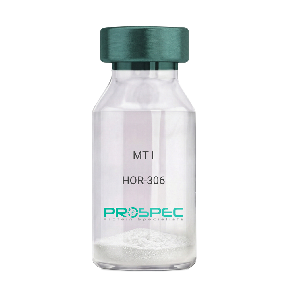
MT IDescription:
Melanotan-I
Melanotan-I, MT-I, Melanotan-1.
Product # :
HOR-306Price :
Quantity :
Shipping Method :
Shipped at Room temp
More Info
- description
- formulation
- purity
- More Info
Description
Melanotan-I has the amino acid sequence of Ser-Tyr-Ser-Nle-Glu-His-D-Phe-Arg-Trp-Gly-Lys-Pro-Val and a molecular weight of 1647.4 Dalton.
Formulation
The protein (1mg/ml) was lyophilized with no additives.
Purity
Greater than 98.0% as determined by RP-HPLC.
More Info
-
Synonyms
Melanotan-I, MT-I, Melanotan-1.
-
Physical Appearance
Sterile Filtered off-White lyophilized (freeze-dried) powder.
-
Stability
Lyophilized Melanotan-I although stable at room temperature for 3 weeks, should be stored desiccated below -18°C. Upon reconstitution MT-I should be stored at 4°C between 2-7 days and for future use below -18°C.For long term storage it is recommended to add a carrier protein (0.1% HSA or BSA).Please prevent freeze-thaw cycles.
-
Solubility
It is recommended to reconstitute the lyophilized Melanotan-I in sterile 1% acetic acid not less than 100 µg/ml, which can then be further diluted to other aqueous solutions.
ProSpec's products are furnished for LABORATORY RESEARCH USE ONLY. The product may not be used as drugs, agricultural or pesticidal products, food additives or household chemicals.
- View Data Sheet
Name :
EBI3 HumanDescription:
Epstein Barr Virus Induced 3 Human Recombinant
Interleukin-27 subunit beta, IL-27 subunit beta, IL-27B, Epstein-Barr virus-induced gene 3 protein, EBV-induced gene 3 protein, EBI3, IL27B.
Product # :
CYT-367Price :
Quantity :
Shipping Method :
Shipped at Room temp
More Info
- description
- source
- formulation
- purity
- biological activity
- More Info
Description
EBI3 Human Recombinant produced in E.Coli is a single, non-glycosylated, Polypeptide chain containing 209 amino acids fragment (21-229) having a molecular weight of 23.3kDa. The EBI3 is purified by proprietary chromatographic techniques.
Source
Escherichia Coli.
Formulation
EBI3 Human Recombinant was lyophilized from a solution containing 10mM Acetic Acid and 0.5% Mannitol.
Purity
Greater than 90% as determined by
(a) Analysis by RP-HPLC.
(b) Analysis by SDS-PAGE.Biological Activity
Assay data for Human recombinant EBI3 is based upon qualitative binding to anti-EBI3 antibody.More Info
-
Introduction
EBI3 has an induced expression in B lymphocytes in reaction to Epstein-Barr virus infection. EBI3 encodes a secreted glycoprotein belonging to the hematopoietin receptor family, and heterodimerizes with a 28 kDa protein to form iIL-27. EBI3 drives rapid clonal expansion of naive cd4(+) t-cells. EBI3 strongly synergizes with IL-12 to activate IFN-gamma production of naive cd4(+) t-cells. EBI3 mediates its biologic effects through the cytokine receptor wsx-1/tccr.
-
Synonyms
Interleukin-27 subunit beta, IL-27 subunit beta, IL-27B, Epstein-Barr virus-induced gene 3 protein, EBV-induced gene 3 protein, EBI3, IL27B.
-
Physical Appearance
Sterile Filtered White lyophilized (freeze-dried) powder.
-
Stability
Lyophilized EBI3 although stable at room temperature for 3 weeks, should be stored desiccated below -18°C. Upon reconstitution EBI3 should be stored at 4°C between 2-7 days and for future use below -18°C. Please prevent freeze-thaw cycles.
-
Solubility
It is recommended to reconstitute the lyophilized EBI3 in sterile 10mM Acetic acid not less than 100µg/ml, which can then be further diluted to other aqueous solutions.
-
Amino Acid Sequence
RKGPPAALTLPRVQCRASRYPIAVDCSWTLPPAPNSTSPVSF
IATYRLGMAARGHSWPCLQQTPTSTSCTITDVQLFSMAPYVL
NVTAVHPWGSSSSFVPFITEHIIKPDPPEGVRLSPLAERQLQ
VQWEPPGSWPFPEIFSLKYWIRYKRQGAARFHRVGPIEATSF
ILRAVRPRARYYVQVAAQDLTDYGELSDWSLPATATMSLGK. -
Background
Title: Epstein-Barr Virus Induced 3 Human Recombinant: Unveiling its Role in Epstein-Barr Virus-Associated Diseases
Abstract:
Epstein-Barr Virus Induced 3 (EBI3) is a crucial cytokine involved in the immune response against Epstein-Barr virus (EBV) and various other pathogens. This research paper provides an extensive analysis of human recombinant EBI3, focusing on its production, characterization, and potential applications in understanding EBV-associated diseases. The paper highlights the significance of EBI3 in modulating immune responses and explores its role in the pathogenesis of EBV-related malignancies. Furthermore, it discusses ongoing research and clinical trials investigating the therapeutic potential of recombinant EBI3 in immune disorders and cancer. The information presented in this paper aims to enhance our understanding of human recombinant EBI3 and its utility as a research tool and a potential immunotherapeutic agent.Introduction:
Epstein-Barr Virus Induced 3 (EBI3) is a cytokine that plays a critical role in the immune response against EBV. Human recombinant EBI3, produced through genetic engineering techniques, provides a valuable tool for studying its immunomodulatory properties and exploring its potential therapeutic applications.Production and Characterization:
Recombinant EBI3 is typically generated using expression systems such as mammalian cells or bacteria. The protein is then purified and characterized to ensure its structural integrity and functional activity. Rigorous quality control measures are implemented to confirm the specificity and potency of the recombinant EBI3.Role in EBV-Associated Diseases:
EBI3 is involved in the regulation of immune responses during EBV infection. It acts as a subunit of the heterodimeric cytokine interleukin-27 (IL-27), which plays a crucial role in anti-viral immunity. Recombinant EBI3 serves as a valuable tool for investigating the mechanisms underlying EBI3-mediated immune regulation and its potential implications in EBV-associated diseases, including infectious mononucleosis, nasopharyngeal carcinoma, and EBV-related lymphomas.Therapeutic Implications:
Dysregulation of the immune response is implicated in various immune disorders and cancers. Recombinant EBI3 holds promise as a potential immunotherapeutic agent due to its immunomodulatory properties. Ongoing research and clinical trials are investigating the therapeutic applications of recombinant EBI3 in conditions such as autoimmune diseases, viral infections, and cancer.Conclusion:
Human recombinant EBI3 represents a valuable research tool and a potential immunotherapeutic agent. Its production, characterization, and applications in understanding EBV-associated diseases contribute to our understanding of immune regulation and the development of targeted therapeutic interventions. Continued research and clinical trials exploring the therapeutic potential of recombinant EBI3 offer promising avenues for improving outcomes in immune disorders and EBV-related malignancies.What is the molecular weight/Mw of EBI3 Protein?
EBI3 Protein has a total Mw of 23.3kDa.
What is the source or expression system of EBI3 Protein?
Escherichia Coli.
What is the Purity of EBI3 Protein?
EBI3 Protein is >90% pure as determined by SDS-PAGE.
What is the Biological Activity of EBI3 Protein?
Assay data for Human recombinant EBI3 is based upon qualitative binding to anti-EBI3 antibody.
What is the amino acid sequence of EBI3 Protein?
RKGPPAALTLPRVQCRASRYPIAVDCSWTLPPAPNSTSPVSF
IATYRLGMAARGHSWPCLQQTPTSTSCTITDVQLFSMAPYVL
NVTAVHPWGSSSSFVPFITEHIIKPDPPEGVRLSPLAERQLQ
VQWEPPGSWPFPEIFSLKYWIRYKRQGAARFHRVGPIEATSF
ILRAVRPRARYYVQVAAQDLTDYGELSDWSLPATATMSLGK.
What applications can EBI3 Protein be used in?
EBI3 Protein can probably be used in western blot, ELISA and Lateral Flow.
What is the endotoxin level for EBI3 Protein?
The endotoxin level is minimal, EBI3 Protein was purified using conventional chromatography techniques.
ProSpec's products are furnished for LABORATORY RESEARCH USE ONLY. The product may not be used as drugs, agricultural or pesticidal products, food additives or household chemicals.
- View Data Sheet
Name :
IL 27 MouseDescription:
Interleukin-27 Mouse Recombinant
Interleukin-30, IL-30, IL-27/p28, p28, Interleukin-27, Interleukin-27/p28, IL-27, Interleukin-27 subunit alpha, IL-27 subunit alpha, IL27-A, Il27, Il27a, IL-27p28.
Product # :
CYT-570Price :
Quantity :
Shipping Method :
Shipped at Room temp
More Info
- description
- source
- formulation
- purity
- biological activity
- More Info
Description
Interleukin-27 Mouse Recombinant produced in E.Coli is a single, non-glycosylated polypeptide chain containing 207 amino acids and having a molecular mass of 23.7 kDa. The Murine IL-27 is purified by proprietary chromatographic techniques.
Source
Escherichia Coli.
Formulation
Lyophilized from 10mM NaHCO₃ buffer pH-8.5.
Purity
Greater than 95.0% as determined by SDS-PAGE.
Biological Activity
Mouse p28 biological activity was measured via dose-dependent inhibition of TGF-beta and IL-6-induced IL-17A expression in mouse CD4 splenocytes. 50ng/ml of mouse p28 is capable of inhibiting >25% of IL-17A expression in this assay.
More Info
-
Introduction
Interleukin-27 protein is related to IL-12A & is one of the subunits of a heterodimeric cytokine complex. IL-27 interacts with EBV induced gene 3 also called EBI3, and forms a complex that drives rapid expansion of CD4 (+) T cells.
IL-27 complex is synergizes with IL-12 in order to trigger the cytokine production of IFN-Gamma of CD4 (+) T cells. The biological effect of IL-27 is mediated by class-I cytokine receptor (WSX1/TCRR).
The pro-inflammatory activity of IL-27 is mediated through the growing expression of key molecules involved in the MHC class-I & MHC class-II pathways. Both MHC class-I and MHC-class-II expression are increased in endothelial cells after Interleukin-27 stimulation which suggests that it may play an important role in conferring the immune function on vascular endothelium IL-27p28 subunit can be induced by IFN-beta and during LPS-induced maturation of dendritic cells in type-I IFN-dependent manner through IFN regulatory factor-1 activation. Interleukin-27 regulates Interleukin-12 responsiveness of CD4+ T cells through Stat1-dependent and -independent mechanisms. IL-17 & IL-23 play an important IL-17 role in inflammation. Interleukin-27 possesses potent anti-angiogenic activity that plays an important role in its antitumor and antimetastatic activities.
EBV induced gene 3 plays a role, independently from IL-27, in regulating anti-viral or anti-tumoral immune responses. Interleukin-27 is a potent inhibitor of HIV-1 replication in macrophages, CD4+ T cells, peripheral blood mononuclear cells.
Interleukin-27 triggers STAT activation and gene transcription. -
Synonyms
Interleukin-30, IL-30, IL-27/p28, p28, Interleukin-27, Interleukin-27/p28, IL-27, Interleukin-27 subunit alpha, IL-27 subunit alpha, IL27-A, Il27, Il27a, IL-27p28.
-
Physical Appearance
Sterile Filtered White lyophilized (freeze-dried) powder.
-
Stability
Lyophilized IL-27 although stable at room temperature for 3 weeks, should be stored desiccated below -18°C. Upon reconstitution IL27 should be stored at 4°C between 2-7 days and for future use below -18°C.Please prevent freeze-thaw cycles.
-
Solubility
It is recommended to reconstitute the lyophilized IL-27 in sterile 18MΩ-cm H2O not less than 100µg/ml, which can then be further diluted to other aqueous solutions.
-
Amino Acid Sequence
MFPTDPLSLQ ELRREFTVSL YLARKLLSEV QGYVHSFAES RLPGVNLDLL PLGYHLPNVS LTFQAWHHLS DSERLCFLAT TLRPFPAMLG GLGTQGTWTS SEREQLWAMR LDLRDLHRHL RFQVLAAGFK CSKEEEDKEE EEEEEEEEKK LPLGALGGPN QVSSQVSWPQ LLYTYQLLHS LELVLSRAVR DLLLLSLPRR PGSAWDS.
ProSpec's products are furnished for LABORATORY RESEARCH USE ONLY. The product may not be used as drugs, agricultural or pesticidal products, food additives or household chemicals.