Search results
406 results found for “lymphotactin (xcl1)”
Name
Description
Product #
Price
Quantity
Shipping Method
- View Data Sheet
Name :
ITGB1 AntibodyDescription:
Mouse Anti Human Integrin Beta 1
Integrin beta-1, Fibronectin receptor subunit beta, Glycoprotein IIa, GPIIA, VLA-4 subunit beta, CD29, ITGB1, FNRB, MDF2, MSK12, Integrin beta 1, CD29, VLAB.
Product # :
ANT-715Price :
Quantity :
Shipping Method :
Shipped with Ice Packs
More Info
- formulation
- More Info
- Citations
Formulation
1mg/ml containing PBS, pH-7.4, 10% Glycerol and 0.02% Sodium Azide.
Citations
More Info
-
Introduction
Integrins are heterodimeric proteins consist of alpha and beta subunits. There are more than 18 alpha and 8 beta subunits discovered in mammals. Integrin family members are membrane receptors that participates in cell adhesion and recognition in a variety of processes including embryogenesis, hemostasis, tissue repair, immune response and metastatic diffusion of tumor cells. ITGB1 encodes a beta subunit.
-
Synonyms
Integrin beta-1, Fibronectin receptor subunit beta, Glycoprotein IIa, GPIIA, VLA-4 subunit beta, CD29, ITGB1, FNRB, MDF2, MSK12, Integrin beta 1, CD29, VLAB.
-
Physical Appearance
Sterile filtered colorless solution.
-
Immunogen
Anti-human ITGB1 mAb, is derived from hybridization of mouse F0 myeloma cells with spleen cells from BALB/c mice immunized with a recombinant human ITGB1 protein 21-461 amino acids purified from E. coli.
-
Ig Subclass
Mouse IgG1 heavy chain and k light chain.
-
Clone
PAT47E2AT.
-
Applications
ITGB1 antibody has been tested by ELISA and Western blot analysis to assure specificity and reactivity. Since application varies, however, each investigation should be titrated by the reagent to obtain optimal results.
-
Type
Mouse Anti Human Monoclonal.
-
Storage Procedures
For periods up to 1 month store at 4°C, for longer periods of time, store at -20°C. Prevent freeze thaw cycles.
-
Purification Method
ITGB1 antibody was purified from mouse ascitic fluids by protein-A affinity chromatography.
ProSpec's products are furnished for LABORATORY RESEARCH USE ONLY. The product may not be used as drugs, agricultural or pesticidal products, food additives or household chemicals.
- View Data Sheet
Name :
CCL2 Human, HisDescription:
Monocyte Chemotactic Protein-1 Human Recombinant (CCL2), His Tag
Small inducible cytokine A2, CCL2, Monocyte chemotactic protein 1, MCP-1, Monocyte chemoattractant protein 1, Monocyte chemotactic and activating factor, MCAF, Monocyte secretory protein JE, HC11, chemokine (C-C motif) ligand 2, MCP1, SCYA2, GDCF-2, SMC-CF, HSMCR30, MGC9434, GDCF-2 HC11.
Product # :
CHM-246Price :
Quantity :
Shipping Method :
Shipped with Ice Packs
More Info
- description
- source
- formulation
- purity
- More Info
- SDS-PAGE
- Citations
Description
MCP-1 Human Recombinant also known as Monocyte Chemotactic and Activating Factor (MCAF) produced in E.Coli is a non-glycosylated, Polypeptide chain containing 97 amino acids (24-99) and having a molecular mass of 10.9 kDa. The MCP-1 is fused to 20 amino acids His-Tag at N-terminus and purified by proprietary chromatographic techniques.
Source
Escherichia Coli.
Formulation
The His Tag MCP-1 protein (1mg/ml) solution contains 20mM Tris-HCL pH-8.0, 1mM DTT and 20% glycerol.
Purity
Greater than 95.0% as determined by SDS-PAGE.
SDS-PAGE
Citations
More Info
-
Introduction
Chemokine (C-C motif) ligand 2 (CCL2) is a small cytokine belonging to the CC chemokine family that is also known as monocyte chemotactic protein-1 (MCP-1). It is found at the site of tooth eruption and bone degradation. In the bone, CCL2 is expressed by mature osteoclasts and osteoblasts and is under the control of nuclear factor ?B (NF?B). CCL2 recruits immune cells, such as monocytes, to sites of tissue injury and infection. This chemokine is produced as a protein precursor containing signal peptide of 23 amino acids and a mature peptide of 76 amino acids. It is a monomeric polypeptide, with a molecular weight of approximately 13kDa. As with many other CC chemokines, CCL2 is located on chromosome 17 in humans. The cell surface receptors that bind CCL2 are CCR2 and CCR5.
-
Synonyms
Small inducible cytokine A2, CCL2, Monocyte chemotactic protein 1, MCP-1, Monocyte chemoattractant protein 1, Monocyte chemotactic and activating factor, MCAF, Monocyte secretory protein JE, HC11, chemokine (C-C motif) ligand 2, MCP1, SCYA2, GDCF-2, SMC-CF, HSMCR30, MGC9434, GDCF-2 HC11.
-
Physical Appearance
Sterile Filtered colorless clear solution.
-
Stability
Store at 4°C if entire vial will be used within 2-4 weeks. Store, frozen at -20°C for longer periods of time. Please avoid freeze thaw cycles.
-
Amino Acid Sequence
MGSSHHHHHH SSGLVPRGSH MQPDAINAPV TCCYNFTNRK ISVQRLASYR RITSSKCPKE AVIFKTIVAK EICADPKQKW VQDSMDHLDK QTQTPKT.
-
Background
What is the molecular weight/Mw of CCL2 HUMAN, HIS Protein?
CCL2 HUMAN, HIS Protein has a total Mw of 10.9kDa.
What is the source or expression system of CCL2 HUMAN, HIS Protein?
Escherichia Coli.
What is the Purity of CCL2 HUMAN, HIS Protein?
CCL2 HUMAN, HIS Protein is >95% pure as determined by SDS-PAGE.
What is the Biological Activity of CCL2 HUMAN, HIS Protein?
The biological functionality of CCL2 HUMAN, HIS Protein will be determined in the future.
What is the amino acid sequence of CCL2 HUMAN, HIS Protein?
MGSSHHHHHH SSGLVPRGSH MQPDAINAPV TCCYNFTNRK ISVQRLASYR RITSSKCPKE AVIFKTIVAK EICADPKQKW VQDSMDHLDK QTQTPKT.
What applications can CCL2 HUMAN, HIS Protein be used in?
CCL2 HUMAN, HIS Protein can probably be used in western blot, ELISA and Lateral Flow.
What is the endotoxin level for CCL2 HUMAN, HIS Protein?
The endotoxin level is minimal, CCL2 HUMAN, HIS Protein was purified using conventional chromatography techniques.
Who supplies recombinant CCL2 HUMAN, HIS that is in stock and available to order?
ProSpec has the protein in stock and available for immediate delivery.
Where can I order bulk mg quantities of CCL2 HUMAN, HIS?
ProSpec supplies bulk mg quantities at affordable prices that meets research and academic budgets.
ProSpec's products are furnished for LABORATORY RESEARCH USE ONLY. The product may not be used as drugs, agricultural or pesticidal products, food additives or household chemicals.
- View Data Sheet
Name :
ASNA1 AntibodyDescription:
arsA Arsenite Transporter ATP-Binding, Homolog 1, Mouse Anti Human
ArsA Arsenite Transporter ATP-Binding Homolog 1 (Bacterial), Arsenical Pump-Driving ATPase, Transmembrane Domain Recognition Complex 40kDa, TRC40. ASNA-I, ATPase ASNA1, ARSA-I, GET3, Golgi To ER Traffic 3 Homolog, Arsenite-Stimulated ATPase, Transmembrane Domain Recognition Complex 40 KDa ATPase Subunit, HASNA-I, HARSA-I, ARSA, ARSA1, EC 3.6.3.16, EC 3.6.
Product # :
ANT-516Price :
Quantity :
Shipping Method :
Shipped with Ice Packs
More Info
- formulation
- More Info
- Citations
Formulation
1mg/ml containing PBS, pH-7.4, 10% Glycerol and 0.02% Sodium Azide.
Citations
More Info
-
Introduction
ArsA Arsenite Transporter, ATP-Binding, Homolog 1 (ASNA1) is a part of the arsA ATPase family. ASNA1 is the human homolog of the bacterial arsA gene. ArsA ATPase is the catalytic component of a multisubunit oxyanion pump In E.coli, which is in charge for resistance to arsenicals and antimonials. ASNA1 is also a main component of a transmembrane domain (TMD) recognition complex (TRC) which is involved in the post-translational delivery of tail-anchored (TA) proteins from the cytosol to the endoplasmic reticulum (ER).
-
Synonyms
ArsA Arsenite Transporter ATP-Binding Homolog 1 (Bacterial), Arsenical Pump-Driving ATPase, Transmembrane Domain Recognition Complex 40kDa, TRC40. ASNA-I, ATPase ASNA1, ARSA-I, GET3, Golgi To ER Traffic 3 Homolog, Arsenite-Stimulated ATPase, Transmembrane Domain Recognition Complex 40 KDa ATPase Subunit, HASNA-I, HARSA-I, ARSA, ARSA1, EC 3.6.3.16, EC 3.6.
-
Physical Appearance
Sterile filtered colorless solution.
-
Immunogen
Anti-human ASNA1 mAb, clone PAT2A1AT, is derived from hybridization of mouse F0 myeloma cells with spleen cells from BALB/c mice immunized with a recombinant human ASNA1 protein 1-348 amino acids purified from E. coli.
-
Ig Subclass
Mouse IgG2b heavy chain and k light chain.
-
Clone
PAT2A1AT.
-
Applications
The antibody has been tested by ELISA, Western blot analysis to assure specificity and reactivity. Since application varies, however, each investigation should be titrated by the reagent to obtain optimal results. Recommended starting dilution is 1:1000.
-
Type
Mouse Anti Human Monoclonal.
-
Storage Procedures
For periods up to 1 month store at 4°C, for longer periods of time, store at -20°C. Prevent freeze thaw cycles.
-
Purification Method
ASNA1 antibody was purified from mouse ascitic fluids by protein-A affinity chromatography.
ProSpec's products are furnished for LABORATORY RESEARCH USE ONLY. The product may not be used as drugs, agricultural or pesticidal products, food additives or household chemicals.
- View Data Sheet
Name :
NKp46 AntibodyDescription:
Natural Cytotoxicity Receptor NKp46, Mouse Anti Human
Natural cytotoxicity triggering receptor 1, Natural killer cell p46-related protein, hNKp46, NK-p46, NKp46, NK cell-activating receptor, Lymphocyte antigen 94 homolog, CD335 antigen, NCR1, LY94, NCRNKp46, CD335.
Product # :
ANT-303Price :
Quantity :
Shipping Method :
Shipped with Ice Packs
More Info
- formulation
- More Info
- Citations
Formulation
1mg/ml containing PBS, pH-7.4, & 0.1% Sodium Azide.
Citations
More Info
-
Introduction
A natural cytotoxicity receptor (NCR) NKp46 has been shown to represent a novel NK cell-specific molecule involved in human NK cell activation. The natural cytotoxicity receptors (NCRs) are a recently characterized family of Ig-like activation receptors that appear to be major triggering receptors in tumor cell recognition. The three known NCRs include NKp46 and NKp30, which are expressed on circulating NKcells, and NKp44, which is expressed only on activating NK cells. NKp46 has been implicated in NK cell-mediated lysis of several autologous tumor cells, pathogen-infected cell lines and mononuclear phagocytes infected with an intracellular bacterium. The Lysis of tumor cells by NK-cells involves recognition by NKp46 of heparan sulfate moieties of membrane heparan sulfate proteoglycans. Furthermore, NKp46 is a surface receptor involved in NK-cell cell death by apoptosis. NKp46 has two extracellular Ig-like domains followed by a ~40 residue stalk region, a type I transmembrane domain, and a short cytoplasmic tail. The extracellular Ig-like domain of NKp46 (22-255aa) is purified by FPLC gel-filtration chromatography, after refolding of the isolated inclusion bodies in a redox buffer. In addition, engagement of the antigen with the monoclonal antibody stimulates intracellular calcium levels and the synthesis of cytokines. CD59 is an NKp46 coreceptor (by physical association) together they activate cytotoxicity of human NK-cells, their engagement results in tyrosine phosphorylation of CD3-zeta chains associated with NKp46. Reduced cell surface expression of NKp46 and other NK-cell receptors is linked to the impaired NK-cell cytolytic function in viremic HIV-1 infection.
-
Synonyms
Natural cytotoxicity triggering receptor 1, Natural killer cell p46-related protein, hNKp46, NK-p46, NKp46, NK cell-activating receptor, Lymphocyte antigen 94 homolog, CD335 antigen, NCR1, LY94, NCRNKp46, CD335.
-
Physical Appearance
Sterile Filtered colorless solution.
-
Immunogen
Anti-human NKp46 mAb, is derived from hybridization of mouse SP2/0 myeloma cells with spleen cells from BALB/c mice immunized with recombinant human NKp46 amino acids 22-255 purified from E. coli.
-
Ig Subclass
Mouse IgG1 heavy chain and κ light chain.
-
Clone
Pn1D9AT.
-
Applications
NKp46 antibody has been tested by immunofluorescent staining with flow cytometric analysis and by Western blot analysis to assure specificity and reactivity. Since application varies, however, each investigation should be titrated by the reagent to obtain optimal results.
-
Type
Mouse Anti Human Monoclonal.
-
Storage Procedures
For periods up to 1 month store at 4°C, for longer periods of time, store at -20°C. Prevent freeze thaw cycles.
-
Purification Method
NKp46 antibody was purified from mouse ascitic fluids by protein-G affinity chromatography.
ProSpec's products are furnished for LABORATORY RESEARCH USE ONLY. The product may not be used as drugs, agricultural or pesticidal products, food additives or household chemicals.
- View Data Sheet
Name :
ODC1 antibodyDescription:
Ornithine Decarboxylase 1, Mouse Anti Human
ODC, Ornithine decarboxylase 1, EC 4.1.1.17.
Product # :
ANT-739Price :
Quantity :
Shipping Method :
Shipped with Ice Packs
More Info
- formulation
- More Info
- Citations
Formulation
1mg/ml containing PBS, pH-7.4, 10% Glycerol and 0.02% Sodium Azide.
Citations
More Info
-
Introduction
Ornithine decarboxylase is the preliminary and rate-limiting enzyme in the biosynthetic pathway of polyamines and it takes part in the alteration of ornithine to putrescine. ODC1 is a key member of various biological processes, such as cell growth, transformation, differentiation and apoptosis. Overexpression of the ODC1 gene has a vital part in cell proliferation and the progress of cancer.
-
Synonyms
ODC, Ornithine decarboxylase 1, EC 4.1.1.17.
-
Physical Appearance
Sterile filtered colorless solution.
-
Immunogen
Anti-human ODC1 mAb, is derived from hybridization of mouse F0 myeloma cells with spleen cells from BALB/c mice immunized with recombinant human ODC1 amino acids 1-461 purified from E. coli.
-
Ig Subclass
Mouse IgG2b heavy chain and k light chain.
-
Clone
PAT9B3AT.
-
Applications
ODC1 antibody has been tested by ELISA, Western blot analysis, Flow cytometry and ICC/IF to assure specificity and reactivity. Since application varies, however, each investigation should be titrated by the reagent to obtain optimal results.
-
Type
Mouse Anti Human Monoclonal.
-
Storage Procedures
For periods up to 1 month store at 4°C, for longer periods of time, store at -20°C. Prevent freeze thaw cycles.
-
Purification Method
ODC1 antibody was purified from mouse ascitic fluids by protein-A affinity chromatography.
ProSpec's products are furnished for LABORATORY RESEARCH USE ONLY. The product may not be used as drugs, agricultural or pesticidal products, food additives or household chemicals.
- View Data Sheet
Name :
NFATC1 AntibodyDescription:
Nuclear factor of activated T-cells cytoplasmic 1, Mouse Anti Human
Nuclear factor of activated T-cells cytoplasmic 1, NFAT transcription complex cytosolic component, NF-ATc1, NFATc1, NF-ATc, NFATc, NFAT2, MGC138448.
Product # :
ANT-436Price :
Quantity :
Shipping Method :
Shipped with Ice Packs
More Info
- formulation
- More Info
- Citations
Formulation
1mg/ml containing PBS, pH-7.4, 0.02% Sodium Azide and 10% Glycerol.
Citations
More Info
-
Introduction
NFATC1 is a component of the nuclear factor of activated T cells DNA-binding transcription complex. This complex consists of at least 2 components: a preexisting cytosolic component that translocates to the nucleus upon T cell receptor (TCR) stimulation, and an inducible nuclear component. Proteins belonging to this family of transcription factors play an essential role in inducible gene transcription during immune response. NFATC1 is an inducible nuclear component. NFATC1 functions as a major molecular target for the immunosuppressive drugs such as cyclosporin A. Different isoforms of NFATC1 may regulate inducible expression of different cytokine genes.
-
Synonyms
Nuclear factor of activated T-cells cytoplasmic 1, NFAT transcription complex cytosolic component, NF-ATc1, NFATc1, NF-ATc, NFATc, NFAT2, MGC138448.
-
Physical Appearance
Sterile filtered colorless solution.
-
Immunogen
Anti-human NFATC1 mAb is derived from hybridization of mouse F0 myeloma cells with spleen cells from BALB/c mice immunized with recombinant human NFATC1 amino acids 428-716 purified from E. coli.
-
Ig Subclass
Mouse IgG2a heavy chain and κ light chain.
-
Clone
PAT1C3AT.
-
Applications
NFATC1 antibody has been tested by ELISA and Western blot analysis to assure specificity and reactivity. Since application varies, however, each investigation should be titrated by the reagent to obtain optimal results.
-
Type
Mouse Anti Human Monoclonal.
-
Storage Procedures
For periods up to 1 month store at 4°C, for longer periods of time, store at -20°C. Prevent freeze thaw cycles.
-
Purification Method
NFATC1 antibody was purified from mouse ascitic fluids by protein-G affinity chromatography.
ProSpec's products are furnished for LABORATORY RESEARCH USE ONLY. The product may not be used as drugs, agricultural or pesticidal products, food additives or household chemicals.
- View Data Sheet
Name :
IL1A Rat, His ActiveDescription:
Interleukin-1 alpha Rat Recombinant, His Tag Active
Interleukin-1 alpha, IL-1 alpha.
Product # :
CYT-1100Price :
Quantity :
Shipping Method :
Shipped with Ice Packs
More Info
- description
- source
- formulation
- purity
- biological activity
- More Info
- Citations
Description
IL1A Rat Recombinant produced in E.Coli is a single, non-glycosylated polypeptide chain containing 179 amino acids (115-270 a.a) and having a molecular mass of 20.2kDa.IL1A is fused to a 23 amino acid His-tag at N-terminus & purified by proprietary chromatographic techniques.
Source
E.coli.
Formulation
IL1A protein solution (1mg/ml) contains 10% glycerol & Phosphate Buffered Saline (pH 7.4).
Purity
Greater than 95% as determined by SDS-PAGE.
Biological Activity
Citations
More Info
-
Introduction
IL-1 alpha is produced by activated macrophages.IL-1 alpha stimulates thymocyte proliferation by inducing il-2 release, b-cell maturation and proliferation, and fibroblast growth factor activity. IL1A proteins take part in the inflammatory response, being identified as endogenous pyrogens, and stimulate the release of prostaglandin and collagenase from synovial cells.
-
Synonyms
Interleukin-1 alpha, IL-1 alpha.
-
Physical Appearance
Sterile Filtered colorless solution.
-
Stability
Store at 4°C if entire vial will be used within 2-4 weeks. Store, frozen at -20°C for longer periods of time. For long term storage it is recommended to add a carrier protein (0.1% HSA or BSA).Avoid multiple freeze-thaw cycles.
-
Amino Acid Sequence
MGSSHHHHHH SSGLVPRGSH MGSSAPHSFQ NNLRYKLIRI VKQEFIMNDS LNQNIYVDMD
RIHLKAASLN DLQLEVKFDM YAYSSGGDDSKYPVTLKVSN TQLFVSAQGE DKPVLLKEIP
ETPKLITGSE TDLIFFWEKI NSKNYFTSAA FPELLIATKE QSQVHLARGL PSMIDFQIS. -
Background
What is the molecular weight/Mw of IL1A RAT, HIS ACTIVE Protein?
IL1A RAT, HIS ACTIVE Protein has a total Mw of 20.2kDa.
What is the source or expression system of IL1A RAT, HIS ACTIVE Protein?
Escherichia Coli.
What is the Purity of IL1A RAT, HIS ACTIVE Protein?
IL1A RAT, HIS ACTIVE Protein is >95% pure as determined by SDS-PAGE.
What is the Biological Activity of IL1A RAT, HIS ACTIVE Protein?
The ED50 that was measured in a cell proliferation assay using D10.G4.1 mouse helper T cell is <20 pg/ml.
What is the amino acid sequence of IL1A RAT, HIS ACTIVE Protein?
MGSSHHHHHH SSGLVPRGSH MGSSAPHSFQ NNLRYKLIRI VKQEFIMNDS LNQNIYVDMD
RIHLKAASLN DLQLEVKFDM YAYSSGGDDSKYPVTLKVSN TQLFVSAQGE DKPVLLKEIP
ETPKLITGSE TDLIFFWEKI NSKNYFTSAA FPELLIATKE QSQVHLARGL PSMIDFQIS.
.What applications can IL1A RAT, HIS ACTIVE Protein be used in?
IL1A RAT, HIS ACTIVE Protein can probably be used in western blot, ELISA and Lateral Flow.
What is the endotoxin level for IL1A RAT, HIS ACTIVE Protein?
The endotoxin level is minimal, IL1A RAT, HIS ACTIVE Protein was purified using conventional chromatography techniques.
ProSpec's products are furnished for LABORATORY RESEARCH USE ONLY. The product may not be used as drugs, agricultural or pesticidal products, food additives or household chemicals.
- View Data Sheet
Name :
SPP1 Human, ActiveDescription:
Osteopontin Human Recombinant, BioActive
OPN, SPP-1, BNSP, BSPI, ETA-1, Bone sialoprotein 1, BSP I.Early T lymphocyte activation 1, ETA 1, ETA1, MGC110940, Nephropontin, Secreted phosphoprotein 1, SPP 1, SPP1, urinary stone protein, uropontin.
Product # :
CYT-1166Price :
Quantity :
Shipping Method :
Shipped with Ice Packs
More Info
- description
- source
- formulation
- purity
- biological activity
- More Info
- Citations
Description
SPP1 Human Recombinant produced in E.Coli is a single, non-glycosylated polypeptide chain containing 321 amino acids (17-314 a.a.) and having a molecular mass of 36.2kDa. SPP1 is fused to a 23 amino acid His tag at N-Terminus and purified by proprietary chromatographic techniques.
Source
Escherichia Coli.
Formulation
The SPP1 solution (1mg/ml) contains 20mM Tris-HCl buffer (pH 7.5), 1mM DTT, 10% glycerol and 2mM EDTA.
Purity
Greater than 90.0% as determined by SDS-PAGE.
Biological Activity
Measured by the ability of the immobilized protein to support the adhesion of HEK293 human embryonic kidney cells. When cells are added to OPN coated plates 10ug/ml. This effect is more to 40%.
Citations
More Info
-
Introduction
Osteopontin is a glycoprotein that was primarilyfound in osteoblasts and takes part in bone remodeling, immune functions in fibroblasts, macrophages, & lymphocytes during inflammation and wound healing. SPP1 highlybinds to hydroxyapatite. SPP1 forms an integral part of the mineralized matrix. SPP1 is vital to cell-matrix interaction.
Secreted Phosphoprotein-1 protects against cardiac ischemia-reperfusion injury through late preconditioning. Expression of Ostepontin and CD44 in hepatocellular carcinoma is linked to advanced tumor stage &leads to prognosis information. SPP1 is the most over-expressed gene in intrahepatic cholangiocarcinoma. Secreted Phosphoprotein-1 overexpression is related with interstitial lung diseases. -
Synonyms
OPN, SPP-1, BNSP, BSPI, ETA-1, Bone sialoprotein 1, BSP I.Early T lymphocyte activation 1, ETA 1, ETA1, MGC110940, Nephropontin, Secreted phosphoprotein 1, SPP 1, SPP1, urinary stone protein, uropontin.
-
Physical Appearance
Sterile Filtered clear colorless solution.
-
Stability
Store at 4°C if entire vial will be used within 2-4 weeks. Store, frozen at -20°C for longer periods of time. For long term storage it is recommended to add a carrier protein (0.1% HSA or BSA).Avoid multiple freeze-thaw cycles.
-
Amino Acid Sequence
MGSSHHHHHH SSGLVPRGSH RSMIPVKQAD SGSSEEKQLY NKYPDAVATW LNPDPSQKQN LLAPQNAVSS EETNDFKQET LPSKSNESHD HMDDMDDEDD DDHVDSQDSI DSNDSDDVDD TDDSHQSDES HHSDESDELV TDFPTDLPAT EVFTPVVPTV DTYDGRGDSV VYGLRSKSKK FRRPDIQYPD ATDEDITSHM ESEELNGAYK AIPVAQDLNA PSDWDSRGKD SYETSQLDDQ SAETHSHKQS RLYKRKANDE SNEHSDVIDS QELSKVSREF HSHEFHSHED MLVVDPKSKE EDKHLKFRIS HELDSASSEV N.
ProSpec's products are furnished for LABORATORY RESEARCH USE ONLY. The product may not be used as drugs, agricultural or pesticidal products, food additives or household chemicals.
- View Data Sheet
Name :
CCL13 HumanDescription:
Monocyte Chemotactic Protein-4 Human Recombinant (CCL13)
Small inducible cytokine A13, CCL13, Monocyte chemotactic protein 4, MCP-4, Monocyte chemoattractant protein 4, CK-beta-10, NCC-1, chemokine (C-C motif) ligand 13, NCC1, CKb10, SCYL1, SCYA13, MGC17134.
Product # :
CHM-319Price :
Quantity :
Shipping Method :
Shipped at Room temp
More Info
- description
- source
- formulation
- purity
- biological activity
- More Info
- Citations
Description
Monocyte Chemotactic Protein-4 Human Recombinant produced in E.Coli is a non-glycosylated, Polypeptide chain containing 75 amino acids and having a molecular mass of 8.6 kDa. The MCP-4 is purified by proprietary chromatographic techniques.
Source
Escherichia Coli.
Formulation
The protein was lyophilized from a concentrated (1mg/ml) sterile solution in 20mM PB, pH 7.4, 130mM NaCl.
Purity
Greater than 96.0% as determined by:
(a) Analysis by RP-HPLC.
(b) Analysis by SDS-PAGE.Biological Activity
The specific activity as determined by the ability of MCP-4 to chemoattaract human monocytes at 10-100ng/ml, corresponding to a Specific Activity of 10,000-100,000 units/mg.Citations
More Info
-
Introduction
Chemokine (C-C motif) ligand 13 (CCL13 / MCP-4) is a small cytokine belonging to the CC chemokine family. The MCP-4 gene is located on human chromosome 17 within a large cluster of other CC chemokines. MCP-4 induces chemotaxis in monocytes, eosinophils, T lymphocytes, and basophils by binding cell surface G-protein linked chemokine receptors such as CCR2, CCR3 and CCR5. Activity of the MCP-4 chemokine has been implicated in allergic reactions such as asthma. MCP-4 can be induced by the inflammatory cytokines interleukin-1 and TNF-a.
-
Synonyms
Small inducible cytokine A13, CCL13, Monocyte chemotactic protein 4, MCP-4, Monocyte chemoattractant protein 4, CK-beta-10, NCC-1, chemokine (C-C motif) ligand 13, NCC1, CKb10, SCYL1, SCYA13, MGC17134.
-
Physical Appearance
Sterile Filtered White lyophilized (freeze-dried) powder.
-
Stability
Lyophilized MCP-4 although stable at room temperature for 3 weeks, should be stored desiccated below -18°C. Upon reconstitution MCP-4 should be stored at 4°C between 2-7 days and for future use below -18°C. For long term storage it is recommended to add a carrier protein (0.1% HSA or BSA).Please prevent freeze-thaw cycles.
-
Solubility
It is recommended to reconstitute the lyophilized MCP-4 in sterile 18M-cm H2O not less than 100µg/ml, which can then be further diluted to other aqueous solutions.
-
Amino Acid Sequence
QPDALNVPSTCCFTFSSKKISLQRLKSYVITTSRCPQKAV
IFRTKLGKEICADPKEKWVQNYMKHLGRKAHTLKT. -
Background
What is the molecular weight/Mw of CCL13 HUMAN Protein?
CCL13 HUMAN Protein has a total Mw of 8.6kDa.
What is the source or expression system of CCL13 HUMAN Protein?
Escherichia Coli.
What is the Purity of CCL13 HUMAN Protein?
CCL13 HUMAN Protein is >96% pure as determined by SDS-PAGE.
What is the Biological Activity of CCL13 HUMAN Protein?
The specific activity as determined by the ability of MCP-4 to chemoattaract human monocytes at 10-100ng/ml, corresponding to a Specific Activity of 10,000-100,000 units/mg.
What is the amino acid sequence of CCL13 HUMAN Protein?
QPDALNVPSTCCFTFSSKKISLQRLKSYVITTSRCPQKAV
IFRTKLGKEICADPKEKWVQNYMKHLGRKAHTLKT.
What applications can CCL13 HUMAN Protein be used in?
CCL13 HUMAN Protein can probably be used in western blot, ELISA and Lateral Flow.
What is the endotoxin level for CCL13 HUMAN Protein?
The endotoxin level is minimal, CCL13 HUMAN Protein was purified using conventional chromatography techniques.Who supplies recombinant CCL13 HUMAN that is in stock and available to order?
ProSpec has the protein in stock and available for immediate delivery.
Where can I order bulk mg quantities of CCL13 HUMAN?
ProSpec supplies bulk mg quantities at affordable prices that meets research and academic budgets.
ProSpec's products are furnished for LABORATORY RESEARCH USE ONLY. The product may not be used as drugs, agricultural or pesticidal products, food additives or household chemicals.
- View Data Sheet
Name :
Tamm HorsfallDescription:
Recombinant Human Tamm Horsfall Glycoprotein
Tamm-Horsfall urinary glycoprotein, THP, FJHN, HNFJ, THGP, MCKD2, ADMCKD2, UMOD, Uromodulin.
Product # :
ENZ-1206Price :
Quantity :
Shipping Method :
Shipped at Room temp
More Info
- description
- source
- formulation
- purity
- More Info
- SDS-PAGE
- Citations
Description
Uromodulin Human Recombinant protein produced from HEK Cells, is a polypeptide chain containing 595 amino acids ( 25-613 a.a. ) and having a total Mw of 65 kDa. Uromodulin is expressed with a 6 amino acid His tag at N-Terminus and purified by proprietary chromatographic techniques.
Source
HEK293
Formulation
The UMOD protein was lyophilized from 0.4μm filtered solution containing 50mM NaCl, 0.02M TRIS, pH 7.5.
Purity
Greater than 90.0% as determined by SDS-PAGE analysis.
SDS-PAGE
Citations
More Info
-
Introduction
Uromodulin (Tamm–Horsfall protein) is produced mainly by cells in the kidney’s thick ascending limb and is the most abundant protein in normal urine.
Uromodulin takes part in salt and water regulation, helps prevent urinary tract infections and kidney stones, and influences inflammation and immune activity in the kidney.
Reduced Uromodulin levels is associated with chronic kidney disease.
UMOD mutations causes unproper protein folding and thus transported incorrectly, resulting in its accumulation inside kidney tubular cells that damages the tubules and cause kidney disease (ADTKD-UMOD), associated with high uric acid, gout, and progressive kidney failure.
-
Synonyms
Tamm-Horsfall urinary glycoprotein, THP, FJHN, HNFJ, THGP, MCKD2, ADMCKD2, UMOD, Uromodulin.
-
Physical Appearance
Filtered White lyophilized (freeze-dried) powder.
-
Stability
Lyophilized UMOD although stable at room temperature for 3 weeks, should be stored at -18C. Upon reconstitution UMOD should be stored at 4C between 2-7 days and for future use below -18C.
For long term storage it is recommended to add a carrier protein (0.1% HSA or BSA).
Please prevent freeze-thaw cycles.
-
Solubility
Add deionized water to prepare a working stock solution of approximately 0.5mg/ml and let the lyophilized pellet dissolve completely. Product is not sterile! Please filter the product by an appropriate sterile filter before using it in the cell culture.
-
Amino Acid Sequence
DTSEARWCSE CHSNATCTED EAVTTCTCQE GFTGDGLTCV DLDECAIPGA HNCSANSSCV NTPGSFSCVC PEGFRLSPGL GCTDVDECAE PGLSHCHALA TCVNVVGSYL CVCPAGYRGD GWHCECSPGS CGPGLDCVPE GDALVCADPC QAHRTLDEYW RSTEYGEGYA CDTDLRGWYR FVGQGGARMA ETCVPVLRCN TAAPMWLNGT HPSSDEGIVS RKACAHWSGH CCLWDASVQV KACAGGYYVY NLTAPPECHL AYCTDPSSVE GTCEECSIDE DCKSNNGRWH CQCKQDFNIT DISLLEHRLE CGANDMKVSL GKCQLKSLGF DKVFMYLSDS RCSGFNDRDN RDWVSVVTPA RDGPCGTVLT RNETHATYSN TLYLADEIII RDLNIKINFA CSYPLDMKVS LKTALQPMVS ALNIRVGGTG MFTVRMALFQ TPSYTQPYQG SSVTLSTEAF LYVGTMLDGG DLSRFALLMT NCYATPSSNA TDPLKYFIIQ DRCPHTRDST IQVVENGESS QGRFSVQMFR FAGNYDLVYL HCEVYLCDTM NEKCKPTCSG TRFRSGSVID QSRVLNLGPI TRKGVQATVH HHHHH
-
Background
What is the function of Uromodulin?
Uromodulin is the most abundant protein in normal urine. Its secretion in urine follows proteolytic cleavage of the ectodomain of its glycosyl phosphatidylinosital-anchored counterpart that is situated on the luminal cell surface of the loop of Henle. Uromodulin plays a role as a constitutive inhibitor of calcium crystallization in renal fluids. Secretion of uromodulin in urine provides protection against urinary tract infections caused by uropathogenic bacteria. Defects in Uromodulin expression are associated with the autosomal dominant renal disorders medullary cystic kidney disease-2 (MCKD2) and familial juvenile hyperuricemic nephropathy (FJHN). These disorders are characterized by juvenile onset of hyperuricemia, gout, and progressive renal failure. While several transcript variants may exist for this gene, the full-length natures of only two have been described to date. UMOD is involved in regulating the circulating activity of cytokines as it binds to il-1, il-2 and tnf with high affinity.
What is the molecular weight/Mw of Uromodulin Protein?
UMOD Protein has a total Mw of 65kDa.
What is the source or expression system of Uromodulin Protein?
HEK293.
What is the Purity of Uromodulin Protein?
UMOD Protein is >90% pure as determined by SDS-PAGE.
What is the Biological Activity of Uromodulin Protein?
The biological functionality of UMOD Protein will be determined in the future.
What is the amino acid sequence of Uromodulin Protein?
UMOD Protein is composed from 595 amino acids.
What applications can Uromodulin Protein be used in?
UMOD Protein can probably be used in western blot, ELISA and Lateral Flow.
What is the endotoxin level for Uromodulin Protein?
The endotoxin level is minimal, UMOD Protein was purified using conventional chromatography techniques.
ProSpec's products are furnished for LABORATORY RESEARCH USE ONLY. The product may not be used as drugs, agricultural or pesticidal products, food additives or household chemicals.
- View Data Sheet
Name :
ANXA1 HumanDescription:
Annexin A1 Human Recombinant
ANX1, LPC1, ANXA1, Lipocortin I, Calpactin II, Chrombindin-9, p35, Annexin-1, Phospholipase A2 inhibitory protein, Annexin I, Annexin A1.
Product # :
PRO-679Price :
Quantity :
Shipping Method :
Shipped with Ice Packs
More Info
- description
- source
- formulation
- purity
- More Info
- Citations
Description
ANXA1 produced in E.Coli is a single, non-glycosylated polypeptide chain containing 346 amino acids (1-346 a.a.) and having a molecular mass of 38.7 kDa.ANXA1 is purified by proprietary chromatographic techniques.
Source
Escherichia Coli.
Formulation
The ANXA1 protein solution contains 20mM Tris-HCl, pH-8, 100mM NaCl, 1mM DTT and 10% glycerol.
Purity
Greater than 90.0% as determined by SDS-PAGE.
Citations
More Info
-
Introduction
ANXA1 is part of the family of Ca(2+)-dependent phospholipid binding proteins which have a Mw between 35kDa-40kDa and are situated on the cytosolic face of the plasma membrane. ANXA1 protein has a Mw of 40kDa, with phospholipase A2 inhibitory activity to bind from two to four calcium ions with high affinity. Since phospholipase A2 is necessary for the biosynthesis of the potent mediators of inflammation, prostaglandins and leukotrienes, ANXA1 might have potential anti-inflammatory activity. ANXA1 promotes membrane fusion and iplays a role in exocytosis. The recognition of ANXA1 protein by immunocytochemical leads a simple, highly sensitive and specific assay for diagnosis of hairy cell leukemia.
-
Synonyms
ANX1, LPC1, ANXA1, Lipocortin I, Calpactin II, Chrombindin-9, p35, Annexin-1, Phospholipase A2 inhibitory protein, Annexin I, Annexin A1.
-
Physical Appearance
Sterile filtered colorless solution.
-
Stability
Store at 4°C if entire vial will be used within 2-4 weeks. Store, frozen at -20°C for longer periods of time. For long term storage it is recommended to add a carrier protein (0.1% HSA or BSA).Avoid multiple freeze-thaw cycles.
-
Amino Acid Sequence
MAMVSEFLKQ AWFIENEEQE YVQTVKSSKG GPGSAVSPYP TFNPSSDVAA LHKAIMVKGV DEATIIDILT KRNNAQRQQI KAAYLQETGK
PLDETLKKAL TGHLEEVVLA LLKTPAQFDA DELRAAMKGL GTDEDTLIEI LASRTNKEIR DINRVYREEL KRDLAKDITS DTSGDFRNAL
LSLAKGDRSE DFGVNEDLAD SDARALYEAG ERRKGTDVNV FNTILTTRSY PQLRRVFQKY TKYSKHDMNK VLDLELKGDI EKCLTAIVKCATSKPAFFAE KLHQAMKGVG TRHKALIRIM VSRSEIDMND IKAFYQKMYG ISLCQAILDE TKGDYEKILV ALCGGN.
ProSpec's products are furnished for LABORATORY RESEARCH USE ONLY. The product may not be used as drugs, agricultural or pesticidal products, food additives or household chemicals.
- View Data Sheet
Name :
Borrelia Garinii OspCDescription:
Borrelia Garinii OspC Recombinant
Product # :
BOR-021Price :
Quantity :
Shipping Method :
Shipped with Ice Packs
More Info
- description
- source
- formulation
- purity
- More Info
- Citations
Description
Recombinant Borrelia Garinii Outer Surface Protein C produced in E.coli is a non-glycosylated, polypeptide chain having a calculated molecular mass of 22kDa.Borrelia Garinii OspC is expressed with a 10xHis tag at N-terminus and purified by proprietary chromatographic techniques.
Source
Escherichia Coli.
Formulation
Borrelia Garinii OspC is supplied in 20mM HEPES buffer pH-8.0, 200mM NaCl and 20% glycerol.
Purity
Greater than 95.0% as determined by SDS-PAGE.
Citations
More Info
-
Introduction
Borrelia belongs to a genus of bacteria of the spirochete phylum. Borrelia causes borreliosis, which is a zoonotic, vector-borne disease transmitted mainly by ticks and some by lice, depending on the species. Of the 36 known species of Borrelia, 12 are distinguished to cause Lyme disease or borreliosis and are transmitted by ticks. The main Borrelia species causing Lyme disease are Borrelia burgdorferi, Borrelia afzelii, and Borrelia garinii. The Borrelia genus members have a linear chromosome which is about 900 kbp in length as well as an excess of both linear and circular plasmids in the 5-220 kbp size range. The plasmids are atypical, as compared to most bacterial plasmids, since they contain many paralogous sequences, a large number of pseudogenes and, in some cases, essential genes. Moreover, a number of the plasmids have features suggesting that they are prophages.
-
Physical Appearance
Sterile Filtered clear solution.
-
Stability
Store at 4°C if entire vial will be used within 2-4 weeks. Store, frozen at -20°C for longer periods of time. Avoid multiple freeze-thaw cycles.
-
Immunological Functions
1. Binds IgG- and IgM-type human antibodies.2. Immunodot test with Lyme disease positive/negative plasma.
ProSpec's products are furnished for LABORATORY RESEARCH USE ONLY. The product may not be used as drugs, agricultural or pesticidal products, food additives or household chemicals.
- View Data Sheet
Name :
IL1A HumanDescription:
Interleukin-1 alpha Human Recombinant
Hematopoietin-1, Lymphocyte-activating factor (LAF), Endogenous Pyrogen (EP), Leukocyte Endogenous Mediator (LEM), Mononuclear Cell Factor (MCF), IL-1 alpha,IL1, IL-1A, IL1F1.
Product # :
CYT-253Price :
Quantity :
Shipping Method :
Shipped at Room temp
More Info
- description
- source
- formulation
- purity
- biological activity
- More Info
- Citations
Description
Interleukin-1 alpha Human Recombinant produced in E.Coli is single, a non-glycosylated, Polypeptide chain containing 159 amino acids and having a molecular mass of 18022 Dalton. The IL-1A is purified by proprietary chromatographic techniques.
Source
Escherichia Coli.
Formulation
The protein was lyophilized from a concentrated (1mg/ml) sterile solution containing 25mM Tris-HCl, pH 8.
Purity
Greater than 97.0% as determined by:
(a) Analysis by RP-HPLC.
(b) Analysis by SDS-PAGE.Biological Activity
The ED50 as determined by the dose-dependant stimulation of murine D10S cells is < 0.001 ng/ml, corresponding to a Specific Activity of 1 x 10⁹ IU/mg.Citations
More Info
-
Introduction
IL-1 alpha is produced by activated macrophages, stimulates thymocyte proliferation by inducing il-2 release, b-cell maturation and proliferation, and fibroblast growth factor activity. IL1A proteins are involved in the inflammatory response, being identified as endogenous pyrogens, and are reported to stimulate the release of prostaglandin from synovial cells.
-
Synonyms
Hematopoietin-1, Lymphocyte-activating factor (LAF), Endogenous Pyrogen (EP), Leukocyte Endogenous Mediator (LEM), Mononuclear Cell Factor (MCF), IL-1 alpha,IL1, IL-1A, IL1F1.
-
Physical Appearance
Sterile Filtered White lyophilized (freeze-dried) powder.
-
Stability
Lyophilized Interleukin-1 alpha although stable at room temperature for 3 weeks, should be stored desiccated below -18°C. Upon reconstitution IL-1a should be stored at 4°C between 2-7 days and for future use below -18°C.Please prevent freeze-thaw cycles.
-
Solubility
It is recommended to reconstitute the lyophilized Interleukin 1 alpha in sterile 18MΩ-cm H2O not less than 100µg/ml, which can then be further diluted to other aqueous solutions.
-
Amino Acid Sequence
SAPFSFLSNVKYNFMRIIKYEFILNDALNQSIIRANDQYLTAAALHNLDEAV KFDMGAYKSSKDDAKITVILRISKTQLYVTAQDEDQPVLLKEMPEIPKTITG SETNLLFFWETHGTKNYFTSVAHPNLFIATKQDYWVCLAGGPPSITDFQILE NQA.
-
Background
What is the molecular weight/Mw of IL1A HUMAN Protein?
IL1A HUMAN Protein has a total Mw of 18kDa.
What is the source or expression system of IL1A HUMAN Protein?
Escherichia Coli.
What is the Purity of IL1A HUMAN Protein?
IL1A HUMAN Protein is >97% pure as determined by SDS-PAGE.
What is the Biological Activity of IL1A HUMAN Protein?
The ED50 as determined by the dose-dependant stimulation of murine D10S cells is < 0.001 ng/ml, corresponding to a Specific Activity of 1 x 10⁹ IU/mg.
What is the amino acid sequence of IL1A HUMAN Protein?
SAPFSFLSNVKYNFMRIIKYEFILNDALNQSIIRANDQYLTAAALHNLDEAV KFDMGAYKSSKDDAKITVILRISKTQLYVTAQDEDQPVLLKEMPEIPKTITG SETNLLFFWETHGTKNYFTSVAHPNLFIATKQDYWVCLAGGPPSITDFQILE NQA.
What applications can IL1A HUMAN Protein be used in?
IL1A HUMAN Protein can probably be used in western blot, ELISA and Lateral Flow.
What is the endotoxin level for IL1A HUMAN Protein?
The endotoxin level is minimal, IL1A HUMAN Protein was purified using conventional chromatography techniques.
-
Protein content
Protein quantitation was carried out by two independent methods:1. UV spectroscopy at 280 nm using the absorbency value of 1.13 as the extinction coefficient for a 0.1% (1mg/ml) solution. This value is calculated by the PC GENE computer analysis program of protein sequences (IntelliGenetics). 2. Analysis by RP-HPLC, using a standard solution of IL-1 as a Reference Standard.
ProSpec's products are furnished for LABORATORY RESEARCH USE ONLY. The product may not be used as drugs, agricultural or pesticidal products, food additives or household chemicals.
- View Data Sheet
Name :
IL1RL1 HumanDescription:
Interleukin-1 receptor-like 1 Human Recombinant
Interleukin-1 receptor-like 1, Protein ST2, IL1RL1, DER4, ST2, T1, ST2L, ST2V, FIT-1, MGC32623.
Product # :
CYT-671Price :
Quantity :
Shipping Method :
Shipped with Ice Packs
More Info
- description
- source
- formulation
- purity
- More Info
- Citations
Description
IL1RL1 Human Recombinant produced in E.Coli is a single, non-glycosylated, Polypeptide chain containing 310 amino acids fragment (19-328) corresponding to the soluble IL1RL1 coding sequence, having a molecular weight of 39.5kDa and fused with a 4.5kDa amino-terminal hexahistidine tag. The IL1RL1 is purified by proprietary chromatographic techniques.
Source
Escherichia Coli.
Formulation
IL1RL1 protein solution is supplied in 25mM Sodium Acetate pH 4.8 and 50% glycerol.
Purity
Greater than 95.0% as determined by SDS-PAGE.
Citations
More Info
-
Introduction
The IL1RL1 gene is a member of the IL-1 receptor family, encoding a transmembrane protein with a structure similar to IL-1R1. IL1RL1 is a receptor for interleukin-33, its stimulation recruits MYD88, IRAK1, IRAK4, and TRAF6, followed by phosphorylation of MAPK3/ERK1 and/or MAPK1/ERK2, MAPK14, and MAPK8. IL1RL1 may possibly be involved in helper T-cell function. IL1RL1 is highly expressed in kidney, lung, placenta, stomach, skeletal muscle, colon and small intestine.
A soluble form of the IL1RL1 is produced from the same gene by alternative splicing and was shown to be expressed in several cell types including fibroblasts and mast cells. Soluble IL1RL1 also acts as a negative regulator of Th2 cytokine production and high levels have been reported in several disease states and conditions including asthma, sepsis and myocardial infarction.
Analysis of the similar gene in mouse suggested that the IL1RL1 receptor can be induced by proinflammatory stimuli, and may be involved in the function of helper T cells. -
Synonyms
Interleukin-1 receptor-like 1, Protein ST2, IL1RL1, DER4, ST2, T1, ST2L, ST2V, FIT-1, MGC32623.
-
Physical Appearance
Sterile Filtered clear solution.
-
Stability
Store at 4°C if entire vial will be used within 2-4 weeks. Store, frozen at -20°C for longer periods of time. Please avoid freeze thaw cycles.
-
Background
What is the molecular weight/Mw of IL1RL1 HUMAN Protein?
IL1RL1 HUMAN Protein has a total Mw of 39.5kDa.
What is the source or expression system of IL1RL1 HUMAN Protein?
Escherichia Coli.
What is the Purity of IL1RL1 HUMAN Protein?
IL1RL1 HUMAN Protein is >95% pure as determined by SDS-PAGE.
What is the Biological Activity of IL1RL1 HUMAN Protein?
The biological functionality of IL1RL1 HUMAN Protein will be determined in the future.
What is the amino acid sequence of IL1RL1 HUMAN Protein?
IL1RL1 HUMAN Protein is composed from 310 amino acids.
What applications can IL1RL1 HUMAN Protein be used in?
IL1RL1 HUMAN Protein can probably be used in western blot, ELISA and Lateral Flow.
What is the endotoxin level for IL1RL1 HUMAN Protein?
The endotoxin level is minimal, IL1RL1 HUMAN Protein was purified using conventional chromatography techniques.
Who supplies recombinant IL1RL1 HUMAN that is in stock and available to order?
ProSpec has the protein in stock and available for immediate delivery.
Where can I order bulk mg quantities of IL1RL1 HUMAN ?
ProSpec supplies bulk mg quantities at affordable prices that meets research and academic budgets.
ProSpec's products are furnished for LABORATORY RESEARCH USE ONLY. The product may not be used as drugs, agricultural or pesticidal products, food additives or household chemicals.
- View Data Sheet
Name :
IL1RL1 Human, Sf9Description:
Interleukin-1 Receptor Like-1 Human Recombinant, Sf9
IL33R, Interleukin-1 receptor-like 1, Protein ST2, IL1RL1, DER4, ST2, T1, ST2L, ST2V, FIT-1, MGC32623.
Product # :
CYT-984Price :
Quantity :
Shipping Method :
Shipped with Ice Packs
More Info
- description
- source
- formulation
- purity
- More Info
- sds-page
- Citations
Description
IL 1RL1 produced in Sf9 Baculovirus cells is a single, glycosylated polypeptide chain (19-328 a.a.) and fused to an 8 aa His Tag at C-terminus containing a total of 318 amino acids and having a molecular mass of 36.0kDa.IL 1RL1 shows multiple bands between 40-57kDa on SDS-PAGE, reducing conditions and purified by proprietary chromatographic techniques.
Source
Sf9, Baculovirus cells.
Formulation
IL1RL1 protein solution (0.5mg/ml) contains Phosphate buffered saline (pH7.4) and 10% glycerol.
Purity
Greater than 95.0% as determined by SDS-PAGE.
sds-page
Citations
More Info
-
Introduction
The IL1RL1 gene is a member of the IL-1 receptor family, encoding a transmembrane protein with a structure similar to IL-1R1. IL1RL1 is a receptor for interleukin-33, its stimulation recruits MYD88, IRAK1, IRAK4, and TRAF6, followed by phosphorylation of MAPK3/ERK1 and/or MAPK1/ERK2, MAPK14, and MAPK8. IL1RL1 may possibly be involved in helper T-cell function. IL1RL1 is highly expressed in kidney, lung, placenta, stomach, skeletal muscle, colon and small intestine.A soluble form of the IL1RL1 is produced from the same gene by alternative splicing and was shown to be expressed in several cell types including fibroblasts and mast cells. Soluble IL1RL1 also acts as a negative regulator of Th2 cytokine production and high levels have been reported in several disease states and conditions including asthma, sepsis and myocardial infarction.Analysis of the similar gene in mouse suggested that the IL1RL1 receptor can be induced by proinflammatory stimuli, and may be involved in the function of helper T cells.
-
Synonyms
IL33R, Interleukin-1 receptor-like 1, Protein ST2, IL1RL1, DER4, ST2, T1, ST2L, ST2V, FIT-1, MGC32623.
-
Physical Appearance
Sterile filtered colorless solution.
-
Stability
Store at 4°C if entire vial will be used within 2-4 weeks. Store, frozen at -20°C for longer periods of time. For long term storage it is recommended to add a carrier protein (0.1% HSA or BSA).Avoid multiple freeze-thaw cycles.
-
Amino Acid Sequence
KFSKQSWGLE NEALIVRCPR QGKPSYTVDW YYSQTNKSIP TQERNRVFAS GQLLKFLPAA VADSGIYTCI VRSPTFNRTG YANVTIYKKQ SDCNVPDYLM YSTVSGSEKN SKIYCPTIDL YNWTAPLEWF KNCQALQGSR YRAHKSFLVI DNVMTEDAGD YTCKFIHNEN GANYSVTATR SFTVKDEQGF SLFPVIGAPA QNEIKEVEIG KNANLTCSAC FGKGTQFLAA VLWQLNGTKI TDFGEPRIQQ EEGQNQSFSN GLACLDMVLR IADVKEEDLL LQYDCLALNL HGLRRHTVRL SRKNPIDHHS LEHHHHHH.
-
Background
What is the molecular weight/Mw of IL1RL1 HUMAN, SF9 Protein?
IL1RL1 HUMAN, SF9 Protein has a total Mw of 57kDa.
What is the source or expression system of IL1RL1 HUMAN, SF9 Protein?
Sf9, Baculovirus cells.
What is the Purity of IL1RL1 HUMAN, SF9 Protein?
IL1RL1 HUMAN, SF9 Protein is >95% pure as determined by SDS-PAGE.
What is the Biological Activity of IL1RL1 HUMAN, SF9 Protein?
The biological functionality of IL1RL1 HUMAN, SF9 Protein will be determined in the future.
What is the amino acid sequence of IL1RL1 HUMAN, SF9 Protein?
KFSKQSWGLE NEALIVRCPR QGKPSYTVDW YYSQTNKSIP TQERNRVFAS GQLLKFLPAA VADSGIYTCI VRSPTFNRTG YANVTIYKKQ SDCNVPDYLM YSTVSGSEKN SKIYCPTIDL YNWTAPLEWF KNCQALQGSR YRAHKSFLVI DNVMTEDAGD YTCKFIHNEN GANYSVTATR SFTVKDEQGF SLFPVIGAPA QNEIKEVEIG KNANLTCSAC FGKGTQFLAA VLWQLNGTKI TDFGEPRIQQ EEGQNQSFSN GLACLDMVLR IADVKEEDLL LQYDCLALNL HGLRRHTVRL SRKNPIDHHS LEHHHHHH.
What applications can IL1RL1 HUMAN, SF9 Protein be used in?
IL1RL1 HUMAN, SF9 Protein can probably be used in western blot, ELISA and Lateral Flow.
What is the endotoxin level for IL1RL1 HUMAN, SF9 Protein?
The endotoxin level is minimal, IL1RL1 HUMAN, SF9 Protein was purified using conventional chromatography techniques.
Who supplies recombinant IL1RL1 HUMAN, SF9 that is in stock and available to order?
ProSpec has the protein in stock and available for immediate delivery.
Where can I order bulk mg quantities of IL1RL1 HUMAN, SF9?
ProSpec supplies bulk mg quantities at affordable prices that meets research and academic budgets.
ProSpec's products are furnished for LABORATORY RESEARCH USE ONLY. The product may not be used as drugs, agricultural or pesticidal products, food additives or household chemicals.
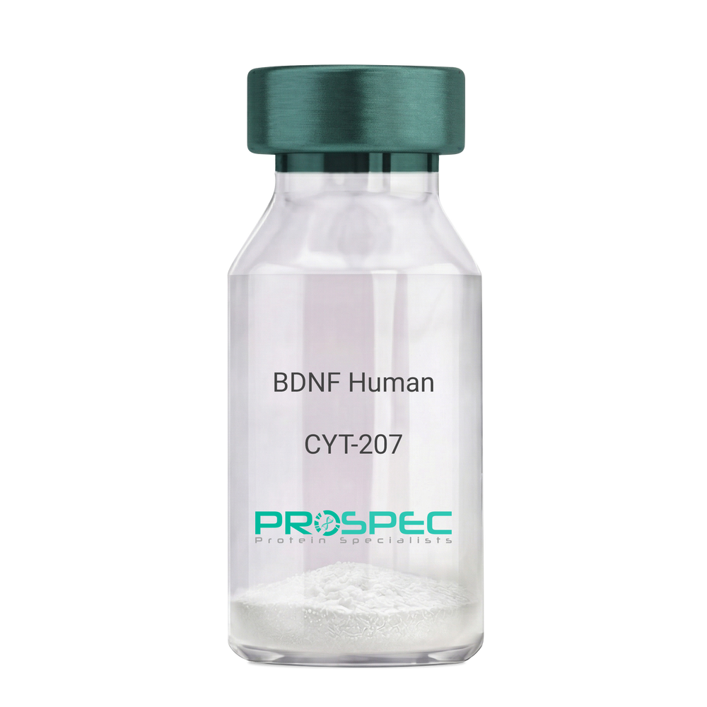
- View Data Sheet
Name :
BDNF HumanDescription:
Brain-Derived Neurotrophic Factor Human Recombinant
Brain-Derived Neurotrophic Factor, BDNF, MGC34632.
Product # :
CYT-207Price :
Quantity :
Shipping Method :
Shipped at Room temp
More Info
- description
- source
- formulation
- purity
- biological activity
- More Info
- Activity
- Citations
Description
BDNF Human Recombinant produced in E.Coli is a homodimer, non-glycosylated, polypeptide chain containing 2 x 119 amino acids (and an N-terminal Met) and having a total molecular mass of 28kDa. BDNF Human Recombinant is purified by proprietary chromatographic techniques.
Source
Escherichia Coli.
Formulation
The protein was lyophilized with 20mM PB and 400mM NaCl, pH 7.2.
Purity
BDNF is greater than 950% as determined SDS-PAGE.
Biological Activity
The activity was determined using Immobilized Human TrkB-His tag protein 2ug/ml (100 μl/well) for its binding to NHS-Biotin BDNF. The ED50 of was found to be ≤20ng/mLActivity
Citations
More Info
-
Introduction
BDNF promotes the survival of neuronal populations that are all located either in the central nervous system or directly connected to it. BDNF is a major regulator of synaptic transmission and plasticity at adult synapses in many regions of the cns. The versatility of BDNF is emphasized by its contribution to a range of adaptive neuronal responses including long-term potentiation (ltp), long-term depression (ltd), certain forms of short-term synaptic plasticity, as well as homeostatic regulation of intrinsic neuronal excitability.
-
Synonyms
Brain-Derived Neurotrophic Factor, BDNF, MGC34632.
-
Physical Appearance
Sterile Filtered White lyophilized (freeze-dried) powder.
-
Stability
Lyophilized BDNF although stable at room temperature for 3 weeks, should be stored desiccated below -18°C. Upon reconstitution BDNF should be stored at 4°C between 2-7 days and for future use below -18°C.For long term storage it is recommended to add a carrier protein (0.1% HSA or BSA).Please prevent freeze-thaw cycles.
-
Solubility
It is recommended to reconstitute the lyophilized BDNF in sterile 18M-cm H2O not less than 100µg/ml, which can then be further diluted to other aqueous solutions.
-
Amino Acid Sequence
HSDPARRGE LSVCDSISEW VTAADKKTAV DMSGGTVTVL EKVPVSKGQL KQYFYETKCN PMGYTKEGCR GIDKRHWNSQ CRTTQSYVRA LTMDSKKRIG WRFIRIDTSC VCTLTIKRGR.
-
Background
Final Thoughts
Although more research is needed on the safety and effectiveness of BDNF human recombinant, trials suggest that this laboratory-produced protein may be effective in managing and treating several neurological and psychiatric disorders. It's important for experts to stay up to date on the latest developments and research to learn more about potential risks and benefits.
What is the molecular weight/Mw of BDNF Protein?
BDNF Protein has a total Mw of 27kDa.
What is the source or expression system of BDNF Protein?
Escherichia Coli.
What is the Purity of BDNF Protein?
BDNF Protein is >97% pure as determined by SDS-PAGE.
What is the Biological Activity of BDNF Protein?
The ED50, as determined by the dose-dependent induction of C6 cells proliferation, is 1.3-2µg/ml.
What is the amino acid sequence of BDNF Protein?
MHSDPARRGE LSVCDSISEW VTAADKKTAV DMSGGTVTVL EKVPVSKGQL KQYFYETKCN PMGYTKEGCR GIDKRHWNSQ CRTTQSYVRA LTMDSKKRIG WRFIRIDTSC VCTLTIKRGR.
What applications can BDNF Protein be used in?
BDNF Protein can probably be used in western blot, ELISA and Lateral Flow.
What is the endotoxin level for BDNF Protein?
The endotoxin level is minimal, BDNF Protein was purified using conventional chromatography techniques.
-
Protein content
BDNF quantitation was carried out by two independent methods:1. UV spectroscopy at 280 nm using the absorbency value of 1.6 as the extinction coefficient for a 0.1% (1mg/ml) solution. This value is calculated by the PC GENE computer analysis program of protein sequences (IntelliGenetics). 2. Analysis by RP-HPLC, using a standard solution of Brain-derived Neurotrophic Factor as a Reference Standard.
ProSpec's products are furnished for LABORATORY RESEARCH USE ONLY. The product may not be used as drugs, agricultural or pesticidal products, food additives or household chemicals.
- View Data Sheet
Name :
Dengue NS1 Paired AntibodyDescription:
Mouse Anti Dengue NS1 Paired
Product # :
ANT-531Price :
Quantity :
Shipping Method :
Shipped with Ice Packs
More Info
- description
- formulation
- purity
- More Info
- Citations
Description
A paired monoclonal antibody has been developed for dengue NS1 antigen lateral flow immunoassay product. It is suggested to use 10µg/ml gold concentration (OD 1.2-1.5) to conjugate with clone 24/c, and 1.2µg/cm coating concentration for clone 2/b. The sensitivity is directly proportional to conjugated gold OD. The specimen used for the test is 80-90 µl. The rapid test developed by these 2 paired antibodies has 90% sensitivity for the samples with OD over 5 and 60% sensitivity for the sample with OD below 5 tested by ELISA. Please note that when ordering for example: 100µg antibody we ship 50µg from each of the antibodies (100µg in total).
Formulation
* Dengue NS1 Conjugation antibody (clone 24/c): (1.2mg/ml) in PBS and Sodium nitrate.
* Dengue NS1 Coating antibody (clone 2/b): (3.4mg/ml) in PBS and Sodium nitrate.Purity
Greater than 95%.
Citations
More Info
-
Introduction
Dengue NS1 is an early marker for dengue infection from the first day of fever and lasting about one week. Dengue IgM is usually detected early on the first day to 5 days of infection. Dengue NS1 could be detected from both primary and secondary infection for all four subtypes.
-
Physical Appearance
2 vials of sterile Filtered clear colorless solution.
-
Stability
Dengue NS1 Paired Antibody should be stored at 4°C.
-
Applications
Lateral flow immunoassay.
-
Assay Conditions
1. Fresh gold particle prepared within 1 to 2 days with maximal homogeneity in particle size, λ 525 to 5529, OD 1.2- 1.4. 2. Conjugation pH 8.5, conjugation time should be 6-8 hours. 3. Blocking condition: adjust OD to 8.0-8.2 for re-suspended ant-dengue NS1 conjugated colloid gold, add casein to the final 0.3-0.4%.
-
Type
Mouse antibody Monoclonal.
-
Purification Method
Purified monoclonal IgG by protein A chromatography.
ProSpec's products are furnished for LABORATORY RESEARCH USE ONLY. The product may not be used as drugs, agricultural or pesticidal products, food additives or household chemicals.
- View Data Sheet
Name :
CCN1 HumanDescription:
Cysteine-Rich Angiogenic Inducer 61 Human Recombinant
CYR61, Protein CYR61, Cysteine-rich angiogenic inducer 61, IGF-binding protein 10, IGFBP-10, IBP-10, Protein GIG1, CCN family member 1, CCN1, GIG1, IGFBP10.
Product # :
CYT-164Price :
Quantity :
Shipping Method :
Shipped at Room temp
More Info
- description
- source
- formulation
- purity
- biological activity
- More Info
- Citations
Description
CYR61 Human Recombinant produced in E.Coli is a single, non-glycosylated, polypeptide chain containing 357 amino acids and having a molecular mass of 39.5kDa.The CYR61 is purified by proprietary chromatographic techniques.
Source
Escherichia Coli.
Formulation
Lyophilized from a 0.2m filtered concentrated solution in PBS, pH 7.4.
Purity
Greater than 95.0% as determined by SDS-PAGE.
Biological Activity
The ED50 was determined by the proliferation of mouse 3T3 cells is < 2.0 ug/ml, corresponding to a specific activity of > 500 units/mg.Citations
More Info
-
Introduction
CYR61 is a growth factor-inducible, immediate-early gene that has multifaceted activities in various cancers. CYR61 is a secreted, cysteine-rich, binding protein which is encoded by a growth factor-inducible immediate-early gene. Acting as an extracellular, matrix-associated signaling molecule, CYR61 promotes the adhesion of endothelial cells through interaction with integrin and enhances growth factor-induced DNA synthesis in the same cell type.
-
Synonyms
CYR61, Protein CYR61, Cysteine-rich angiogenic inducer 61, IGF-binding protein 10, IGFBP-10, IBP-10, Protein GIG1, CCN family member 1, CCN1, GIG1, IGFBP10.
-
Physical Appearance
Sterile Filtered White lyophilized (freeze-dried) powder.
-
Stability
Lyophilized CYR61 Human although stable at room temperature for 3 weeks, should be stored desiccated below -18°C. Upon reconstitution CYR61 should be stored at 4°C between 2-7 days and for future use below -18°C.Please prevent freeze-thaw cycles.
-
Solubility
It is recommended to reconstitute the lyophilized CYR61 in sterile 18M-cm H2O not less than 100µg/ml, which can then be further diluted to other aqueous solutions.
-
Amino Acid Sequence
TCPAACHCPL EAPKCAPGVG LVRDGCGCCK VCAKQLNEDC SKTQPCDHTK GLECNFGASS TALKGICRAQ SEGRPCEYNS RIYQNGESFQ PNCKHQCTCI DGAVGCIPLC PQELSLPNLG CPNPRLVKVT GQCCEEWVCD EDSIKDPMED QDGLLGKELG FDASEVELTR NNELIAVGKG SSLKRLPVFG MEPRILYNPL QGQKCIVQTT SWSQCSKTCG TGISTRVTND NPECRLVKET RICEVRPCGQ PVYSSLKKGK KCSKTKKSPE PVRFTYAGCL SVKKYRPKYC GSCVDGRCCT
PQLTRTVKMR FRCEDGETFS KNVMMIQSCK CNYNCPHANE AAFPFYRLFN DIHKFRD -
Background
Title: Cysteine-Rich Angiogenic Inducer 61 Human Recombinant: A Potential Regulator of Angiogenesis
Abstract:
Cysteine-rich angiogenic inducer 61 (CYR61) is an important extracellular matrix-associated protein that plays a significant role in angiogenesis and cell adhesion. This research paper provides a comprehensive analysis of human recombinant CYR61, focusing on its production, characterization, and potential applications in regulating angiogenesis. The paper discusses the significance of CYR61 in physiological and pathological angiogenesis, including wound healing, tumor development, and cardiovascular diseases. Furthermore, it explores ongoing research and clinical trials investigating the therapeutic potential of recombinant CYR61 in various pathological conditions. The information presented in this paper aims to enhance our understanding of human recombinant CYR61 and its utility as a research tool and a potential regulator of angiogenesis.Introduction:
Cysteine-rich angiogenic inducer 61 (CYR61) is an extracellular matrix-associated protein that plays a crucial role in angiogenesis, the formation of new blood vessels from pre-existing ones. Human recombinant CYR61, produced through genetic engineering techniques, provides researchers with a valuable tool to study its biological functions and explore its therapeutic potential.Production and Characterization:
Recombinant CYR61 is typically generated using expression systems such as mammalian cells or bacteria. The protein is then purified and characterized to ensure its structural integrity and functional activity. Quality control measures are implemented to confirm the specificity and bioactivity of the recombinant CYR61.Role in Angiogenesis:
CYR61 is involved in various aspects of angiogenesis, including endothelial cell proliferation, migration, and tube formation. It interacts with integrins and other cell surface receptors to modulate signaling pathways involved in angiogenic processes. Recombinant CYR61 serves as a valuable tool for studying the mechanisms underlying angiogenesis and exploring its potential as a therapeutic target.Therapeutic Implications:
The dysregulation of angiogenesis is associated with several pathological conditions, including cancer, cardiovascular diseases, and chronic wounds. Recombinant CYR61 has shown promise as a potential regulator of angiogenesis and a therapeutic agent. It can be used to promote or inhibit angiogenesis, depending on the specific context. Ongoing research and clinical trials are investigating the therapeutic applications of recombinant CYR61 in various diseases, including cancer and ischemic disorders.Conclusion:
Human recombinant CYR61 is a valuable research tool and a potential regulator of angiogenesis. Its production, characterization, and applications in modulating angiogenic processes contribute to our understanding of angiogenesis and the development of novel therapeutic interventions. Continued research and clinical trials exploring the therapeutic potential of recombinant CYR61 offer promising prospects for improving outcomes in cancer, cardiovascular diseases, and wound healing.What is the molecular weight/Mw of CCN1 Protein?
CCN1 Protein has a total Mw of 39.5kDa.
What is the source or expression system of CCN1 Protein?
Escherichia Coli.
What is the Purity of CCN1 Protein?
CCN1 Protein is >95% pure as determined by SDS-PAGE.
What is the Biological Activity of CCN1 Protein?
The ED50 was determined by the proliferation of mouse 3T3 cells is < 2.0 ug/ml, corresponding to a specific activity of > 500 units/mg.
What is the amino acid sequence of CCN1 Protein?
TCPAACHCPL EAPKCAPGVG LVRDGCGCCK VCAKQLNEDC SKTQPCDHTK GLECNFGASS TALKGICRAQ SEGRPCEYNS RIYQNGESFQ PNCKHQCTCI DGAVGCIPLC PQELSLPNLG CPNPRLVKVT GQCCEEWVCD EDSIKDPMED QDGLLGKELG FDASEVELTR NNELIAVGKG SSLKRLPVFG MEPRILYNPL QGQKCIVQTT SWSQCSKTCG TGISTRVTND NPECRLVKET RICEVRPCGQ PVYSSLKKGK KCSKTKKSPE PVRFTYAGCL SVKKYRPKYC GSCVDGRCCT
PQLTRTVKMR FRCEDGETFS KNVMMIQSCK CNYNCPHANE AAFPFYRLFN DIHKFRD
What applications can CCN1 Protein be used in?
CCN1 Protein can probably be used in western blot, ELISA and Lateral Flow.
What is the endotoxin level for CCN1 Protein?
The endotoxin level is minimal, CCN1 Protein was purified using conventional chromatography techniques.
ProSpec's products are furnished for LABORATORY RESEARCH USE ONLY. The product may not be used as drugs, agricultural or pesticidal products, food additives or household chemicals.
- View Data Sheet
Name :
CXCL8 Human, PichiaDescription:
Interleukin-8 (1-77 a.a.) Human Recombinant, (CXCL8) Pichia
IL-8, CXCL8, Monocyte-derived neutrophil chemotactic factor, MDNCF, T-cell chemotactic factor, Neutrophil-activating protein 1, NAP-1, Protein 3-10C, Granulocyte chemotactic protein 1, GCP-1, Monocyte-derived neutrophil-activating peptide, MONAP, Emoctakin, K60, NAF, LECT, LUCT, 3-10C, LYNAP, SCYB8, TSG-1, AMCF-I, b-ENAP.
Product # :
CHM-349Price :
Quantity :
Shipping Method :
Shipped at Room temp
More Info
- description
- source
- formulation
- purity
- biological activity
- More Info
- Citations
Description
Interleukin-8 Human Recombinant produced in Yeast is a single, glycosylated polypeptide chain containing 79 amino acids and having a molecular mass of 9 kDa. The IL-8 is purified by proprietary chromatographic techniques.
Source
Pichia Pastoris.
Formulation
Lyophilized from a concentrated (1mg/ml) solution in water containing 20mM sodium phosphate buffer pH-8.
Purity
Greater than 98.0% as determined by SDS-PAGE.
Biological Activity
Chemotactic activity was reached at 25ng/ml on human neutrophils.
Citations
More Info
-
Introduction
Interleukin-8 (IL-8) is a chemokine produced by macrophages and other cell types such as epithelial cells. It is also synthesized by endothelial cells, which store IL-8 in their storage vesicles, the Weibel-Palade bodies.
When first encountering an antigen, the primary cells to encounter it are the macrophages who phagocytose the particle. Upon processing, they release chemokines to signal other immune cells to come in to the site of inflammation. IL-8 is one such chemokine. It serves as a chemical signal that attracts neutrophils at the site of inflammation, and therefore is also known as Neutrophil Chemotactic Factor. -
Synonyms
IL-8, CXCL8, Monocyte-derived neutrophil chemotactic factor, MDNCF, T-cell chemotactic factor, Neutrophil-activating protein 1, NAP-1, Protein 3-10C, Granulocyte chemotactic protein 1, GCP-1, Monocyte-derived neutrophil-activating peptide, MONAP, Emoctakin, K60, NAF, LECT, LUCT, 3-10C, LYNAP, SCYB8, TSG-1, AMCF-I, b-ENAP.
-
Physical Appearance
Sterile Filtered White lyophilized (freeze-dried) powder.
-
Stability
Lyophilized Interleukin-8 although stable at room temperature for 3 weeks, should be stored desiccated below -18°C. Upon reconstitution CXCL8 should be stored at 4°C between 2-7 days and for future use below -18°C.For long term storage it is recommended to add a carrier protein (0.1% HSA or BSA).Please prevent freeze-thaw cycles.
-
Solubility
It is recommended to reconstitute the lyophilized Interleukin 8 in sterile 18MΩ-cm H2O not less than 100µg/ml, which can then be further diluted to other aqueous solutions.
-
Background
What is the molecular weight/Mw of CXCL8 HUMAN, PICHIA Protein?
CXCL8 HUMAN, PICHIA Protein has a total Mw of 9kDa.
What is the source or expression system of CXCL8 HUMAN, PICHIA Protein?
Pichia Pastoris.
What is the Purity of CXCL8 HUMAN, PICHIA Protein?
CXCL8 HUMAN, PICHIA Protein is >98% pure as determined by SDS-PAGE.
What is the Biological Activity of CXCL8 HUMAN, PICHIA Protein?
Chemotactic activity was reached at 25ng/ml on human neutrophils.
What is the amino acid sequence of CXCL8 HUMAN, PICHIA Protein?
CXCL8 HUMAN, PICHIA Protein is composed from 79 amino acids.
What applications can CXCL8 HUMAN, PICHIA Protein be used in?
CXCL8 HUMAN, PICHIA Protein can probably be used in western blot, ELISA and Lateral Flow.
What is the endotoxin level for CXCL8 HUMAN, PICHIA Protein?
The endotoxin level is minimal, CXCL8 HUMAN, PICHIA Protein was purified using conventional chromatography techniques.
Who supplies recombinant CXCL8 HUMAN, PICHIA that is in stock and available to order?
ProSpec has the protein in stock and available for immediate delivery.
Where can I order bulk mg quantities of CXCL8 HUMAN, PICHIA?
ProSpec supplies bulk mg quantities at affordable prices that meets research and academic budgets.
ProSpec's products are furnished for LABORATORY RESEARCH USE ONLY. The product may not be used as drugs, agricultural or pesticidal products, food additives or household chemicals.
- View Data Sheet
Name :
MEMO1 AntibodyDescription:
Mediator of Cell Motility 1, Mouse Anti Human
Protein MEMO1, C21orf19-like protein, Hepatitis C virus NS5A-transactivated protein 7, HCV NS5A-transactivated protein 7, Mediator of ErbB2-driven cell motility 1, Mediator of cell motility 1, Memo-1, MEMO1, C2orf4, MEMO, NS5ATP7, CGI-27.
Product # :
ANT-090Price :
Quantity :
Shipping Method :
Shipped with Ice Packs
More Info
- formulation
- More Info
- Citations
Formulation
1mg/ml containing PBS, pH-7.4, & 0.1% Sodium Azide.
Citations
More Info
-
Introduction
Mediator of ErbB2-driven cell motility 1 (MEMO1) is a member of the UPF0103 family. MEMO1 regulates cell migration by transmitting extracellular chemotactic signals to the microtubule cytoskeleton. Furthermore, MEMO1 controls the localization of APC and CLASP2 to the cell membrane, using the regulation of GSK3B activity. MEMO1 is essential for breast carcinoma cell migration, suggesting a key role in tumorigenesis. MEMO1 is also a mediator of ERBB2 signaling.
-
Synonyms
Protein MEMO1, C21orf19-like protein, Hepatitis C virus NS5A-transactivated protein 7, HCV NS5A-transactivated protein 7, Mediator of ErbB2-driven cell motility 1, Mediator of cell motility 1, Memo-1, MEMO1, C2orf4, MEMO, NS5ATP7, CGI-27.
-
Immunogen
Anti-human MEMO1 mAb, is derived from hybridization of mouse FO myeloma cells with spleen cells from BALB/c mice immunized with recombinant human MEMO1 amino acids 1-297 purified from E. coli.
-
Ig Subclass
Mouse IgG1 heavy chain and kappa light chain.
-
Clone
P1E9AT.
-
Type
Mouse Anti Human Monoclonal.
-
Storage Procedures
For periods up to 1 month store at 4°C, for longer periods of time, store at -20°C. Prevent freeze thaw cycles.
-
Purification Method
MEMO1 antibody was purified from mouse ascitic fluids by protein-G affinity chromatography.
ProSpec's products are furnished for LABORATORY RESEARCH USE ONLY. The product may not be used as drugs, agricultural or pesticidal products, food additives or household chemicals.
- View Data Sheet
Name :
Visfatin HumanDescription:
Visfatin Human Recombinant
PBEF, Pre-B cell colony-enhancing factor, Nicotinamide phosphoribosyltransferase NAmPRTase, Nampt, MGC117256, DKFZP666B131, 1110035O14Rik.
Product # :
CYT-318Price :
Quantity :
Shipping Method :
Shipped at Room temp
More Info
- description
- source
- formulation
- purity
- biological activity
- More Info
- Citations
Description
Visfatin Human Recombinant produced in E.Coli is a single, non-glycosylated, polypeptide chain containing 466 amino acids. The total molecular mass is 52.6kDa (calculated). The Visfatin is purified by Flag-affinity chromatography.
Source
Escherichia Coli.
Formulation
Visfatin was lyophilized with no additives.
Purity
Greater than 90.0% as determined by SDS-PAGE.
Biological Activity
The activity is determined by its ability to induce IL-6, IL-1 beta and TNF alpha production from human PBMCs at 100ng/ml.Citations
More Info
-
Introduction
Excess adiposity is the most important risk in the development of type 2 diabetes mellitus (T2DM). Adipose tissue produces several proteins (adipocytokines) such as leptin, adiponectin, resistin, tumor necrosis factor-a, and IL-6, that modulate sensitivity and appear to play an important role in the pathogenesis, diabetes, dyslipidemia, inflammation, and atherosclerosis. Visfatin, also known as pre-B cell colony-enhancing factor (PBEF), is a cytokine that is highly expressed in visceral fat and was originally isolated as a secreted factor that synergizes with IL-7 and stem cell factors to promote the growth of B cell precursors. Visfatin homologs have been identified in carp, invertebrate mollusks, and bacteria, as well as in vertebrates, including humans and the mouse. It has been postulated to play a role in innate immunity.
Visfatin exerts mimetic effects that are dose-dependent and quantitatively similar to stimulating muscle and adipocyte glucose transport, and in inhibiting hepatocyte glucose production. Intravenous injection of recombinant visfatin in mice decreased plasma glucose in a dose-dependent fashion. In keeping with its mimetic effects, visfatin was as effective in reducing hyperglycemia in deficient diabetic mice. Visfatin was also found to be bound to and activate receptor, causing receptor phosphorylation and the activation of downstream signaling molecules. However, visfatin did not compete for binding to the receptor, indicating that the two proteins were recognized by different regions of the receptor. Thus, visfatin might play a role in glucose homeostasis and dysregulation in biosynthesis or signal transduction, and might contribute to the pathogenesis of diabetes. -
Synonyms
PBEF, Pre-B cell colony-enhancing factor, Nicotinamide phosphoribosyltransferase NAmPRTase, Nampt, MGC117256, DKFZP666B131, 1110035O14Rik.
-
Physical Appearance
Filtered White lyophilized (freeze-dried) powder.
-
Stability
Lyophilized Visfatin although stable at room temperature for 3 weeks, should be stored desiccated below -18°C. Upon reconstitution Visfatin should be stored at 4°C between 2-7 days and for future use below -18°C.For long term storage it is recommended to add a carrier protein (0.1% HSA or BSA).Please prevent freeze-thaw cycles.
-
Solubility
Centrifuge vial before opening. When reconstituting the product, gently pipet and wash down the sides of the vial to ensure full recovery of the protein into solution. It is recommended to reconstitute the lyophilized product with 20 mM HCl at a concentration of 0.1 mg/mL, which can be further diluted into other aqueous solutions. Wait several minutes for full reconstitution and solubility.
-
Amino Acid Sequence
MPPNTSKVYS YFECREKKTE NSKLRKVKYE ETVFYGLQYI LNKYLKGKVV TKEKIQEAKD VYKEHFQDDV FNEKGWNYIL EKYDGHLPIE IKAVPEGFVI PRGNVLFTVE NTDPECYWLT NWIETILVQS WYPITVATNS REQKKILAKY LLETSGNLDG LEYKLHDFGY RGVSSQETAG IGASAHLVNF KGTDTVAGLA LIKKYYGTKD PVPGYSVPAA EHSTITAWGK DHEKDAFEHI VTQFSSVPVS VVSDSYDIYN ACEKIWGEDL RHLIVSRSTQ APLIIRPDSG NPLDTVLKVL EILGKKFPVT ENSKGYKLLP PYLRVIQGDG VDINTLQEIV EGMKQKMWSI ENIAFGSGGG LLQKLTRDLL NCSFKCSYVV TNGLGINVFK DPVADPNKRS KKGRLSLHRT PAGNFVTLEE GKGDLEEYGQ DLLHTVFKNG KVTKSYSFDE IRKNAQLNIE LEAAHH.
-
Background
About Visfatin Human
Visfatin is a cytokine expressed in visceral fat that was originally isolated as a secreted
element that synergized with stem cell factors and IL-7. One of its main functions is to
enhance the development of B cell precursors.The cytokine is also known as the “Pre-B Cell Colony-Enhancing Factor (PBEF).” It has been
identified in vertebrates, including mice and humans, and it’s being studied due to its link
to inflammatory conditions, beta cell function, and cardiovascular disease.
What’s the Function of Visfatin Human Recombinant?Visfatin human recombinant is produced in E. Coli. It’s a single, non-glycosylated,
polypeptide chain that contains 466 amino acids, it’s purified by FLAG-affinity
chromatography, and it contains a total molecular mass of 52.6 kDa.
What Are the Main Applications of Visfatin Human Recombinant?The cytokine is being researched because of its involvement in glucose homeostasis,
dysregulation in biosynthesis and signal transduction, and the pathogenesis of diabetes.
Visfatin human recombinant is tailored exclusively for laboratory research, ensuring
experts can get further answers regarding the cytokine’s involvement in different
processes, including pathogenesis, diabetes, inflammation, dyslipidemia, and
atherosclerosis.Findings can also help during the identification of high-risk people for cardiovascular
disease and diabetes.
ProSpec's products are furnished for LABORATORY RESEARCH USE ONLY. The product may not be used as drugs, agricultural or pesticidal products, food additives or household chemicals.
- View Data Sheet
Name :
Vimentin AntibodyDescription:
Vimentin, Mouse Anti Human Antibody
Product # :
ANT-245Price :
Quantity :
Shipping Method :
Shipped at Room temp
More Info
- formulation
- More Info
- Citations
Formulation
1mg/ml in PBS (after reconstitution).
Citations
More Info
-
Introduction
Vimentin expression in human malignant glioma cells depends on cellular density, algorithms of drug delivery and chemo/radio treatment. Vimentin and detyrosinated microtubules provide structural support for the extensive microtentacles observed in detached tumor cells and a mechanism to promote successful metastatic spread. Primary colorectal carcinomas display aberrant expression of vimentin, and have activated Notch and TGFbeta signaling pathways. Vimentin is a strong arterial substrate for transglutaminases. Transglutaminase-mediated vimentin dimerization results in a novel unifying pathway by which vasodilatory and remodeling responses may be regulated. Ablation of vimentin expression inhibits migration and invasion of colon and breast cancer cell lines.
-
Solubility
Reconstitute with H20. Mix gently, wash the sides of the vial and wait 30-60 seconds before use.
-
Immunogen
Purified Human Vimentin.
-
Ig Subclass
mouse IgG1.
-
Clone
hVimentin.
-
Applications
Direct ELISA, Western Blot, Immuneprecipitation, immunohistochemistry.
-
Cross reactivity
Vimentin antibody will recognize most mammalian Vimentin except mouse and rat.
-
Titer
For intra-cellular staining, use 5-10µl/1,000,000 cells. Wash twice with PBS/Azide before adding the appropriate amount of secondary fluorescent reagent.
-
Shipping Conditions
Antibody is shipped lyophilized at ambient temperature.
-
Available Conjugates
This antibody is also available conjugated to biotin and FITC.
-
Type
Mouse Anti Human Monoclonal Antibody.
-
Storage Procedures
In lyophilized form, for long periods, store at 4°C in a dry environment. After reconstitution, if not intended for use within a month, aliquot and store at -20°C.
-
Purification Method
Ion exchange column.
ProSpec's products are furnished for LABORATORY RESEARCH USE ONLY. The product may not be used as drugs, agricultural or pesticidal products, food additives or household chemicals.
- View Data Sheet
Name :
FAS Blocking AntibodyDescription:
FAS Blocking Antibody (CD95), Mouse anti Human
FASLG receptor, Apoptosis-mediating surface antigen FAS, Apo-1 antigen, CD95, Tumor necrosis factor receptor superfamily member 6 TNR6, APT1, FAS1, TNFRSF6.
Product # :
ANT-205Price :
Quantity :
Shipping Method :
Shipped at Room temp
More Info
- More Info
- Citations
Citations
More Info
-
Introduction
The Fas receptor (CD95) mediates apoptotic signaling by Fas-ligand expressed on the surface of other cells. The Fas-FasL interaction plays an important role in the immune system and lack of this system leads to autoimmunity, indicating that Fas-mediated apoptosis removes self-reactive lymphocytes. Fas signaling is also involved in immune surveillance to remove transformed cells and virus infected cells. Binding of FAS to oligimerized FasL on another cell activates apoptotic signaling through a cytoplasmic domain termed the death domain that interacts with signaling adaptors including FAF, FADD and DAX to activate the caspase proteolytic cascade. Caspase-8 and caspase-10 are first activated, to then cleave and activate downstream caspases, and a variety of cellular substrates that lead to cell death. Caspases cleave nuclear lamins, causing the nucleus to break down and lose its normal structure and another caspase substrate is DFF, inducing cleavage and degradation of the genome. Other caspase substrates are involved in cytoskeletal structure, cell cycle regulation and signaling pathways. Activation of JNK kinase, activation of Jun, and production of ceramide may also play roles in Fas-mediated apoptosis. Activation of fas-mediated apoptosis is opposed by I-FLICE and FAP. Viruses and tumors may escape immune surveillance in part through suppression of fas-mediated apoptosis using similar mechanisms.
-
Synonyms
FASLG receptor, Apoptosis-mediating surface antigen FAS, Apo-1 antigen, CD95, Tumor necrosis factor receptor superfamily member 6 TNR6, APT1, FAS1, TNFRSF6.
-
Solubility
Reconstitute with H20. Mix gently, wash the sides of the vial and wait 30-60 seconds before use.
-
Immunogen
Recombinant Human FAS.
-
Ig Subclass
mouse IgG1.
-
Clone
NYRhFAS.
-
Note
This is a BLOCKING antibody and will block FAS-mediated apoptosis.
-
Titer
By direct ELISA, 1:10,000 dilution will yield 0.5 O.D using alkaline phosphatase conjugated rabbit anti-mouse Ig (Jackson Laboratories).
-
Shipping Conditions
Antibody is shipped lyophilized at ambient temperature.
-
Type
Mouse Anti Human Monoclonal.
-
Storage Procedures
In lyophilized form, for long periods, store at 4°C in a dry environment. After reconstitution, if not intended for use within a month, aliquot and store at -20°C.
-
Purification Method
Ion exchange column Protein concentration1 mg/ml in PBS (after reconstitution).
ProSpec's products are furnished for LABORATORY RESEARCH USE ONLY. The product may not be used as drugs, agricultural or pesticidal products, food additives or household chemicals.
- View Data Sheet
Name :
IL1A Rat, HisDescription:
Interleukin-1 alpha Rat Recombinant, His Tag
Interleukin-1 alpha, IL-1 alpha.
Product # :
CYT-913Price :
Quantity :
Shipping Method :
Shipped with Ice Packs
More Info
- description
- source
- formulation
- purity
- biological activity
- More Info
- Citations
Description
IL 1 alpha Rat Recombinant produced in E.Coli is a single, non-glycosylated polypeptide chain containing 179 amino acids (115-270 a.a) and having a molecular mass of 20.2kDa. IL 1 alpha is fused to a 23 amino acid His-tag at N-terminus & purified by proprietary chromatographic techniques.
Source
Escherichia Coli.
Formulation
IL 1 alpha protein solution (1mg/ml) containing Phosphate Buffered Saline (pH 7.4) and 10% glycerol.
Purity
Greater than 95.0% as determined by SDS-PAGE.
Biological Activity
The biological activity was Rat IL1 alpha was measured in a cell proliferation assay using D10.G4.1 mouse helper T cell. The ED50 for this effect is less or equal to 20 pg/ml.
Citations
More Info
-
Introduction
IL-1 alpha is produced by activated macrophages, stimulates thymocyte proliferation by inducing il-2 release, b-cell maturation and proliferation, and fibroblast growth factor activity. IL1A proteins are involved in the inflammatory response, being identified as endogenous pyrogens, and are reported to stimulate the release of prostaglandin and collagenase from synovial cells.
-
Synonyms
Interleukin-1 alpha, IL-1 alpha.
-
Physical Appearance
Sterile Filtered colorless solution.
-
Stability
Store at 4°C if entire vial will be used within 2-4 weeks. Store, frozen at -20°C for longer periods of time. For long term storage it is recommended to add a carrier protein (0.1% HSA or BSA). Avoid multiple freeze-thaw cycles.
-
Amino Acid Sequence
MGSSHHHHHH SSGLVPRGSH MGSSAPHSFQ NNLRYKLIRI VKQEFIMNDS LNQNIYVDMD RIHLKAASLN DLQLEVKFDM YAYSSGGDDS KYPVTLKVSN TQLFVSAQGE DKPVLLKEIP ETPKLITGSE TDLIFFWEKI NSKNYFTSAA FPELLIATKE QSQVHLARGL PSMIDFQIS.
-
Background
What is the molecular weight/Mw of IL1A RAT, HIS Protein?
IL1A RAT, HIS Protein has a total Mw of 20.2kDa.
What is the source or expression system of IL1A RAT, HIS Protein?
Escherichia Coli.
What is the Purity of IL1A RAT, HIS Protein?
IL1A RAT, HIS Protein is >95% pure as determined by SDS-PAGE.
What is the Biological Activity of IL1A RAT, HIS Protein?
The biological activity was Rat IL1 alpha was measured in a cell proliferation assay using D10.G4.1 mouse helper T cell. The ED50 for this effect is less or equal to 20 pg/ml.
What is the amino acid sequence of IL1A RAT, HIS Protein?
MGSSHHHHHH SSGLVPRGSH MGSSAPHSFQ NNLRYKLIRI VKQEFIMNDS LNQNIYVDMD RIHLKAASLN DLQLEVKFDM YAYSSGGDDS KYPVTLKVSN TQLFVSAQGE DKPVLLKEIP ETPKLITGSE TDLIFFWEKI NSKNYFTSAA FPELLIATKE QSQVHLARGL PSMIDFQIS.
What applications can IL1A RAT, HIS Protein be used in?
IL1A RAT, HIS Protein can probably be used in western blot, ELISA and Lateral Flow.
What is the endotoxin level for IL1A RAT, HIS Protein?
The endotoxin level is minimal, IL1A RAT, HIS Protein was purified using conventional chromatography techniques.
ProSpec's products are furnished for LABORATORY RESEARCH USE ONLY. The product may not be used as drugs, agricultural or pesticidal products, food additives or household chemicals.