Search results
1000 results found for “lyase”
Name
Description
Product #
Price
Quantity
Shipping Method
- View Data Sheet
Name :
GLRX1 YeastDescription:
Glutaredoxin 1 Yeast Recombinant
Thioltransferase, GRX, GLRX1, GRX1, GRX-1, GLRX-1, Glutathione-dependent oxidoreductase 1, Glutaredoxin 1.
Product # :
ENZ-361Price :
Quantity :
Shipping Method :
Shipped with Ice Packs
More Info
- description
- source
- formulation
- purity
- More Info
- Citations
Description
Glutaredoxin Saccharamyces cerevisiae Recombinant containing 6x His tag at C-Terminus produced in E.Coli is a single, non-glycosylated, Polypeptide chain having a molecular mass of 16 kDa.
Source
Escherichia Coli.
Formulation
Glutaredoxin solution contains PBS, pH-7.5 & 0.01% Na Azide.
Purity
Greater than 95% as determined by SDS-PAGE.
Citations
More Info
-
Introduction
GLRX1 has a glutathione-disulfide oxidoreductase activity in the presence of nadph and glutathione reductase. reduces low molecular weight disulfides and proteins. Glutaredoxin is a glutathione (GSH)-dependent hydrogen donor for ribonucleotide reductase and also catalyzes glutathione-disulfide oxidoreduction reactions in the presence of NADPH and glutathione reductase. GLRX1 is multifunctional enzyme with glutathione-dependent oxidoreductase, glutathione peroxidase and glutathione S-transferase (GST) activity. The disulfide bond functions as an electron carrier in the glutathione-dependent synthesis of deoxyribonucleotides by the enzyme ribonucleotide reductase. In addition, it is also involved in reducing cytosolic protein- and non-protein-disulfides in a coupled system with glutathione reductase. Required for resistance to reactive oxygen species (ROS) by directly reducing hydroperoxides and for the detoxification of ROS-mediated damage.
-
Synonyms
Thioltransferase, GRX, GLRX1, GRX1, GRX-1, GLRX-1, Glutathione-dependent oxidoreductase 1, Glutaredoxin 1.
-
Physical Appearance
Sterile Filtered clear colorless solution.
-
Stability
1 week at 2-10°C. For long term store at -20 to -80°C.
ProSpec's products are furnished for LABORATORY RESEARCH USE ONLY. The product may not be used as drugs, agricultural or pesticidal products, food additives or household chemicals.
- View Data Sheet
Name :
RNASEH2A E.ColiDescription:
Ribonuclease H2A E.Coli Recombinant
AGS4, JUNB, RNASEHI, RNHIA, RNHL , Ribonuclease H2 subunit A, Aicardi-Goutieres syndrome 4 protein, RNase H(35), Ribonuclease HI large subunit, RNase HI large subunit, EC=3.1.26.4.
Product # :
ENZ-713Price :
Quantity :
Shipping Method :
Shipped with Ice Packs
More Info
- description
- source
- formulation
- purity
- More Info
- Citations
Description
RNASEH2A Recombinant produced in E.coli is a single, non-glycosylated polypeptide chain containing 322 amino acids (1-299) and having a molecular mass of 35.8kDa.RNASEH2A is fused to a 23 amino acid His-tag at N-terminus & purified by proprietary chromatographic techniques.
Source
Escherichia Coli.
Formulation
The RNASEH2A solution (0.5mg/ml) contains 20mM Tris-HCl buffer (pH 8.0), 0.4M Urea and 10% glycerol.
Purity
Greater than 85% as determined by SDS-PAGE.
Citations
More Info
-
Introduction
Ribonuclease H2 subunit A (RNASEH2A) is a member of the RNase HII family and eukaryotic subfamily. RNASEH2A Plays a part in DNA replication, probably by mediating the removal of lagging-strand Okazaki fragment RNA primers throughout DNA replication. RNASEH2A catalyzes the endonucleolytic cleavage of RNA to a 5’-phosphomonoester and is capable to bind magnesium or manganese as cofactors. Aicardi-Goutieres syndrome type 4 (AGS4) caused by defects in RNASEH2A.
-
Synonyms
AGS4, JUNB, RNASEHI, RNHIA, RNHL , Ribonuclease H2 subunit A, Aicardi-Goutieres syndrome 4 protein, RNase H(35), Ribonuclease HI large subunit, RNase HI large subunit, EC=3.1.26.4.
-
Physical Appearance
Sterile filtered colorless solution.
-
Stability
Store at 4°C if entire vial will be used within 2-4 weeks. Store, frozen at -20°C for longer periods of time. For long term storage it is recommended to add a carrier protein (0.1% HSA or BSA).Avoid multiple freeze-thaw cycles.
-
Amino Acid Sequence
MGSSHHHHHH SSGLVPRGSH MGSMDLSELE RDNTGRCRLS SPVPAVCRKE PCVLGVDEAG RGPVLGPMVY AICYCPLPRL ADLEALKVAD SKTLLESERE RLFAKMEDTD FVGWALDVLS PNLISTSMLG RVKYNLNSLS HDTATGLIQY ALDQGVNVTQ VFVDTVGMPE TYQARLQQSF PGIEVTVKAK ADALYPVVSA ASICAKVARD QAVKKWQFVE KLQDLDTDYG SGYPNDPKTK AWLKEHVEPV FGFPQFVRFS WRTAQTILEK EAEDVIWEDS ASENQEGLRK ITSYFLNEGS QARPRSSHRY FLERGLESAT SL.
ProSpec's products are furnished for LABORATORY RESEARCH USE ONLY. The product may not be used as drugs, agricultural or pesticidal products, food additives or household chemicals.
- View Data Sheet
Name :
AminopeptidaseDescription:
Aminopeptidase Aeromonas Recombinant
Bacterial leucyl aminopeptidase, EC 3.4.11.10.
Product # :
ENZ-275Price :
Quantity :
Shipping Method :
Shipped with Ice Packs
More Info
- description
- source
- formulation
- purity
- biological activity
- More Info
- Citations
Description
The 29 kDa Aeromonas Aminopeptidase is produced by genetic engineering and can be used for physical & structural investigations, sequence and amino-terminal determinations. This exopeptidase recognizes a specific stop sign at –X- Pro and requires a free a-amino group in the L-configuration. It is therefore suitable for the removal of the redundant N-terminal methionine often added to engineered recombinant proteins.
Source
Aeromonas Proteolytica.
Formulation
Buffered solution containing 10mM Tris-HCl, 100mM NaCl and 5µM ZnSO4, pH 8.0.
Purity
Greater than 95.0% as determined by SEC-HPLC.
Biological Activity
Recombinant Aeromonas Aminopeptidase was found to have an activity of 108 Units/mg protein.
Citations
More Info
-
Synonyms
Bacterial leucyl aminopeptidase, EC 3.4.11.10.
-
Physical Appearance
Sterile filtered liquid formulation.
-
Stability
Two years when stored at -20°C, 2 weeks at 4°C.
-
Unit Definition
One unit of aminopeptidase activity is defined as the amount of enzyme that releases 1 μmole p-nitroaniline at 25°C in 1 minute.
ProSpec's products are furnished for LABORATORY RESEARCH USE ONLY. The product may not be used as drugs, agricultural or pesticidal products, food additives or household chemicals.
- View Data Sheet
Name :
UricaseDescription:
Urate Oxidase Recombinant
Urate Oxidase, Uricase, Urate Oxygen, Oxidoreductase, UOX, UO, EC 1.7.3.
Product # :
ENZ-312Price :
Quantity :
Shipping Method :
Shipped at Room temp
More Info
- description
- source
- formulation
- purity
- biological activity
- More Info
- Citations
Description
Urate Oxidase Recombinant produced in E.Coli is a tetrameric, non-glycosylated polypeptide chain containing 302 amino acids, having a molecular formula of C1523H2383N417O462S7 and a molecular mass of 34,247 Dalton.The cDNA coding for urate-oxidase was cloned from a strain of Aspergillus flavus . The monomer protein has no intra- or inter-disulfide bridges.
Source
Escherichia Coli.
Formulation
Each 1.5mg Uricase contains 5mg sucrose, 25mg glycine, 0.1mg Tween-80, 13.6 mg Na2HPO4*12H20 and 0.33 mg NaH2PO4*2H20.
Purity
Greater than 90.0% as determined by SDS-PAGE.
Biological Activity
The specific activity was found to be 10U/mg.
One Unit oxidizes one micromole of uric acid per minute at 25°C, at pH 8.5.Citations
More Info
-
Introduction
Urate oxidase catalyzes the enzymatic oxidation (degrades) of uric acid into allantoin, an inactive and soluble metabolite, which is 5 to 10 fold more soluble than uric acid . Urate oxidase is an enzyme of the purine breakdown pathway that catalyses the oxidation of uric acid to allantoin. Uricase is present in numerous diverse organisms, but not in higher primates including human. Hyperuricaemia is most commonly associated with gout and also occurs in mammalians with malignancy, especially those with lymphoid malignancies due to rapid cell turnover and an increased rate of purine metabolism. Uricase is effective in the prevention and treatment of hyperuricaemia in mammalians with malignancy and in those who have undergone transplantation. It appears to act rapidly, safely and induces a more dramatic decrease in plasma levels of uric acid.
-
Synonyms
Urate Oxidase, Uricase, Urate Oxygen, Oxidoreductase, UOX, UO, EC 1.7.3.
-
Physical Appearance
Sterile Filtered White lyophilized (freeze-dried) powder.
-
Stability
Lyophilized Urate Oxidase although stable at room temperature for 3 weeks, should be stored desiccated below -18°C. Upon reconstitution Uricase should be stored at 4°C between 2-7 days and for future use below -18°C.Please prevent freeze-thaw cycles.
-
Solubility
We highly recommend reconstituting the lyophilized Uricase in 50mM borate buffer containing 0.001%Triton X-100 and 1.0mM EDTA, pH 8.5 for activity assay.
-
Amino Acid Sequence
msavkaaryg kdnvrvykvh kdektgvqtv yemtvcvlle geietsytka dnsvivatds ikntiyitak qnpvtppelf gsilgthfie kynhihaahv nivchrwtrm didgkphphs firdseekrn vqvdvvegkg idiksslsgl tvlkstnsqf wgflrdeytt lketwdrils tdvdatwqwk nfsglqevrs hvpkfdatwa tarevtlktf aednsasvqa tmykmaeqil arqqlietve yslpnkhyfe idlswhkglq ntgknaevfa pqsdpnglik ctvgrsslks kl.
ProSpec's products are furnished for LABORATORY RESEARCH USE ONLY. The product may not be used as drugs, agricultural or pesticidal products, food additives or household chemicals.
- View Data Sheet
Name :
MMP 3 Human, HEKDescription:
Matrix Metalloproteinase-3 Human Recombinant, HEK
Stromelysin-1, EC 3.4.24.17, Matrix metalloproteinase-3, MMP-3, Transin-1, SL-1, STMY, STR1, STMY1, MGC126102, MGC126103, MGC126104.
Product # :
ENZ-284Price :
Quantity :
Shipping Method :
Shipped with Ice Packs
More Info
- description
- source
- formulation
- purity
- biological activity
- More Info
- sds-page
- Citations
Description
MMP-3 Human Recombinant produced in HEK293 cells is a proform of the Human MMP3 [Tyr18-Cys477 (Lys45Glu)] and fused with a ployhistide tag at the C-terminus, having an Mw of 52kDa. MMP-3 is purified by proprietary chromatographic techniques.
Source
HEK293 cells.
Formulation
The MMP-3 is supplied as a 0.2µm filtered solution in 20mM Tris-HCl, 150mM NaCl and 0.05% Brij35, pH 7.5.
Purity
Greater than 95% as determined by SDS-PAGE.
Biological Activity
The activity was measured by its ability to cleave the fluorogenic peptide substrate, Mca-RPKPVE-Nval-WRK(Dnp)-NH2. The specific activity is > 150 pmoles/min/µg.
Recombinant Human MMP-3 protein pro form needs to be activated with Chymotrypsin.
Activation Protocol:
1. Dilute MMP3 to 20µg/ml in the Assay Buffer: 50mM Tris, 10mM CaCl2, 150mM NaCl, 0.05% (w/v) and Brij 35, pH 7.5.
2. Activate MMP3 by adding Chymotrypsin(Sigma, Catalog#C3142,1mg/ml stock in 1mM HCl) to a final concentration of 5ug/ml.
3. Incubate at 37°C for 30 minutes.
4. Stop activation with 2mM PMSF. Pre-warm the PMSF to 37°C prior to adding to sample.sds-page
Citations
More Info
-
Introduction
MMP-3 enzyme is also known as Stromelysin-1or as Transin-1 which hydrolyzes natural collagen at physiological pH and temperature. It dissolves the intervertebral nucleus pulposus and annulus fibrosus of Herniated Lumbar Intervertebral Disk . MMP-3 hydrolyzes components of the extracellular matrix like proteoglycan, laminin, fibronectin, gelatin and collagen types III, IV and IX. It also activates pro-MMP-9 and pro-MMP-8 and superactivates plasmin activated MMP-1. MMP-3 is secreted as a latent proenzyme and is activated by a variety of proteinases, e.g. plasmin, trypsin, chymotrypsin, cathepsin G or human neutrophil elastase. MMP-3 was found to be capable of activating the precursor of IL1-beta.
-
Synonyms
Stromelysin-1, EC 3.4.24.17, Matrix metalloproteinase-3, MMP-3, Transin-1, SL-1, STMY, STR1, STMY1, MGC126102, MGC126103, MGC126104.
-
Physical Appearance
Sterile Filtered colorless solution.
-
Stability
Store at 4°C if entire vial will be used within 2-4 weeks. Store, frozen at -20°C for longer periods of time.Avoid multiple freeze-thaw cycles.
ProSpec's products are furnished for LABORATORY RESEARCH USE ONLY. The product may not be used as drugs, agricultural or pesticidal products, food additives or household chemicals.
- View Data Sheet
Name :
MMP-2 Human, HEKDescription:
Matrix Metalloproteinase-2 Human Recombinant, HEK
72 kDa type IV collagenase, 72 kDa gelatinase, Gelatinase A, Matrix metalloproteinase-2, MMP-2, TBE-1, MMP2, CLG4A, CLG4, MONA, MMP-II.
Product # :
ENZ-100Price :
Quantity :
Shipping Method :
Shipped with Ice Packs
More Info
- description
- source
- formulation
- purity
- biological activity
- More Info
- sds-page
- Citations
Description
MMP-2 Human Recombinant produced in HEK293 cells is a proform of the Human MMP-2 (Ala30-Cys660) and fused with a ployhistide tag at the C-terminus, having an Mw of 71kDa. MMP-2 is purified by proprietary chromatographic techniques.
Source
HEK293 cells.
Formulation
The MMP-2 is supplied as a 0.2µm filtered solution in 20mM Tris-HCl, 150mM NaCl and 0.05% Brij 35, pH 7.4.
Purity
Greater than 95% as determined by SDS-PAGE.
Biological Activity
The activity was measured by its ability to cleave fluorogenic peptide substrate, Mca-PLGL-Dpa-AR-NH2 (RND,Catalog # ES001)., The specific activity is > 1,000 pmoles/min/µg.
Recombinant Human MMP-2 protein pro form needs to be activated with p-aminophenylmercuric acetate (APMA).
Activation Protocol:
1. Dilute MMP2 to 100µg/ml in the Assay Buffer: 50mM Tris, 10mM CaCl2, 150mM NaCl, 0.05% (w/v) and Brij 35, pH 7.5.
2. Activate MMP2 by adding APMA to a final concentration of 1mM. (Sigma, Catalog # A9563) and 100mM stock in DMSO.
3. Incubate at 37°C for 1 hour.sds-page
Citations
More Info
-
Introduction
Matrix metalloproteinase-2 (MMP-2) is a type IV collagenase, which is involved in endometrial menstrual breakdown, regulation of vascularization and the inflammatory response. MMP-2 contains a number of distinct domains: a prodomain that is cleaved upon activation; a catalytic domain containing the zinc binding site; a fibronectin like domain believed to have a role in substrate targeting; and a carboxyl terminal (hemopexin like) domain containing 2 N-linked glycosylation. The MMP-2 can degrade an extensive array of substrates including type IV, V, VII and X collagens as well as gelatin type I. In addition, MMP-2 interacts with THBS2, TIMP2, Thrombospondin 1, CCL7 and TIMP4. MMP-2 autocatalytic cleavage in the C-terminal generates the anti-angiogenic peptide, PEX. This process seems to be made possible by binding integrinv/beta3. Defects in the MMP-2 are the cause of Torg-Winchester syndrome (TWS), aka multicentric osteolysis nodulosis and arthropathy (MONA).
-
Synonyms
72 kDa type IV collagenase, 72 kDa gelatinase, Gelatinase A, Matrix metalloproteinase-2, MMP-2, TBE-1, MMP2, CLG4A, CLG4, MONA, MMP-II.
-
Physical Appearance
The MMP-2 is supplied as a sterile Filtered colorless solution.
-
Stability
Store MMP-2 at 4°C if entire vial will be used within 2-4 weeks. Store frozen at -20°C for longer periods of time.Avoid multiple freeze-thaw cycles.
ProSpec's products are furnished for LABORATORY RESEARCH USE ONLY. The product may not be used as drugs, agricultural or pesticidal products, food additives or household chemicals.
- View Data Sheet
Name :
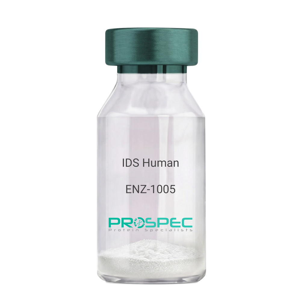
IDS HumanDescription:
Iduronate 2-Sulfatase Human Recombinant
Iduronate 2-Sulfatase, Alpha-L-Iduronate Sulfate Sulfatase, SIDS, Iduronate 2-Sulfatase 14 KDa Chain, Iduronate 2-Sulfatase 42 KDa Chain, Hunter Syndrome, EC 3.1.6.13, MPS2, Iduronate 2-sulfatase, Alpha-L-iduronate sulfate sulfatase.
Product # :
ENZ-1005Price :
Quantity :
Shipping Method :
Shipped with Ice Packs
More Info
- description
- source
- formulation
- purity
- More Info
- Citations
Description
IDS Human Recombinant produced in Sf9 Baculovirus cells is a single, glycosylated polypeptide chain containing 533 amino acids (26-550a.a) and having a molecular mass of 60.3kDa. (Molecular size on SDS-PAGE will appear at approximately 35-70kDa). IDS is fused to an 8 amino acid His-tag at C-terminus & purified by proprietary chromatographic techniques.
Source
Sf9, Baculovirus cells.
Formulation
IDS protein solution (0.25mg/ml) containing Phosphate Buffered Saline (pH 7.4) and 10% glycerol.
Purity
Greater than 90% as determined by SDS-PAGE.
Citations
More Info
-
Introduction
Iduronate 2-Sulfatase also known as IDS, belongs to the highly-conserved sulfatase family of enzymes which catalyze the hydrolysis of O-sulfate and N-salfate esters from a variety of substrates. IDS is essential for the lysosomal degradation of the glycosaminoglycans (GAG) heparan sulfate as well as dermatan sulfate. Furthermore, IDS hydrolyzes the 2-sulfate group of the IDS units of the GAG.
-
Synonyms
Iduronate 2-Sulfatase, Alpha-L-Iduronate Sulfate Sulfatase, SIDS, Iduronate 2-Sulfatase 14 KDa Chain, Iduronate 2-Sulfatase 42 KDa Chain, Hunter Syndrome, EC 3.1.6.13, MPS2, Iduronate 2-sulfatase, Alpha-L-iduronate sulfate sulfatase.
-
Physical Appearance
Sterile Filtered clear solution.
-
Stability
Store at 4°C if entire vial will be used within 2-4 weeks. Store, frozen at -20°C for longer periods of time. For long term storage it is recommended to add a carrier protein (0.1% HSA or BSA).Avoid multiple freeze-thaw cycles.
-
Amino Acid Sequence
SETQANSTTD ALNVLLIIVD DLRPSLGCYG DKLVRSPNID QLASHSLLFQ NAFAQQAVCA PSRVSFLTGR RPDTTRLYDF NSYWRVHAGN FSTIPQYFKE NGYVTMSVGK VFHPGISSNH TDDSPYSWSF PPYHPSSEKY ENTKTCRGPD GELHANLLCP VDVLDVPEGT LPDKQSTEQA IQLLEKMKTS ASPFFLAVGY HKPHIPFRYP KEFQKLYPLE NITLAPDPEV PDGLPPVAYN PWMDIRQRED VQALNISVPY GPIPVDFQRK IRQSYFASVS YLDTQVGRLL SALDDLQLAN STIIAFTSDH GWALGEHGEW AKYSNFDVAT HVPLIFYVPG RTASLPEAGE KLFPYLDPFD SASQLMEPGR QSMDLVELVS LFPTLAGLAG LQVPPRCPVP SFHVELCREG KNLLKHFRFR DLEEDPYLPG NPRELIAYSQ YPRPSDIPQW NSDKPSLKDI KIMGYSIRTI DYRYTVWVGF NPDEFLANFS DIHAGELYFV DSDPLQDHNM YNDSQGGDLF QLLMPLEHHH HHH.
ProSpec's products are furnished for LABORATORY RESEARCH USE ONLY. The product may not be used as drugs, agricultural or pesticidal products, food additives or household chemicals.
- View Data Sheet
Name :
Chitinase ProteinDescription:
Chitinase Clostridium Paraputrificum Recombinant
Product # :
ENZ-031Price :
Quantity :
Shipping Method :
Shipped at Room temp
More Info
- description
- source
- formulation
- purity
- More Info
- Citations
Description
Chitinase Clostridium Paraputrificum Recombinant fused with a His tag at N-terminus produced in E.Coli is a single, non-glycosylated, polypeptide chain containing 582 amino acids and having a molecular mass of 64.2kDa. The Chitinase is purified by proprietary chromatographic techniques.
Source
Escherichia Coli.
Formulation
Chitinase lyophilized from a 0.2µm filtered concentrated solution in PBS.
Purity
Greater than 95.0% as determined by SDS-PAGE.
Citations
More Info
-
Introduction
Chitinase is a digestive enzyme which breaks down glycosidic bonds in chitin. Due to chitin being a component of the cell walls of fungi and exoskeletal elements of some animals (including worms and arthropods), chitinases are usually found in organisms that either need to remake their own chitin or to dissolve and digest the chitin of fungi or animals. Chitinivorous organisms include many bacteria genuses such as Aeromonas, Bacillus, Vibrio, among others, which may be pathogenic or detritivorous. Chitinase expression is mediated by the NPR1 gene and the salicylic acid pathway, both of which are involved in resisting fungal and insect attack. Human chitinases appear in gastric juices. They are likely to be digestive chitinases, for catabolic activity. Chitinase activity is identified systemically in humans, in the blood, and possibly cartilage. Chitinase has been related to allergies, asthma in particular has been linked to enhanced chitinase expression levels, also dust mites and mold spores which are both chitin covered.
-
Physical Appearance
Sterile Filtered White lyophilized (freeze-dried) powder.
-
Stability
Lyophilized Chitinase although stable at room temperature for 3 weeks, should be stored desiccated below -18°C. Upon reconstitution Chitinase should be stored at 4°C between 2-7 days and for future use below -18°C.For long term storage it is recommended to add a carrier protein (0.1% HSA or BSA).Please prevent freeze-thaw cycles.
-
Solubility
It is recommended to reconstitute the lyophilized Chitinase in sterile 18MΩ-cm H2O not less than 100µg/ml, which can then be further diluted to other aqueous solutions.
-
Amino Acid Sequence
MRGSGSHHHH HHMYYGDWSI WGGQGNFYPK DIPADKLTHL NFAFMDFNSS GELIYCDKDA AIGHPLGNLG VTYGDVNGGI LNAFQVLKSE NPNLKIGVSL GGWSKSGDFS TIAATPSIRA KFVENVMKFI KYTNMDFVDI DWEYPGDYRE PDKTDNINDE GTPNASAGDK ENYILLLQDL KEALNKQGKE LGKVYELSVA LPAGVSKIEK GIDVDKLFNI VDFANIMTYD MAGAWSTTSG HQTALYTNPN APEEYKGLSV DESVKYYISQ GAEREKIVVG AAYYTRGWEQ VSDKGTDPNN PGLFGEAAVV NKDADLSPTP GALNEAPMKN GEGGRAGGVW GYNALDKLKS KYTGLKEYWD DSAKAPYLYN SETGAFFTYD NIRSIQEKAK YVKENNLGGI IGWMASQDAT TNSTKRDELT TATKESLFGK EDLPKYEIKY TENDITCTVT PVKQSWGSGG VLKMSITNNE KLDESGEVLS TVETSAKTVK NMKVYIKTDG IAITGSQYPA GPVTKEGDYY VIDFGKISDG KLMKAGITFT FDLNLDKAIE DTNNIISIEV SQRMYQTSPE FNRQTIWENT NS.
ProSpec's products are furnished for LABORATORY RESEARCH USE ONLY. The product may not be used as drugs, agricultural or pesticidal products, food additives or household chemicals.
- View Data Sheet
Name :
HRPDescription:
Horseradish Peroxidase
Horseradish Peroxidase, HRP, EC 1.11.1.7.
Product # :
ENZ-321Price :
Quantity :
Shipping Method :
Shipped at Room temp
More Info
- description
- source
- purity
- biological activity
- More Info
- Citations
Description
HRP consists of the basic isoenzyme having a molecular weight of 44 kDa.The Horseradish Peroxidase is purified by affinity chromatography, which results in an enzyme of high specific activity and purity.
Source
Root extracts of horseradish.
Purity
(A403/A275) = RZ: 3.0.
Biological Activity
276 U/mg (25°C, guaiacol as the hydrogen donor, pH-7 and H2O2 as substrates).
Citations
More Info
-
Introduction
The enzyme horseradish peroxidase, found in horseradish, is used extensively in molecular biologyand in antibody amplification and detection, among other things. For example, "In recent years the technique of marking neurons with the enzyme horseradish peroxidase (HRP) has become a major tool. In its brief history, this method has probably been used by more neurobiologists than have used the Golgi stainsince its discovery in 1870." Horseradish peroxidase is also highly used in techniques such as Western blottingand ELISAs.
HRP is widely used as an enzymatic label in immunoassays. Usually, the enzyme is coupled to antibodies, lectins or haptens. Coupling to antibodies etc. may be performed through the carbohydrate side chains of the HRP. -
Synonyms
Horseradish Peroxidase, HRP, EC 1.11.1.7.
-
Physical Appearance
Sterile Filtered red-brown lyophilized powder.
-
Stability
Lyophilized HRP although stable at room temperature for 3 weeks, should be stored desiccated below -18°C. Upon reconstitution HRP should be stored at 4°C between 2-7 days and for future use below -18°C.Please prevent freeze-thaw cycles.
-
Solubility
It is recommended to reconstitute the lyophilized HRP in sterile 18MΩ-cm H2O not less than 100 µg/ml.
ProSpec's products are furnished for LABORATORY RESEARCH USE ONLY. The product may not be used as drugs, agricultural or pesticidal products, food additives or household chemicals.
- View Data Sheet
Name :
ECH1 HumanDescription:
Enoyl CoA Hydratase 1, Peroxisomal Human Recombinant
peroxisomal, enoyl Coenzyme A hydratase 1.
Product # :
ENZ-562Price :
Quantity :
Shipping Method :
Shipped with Ice Packs
More Info
- description
- source
- formulation
- purity
- More Info
- Citations
Description
ECH1 produced in E.Coli is a single, non-glycosylated polypeptide chain containing 316 amino acids (34-328a.a.) and having a molecular mass of 34.4kDa.ECH1 is fused to a 21 amino acid His-tag at N-terminus & purified by proprietary chromatographic techniques.
Source
Escherichia Coli.
Formulation
The ECH1 protein solution (1mg/1ml) is formulated in 20mM Tris-HCl buffer (pH8.0) 1mM DTT, 50mM NaCl, and 10% glycerol.
Purity
Greater than 90% as determined by SDS-PAGE.
Citations
More Info
-
Introduction
ECH1 is a member of the hydratase/isomerase superfamily. ECH1 demonstrates high sequence similarity to enoyl-coenzyme A (CoA) hydratases of more than a few species, mostly within a conserved domain characteristic of these proteins. ECH1 contains a C-terminal peroxisomal targeting sequence, localizes to both the peroxisome and the mitochondria. peroxisomal takes part in the auxiliary step of the fatty acid beta-oxidation pathway specifically functioning to catalyze the isomerization of 3-trans, 5-cis-dienoyl-CoA to 2-trans, 4-transdienoyl-CoA.
-
Synonyms
peroxisomal, enoyl Coenzyme A hydratase 1.
-
Physical Appearance
Sterile Filtered clear solution.
-
Stability
Store at 4°C if entire vial will be used within 2-4 weeks. Store, frozen at -20°C for longer periods of time. For long term storage it is recommended to add a carrier protein (0.1% HSA or BSA).Avoid multiple freeze-thaw cycles.
-
Amino Acid Sequence
MGSSHHHHHH SSGLVPRGSH MTGSSAQEAA SGVALGEAPD HSYESLRVTS AQKHVLHVQL NRPNKRNAMN KVFWREMVEC FNKISRDADC RAVVISGAGK MFTAGIDLMD MASDILQPKG DDVARISWYL RDIITRYQET FNVIERCPKP VIAAVHGGCI GGGVDLVTAC DIRYCAQDAF FQVKEVDVGL AADVGTLQRL PKVIGNQSLV NELAFTARKM MADEALGSGL VSRVFPDKEV MLDAALALAA EISSKSPVAV QSTKVNLLYS RDHSVAESLN YVASWNMSML QTQDLVKSVQ ATTENKELKT VTFSKL.
ProSpec's products are furnished for LABORATORY RESEARCH USE ONLY. The product may not be used as drugs, agricultural or pesticidal products, food additives or household chemicals.
- View Data Sheet
Name :
ST6GAL1 HumanDescription:
ST6 Beta-Galactosamide Alpha-2,6-Sialyltranferase 1 Human Recombinant
ST6 Beta-Galactosamide Alpha-2,6-Sialyltranferase 1, ST6Gal I, SIAT1, CMP-N-Acetylneuraminate-Beta-Galactosamide-Alpha-2,6-Sialyltransferase 1, B-Cell Antigen CD75, Alpha 2,6-ST 1, EC 2.4.99.1, ST6GalI, CMP-N-Acetylneuraminate Beta-Galactosamide Alpha-2,6-Sialyltransferase, Sialyltransferase 1 (Beta-Galactoside Alpha-2,6-Sialyltransferase), Sialyltransferase 1 (Beta-Galactoside Alpha-2,6-Sialytransferase),Beta-Galactoside Alpha-2,6-Sialyltransferase 1, Sialyltransferase 1, ST6N, ST6GAL1.
Product # :
ENZ-893Price :
Quantity :
Shipping Method :
Shipped with Ice Packs
More Info
- description
- source
- formulation
- purity
- More Info
- Citations
Description
ST6GAL1 Human Recombinant produced in E.Coli is a single, non-glycosylated polypeptide chain containing 403 amino acids (27-406 a.a) and having a molecular mass of 46kDa.ST6GAL1 is fused to a 23 amino acid His-tag at N-terminus & purified by proprietary chromatographic techniques.
Source
Escherichia Coli.
Formulation
ST6GAL1 protein solution (1mg/ml) containing 20mM Tris-HCl (pH 8.0) and 10% glycerol.
Purity
Greater than 90.0% as determined by SDS-PAGE.
Citations
More Info
-
Introduction
ST6GAL1 also known as ST6 Beta-Galactosamide Alpha-2,6-Sialyltranferase 1, is part of the glycosyltransferase family 29. ST6GAL1 is a type II membrane protein which catalyzes the transfer of sialic acid from CMP-sialic acid to galactose-containing substrates. Furthermore, ST6GAL1 is normally found in the Golgi however it can be proteolytically processed to a soluble form, ST6GAL1 is also involved in the generation of the cell-surface carbohydrate determinants as well differentiation antigens HB-6, CD75, and CD76.
-
Synonyms
ST6 Beta-Galactosamide Alpha-2,6-Sialyltranferase 1, ST6Gal I, SIAT1, CMP-N-Acetylneuraminate-Beta-Galactosamide-Alpha-2,6-Sialyltransferase 1, B-Cell Antigen CD75, Alpha 2,6-ST 1, EC 2.4.99.1, ST6GalI, CMP-N-Acetylneuraminate Beta-Galactosamide Alpha-2,6-Sialyltransferase, Sialyltransferase 1 (Beta-Galactoside Alpha-2,6-Sialyltransferase), Sialyltransferase 1 (Beta-Galactoside Alpha-2,6-Sialytransferase),Beta-Galactoside Alpha-2,6-Sialyltransferase 1, Sialyltransferase 1, ST6N, ST6GAL1.
-
Physical Appearance
Sterile Filtered colorless solution.
-
Stability
Store at 4°C if entire vial will be used within 2-4 weeks. Store, frozen at -20°C for longer periods of time. For long term storage it is recommended to add a carrier protein (0.1% HSA or BSA).Avoid multiple freeze-thaw cycles.
-
Amino Acid Sequence
MGSSHHHHHH SSGLVPRGSH MGSKEKKKGS YYDSFKLQTK EFQVLKSLGK LAMGSDSQSV SSSSTQDPHR GRQTLGSLRG LAKAKPEASF QVWNKDSSSK NLIPRLQKIW KNYLSMNKYK VSYKGPGPGI KFSAEALRCH LRDHVNVSMV EVTDFPFNTS EWEGYLPKES IRTKAGPWGR CAVVSSAGSL KSSQLGREID DHDAVLRFNG APTANFQQDV GTKTTIRLMN SQLVTTEKRF LKDSLYNEGI LIVWDPSVYH SDIPKWYQNP DYNFFNNYKT YRKLHPNQPF YILKPQMPWE LWDILQEISP EEIQPNPPSS GMLGIIIMMT LCDQVDIYEF LPSKRKTDVC YYYQKFFDSA CTMGAYHPLL YEKNLVKHLN QGTDEDIYLL GKATLPGFRT IHC
ProSpec's products are furnished for LABORATORY RESEARCH USE ONLY. The product may not be used as drugs, agricultural or pesticidal products, food additives or household chemicals.
- View Data Sheet
Name :
UBE2L3 Human HisDescription:
Ubiquitin-Conjugating Enzyme E2L 3 Human Recombinant, His Tag
Ubiquitin-conjugating enzyme E2 L3, EC 6.3.2.19, Ubiquitin-protein ligase L3,Ubiquitin carrier protein L3, UbcH7, E2-F1, L-UBC, UbcM4.
Product # :
ENZ-344Price :
Quantity :
Shipping Method :
Shipped at Room temp
More Info
- description
- source
- formulation
- purity
- More Info
- Citations
Description
Ubiquitin-Conjugating Enzyme E2L 3 Human Recombinant produced in E.coli is an 18.9 kDa protein containing 162 amino acids.The UBE2L3 protein contains 6xHis tag and is purified by proprietary chromatographic techniques.
Source
Escherichia Coli.
Formulation
Lyophilized from a 0.2μm filtered concentrated (1 mg/ml) solution in 1X PBS and 1mM DTT, pH 7.5.
Purity
Greater than 95.0% as determined by: (a) Analysis by RP-HPLC.
(b) Analysis by SDS-PAGE.Citations
More Info
-
Introduction
Human Ubquitin Conjugating Enzyme 7 (UbcH7) is a class I enzyme which functions in the stress response and the control of transcription factors. The enzyme is ubiquitously expressed with high levels of expression seen in adult muscle. UbcH7 mediates the selective degradation of short-lived and abnormal proteins and is highly homologous to UbcH5. It has been demonstrated to participate in the ubiquitinylation of p53, c-Fos and NF-?B. UbcH7 is one of two E2s (UbcH5 being the other) with which HECT domain proteins interact with UbcH7 being able to efficiently substitute for UbcH5 in E6-AP-dependent ubiquitinylation.
-
Synonyms
Ubiquitin-conjugating enzyme E2 L3, EC 6.3.2.19, Ubiquitin-protein ligase L3,Ubiquitin carrier protein L3, UbcH7, E2-F1, L-UBC, UbcM4.
-
Physical Appearance
Sterile Filtered white lyophilized powder.
-
Stability
Lyophilized UBE2L3 although stable at room temperature for 3 weeks, should be stored desiccated below -18°C. Upon reconstitution UBE2L3 should be stored at 4°C between 2-7 days and for future use below -18°C.For long term storage it is recommended to add a carrier protein (0.1% HSA or BSA).Please prevent freeze-thaw cycles.
-
Solubility
It is recommended to reconstitute the lyophilized UBE2L3 in sterile water not less than 100µg/ml, which can then be further diluted to other aqueous solutions.
-
Amino Acid Sequence
MHHHHHHAMAASRRLMKELEEIRKCGMKNFRNIQVDEANLLTWQGLIVP
DNPPYDKGAFRIEINFPAEYPFKPPKITFKTKIYHPNIDEKGQVCLPVISAEN
WKPATKTDQVIQSLIALVNDPQPEHPLRADLAEEYSKDRKKFCKNAEEFT
KKYGEKRPVD.
ProSpec's products are furnished for LABORATORY RESEARCH USE ONLY. The product may not be used as drugs, agricultural or pesticidal products, food additives or household chemicals.
- View Data Sheet
Name :
LY6G Antibody, BiotinDescription:
LY6G, Rat Anti Mouse Antibody, Biotin
Lymphocyte antigen 6G, Ly-6G, Ly-6G.1, Ly6g, Gr1, Gr-1.
Product # :
ANT-074Price :
Quantity :
Shipping Method :
Shipped at Room temp
More Info
- formulation
- More Info
- Citations
Formulation
1mg/ml in PBS (after reconstitution).
Citations
More Info
-
Introduction
Myeloid differentiation antigen Gr1 (Ly6G) is a GPI-anchored protein, which is briefly expressed on monocytes in the bone marrow. The level of the Ly6G expression in the bone marrow completely correlates with granulocyte differentiation and maturation. Ly6G ligation on murine neutrophils inhibits neutrophil recruitment, thus providing the first evidence of a function for the Ly6G molecule. Ly6G is seen primarily on neutrophils, also in a subgroup of eosinophils, differentiating pre-monocytes and plasmacytoid dendritic cells.
-
Synonyms
Lymphocyte antigen 6G, Ly-6G, Ly-6G.1, Ly6g, Gr1, Gr-1.
-
Solubility
Reconstitute with H20. Mix gently, wash the sides of the vial and wait 30-60 seconds before use.
-
Immunogen
Ly-6G-transfected cell line.
-
Ig Subclass
Rat IgG2a.
-
Clone
YRmLy-6G.
-
Applications
Flow cytometry, immunohistochemistry, cell depletion.
This antibody recognizes only Ly-6G which is expressed on most myeloid cells in the bone marrow and all peripheral granulocytes. For flow cytometry, use 10 µl/106 cells. Exact titer for cell depletion in vivo (cytotoxicity) should be determined by the investigator. -
Shipping Conditions
Antibody is shipped lyophilized at ambient temperature.
-
Available Conjugates
This antibody is also available unconjugated and conjugated to FITC.
-
Type
Rat Anti Mouse Monoclonal.
-
Storage Procedures
In lyophilized form, for long periods, store at 4°C in a dry environment. After reconstitution, if not intended for use within a month, aliquot and store at -20°C.
-
Purification Method
Protein A column.
ProSpec's products are furnished for LABORATORY RESEARCH USE ONLY. The product may not be used as drugs, agricultural or pesticidal products, food additives or household chemicals.
- View Data Sheet
Name :
LYVE1 Human 25-235 a.a.Description:
Lymphatic Vessel Endothelial Hyaluronic Acid Receptor 1 (25-235 a.a) Human Recombinant
HAR, XLKD1, LYVE-1, CRSBP-1, LYVE1, Lymphatic vessel endothelial hyaluronic acid receptor 1 precursor, Cell surface retention sequence-binding protein 1, Hyaluronic acid receptor, Extracellular link domain-containing protein.
Product # :
PKA-349Price :
Quantity :
Shipping Method :
Shipped with Ice Packs
More Info
- description
- source
- formulation
- purity
- More Info
- Citations
Description
LYVE1 Human Recombinant produced in insect cells is a single, glycosylated polypeptide chain containing 229 amino acids and having a molecular mass of 24.8 kDa. As a result of glycosylation, the LYVE1 migrates on SDS-PAGE at approximately 50 kDa. LYVE1 is expressed with 15 amino acids His tag at C-Terminus and purified by proprietary chromatographic techniques.
Source
High Five insect cells.
Formulation
The LYVE1 protein solution contains 20mM Tris buffer pH-7.5 and 10% Glycerol.
Purity
Greater than 90.0% as determined by:(a) Analysis by RP-HPLC.(b) Analysis by SDS-PAGE.
Citations
More Info
-
Introduction
LYVE1 is a selective marker of the lymphatic endothelium & a surface endocytic receptor for both soluble and immobilized hyaluronan, LYVE1 is an extracellular glycosaminoglycan that plays a role in cell adhesion and migration. LYVE1 functions in lympathic hyaluronan transport and is involved in tumor metastasis. Recombinant human LYVE1 was expressed in and purified by conventional chromatography techniques. The normal adult human choroid is endowed with a significant number of LYVE-1 positive macrophages. LYVE-1 is expressed in a reticulum cell neoplasm in an axillary lymph node. This reticulum cell sarcoma is a lymphatic sinus lining cell sarcoma which might represent another subtype of reticulum cell sarcomas.
LYVE-1 immunohistochemistry is a functional method for detecting lymphatics invaded by cancer cells, and detailed examination of the submucosa around the tumor is important for predicting LN metastasis. -
Synonyms
HAR, XLKD1, LYVE-1, CRSBP-1, LYVE1, Lymphatic vessel endothelial hyaluronic acid receptor 1 precursor, Cell surface retention sequence-binding protein 1, Hyaluronic acid receptor, Extracellular link domain-containing protein.
-
Physical Appearance
Sterile Filtered colorless solution.
-
Stability
Store at 4°C if entire vial will be used within 2-4 weeks. Store, frozen at -20°C for longer periods of time. For long term storage it is recommended to add a carrier protein (0.1% HSA or BSA).Avoid multiple freeze-thaw cycles.
-
Amino Acid Sequence
DPLRAEELS IQVSCRIMGI TLVSKKANQQ LNFTEAKEAC RLLGLSLAGK DQVETALKAS FETCSYGWVG DGFVVISRIS PNPKCGKNGV GVLIRKVPVS RQFAAYCYNS SDTWTNSCIP EIITTKDPIF NTQTATQTTE FIVSDSTYSV ASPYSTIPAP TTTPPAPAST SIPRRKKLIC VTEVFMETST MSTETEPFVE NKAAFKNEAA GFGGSGRLVP RGSHHHHHH.
ProSpec's products are furnished for LABORATORY RESEARCH USE ONLY. The product may not be used as drugs, agricultural or pesticidal products, food additives or household chemicals.
- View Data Sheet
Name :
CTSZ Human, Sf9Description:
Cathepsin-Z Human Recombinant, Sf9
Cathepsin Z, Cathepsin X, Cysteine-Type Carboxypeptidase, Lysosomal Carboxypeptidase B, Carboxypeptidase LB, Cathepsin B2, Cathepsin IV, Cathepsin Z1, Cathepsin P, Cathepsin Y, EC 3.4.18.1, Preprocathepsin P, CTSX.
Product # :
ENZ-1097Price :
Quantity :
Shipping Method :
Shipped with Ice Packs
More Info
- description
- source
- formulation
- purity
- biological activity
- More Info
- Citations
Description
CTSZ produced in Sf9 Baculovirus cells is a single, glycosylated polypeptide chain containing 288 amino acids (24-303.a.) and having a molecular mass of 32.5kDa. (Molecular size on SDS-PAGE will appear at approximately 28-40kDa).CTSZ is expressed with an 8 amino acid His tag at C-Terminus and purified by proprietary chromatographic techniques.
Source
Sf9, Baculovirus cells.
Formulation
CTSZ protein solution (0.5mg/ml) contains 10% glycerol & Phosphate Buffered Saline (pH 7.4).
Purity
Greater than 95% as determined by SDS-PAGE.
Biological Activity
The specific activity is determent as the ability of 1 unit to convert 1.0 pmole of Mca-PLGL-Dpa-AR-NH2 to MCA- Pro-Leu-OH per minute at pH 3.5 at 25C and is > 1,400 pmol/min/ug.
Citations
More Info
-
Introduction
Cathepsin-Z (CTSZ) is a lysosomal cysteine proteinase and a member of the peptidase C1 family. CTSZ, which is known also as cathepsin X and cathepsin P, exhibits carboxy-monopeptidase and carboxy-dipeptidase activities. CTSZ is expressed ubiquitously in cancer cell lines and primary tumors and as other members of this family, takes part in tumorigenesis.
-
Synonyms
Cathepsin Z, Cathepsin X, Cysteine-Type Carboxypeptidase, Lysosomal Carboxypeptidase B, Carboxypeptidase LB, Cathepsin B2, Cathepsin IV, Cathepsin Z1, Cathepsin P, Cathepsin Y, EC 3.4.18.1, Preprocathepsin P, CTSX.
-
Physical Appearance
Sterile Filtered clear solution.
-
Stability
Store at 4°C if entire vial will be used within 2-4 weeks. Store, frozen at -20°C for longer periods of time.For long term storage it is recommended to add a carrier protein (0.1% HSA or BSA).Please avoid freeze thaw cycles.
-
Amino Acid Sequence
GLYFRRGQTC YRPLRGDGLA PLGRSTYPRP HEYLSPADLP KSWDWRNVDG VNYASITRNQ
HIPQYCGSCW AHASTSAMAD RINIKRKGAW PSTLLSVQNV IDCGNAGSCE GGNDLSVWDY
AHQHGIPDET CNNYQAKDQE CDKFNQCGTC NEFKECHAIR NYTLWRVGDY GSLSGREKMM
AEIYANGPIS CGIMATERLA NYTGGIYAEY QDTTYINHVV SVAGWGISDG TEYWIVRNSW
GEPWGERGWL RIVTSTYKDG KGARYNLAIE EHCTFGDPIV LEHHHHHH
ProSpec's products are furnished for LABORATORY RESEARCH USE ONLY. The product may not be used as drugs, agricultural or pesticidal products, food additives or household chemicals.
- View Data Sheet
Name :
DUSP6 HumanDescription:
Dual Specificity Phosphatase 6 Human Recombinant
Dual specificity protein phosphatase 6, Dual specificity protein phosphatase PYST1, Mitogen-activated protein kinase phosphatase 3, MAP kinase phosphatase 3, MKP-3, DUSP6, MKP3, PYST1, Dual Specificity Phosphatase 6, Dual specificity phosphatase 6 isoform a.
Product # :
ENZ-817Price :
Quantity :
Shipping Method :
Shipped with Ice Packs
More Info
- description
- source
- formulation
- purity
- More Info
- Citations
Description
DUSP6 Human Recombinant produced in E.Coli is a single, non-glycosylated polypeptide chain containing 401 amino acids (1-381 a.a) and having a molecular mass of 44.4kDa.DUSP6 is fused to a 20 amino acid His-tag at N-terminus & purified by proprietary chromatographic techniques.
Source
Escherichia Coli.
Formulation
DUSP6 protein solution (0.25mg/ml) containing 20mM Tris-HCl buffer (pH 8.0), 0.15M NaCl, 10% glycerol and 1mM DTT.
Purity
Greater than 85.0% as determined by SDS-PAGE.
Citations
More Info
-
Introduction
Dual Specificity Phosphatase 6, also known as DUSP6 belongs to the dual specificity protein phosphatase subfamily. DUSP6 is a Protein coding gene which inactivates MAP kinases. Various members of the family of dual specificity phosphatases show diverse tissue distribution, subcellular localization, and different modes of inducibility of their expression by extracellular stimuli.
-
Synonyms
Dual specificity protein phosphatase 6, Dual specificity protein phosphatase PYST1, Mitogen-activated protein kinase phosphatase 3, MAP kinase phosphatase 3, MKP-3, DUSP6, MKP3, PYST1, Dual Specificity Phosphatase 6, Dual specificity phosphatase 6 isoform a.
-
Physical Appearance
Sterile Filtered colorless solution.
-
Stability
Store at 4°C if entire vial will be used within 2-4 weeks. Store, frozen at -20°C for longer periods of time. For long term storage it is recommended to add a carrier protein (0.1% HSA or BSA).Avoid multiple freeze-thaw cycles.
-
Amino Acid Sequence
MGSSHHHHHH SSGLVPRGSH MIDTLRPVPF ASEMAISKTV AWLNEQLELG NERLLLMDCR PQELYESSHI ESAINVAIPG IMLRRLQKGN LPVRALFTRG EDRDRFTRRC GTDTVVLYDE SSSDWNENTG GESVLGLLLK KLKDEGCRAF YLEGGFSKFQ AEFSLHCETN LDGSCSSSSP PLPVLGLGGL RISSDSSSDI ESDLDRDPNS ATDSDGSPLS NSQPSFPVEI LPFLYLGCAK DSTNLDVLEE FGIKYILNVT PNLPNLFENA GEFKYKQIPI SDHWSQNLSQ FFPEAISFID EARGKNCGVL VHCLAGISRS VTVTVAYLMQ KLNLSMNDAY DIVKMKKSNI SPNFNFMGQL LDFERTLGLS SPCDNRVPAQ QLYFTTPSNQ NVYQVDSLQS T.
ProSpec's products are furnished for LABORATORY RESEARCH USE ONLY. The product may not be used as drugs, agricultural or pesticidal products, food additives or household chemicals.
- View Data Sheet
Name :
NPEPPS HumanDescription:
Aminopeptidase Puromycin Sensitive Human Recombinant
PSA, MP100, AAP-S, Puromycin-sensitive aminopeptidase, Cytosol alanyl aminopeptidase, aminopeptidase puromycin sensitive.
Product # :
ENZ-1196Price :
Quantity :
Shipping Method :
Shipped with Ice Packs
More Info
- description
- source
- formulation
- purity
- biological activity
- More Info
- Citations
Description
NPEPPS Human Recombinant produced in HEK293 Cells is a single, glycosylated polypeptide chain containing 925 amino acids (1-919 a.a.) and having a molecular mass of 104kDa. NPEPPS is fused to a 6 amino acid His-tag at C-terminus and is purified by proprietary chromatographic techniques.
Source
HEK293 Cells.
Formulation
NPEPPS protein solution (0.25mg/ml) containing 20% Glycerol and Phosphate-Buffered Saline (pH 7.4).
Purity
Greater than 85.0% as determined by SDS-PAGE.
Biological Activity
Specific activity is > 800 pmol/min/ug and is defined as the amount of enzyme that cleaves 1 pmole of H-Leu[1]AMC per minute at pH7.0 at 37°C.
Citations
More Info
-
Synonyms
PSA, MP100, AAP-S, Puromycin-sensitive aminopeptidase, Cytosol alanyl aminopeptidase, aminopeptidase puromycin sensitive.
-
Physical Appearance
Sterile filtered colorless solution.
-
Stability
Store at 4°C if entire vial will be used within 2-4 weeks. Store, frozen at -20°C for longer periods of time. For long term storage it is recommended to add a carrier protein (0.1% HSA or BSA). Avoid multiple freeze-thaw cycles.
-
Amino Acid Sequence
MWLAAAAPSL ARRLLFLGPP PPPLLLLVFS RSSRRRLHSL GLAAMPEKRP FERLPADVSP INYSLCLKPD LLDFTFEGKL EAAAQVRQAT NQIVMNCADI DIITASYAPE GDEEIHATGF NYQNEDEKVT LSFPSTLQTG TGTLKIDFVG ELNDKMKGFY RSKYTTPSGE VRYAAVTQFE ATDARRAFPC WDEPAIKATF DISLVVPKDR VALSNMNVID RKPYPDDENL VEVKFARTPV MSTYLVAFVV GEYDFVETRS KDGVCVRVYT PVGKAEQGKF ALEVAAKTLP FYKDYFNVPY PLPKIDLIAI ADFAAGAMEN GLVTYRETA LLIDPKNSCS SSRQWVALVV GHELAHQWFG NLVTMEWWTH LWLNEGFASW IEYLCVDHCF PEYDIWTQFV SADYTRAQEL DALDNSHPIE VSVGHPSEVD EIFDAISYSK GASVIRMLHD YIGDKDFKKG MNMYLTKFQQ KNAATEDLWE SLENASGKPI AAVMNTWTKQ MGFPLIYVEA EQVEDDRLLR LSQKKFCAGG SYVGEDCPQW MVPITISTSE DPNQAKLKIL MDKPEMNVVL KNVKPDQWVK LNLGTVGFYR TQYSSAMLES LLPGIRDLSL PPVDRLGLQN DLFSLARAGI ISTVEVLKVM EAFVNEPNYT VWSDLSCNLG ILSTLLSHTD FYEEIQEFVK DVFSPIGERL GWDPKPGEGH LDALLRGLVL GKLGKAGHKA TLEEARRRFK DHVEGKQILS ADLRSPVYLT VLKHGDGTTL DIMLKLHKQA DMQEEKNRIE RVLGATLLPD LIQKVLTFAL SEEVRPQDTV SVIGGVAGGS KHGRKAAWKF IKDNWEELYN RYQGGFLISR LIKLSVEGFA VDKMAGEVKA FFESHPAPSA ERTIQQCCEN ILLNAAWLKR DAESIHQYLL QRKASPPTVH HHHHH.
-
Background
NPEPPS is involved in the proteolytic degradation of misfolded or damaged proteins, contributing to the maintenance of protein homeostasis within the cell. It specifically removes N-terminal amino acids from peptides, thereby regulating their activity and facilitating their further degradation by other proteases.
This enzyme is particularly important in the nervous system, where it degrades neuropeptides and helps regulate synaptic signaling and neuronal communication.
ProSpec's products are furnished for LABORATORY RESEARCH USE ONLY. The product may not be used as drugs, agricultural or pesticidal products, food additives or household chemicals.
- View Data Sheet
Name :
GLB1 HumanDescription:
Galactosidase-Beta 1 Human Recombinant
lacZ, beta-gal, b-gal, Acid beta-galactosidase, Lactase, Elastin receptor 1
Product # :
ENZ-1136Price :
Quantity :
Shipping Method :
Shipped with Ice Packs
More Info
- description
- source
- formulation
- purity
- More Info
- Citations
Description
GLB1 Human produced in Sf9 Baculovirus cells is a single, glycosylated polypeptide chain containing 662 amino acids (24-677 a.a.) and having a molecular mass of 74.6 kDa.GLB1 is fused to an 8 amino acid His tag at C-Terminus and purified by proprietary chromatographic techniques.
Source
Sf9, Baculovirus cells.
Formulation
GLB1 Human protein (0.25mg/ml) is formulated in 10% glycerol and Phosphate-Buffered Saline (pH 7.4).
Purity
Greater than 90.0% as determined by SDS-PAGE.
Citations
More Info
-
Introduction
Beta-galactosidase or GLB1 is a B-galactosidase found in the lysosome. This enzyme hydrolyzes the finale B-galactose from the precursors ganglioside & keratin sulfate. GLB1 has a crucial part in the creation of elastogenesis (extracellular elastic fibers) and connective tissue development. This enzyme is similar to elastin-binding protein which is a key part in non-integrin cell surface receptor. In cells that extort elastin GLB1 is linked to tropoelastin intracellularly and act as recycling molecular chaperone.
-
Synonyms
lacZ, beta-gal, b-gal, Acid beta-galactosidase, Lactase, Elastin receptor 1
-
Physical Appearance
Sterile Filtered colorless solution.
-
Stability
Store at 4°C if entire vial will be used within 2-4 weeks. Store, frozen at -20°C for longer periods of time. For long term storage it is recommended to add a carrier protein (0.1% HSA or BSA).Avoid multiple freeze-thaw cycles.
-
Amino Acid Sequence
LRNATQRMFE IDYSRDSFLK DGQPFRYISG SIHYSRVPRF YWKDRLLKMK MAGLNAIQTY VPWNFHEPWP GQYQFSEDHD VEYFLRLAHE LGLLVILRPG PYICAEWEMG GLPAWLLEKE SILLRSSDPD YLAAVDKWLG VLLPKMKPLL YQNGGPVITV QVENEYGSYF ACDFDYLRFL QKRFRHHLGD DVVLFTTDGA HKTFLKCGAL QGLYTTVDFG TGSNITDAFL SQRKCEPKGP LINSEFYTGW LDHWGQPHST IKTEAVASSL YDILARGASV NLYMFIGGTN FAYWNGANSP YAAQPTSYDY DAPLSEAGDL TEKYFALRNI IQKFEKVPEG PIPPSTPKFA YGKVTLEKLK TVGAALDILC PSGPIKSLYP LTFIQVKQHY GFVLYRTTLP QDCSNPAPLS SPLNGVHDRA YVAVDGIPQG VLERNNVITL NITGKAGATL DLLVENMGRV NYGAYINDFK GLVSNLTLSS NILTDWTIFP LDTEDAVRSH LGGWGHRDSG HHDEAWAHNS SNYTLPAFYM GNFSIPSGIP DLPQDTFIQF PGWTKGQVWI NGFNLGRYWP ARGPQLTLFV PQHILMTSAP NTITVLELEW APCSSDDPEL CAVTFVDRPV IGSSVTYDHP SKPVEKRLMP PPPQKNKDSW LDHVLEHHHH HH
ProSpec's products are furnished for LABORATORY RESEARCH USE ONLY. The product may not be used as drugs, agricultural or pesticidal products, food additives or household chemicals.
- View Data Sheet
Name :
LPCAT1 HumanDescription:
Lysophosphatidylcholine Acyltransferase Human Recombinant
AYTL2, lpcat, PFAAP3, Lysophosphatidylcholine acyltransferase 1, LPC acyltransferase 1, LPCAT-1, LysoPC acyltransferase 1, 1-acylglycerophosphocholine O-acyltransferase, 1-alkylglycerophosphocholine O-acetyltransferase, Acetyl-CoA:lyso-platelet-activating factor acetyltransferase, Acetyl-CoA:lyso-PAF acetyltransferase, Lyso-PAF acetyltransferase, LysoPAFAT, Acyltransferase-like 2, Phosphonoformate immuno-associated protein 3.
Product # :
ENZ-695Price :
Quantity :
Shipping Method :
Shipped with Ice Packs
More Info
- description
- source
- formulation
- purity
- More Info
- Citations
Description
LPCAT1 Human Recombinant produced in E.coli is a single, non-glycosylated polypeptide chain containing 479 amino acids (79-534a.a) and having a molecular mass of 53.4kDa. LPCAT1 is fused to a 23 amino acid His-tag at N-terminus & purified by proprietary chromatographic techniques.
Source
Escherichia Coli.
Formulation
The LPCAT1 solution (0.25mg/ml) contains 20mM Tris-HCl buffer (pH 8.0), 1M Urea and 10% glycerol.
Purity
Greater than 90.0% as determined by SDS-PAGE.
Citations
More Info
-
Introduction
Lysophosphatidylcholine acyltransferase 1 (LPCAT1) is a part of the 1-acyl-sn-glycerol-3-phosphate acyltransferase family. LPCAT1 is a key enzyme for remodeling phospholipids, including phosphatidylcholine. LPCAT1 possesses both acyltransferase and acetyltransferase activities and also mediates the conversion of 1-acyl-sn-glycero-3-phosphocholine (LPC) into phosphatidylcholine (PC). LPCAT1 presents a clear preference for saturated fatty acyl-CoAs, and 1-myristoyl or 1-palmitoyl LPC as acyl donors and acceptors, respectively. LPCAT1 synthesizes phosphatidylcholine in pulmonary surfactant and therefore playing an important role in respiratory physiology.
-
Synonyms
AYTL2, lpcat, PFAAP3, Lysophosphatidylcholine acyltransferase 1, LPC acyltransferase 1, LPCAT-1, LysoPC acyltransferase 1, 1-acylglycerophosphocholine O-acyltransferase, 1-alkylglycerophosphocholine O-acetyltransferase, Acetyl-CoA:lyso-platelet-activating factor acetyltransferase, Acetyl-CoA:lyso-PAF acetyltransferase, Lyso-PAF acetyltransferase, LysoPAFAT, Acyltransferase-like 2, Phosphonoformate immuno-associated protein 3.
-
Physical Appearance
Sterile Filtered colorless solution.
-
Stability
Store at 4°C if entire vial will be used within 2-4 weeks. Store, frozen at -20°C for longer periods of time. For long term storage it is recommended to add a carrier protein (0.1% HSA or BSA).Avoid multiple freeze-thaw cycles.
-
Amino Acid Sequence
MGSSHHHHHH SSGLVPRGSH MGSSAEKEPE QPPALWRKVV DFLLKAIMRT MWFAGGFHRV AVKGRQALPT EAAILTLAPH SSYFDAIPVT MTMSSIVMKA ESRDIPIWGT LIQYIRPVFV SRSDQDSRRK TVEEIKRRAQ SNGKWPQIMI FPEGTCTNRT CLITFKPGAF IPGAPVQPVV LRYPNKLDTI TWTWQGPGAL EILWLTLCQF HNQVEIEFLP VYSPSEEEKR NPALYASNVR RVMAEALGVS VTDYTFEDCQ LALAEGQLRL PADTCLLEFA RLVRGLGLKP EKLEKDLDRY SERARMKGGE KIGIAEFAAS LEVPVSDLLE DMFSLFDESG SGEVDLRECV VALSVVCRPA RTLDTIQLAF KMYGAQEDGS VGEGDLSCIL KTALGVAELT VTDLFRAIDQ EEKGKITFAD FHRFAEMYPA FAEEYLYPDQ THFESCAETS PAPIPNGFCA DFSPENSDAG RKPVRKKLD.
ProSpec's products are furnished for LABORATORY RESEARCH USE ONLY. The product may not be used as drugs, agricultural or pesticidal products, food additives or household chemicals.
- View Data Sheet
Name :
LY6C/G Antibody, BiotinDescription:
LY6C/G, Rat Anti Mouse Antibody, Biotin
Product # :
ANT-077Price :
Quantity :
Shipping Method :
Shipped at Room temp
More Info
- formulation
- More Info
- Citations
Formulation
1mg/ml in PBS (after reconstitution).
Citations
More Info
-
Solubility
Reconstitute with H20. Mix gently, wash the sides of the vial and wait 30-60 seconds before use.
-
Immunogen
BM-enriched myeloid cells.
-
Ig Subclass
Rat IgG2b.
-
Clone
YRmLy-6C/G.
-
Applications
Flow cytometry, immunohistochemistry.
This antibody recognizes both Ly-6G and Ly-6C (granulocyte receptor 1- Gr-1). Ly-6G is expressed on most myeloid cells in the bone marrow and all peripheral granulocytes. Ly-6C is also expressed on subsets of lymphocytes and monocytes. For flow cytometry, use 10 µl/106 cells. Exact titer for IHC should be determined by the investigator. -
Shipping Conditions
Antibody is shipped lyophilized at ambient temperature.
-
Available Conjugates
This antibody is also available unconjugated and conjugated to FITC.
-
Type
Rat Anti Mouse Monoclonal.
-
Storage Procedures
In lyophilized form, for long periods, store at 4°C in a dry environment. After reconstitution, if not intended for use within a month, aliquot and store at -20°C.
-
Purification Method
Protein A column.
ProSpec's products are furnished for LABORATORY RESEARCH USE ONLY. The product may not be used as drugs, agricultural or pesticidal products, food additives or household chemicals.
- View Data Sheet
Name :
Heparanase 1 Clone HP3/17Description:
Heparanase 1 (HPA1), Monoclonal Anti-Human Antibody
Product # :
ANT-154Price :
Quantity :
Shipping Method :
Shipped with Ice Packs
More Info
- source
- formulation
- purity
- More Info
- Citations
Source
Mab HP3/17 is a Protein G affinity purified monoclonal antibody raised against a polypeptide from the 50 kDa subunit of Heparanase.
Formulation
Each vial contains 50, 100 or 150 μg in 14, 28 or 42 μl respectively, of 0.22 micron filtered solution of 20 mM Sodium Phosphate; 150 mM NaCl; pH 7.2, containing 0.01% Thimerosal.
Purity
>98% on SDS-PAGE when loaded 50 μg/lane.
Citations
More Info
-
Introduction
Heparanase is an endo-β-D-glucuronidase, which degrades heparan sulfate side chains of heparan sulfate proteoglycans (HSPGs) in the extracellular matrix. Heparanase plays an important role in ECM degradation, facilitating the migration and extravasation of tumor cells and inflammatory leukocytes (1,2,3). Upon degradation, heparanase releases growth factors and cytokines that stimulate cell proliferation and chemotaxis (4,5). Heparanase is a heterodimer comprised of a 50 kDa subunit harboring the active site and a 8 kDa subunit. It is produced as a latent 65 kDa precursor and proteolytically processed to its active form (1,6). Heparanase is highly expressed in myeloid leukocytes (i.e. neutrophils) in platelets and in human placenta. Human heparanase was found to be upregulated in various types of primary tumors, correlating in some cases with increased tumor invasiveness and vascularity and with poor prospective survival (7,8).
-
Stability
Store at 4°C. Stable for six months from the date of shipment. For extended storage, freeze in working aliquots at -20°C. Avoid repeated freeze-thaw cycles.
-
Ig Subclass
Mouse IgG2Bκ
-
Applications
Western blot
Immunohistochemistry -
Data Sheet
To view the FULL VERSION click Monoclonal Anti-Human Heparanase 1:
-
Patent Protected Countries
Anti-heparanase antibodies and their uses, including HP3/17 and its uses, are protected by US. Patents No. 6,177,545; 6,531,129, additional US patent applications and patents and patent applications worldwide.
-
Specificity
HP3/17 reacts with the 50 kDa subunit and with the 65 kDa precursor of human or mouse Heparanase by Western blotting and immunohistochemistry.Recommended dilution range for Western blot analysis: 1:4000.Recommended dilution range for immunohistochemistry: 1:40.
ProSpec's products are furnished for LABORATORY RESEARCH USE ONLY. The product may not be used as drugs, agricultural or pesticidal products, food additives or household chemicals.
- View Data Sheet
Name :
L-AsparaginaseDescription:
L-Asparaginase
Product # :
ENZ-287Price :
Quantity :
Shipping Method :
Shipped at Room temp
More Info
- description
- source
- formulation
- purity
- biological activity
- More Info
- Citations
Description
L-asparaginase was purified from E.coli ASI.357.
Source
Escherichia Coli.
Formulation
The enzyme was lyophilized with no additives.
Purity
Greater than 96.0% as determined by SDS-PAGE.
Biological Activity
One IU of L- Asparaginase is defined as that amount of enzyme required to generate 1 µmol of ammonia per minute at pH 7.3 and 37°C.
Citations
More Info
-
Introduction
L-Asparaginase is an enzyme that depletes L-Asparagine "an important nutrient for cancer cells" resulting in cancer/tumor cell starvation. L-asparaginase is an anti-tumor agent derived from E.coli.,which can inhibit the growth of malignant cells. It is used mainly for the induction of remission in acute lymphoblastic leukaemia. Because of the lymph node origin of malignant B cells in Multiple Myeloma, L-Asparagine is an essential amino acid for their cell metabolism, and, consequently, L-Asparaginase may be of value in managing the disease.
The rationale behind asparaginase is that it takes advantage of the fact that ALL cellsare unable to synthesize the non-essential amino acidasparaginewhereas normal cells are able to make their own asparagine. These leukemic cells depend on circulating asparagine. Asparaginase however catalyzes the conversion of L-asparagine to aspartic acidand ammonia. This deprives the leukemic cell of circulating asparagine. -
Physical Appearance
Sterile Filtered White lyophilized (freeze-dried) powder.
-
Stability
Lyophilized L-Asparaginase although stable at room temperature for 3 weeks, should be stored desiccated below -18°C. Upon reconstitution L-Asparaginase should be stored at 4°C between 2-7 days and for future use below -18°C. Please prevent freeze-thaw cycles.
-
Solubility
It is recommended to reconstitute the lyophilized L-Asparaginase in 18M-cm H2O at 1mg/ml.
-
Background
L-Asparaginase Enzyme: Unraveling Its Therapeutic Potential in Cancer Treatment
Abstract: L-Asparaginase is an enzyme that plays a crucial role in cancer therapy by depleting the levels of asparagine in the blood, thereby inhibiting the growth of cancer cells.
This research paper provides a comprehensive analysis of L-Asparaginase, including its biochemical properties, mechanisms of action, therapeutic applications, and clinical implications.
The paper explores the significance of L-Asparaginase as a key enzyme in cancer treatment and highlights its potential for improving patient outcomes.
- Introduction L-Asparaginase is an enzyme widely used in cancer therapy, particularly in the treatment of acute lymphoblastic leukemia (ALL). This section introduces L-Asparaginase and its role in cancer treatment, emphasizing its importance in medical research and clinical practice.
- Biochemical Properties of L-Asparaginase L-Asparaginase belongs to the class of enzymes known as hydrolases and catalyzes the hydrolysis of asparagine to aspartic acid and ammonia. This section discusses the biochemical properties of L-Asparaginase, including its structure, catalytic mechanism, and factors influencing its activity and stability.
- Mechanisms of Action L-Asparaginase exerts its anti-cancer effects by depleting circulating asparagine, an essential amino acid for cancer cell survival. This section delves into the mechanisms of action of L-Asparaginase, including its ability to selectively target cancer cells and induce metabolic stress, leading to cell death. The impact of asparagine deprivation on cancer cell metabolism and survival is also explored.
- Therapeutic Applications of L-Asparaginase L-Asparaginase has demonstrated therapeutic efficacy in the treatment of various malignancies, including ALL and certain solid tumors. This section provides an overview of the therapeutic applications of L-Asparaginase, highlighting its use as a first-line treatment in ALL and its potential in other cancer types. The challenges and limitations associated with L-Asparaginase therapy are also discussed.
- Clinical Implications and Future Perspectives L-Asparaginase therapy has shown promising results in improving patient outcomes, but it is not without side effects and challenges. This section discusses the clinical implications of L-Asparaginase treatment, including its impact on patient survival, toxicity profile, and the development of resistance. Additionally, the future prospects of L-Asparaginase therapy, such as the development of novel formulations and combination strategies, are explored.
- Conclusion L-Asparaginase is a vital enzyme in cancer treatment, particularly in the management of ALL. This research paper has provided a comprehensive analysis of L-Asparaginase, highlighting its biochemical properties, mechanisms of action, therapeutic applications, and clinical implications. Further research on L-Asparaginase and its optimization in cancer therapy will enhance our understanding and pave the way for improved treatments.
-
Unit Definition
One unit of enzyme catalyzes hydrolyzation of 10 nanomoles of dUTP to dUMP in one hour at 85 Centigrade.
-
Specific Activity
250IU/mg.
ProSpec's products are furnished for LABORATORY RESEARCH USE ONLY. The product may not be used as drugs, agricultural or pesticidal products, food additives or household chemicals.
- View Data Sheet
Name :
Alkaline Phosphatase BovineDescription:
Alkaline Phosphatase Bovine Intestinal
EC 3.1.3.1, IAP, AP, ALPI, Intestinal-type alkaline phosphatase, Intestinal alkaline phosphatase.
Product # :
ENZ-322Price :
Quantity :
Shipping Method :
Shipped with Ice Packs
More Info
- description
- source
- formulation
- purity
- biological activity
- More Info
- Citations
Description
The Alkaline Phosphatase is purified by affinity chromatography, which results in an enzyme of high specific activity and purity. Alkaline Phosphatase is Dimeric protein having a molecular weight of 140 kDa, one Zn++ ion is tightly bound to each subunit, and another less tightly bound is involved in the catalytic reaction. Mg++ stimulates the catalysis. The binding site for Mg++ is different to that of Zn++, but will be occupied by excess Zn++ followed by loss of enzyme activity.
Source
Calf Intestine.
Formulation
50% glycerol, 5mM MgCl2, 0.1mM ZnCl2 and 5mM TRIS, pH 7.0.
Purity
95% pure by Gel Filtration.
Biological Activity
>1500 U/mg (pH 9.6), 25°C and 0.025M glycin, 10% glycerol as buffer.
Citations
More Info
-
Synonyms
EC 3.1.3.1, IAP, AP, ALPI, Intestinal-type alkaline phosphatase, Intestinal alkaline phosphatase.
-
Physical Appearance
Sterile Filtered clear colorless solution.
-
Stability
AP should be stored at 4°C. Please Do-Not freeze.
ProSpec's products are furnished for LABORATORY RESEARCH USE ONLY. The product may not be used as drugs, agricultural or pesticidal products, food additives or household chemicals.
- View Data Sheet
Name :
GLSA1 E.ColiDescription:
Glutaminase 1 E.Coli Recombinant
Glutaminase 1, ybaS.
Product # :
ENZ-133Price :
Quantity :
Shipping Method :
Shipped with Ice Packs
More Info
- description
- source
- formulation
- purity
- More Info
- Citations
Description
GLSA1 E.Coli Recombinant fused with a 20 amino acid His tag at N-terminus produced in E.Coli is a single, non-glycosylated, polypeptide chain containing 330 amino acids (1-310a.a.) and having a molecular mass of 35.0kDa. The GLSA1 is purified by proprietary chromatographic techniques.
Source
Escherichia Coli.
Formulation
The GLSA1 solution (1mg/1ml) contains 20mM Tris-HCl buffer (pH8.0), 0.1M NaCl, 1mM DTT and 10% glycerol.
Purity
Greater than 95.0% as determined by SDS-PAGE.
Citations
More Info
-
Introduction
GlsA1 is a member of the great superfamily of serine-dependent beta-lactamases and penicillin-binding proteins which exist almost in every bacteria and eukaryotes and catalyze the hydrolytic deamidation of l-glutamine to l-glutamate and free NH4+.
-
Synonyms
Glutaminase 1, ybaS.
-
Physical Appearance
Sterile Filtered colorless solution.
-
Stability
Store at 4°C if entire vial will be used within 2-4 weeks. Store, frozen at -20°C for longer periods of time. For long term storage it is recommended to add a carrier protein (0.1% HSA or BSA).Avoid multiple freeze-thaw cycles.
-
Amino Acid Sequence
MGSSHHHHHH SSGLVPRGSH MLDANKLQQA VDQAYTQFHS LNGGQNADYI PFLANVPGQL AAVAIVTCDG NVYSAGDSDY RFALESISKV CTLALALEDV GPQAVQDKIG ADPTGLPFNS VIALELHGGK PLSPLVNAGA IATTSLINAE NVEQRWQRIL HIQQQLAGEQ VALSDEVNQS EQTTNFHNRA IAWLLYSAGY LYCDAMEACD VYTRQCSTLL NTIELATLGA TLAAGGVNPL THKRVLQADN VPYILAEMMM EGLYGRSGDW AYRVGLPGKS GVGGGILAVV PGVMGIAAFS PPLDEDGNSV RGQKMVASVA KQLGYNVFKG
ProSpec's products are furnished for LABORATORY RESEARCH USE ONLY. The product may not be used as drugs, agricultural or pesticidal products, food additives or household chemicals.