Skip to content

High-quality research proteins.

  • 1-800-805-6531

  • Mon - Sat 8.00-18.00, Sun - Closed

  • info@prospecbio.com

  • PO Box 6591 East Brunswick NJ 08816

  • Products
    • Cytokines
    • Growth factors
    • Chemokines
    • Cd antigens
    • Neurotrophins
    • Hormones
    • Enzymes
    • Viral antigens
    • Recombinant proteins
    • Natural proteins
    • Monoclonal antibodies
    • Polyclonal antibodies
    • Thermal Packaging
  • About
    • Company
    • Founder
    • Contribution
  • Customers
    • Eastern Asia
    • Eastern Europe
    • North America
    • South America
    • South East Asia
    • Southern Asia
    • Western Europe
  • Ordering
    • Ordering Method
    • Ordering - Credit Card
    • Ordering - PO
    • Shipping Fees
    • FAQs
  • Services
    • DNA Cloning
    • Protein Expression
    • Monoclonal Antibody
    • Fermentation
    • Technology License
  • Contact us
Log in

Country/region

  • USD $ | Albania
  • USD $ | Andorra
  • USD $ | Argentina
  • USD $ | Armenia
  • USD $ | Australia
  • USD $ | Austria
  • USD $ | Azerbaijan
  • USD $ | Belarus
  • USD $ | Belgium
  • USD $ | Bolivia
  • USD $ | Brazil
  • USD $ | Bulgaria
  • USD $ | Cambodia
  • USD $ | Canada
  • USD $ | Chile
  • USD $ | China
  • USD $ | Colombia
  • USD $ | Costa Rica
  • USD $ | Croatia
  • USD $ | Cyprus
  • USD $ | Czechia
  • USD $ | Denmark
  • USD $ | Estonia
  • USD $ | Finland
  • USD $ | France
  • USD $ | Georgia
  • USD $ | Germany
  • USD $ | Greece
  • USD $ | Hong Kong SAR
  • USD $ | Hungary
  • USD $ | Iceland
  • USD $ | India
  • USD $ | Ireland
  • USD $ | Israel
  • USD $ | Italy
  • USD $ | Japan
  • USD $ | Kazakhstan
  • USD $ | Latvia
  • USD $ | Liechtenstein
  • USD $ | Lithuania
  • USD $ | Madagascar
  • USD $ | Malta
  • USD $ | Mexico
  • USD $ | Moldova
  • USD $ | Monaco
  • USD $ | Netherlands
  • USD $ | New Zealand
  • USD $ | North Macedonia
  • USD $ | Norway
  • USD $ | Panama
  • USD $ | Paraguay
  • USD $ | Peru
  • USD $ | Philippines
  • USD $ | Poland
  • USD $ | Portugal
  • USD $ | Romania
  • USD $ | Singapore
  • USD $ | Slovakia
  • USD $ | Slovenia
  • USD $ | South Africa
  • USD $ | South Korea
  • USD $ | Spain
  • USD $ | Sri Lanka
  • USD $ | Sweden
  • USD $ | Switzerland
  • USD $ | Taiwan
  • USD $ | Thailand
  • USD $ | Türkiye
  • USD $ | Ukraine
  • USD $ | United Kingdom
  • USD $ | United States
  • USD $ | Uruguay
  • USD $ | Vatican City
  • USD $ | Venezuela
  • USD $ | Vietnam
  • Facebook
  • Instagram
logoProSpec - Recombinant Protein Manufacturer for academic and R&D industry
Become a distributor
Account Log in
Wishlist
Cart Cart(0)
  • Products
    +
    • Cytokines
    • Growth factors
    • Chemokines
    • Cd antigens
    • Neurotrophins
    • Hormones
    • Enzymes
    • Viral antigens
    • Recombinant proteins
    • Natural proteins
    • Monoclonal antibodies
    • Polyclonal antibodies
    • Thermal Packaging
  • About
    +
    • Company
    • Founder
    • Contribution
  • Customers
    +
    • Eastern Asia
    • Eastern Europe
    • North America
    • South America
    • South East Asia
    • Southern Asia
    • Western Europe
  • Ordering
    +
    • Ordering Method
    • Ordering - Credit Card
    • Ordering - PO
    • Shipping Fees
    • FAQs
  • Services
    +
    • DNA Cloning
    • Protein Expression
    • Monoclonal Antibody
    • Fermentation
    • Technology License
  • Contact us

Item added to your cart

View cart
View All
  • Cytokines
  • Growth factors
  • Chemokines
  • Cd antigens
  • Neurotrophins
  • Hormones
  • Enzymes
  • Viral antigens
  • Recombinant proteins
  • Natural proteins
  • Monoclonal antibodies
  • Polyclonal antibodies
  • Thermal Packaging
  • Tumor Necrosis Factor

    Tumor Necrosis Factor

    View All
  • 4-1BB

    4-1BB

    View All
  • Adiponectin

    Adiponectin

    View All
  • AIF1

    AIF1

    View All
  • Other Cytokines

    Other Cytokines

    View All
  • AITRL

    AITRL

    View All
  • Angiopoietin

    Angiopoietin

    View All
  • Apolipoprotein

    Apolipoprotein

    View All
  • B-Cell Activating Factor

    B-Cell Activating Factor

    View All
  • Beta Defensin

    Beta Defensin

    View All
  • Bone Morphogenetic Protein

    Bone Morphogenetic Protein

    View All
  • B type Natriuretic Peptide

    B type Natriuretic Peptide

    View All
  • BST

    BST

    View All
  • Betacellulin

    Betacellulin

    View All
  • Cardiotrophin

    Cardiotrophin

    View All
  • Activin

    Activin

    View All
  • Fibroblast Growth Factor

    Fibroblast Growth Factor

    View All
  • Other Growth Factors

    Other Growth Factors

    View All
  • CSF

    CSF

    View All
  • CTGF

    CTGF

    View All
  • IGFBP

    IGFBP

    View All
  • VEGF Protein

    VEGF Protein

    View All
  • EGF

    EGF

    View All
  • Epigen

    Epigen

    View All
  • Erythropoietin

    Erythropoietin

    View All
  • Insulin-Like Growth Factor

    Insulin-Like Growth Factor

    View All
  • Growth Hormone

    Growth Hormone

    View All
  • HDGF

    HDGF

    View All
  • Hepatocyte Growth Factor

    Hepatocyte Growth Factor

    View All
  • Insulin

    Insulin

    View All
  • BCA-1/ BLC (CXCL13)

    BCA-1/ BLC (CXCL13)

    View All
  • BRAK (CXCL14)

    BRAK (CXCL14)

    View All
  • C-10 (CCL6)

    C-10 (CCL6)

    View All
  • Other Chemokines

    Other Chemokines

    View All
  • MEC (CCL28)

    MEC (CCL28)

    View All
  • LD78-beta (CCL3L1)

    LD78-beta (CCL3L1)

    View All
  • CTACK (CCL27)

    CTACK (CCL27)

    View All
  • CXCL16

    CXCL16

    View All
  • CXCL17

    CXCL17

    View All
  • Platelet Factor-4 (CXCL4)

    Platelet Factor-4 (CXCL4)

    View All
  • ENA-78 (CXCL5)

    ENA-78 (CXCL5)

    View All
  • Eotaxin (CCL11,24,26)

    Eotaxin (CCL11,24,26)

    View All
  • Exodus-2 (CCL21)

    Exodus-2 (CCL21)

    View All
  • Fractalkine (CX3CL1)

    Fractalkine (CX3CL1)

    View All
  • CXCL6

    CXCL6

    View All
  • Other CD Antigens

    Other CD Antigens

    View All
  • CD14

    CD14

    View All
  • CD164

    CD164

    View All
  • CD1

    CD1

    View All
  • CD2

    CD2

    View All
  • CD200

    CD200

    View All
  • CD207

    CD207

    View All
  • CD226

    CD226

    View All
  • CD23

    CD23

    View All
  • CD244

    CD244

    View All
  • CD247

    CD247

    View All
  • CD27

    CD27

    View All
  • CD274

    CD274

    View All
  • CD300

    CD300

    View All
  • CD33

    CD33

    View All
  • Other Neurotrophins

    Other Neurotrophins

    View All
  • Beta-NGF

    Beta-NGF

    View All
  • BDNF

    BDNF

    View All
  • CDNF

    CDNF

    View All
  • CNTF

    CNTF

    View All
  • GDNF

    GDNF

    View All
  • Glia Maturation Factor

    Glia Maturation Factor

    View All
  • MANF

    MANF

    View All
  • Midkine

    Midkine

    View All
  • Neuroglobin

    Neuroglobin

    View All
  • Neurotrophic factors

    Neurotrophic factors

    View All
  • Neuregulin

    Neuregulin

    View All
  • Neuropilin

    Neuropilin

    View All
  • Pigment Epithelium-Derived Factor

    Pigment Epithelium-Derived Factor

    View All
  • Pleiotrophin

    Pleiotrophin

    View All
  • Peptide Hormones

    Peptide Hormones

    View All
  • Other Hormones

    Other Hormones

    View All
  • HCG

    HCG

    View All
  • Endothelin

    Endothelin

    View All
  • Exendin

    Exendin

    View All
  • FSH

    FSH

    View All
  • Glucagon

    Glucagon

    View All
  • GHRP

    GHRP

    View All
  • GLP

    GLP

    View All
  • Thyrostimulin

    Thyrostimulin

    View All
  • Inhibin A

    Inhibin A

    View All
  • LHRH

    LHRH

    View All
  • Melanotan

    Melanotan

    View All
  • Procalcitonin

    Procalcitonin

    View All
  • PTH

    PTH

    View All
  • Peroxiredoxin

    Peroxiredoxin

    View All
  • Peptidase

    Peptidase

    View All
  • Synthetase

    Synthetase

    View All
  • Transferase

    Transferase

    View All
  • Hydrolase

    Hydrolase

    View All
  • Dehydrogenase

    Dehydrogenase

    View All
  • ACE2

    ACE2

    View All
  • Esterase

    Esterase

    View All
  • Protein Kinases

    Protein Kinases

    View All
  • Other Enzymes

    Other Enzymes

    View All
  • Phosphatase

    Phosphatase

    View All
  • Deaminase

    Deaminase

    View All
  • Oxygenase

    Oxygenase

    View All
  • Adenylate Kinase

    Adenylate Kinase

    View All
  • Lyase

    Lyase

    View All
  • Chagas

    Chagas

    View All
  • SARS

    SARS

    View All
  • Influenza

    Influenza

    View All
  • ASFV

    ASFV

    View All
  • Astrovirus

    Astrovirus

    View All
  • Borrelia

    Borrelia

    View All
  • Helicobacter Pylori

    Helicobacter Pylori

    View All
  • Chikungunya

    Chikungunya

    View All
  • Chlamydia

    Chlamydia

    View All
  • Cytomegalo

    Cytomegalo

    View All
  • Dengue

    Dengue

    View All
  • EBV

    EBV

    View All
  • Ebola

    Ebola

    View All
  • Feline Leukemia Virus

    Feline Leukemia Virus

    View All
  • Hepatitis A

    Hepatitis A

    View All
  • Other

    Other

    View All
  • Actin

    Actin

    View All
  • ADAM

    ADAM

    View All
  • Complement Component

    Complement Component

    View All
  • Ag85

    Ag85

    View All
  • Eukaryotic Translation Initiation Factor

    Eukaryotic Translation Initiation Factor

    View All
  • Anterior Gradient Protein

    Anterior Gradient Protein

    View All
  • Heat Shock Protein

    Heat Shock Protein

    View All
  • Alpha-2-HS-Glycoprotein

    Alpha-2-HS-Glycoprotein

    View All
  • Hemoglobin

    Hemoglobin

    View All
  • Allergy

    Allergy

    View All
  • Anaplasma

    Anaplasma

    View All
  • Angiogenin

    Angiogenin

    View All
  • Ankyrin Repeat Domain

    Ankyrin Repeat Domain

    View All
  • Annexin

    Annexin

    View All
  • Other Natural Proteins

    Other Natural Proteins

    View All
  • Aprotinin

    Aprotinin

    View All
  • Transferrin

    Transferrin

    View All
  • Avidin

    Avidin

    View All
  • Anti Coagulation Factors

    Anti Coagulation Factors

    View All
  • Natural Albumin

    Natural Albumin

    View All
  • Natural Coagulation Factors

    Natural Coagulation Factors

    View All
  • Fibronectin

    Fibronectin

    View All
  • Thrombin

    Thrombin

    View All
  • Anti Human Enzyme

    Anti Human Enzyme

    View All
  • Other Monoclonal Antibodies

    Other Monoclonal Antibodies

    View All
  • Anti Human Cytokine

    Anti Human Cytokine

    View All
  • Anti Human Heat Shock Protein

    Anti Human Heat Shock Protein

    View All
  • Anti Mouse Lymphocyte

    Anti Mouse Lymphocyte

    View All
  • Anti Human Chemokine

    Anti Human Chemokine

    View All
  • Anti Human Lymphocyte

    Anti Human Lymphocyte

    View All
  • Anti Viral Monoclonal

    Anti Viral Monoclonal

    View All
  • Anti Mouse Cytokine

    Anti Mouse Cytokine

    View All
  • Anti-GST Monoclonal

    Anti-GST Monoclonal

    View All
  • Other Polyclonal Antibodies

    Other Polyclonal Antibodies

    View All
  • Anti Viral

    Anti Viral

    View All
  • Anti-GST Monoclonal

    Anti-GST Monoclonal

    View All
Back

Search results

903 results found for “hypoxia-inducible factor”

Name

Description

Product #

Price

Quantity

Shipping Method

  • View Data Sheet

    Name :

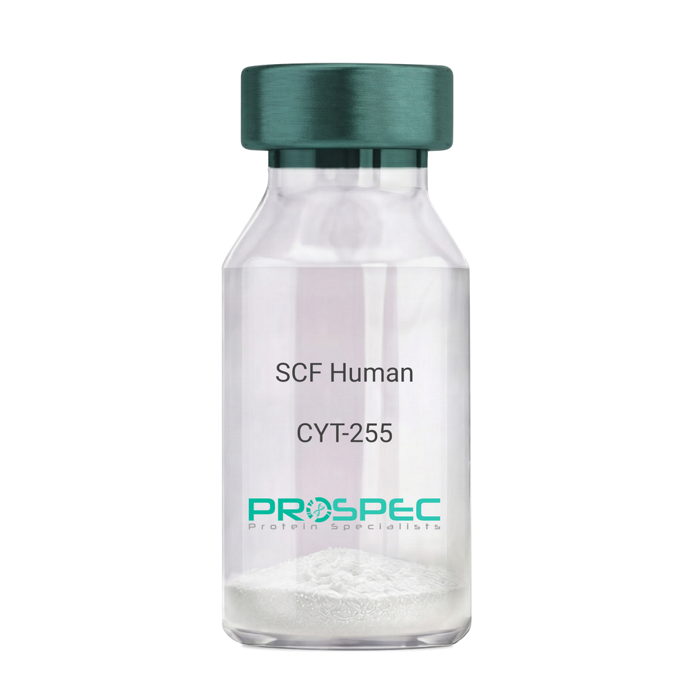
    SCF Human

    Description:

    Stem Cell Factor Human Recombinant

    Kit ligand Precursor, C-kit ligand, SCF, Mast cell growth factor, MGF, SF, KL-1, Kitl, DKFZp686F2250.

    Product # :

    CYT-255

    Price :

    Quantity :

    Shipping Method :

    Room Temp Icon

    Shipped at Room temp

    Add To Cart

    More Info

    • description
    • source
    • formulation
    • purity
    • biological activity
    • More Info

    Description

    Stem Cell Factor Human Recombinant produced in E.Coli is a single, non-glycosylated polypeptide chain containing 165 amino acids and having a molecular mass of 18409 Dalton. The SCF is purified by proprietary chromatographic techniques.

    Source

    Escherichia Coli.

    Formulation

    Lyophilized from a concentrated (1mg/ml) solution in water containing 0.02% NaHCO3.

    Purity

    Greater than 98.0% as determined by:
    (a) Analysis by SEC-HPLC.
    (b) Analysis by SDS-PAGE.

    Biological Activity

    The ED50 as determined by the dose-dependant stimulation of Human TF-1 cells is < 2 ng/ml, corresponding to a Specific Activity of 500,000 IU/mg.

    More Info

    • Introduction

      Stem cell factor / KIT ligand (SCF) is a cytokine which binds CD117(c-Kit). SCF is also known as "steel factor" or "c-kit ligand". SCF exists in two forms, cell surface bound SCF and soluble (or free) SCF. Soluble SCF is produced by the cleavage of surface bound SCF by metalloproteases. SCF is a growth factor important for the survival, proliferation, and differentiation of hematopoietic stem cells and other hematopoietic progenitor cells. One of its roles is to change the BFU-E (burst-forming unit-erythroid) cells, which are the earliest erythrocyte precursors in the erythrocytic series, into the CFU-E (colony-forming unit-erythroid).

    • Synonyms

      Kit ligand Precursor, C-kit ligand, SCF, Mast cell growth factor, MGF, SF, KL-1, Kitl, DKFZp686F2250.

    • Physical Appearance

      Sterile Filtered White lyophilized (freeze-dried) powder.

    • Stability

      Lyophilized KIT ligand although stable at room temperature for 3 weeks, should be stored desiccated below -18°C. Upon reconstitution SCF should be stored at 4°C between 2-7 days and for future use below -18°C.For long term storage it is recommended to add a carrier protein (0.1% HSA or BSA).Please prevent freeze-thaw cycles.

    • Solubility

      It is recommended to reconstitute the lyophilized Stem Cell Factor in sterile 18MΩ-cm H2O not less than 100µg/ml, which can then be further diluted to other aqueous solutions.

    • Amino Acid Sequence

      The sequence of the first five N-terminal amino acids was determined and was found to be Met-Glu-Gly-Ile-Cys.

    • Protein content

      Protein quantitation was carried out by two independent methods1. UV spectroscopy at 280 nm using the absorbency value of 0.52 as the extinction coefficient for a 0.1% (1mg/ml) solution. This value is calculated by the PC GENE computer analysis program of protein sequences (IntelliGenetics). 2. Analysis by RP-HPLC, using a standard solution of Stem Cell Factor as a Reference Standard.

    ProSpec's products are furnished for LABORATORY RESEARCH USE ONLY. The product may not be used as drugs, agricultural or pesticidal products, food additives or household chemicals.

    Scf Human
  • View Data Sheet

    Name :

    BAFFR Human, HEK

    Description:

    BAFF (BLyS) Receptor Human Recombinant, HEK

    TNFRSF13C, CD268, BAFF-R, MGC138235, B cell-activating factor receptor.

    Product # :

    CYT-1224

    Price :

    Quantity :

    Shipping Method :

    Ice Icon

    Shipped with Ice Packs

    Add To Cart

    More Info

    • description
    • source
    • formulation
    • purity
    • biological activity
    • More Info

    Description

    BAFFR Human Recombinant is a single, glycosylated, polypeptide chain (1-78 a.a) containing a total of 314 amino acids and having a molecular mass of 34.4 kDa. BAFFR is fused to 233 a.a hIgG-Tag at C-terminus and is purified by proprietary chromatographic techniques.

    Source

    HEK293 Cells.

    Formulation

    The BAFFR solution (1mg/ml) contains 10% Glycerol and Phosphate-Buffered Saline (pH 7.4).

    Purity

    Greater than 95.0% as determined by SDS-PAGE.

    Biological Activity

    The ED50 is ≤0.7 ug/ml, measured by its ability in a functional ELISA with BAFF Human.

    More Info

    • Synonyms

      TNFRSF13C, CD268, BAFF-R, MGC138235, B cell-activating factor receptor.

    • Physical Appearance

      Sterile Filtered colorless solution.

    • Stability

      Store at 4°C if entire vial will be used within 2-4 weeks. Store, frozen at -20°C for longer periods of time. For long term storage it is recommended to add a carrier protein (0.1% HSA or BSA).Avoid multiple freeze-thaw cycles.

    • Amino Acid Sequence

      DGSMRRGPRS LRGRDAPAPT PCVPAECFDL LVRHCVACGL LRTPRPKPAG ASSPAPRTAL QPQESVGAGA GEAALPLPGL LLEPKSCDKT HTCPPCPAPE LLGGPSVFLF PPKPKDTLMI SRTPEVTCVV VDVSHEDPEV KFNWYVDGVE VHNAKTKPRE EQYNSTYRVV SVLTVLHQDW LNGKEYKCKV SNKALPAPIE KTISKAKGQP REPQVYTLPP SRDELTKNQV SLTCLVKGFY PSDIAVEWES NGQPENNYKT TPPVLDSDGS FFLYSKLTVD KSRWQQGNVF SCSVMHEALH NHYTQKSLSL SPGK.

    • Background

      B-cell Activating Factor (BAFF) and its corresponding receptor, BAFF-R, are integral components of the immune system, orchestrating crucial processes in B-cell survival, maturation, and differentiation. As we delve into the intricate world of immunology, the study of BAFF and its receptor has unveiled essential pathways that govern the immune responses in health and disease. This research investigates the multifaceted role of BAFF Receptor Protein, shedding light on its structural complexities, signaling mechanisms, and its pivotal contributions to immune regulation. By exploring the interactions between BAFF and its receptor, scientists aim to decipher the delicate balance that underlies immune homeostasis and explore potential therapeutic avenues.

      Structural Architecture of BAFF Receptor Protein:

      BAFF Receptor, a transmembrane protein predominantly expressed on B cells, belongs to the tumor necrosis factor receptor (TNFR) superfamily. Its intricate structure involves various domains, each playing a unique role in ligand binding, receptor activation, and downstream signaling. Understanding the structural intricacies of BAFF Receptor is paramount to unraveling the molecular events that govern B-cell fate decisions and immune responses.

      Physiological Significance in B-Cell Biology:

      BAFF Receptor, upon binding with its ligand BAFF, initiates a cascade of events critical for B-cell survival and function. This interaction promotes B-cell maturation, prevents premature apoptosis, and influences the formation of immune synapses. Additionally, BAFF Receptor signaling is tightly regulated to prevent excessive B-cell activation, ensuring immune tolerance and preventing autoimmune responses. Disruptions in these pathways can lead to autoimmune disorders, underscoring the crucial role of BAFF Receptor in maintaining immune equilibrium.

      Regulation of Immune Responses:

      BAFF Receptor signaling not only affects B-cell development but also has broader implications for immune responses. By modulating antibody production, B-cell activation, and immune memory, BAFF Receptor plays a vital role in shaping adaptive immunity. Its dysregulation has been implicated in various autoimmune conditions, making it an attractive target for therapeutic interventions aimed at restoring immune balance.

      BAFF Receptor as a Therapeutic Target:

      The intricate involvement of BAFF Receptor in autoimmune diseases, such as rheumatoid arthritis and systemic lupus erythematosus, has positioned it as a promising therapeutic target. Researchers are exploring monoclonal antibodies and other targeted therapies that aim to modulate BAFF Receptor signaling, providing a new frontier in autoimmune disease management. Additionally, understanding the BAFF-BAFF Receptor axis offers potential insights into the development of vaccines and immunotherapies, fostering innovative approaches in the fight against infectious diseases and malignancies.

      BAFF Receptor Protein, as a key player in immune regulation, embodies the complexities of immunology. Its interactions with BAFF orchestrate fundamental processes in B-cell biology and adaptive immunity. As scientists unravel the intricate signaling pathways and structural nuances of BAFF Receptor, they pave the way for novel therapeutic strategies and innovative treatments for autoimmune disorders and beyond. This research not only deepens our understanding of immune regulation but also holds the promise of transformative advancements in immunotherapy, ultimately shaping the future of immune-related healthcare.

      What is the molecular weight/Mw of BAFF-R Protein?
      BAFF-R Protein has a total Mw of 34.4kDa.

      What is the source or expression system of BAFF-R Protein?
      HEK293 Cells.

      What is the Purity of BAFF-R Protein?
      BAFF-R Protein is >95% pure as determined by SDS-PAGE.

      What is the Biological Activity of BAFF-R Protein?
      The ED50 is ≤0.7 ug/ml, measured by its ability in a functional ELISA with BAFF Human.

      What is the amino acid sequence of BAFF-R Protein?
      DGSMRRGPRS LRGRDAPAPT PCVPAECFDL LVRHCVACGL LRTPRPKPAG ASSPAPRTAL QPQESVGAGA GEAALPLPGL LLEPKSCDKT HTCPPCPAPE LLGGPSVFLF PPKPKDTLMI SRTPEVTCVV VDVSHEDPEV KFNWYVDGVE VHNAKTKPRE EQYNSTYRVV SVLTVLHQDW LNGKEYKCKV SNKALPAPIE KTISKAKGQP REPQVYTLPP SRDELTKNQV SLTCLVKGFY PSDIAVEWES NGQPENNYKT TPPVLDSDGS FFLYSKLTVD KSRWQQGNVF SCSVMHEALH NHYTQKSLSL SPGK.

      What applications can BAFF-R Protein be used in?
      BAFF-R Protein can probably be used in western blot, ELISA and Lateral Flow.

      What is the endotoxin level for BAFF-R Protein?
      The endotoxin level is minimal, BAFF-R Protein was purified using conventional chromatography techniques.

    ProSpec's products are furnished for LABORATORY RESEARCH USE ONLY. The product may not be used as drugs, agricultural or pesticidal products, food additives or household chemicals.

    Baff Receptor Human
  • View Data Sheet

    Name :

    OPG Fc Human

    Description:

    Osteoprotegerin Human Recombinant /Fc Chimera

    TNFRSF11B, OPG, OCIF, Osteoclastogenesis inhibitory factor, TR1, MGC29565.

    Product # :

    CYT-266

    Price :

    Quantity :

    Shipping Method :

    Room Temp Icon

    Shipped at Room temp

    Add To Cart

    More Info

    • description
    • source
    • formulation
    • purity
    • biological activity
    • More Info

    Description

    Recombinant OPG produced in yeast contains 2x412 amino acid residues, including 180 residues from mature OPG (a.a 22-201) and 232 residues from the Fc protein of human IgG1, and has a calculated molecular mass of 109.6kDa.

    Source

    Pichia Pastoris.

    Formulation

    OPG was lyophilized from a 0.2µm filtered concentrated solution in 20mM PB, pH 6.0, 150mM NaCl and 0.02 % Tween-80.

    Purity

    Greater than 90.0% as determined by:
    (a) Analysis by RP-HPLC.
    (b) Analysis by SDS-PAGE.

    Biological Activity

    The ED50 as determined by neutralizing the stimulation of U937 cells is less tha10ng/ml, corresponding to a specific activity of > 1.0 × 10⁵ IU/mg in the presence of 10ng/ml soluble Human RANKL (sRANKL).

    More Info

    • Introduction

      Osteoprotegerin acts as decoy receptor for rankl and thereby neutralizes its function in osteoclastogenesis. OPG inhibits the activation of osteoclasts and promotes osteoclast apoptosis in vitro. Bone homeostasis seems to depend on the local rankl/opg ratio. Osteoprotegerin may also play a role in preventing arterial calcification. May act as decoy receptor for trail and protect against apoptosis. Trail binding blocks the inhibition of osteoclastogenesis.

    • Synonyms

      TNFRSF11B, OPG, OCIF, Osteoclastogenesis inhibitory factor, TR1, MGC29565.

    • Physical Appearance

      Sterile Filtered White lyophilized (freeze-dried) powder.

    • Stability

      Lyophilized Osteoprotegerin although stable at room temperature for 3 weeks, should be stored desiccated below -18°C. Upon reconstitution OCIF should be stored at 4°C between 2-7 days and for future use below -18°C. For long term storage it is recommended to add a carrier protein (0.1% HSA or BSA).Please prevent freeze-thaw cycles.

    • Solubility

      It is recommended to reconstitute the lyophilized Osteoprotegerin in sterile 18MΩ-cm H2O not less than 100µg/ml, which can then be further diluted to other aqueous solutions.

    • Amino Acid Sequence

      OPG 22-201 ETFPPKYLHY DEETSHQLLC DKCPPGTYLK QHCTAKWKTV CAPCPDHYYT DSWHTSDECL YCSPVCKELQ YVKQECNRTH NRVCECKEGR YLEIEFCLKH RSCPPGFGVV QAGTPERNTV CKRCPDGFFS NETSSKAPCR KHTNCSVFGL LLTQKGNATH DNICSGNSES TQKCGIDVTL
      Fc232EPKSSDKTHT CPPCPAPEFE GAPSVFLFPP KPKDTLMISR TPEVTCVVVD VSHEDPEVKF NWYVDGVEVH NAKTKPREEQ YNSTYRVVSV LTVLHQDWLN GKEYKCKVSN KALPTPIEKTISKAKGQPRE PQVYTLPPSR DELTKNQVSL TCLVKGFYPS DIAVEWESNG QPENNYKTTP PVLDSDGSFF LYSKLTVDKS RWQQGNVFSC SVMHEALHNH YTQKSLSLSP GK

    ProSpec's products are furnished for LABORATORY RESEARCH USE ONLY. The product may not be used as drugs, agricultural or pesticidal products, food additives or household chemicals.

    Osteoprotegerin Human
  • View Data Sheet

    Name :

    ATF3 Human

    Description:

    Activating Transcription Factor-3 Human Recombinant

    Activating transcription factor 3, cyclic AMP-dependent transcription factor ATF-3, FLJ41705.

    Product # :

    PKA-270

    Price :

    Quantity :

    Shipping Method :

    Ice Icon

    Shipped with Ice Packs

    Add To Cart

    More Info

    • description
    • source
    • formulation
    • purity
    • More Info

    Description

    ATF3 Human Recombinant produced in E.coli is a single, non-glycosylated polypeptide chain containing 181 amino acids (1-181) and having a molecular mass of 20.6 kDa.The ATF3 is purified by proprietary chromatographic techniques.

    Source

    Escherichia Coli.

    Formulation

    ATF3 protein (1mg/ml) is supplied in 20mM Tris-HCl, pH-8, 2M Urea and 20% Glycerol.

    Purity

    Greater than 90.0% as determined by SDS-PAGE.

    More Info

    • Introduction

      ATF3 belongs to the mammalian activation transcription factor/cAMP responsive element-binding (CREB) protein family of transcription factors. ATF3 binds the cAMP response element (CRE) (consensus: 5'-GTGACGT[AC][AG]-3'), a sequence present in numerous viral and cellular promoters. Multiple transcript variants encoding two different isoforms are known for this gene. The longer isoform represses transcription from promoters with ATF binding elements instead of activating it. The shorter isoform (deltaZip2) which stimulates transcription by sequestering inhibitory co-factors away from the promoter has no leucine zipper protein-dimerization motif and does not bind to DNA.

    • Synonyms

      Activating transcription factor 3, cyclic AMP-dependent transcription factor ATF-3, FLJ41705.

    • Physical Appearance

      ATF3 is supplied as a sterile filtered clear solution.

    • Stability

      Store at 4°C if entire vial will be used within 2-4 weeks. Store, frozen at -20°C for longer periods of time. For long term storage it is recommended to add a carrier protein (0.1% HSA or BSA).Avoid multiple freeze-thaw cycles.

    • Amino Acid Sequence

      MMLQHPGQVS ASEVSASAIV PCLSPPGSLV FEDFANLTPF VKEELRFAIQ NKHLCHRMSS ALESVTVSDR PLGVSITKAE VAPEEDERKK RRRERNKIAA AKCRNKKKEK TECLQKESEK LESVNAELKA QIEELKNEKQ HLIYMLNLHR PTCIVRAQNG RTPEDERNLF IQQIKEGTLQ S.

    ProSpec's products are furnished for LABORATORY RESEARCH USE ONLY. The product may not be used as drugs, agricultural or pesticidal products, food additives or household chemicals.

    Atf3 Human
  • View Data Sheet

    Name :

    EFNA1 Human

    Description:

    Ephrin A1 Human Recombinant

    Ephrin-A1, EPLG1, TNFAIP4, LERK1, EFL1, ECKLG, EPH-related receptor tyrosine kinase ligand 1, Immediate early response protein B61, Tumor necrosis factor alpha-induced protein 4, TNF alpha-induced protein 4, ligand of eph-related kinase 1, tumor necrosis factor, alpha-induced protein 4.

    Product # :

    PRO-971

    Price :

    Quantity :

    Shipping Method :

    Ice Icon

    Shipped with Ice Packs

    Add To Cart

    More Info

    • description
    • source
    • formulation
    • purity
    • More Info

    Description

    EFNA1 Human Recombinant produced in E.coli is a single, non-glycosylated polypeptide chain containing 185 amino acids (19-182) and having a molecular mass of 21.6 kDa.EFNA1 is fused to a 21 amino acid His-tag at N-terminus & purified by proprietary chromatographic techniques.

    Source

    E.coli.

    Formulation

    The EFNA1 solution (1mg/ml) contains 20mM Tris-HCl buffer (pH 8.0), 0.4M Urea and 10% glycerol.

    Purity

    Greater than 90% as determined by SDS-PAGE.

    More Info

    • Introduction

      EFNA1 belongs to the ephrin (EPH) family. The EPH subfamily is the biggest group of receptor protein kinases and they take part in vital nervous system function and development.

    • Synonyms

      Ephrin-A1, EPLG1, TNFAIP4, LERK1, EFL1, ECKLG, EPH-related receptor tyrosine kinase ligand 1, Immediate early response protein B61, Tumor necrosis factor alpha-induced protein 4, TNF alpha-induced protein 4, ligand of eph-related kinase 1, tumor necrosis factor, alpha-induced protein 4.

    • Physical Appearance

      Sterile Filtered colorless solution.

    • Stability

      Store at 4°C if entire vial will be used within 2-4 weeks. Store, frozen at -20°C for longer periods of time. For long term storage it is recommended to add a carrier protein (0.1% HSA or BSA).Avoid multiple freeze-thaw cycles.

    • Amino Acid Sequence

      MGSSHHHHHH SSGLVPRGSH MDRHTVFWNS SNPKFRNEDY TIHVQLNDYV DIICPHYEDH SVADAAMEQY ILYLVEHEEY QLCQPQSKDQ VRWQCNRPSA KHGPEKLSEK FQRFTPFTLG KEFKEGHSYY YISKPIHQHE DRCLRLKVTV SGKITHSPQA HVNPQEKRLA ADDPEVRVLH SIGHS

    ProSpec's products are furnished for LABORATORY RESEARCH USE ONLY. The product may not be used as drugs, agricultural or pesticidal products, food additives or household chemicals.

    Efna1 Human
  • View Data Sheet

    Name :

    TNFSF7 Human, sf9

    Description:

    CD70 Human Recombinant, sf9

    CD70 Molecule, Tumor Necrosis Factor (Ligand) Superfamily, Member 7, Tumor Necrosis Factor Ligand Superfamily Member 7 , CD27 Ligand, CD27LG, TNFSF7, CD27-L, CD27L, Tumor Necrosis Factor Ligand 8A, Surface Antigen CD70, Ki-24 Antigen, CD70 Antigen, TNLG8A.

    Product # :

    CYT-953

    Price :

    Quantity :

    Shipping Method :

    Ice Icon

    Shipped with Ice Packs

    Add To Cart

    More Info

    • description
    • source
    • formulation
    • purity
    • More Info

    Description

    TNFSF7 produced in Sf9 Baculovirus cells is a single, glycosylated polypeptide chain containing 164 amino acids (39-193 a.a.) and having a molecular mass of 18.2kDa. TNFSF7 is expressed with a 6 amino acid His tag at C-Terminus and purified by proprietary chromatographic techniques.

    Source

    Sf9, Baculovirus cells.

    Formulation

    TNFSF7 protein solution (0.25mg/ml) contains Phosphate Buffered Saline (pH 7.4) and 10% glycerol.

    Purity

    Greater than 90.0% as determined by SDS-PAGE.

    More Info

    • Introduction

      CD70 also known as TNFSF7 is a cytokine which binds to CD27. TNFSF7 takes part in T-cell activation as well as induces the proliferation of costimulated T-cells. Moreover, TNFSF7 enhances the generation of cytolytic T-cells. Among the diseases which are associated with TNFSF7: Include acute myocarditis & Myocarditis.

    • Synonyms

      CD70 Molecule, Tumor Necrosis Factor (Ligand) Superfamily, Member 7, Tumor Necrosis Factor Ligand Superfamily Member 7 , CD27 Ligand, CD27LG, TNFSF7, CD27-L, CD27L, Tumor Necrosis Factor Ligand 8A, Surface Antigen CD70, Ki-24 Antigen, CD70 Antigen, TNLG8A.

    • Physical Appearance

      Sterile Filtered colorless solution.

    • Stability

      Store at 4°C if entire vial will be used within 2-4 weeks. Store, frozen at -20°C for longer periods of time. For long term storage it is recommended to add a carrier protein (0.1% HSA or BSA). Avoid multiple freeze-thaw cycles.

    • Amino Acid Sequence

      ADPQRFAQAQ QQLPLESLGW DVAELQLNHT GPQQDPRLYW QGGPALGRSF LHGPELDKGQ LRIHRDGIYM VHIQVTLAIC SSTTASRHHP TTLAVGICSP ASRSISLLRL SFHQGCTIAS QRLTPLARGD TLCTNLTGTL LPSRNTDETF FGVQWVRPHH HHHH.

    ProSpec's products are furnished for LABORATORY RESEARCH USE ONLY. The product may not be used as drugs, agricultural or pesticidal products, food additives or household chemicals.

    Tnfsf7 Human Sf9
  • View Data Sheet

    Name :

    Eotaxin Human

    Description:

    Eotaxin Human Recombinant (CCL11)

    Small inducible cytokine A11, CCL11, Eosinophil chemotactic protein, chemokine (C-C motif) ligand 11, SCYA11, MGC22554.

    Product # :

    CHM-256

    Price :

    Quantity :

    Shipping Method :

    Room Temp Icon

    Shipped at Room temp

    Add To Cart

    More Info

    • description
    • source
    • formulation
    • purity
    • biological activity
    • More Info

    Description

    Eotaxin Human Recombinant produced in E.Coli is a single, non-glycosylated polypeptide chain containing 74 amino acids and having a molecular mass of 8345.9 Dalton. The CCL11 is purified by proprietary chromatographic techniques.

    Source

    Escherichia Coli.

    Formulation

    Lyophilized from a 0.2µm filtered concentrated (1.0mg/ml) solution in 20mM PB, pH 7.4, 150mM NaCl.

    Purity

    Greater than 97.0% as determined by:
    (a) Analysis by RP-HPLC.
    (b) Analysis by SDS-PAGE.

    Biological Activity

    The activity is determined by the chemoattract of human PBE (peripheral blood eosinophils) at a concentration between 0.1-10 ng/ml corresponding to a Specific Activity of 100,000-10,000,000IU/mg.

    More Info

    • Introduction

      Chemokine (C-C motif) ligand 11 (CCL11) is a small cytokine belonging to the CC chemokine family that is also known as eotaxin. CCL11 selectively recruits eosinophils by inducing their chemotaxis, and therefore, is implicated in allergic responses. The effects of CCL11 are mediated by its binding to a G-protein-linked receptor known as a chemokine receptor. Chemokine receptors for which CCL11 is a ligand include CCR2, CCR3 and CCR5. The gene for human CCL11 (scya11) is encoded on three exons and is located on chromosome 17.

    • Synonyms

      Small inducible cytokine A11, CCL11, Eosinophil chemotactic protein, chemokine (C-C motif) ligand 11, SCYA11, MGC22554.

    • Physical Appearance

      Sterile Filtered White lyophilized (freeze-dried) powder.

    • Stability

      Lyophilized Eotaxin although stable at room temperature for 3 weeks, should be stored desiccated below -18°C. Upon reconstitution CCL11 should be stored at 4°C between 2-7 days and for future use below -18°C.For long term storage it is recommended to add a carrier protein (0.1% HSA or BSA).Please prevent freeze-thaw cycles.

    • Solubility

      It is recommended to reconstitute the lyophilized Eotaxin Human Recombinant in sterile 18MΩ-cm H2O not less than 100µg/ml, which can then be further diluted to other aqueous solutions.

    • Amino Acid Sequence

      GPASVPTTCC FNLANRKIPL QRLESYRRIT SGKCPQKAVI FKTKLAKDICADPKKKWVQD

      SMKYLDQKSP TPKP.

    • Background

      What is the molecular weight/Mw of EOTAXIN HUMAN Protein?
      EOTAXIN HUMAN Protein has a total Mw of 8.3459kDa.

      What is the source or expression system of EOTAXIN HUMAN Protein?
      Escherichia Coli.

      What is the Purity of EOTAXIN HUMAN Protein?
      EOTAXIN HUMAN Protein is >97% pure as determined by SDS-PAGE.

      What is the Biological Activity of EOTAXIN HUMAN Protein?
      The activity is determined by the chemoattract of human PBE (peripheral blood eosinophils) at a concentration between 0.1-10 ng/ml corresponding to a Specific Activity of 100,000-10,000,000IU/mg.

      What is the amino acid sequence of EOTAXIN HUMAN Protein?
      GPASVPTTCC FNLANRKIPL QRLESYRRIT SGKCPQKAVI FKTKLAKDICADPKKKWVQD
      SMKYLDQKSP TPKP.

      What applications can EOTAXIN HUMAN Protein be used in?
      EOTAXIN HUMAN Protein can probably be used in western blot, ELISA and Lateral Flow.

      What is the endotoxin level for EOTAXIN HUMAN Protein?
      The endotoxin level is minimal, EOTAXIN HUMAN Protein was purified using conventional chromatography techniques.


    ProSpec's products are furnished for LABORATORY RESEARCH USE ONLY. The product may not be used as drugs, agricultural or pesticidal products, food additives or household chemicals.

    Eotaxin Human
  • View Data Sheet

    Name :

    VEGI Human, His

    Description:

    Vascular Endothelial Growth InhibitorHuman Recombinant, His Tag

    Tumor necrosis factor ligand superfamily member 15, TNFSF-15, TNFSF15, TNF ligand-related molecule 1, VEGI, TL-1, TL1, TL1A, VEGI192A, VEGI-192, MGC129934, MGC129935.

    Product # :

    CYT-589

    Price :

    Quantity :

    Shipping Method :

    Ice Icon

    Shipped with Ice Packs

    Add To Cart

    More Info

    • description
    • source
    • formulation
    • purity
    • More Info

    Description

    VEGI Human Recombinant fused with a 21 amino acid His tag at N-terminus produced in E.Coli is a single, non-glycosylated, polypeptide chain containing 201 amino acids(72-251a.a.) and having a molecular mass of 22.7kDa. The VEGI is purified by proprietary chromatographic techniques.

    Source

    Escherichia Coli.

    Formulation

    The VEGI solution (1mg/1ml) contains 20mM Tris-HCl buffer (pH 8.0), 50% glycerol, 1mM DTT and 0.2M NaCl.

    Purity

    Greater than 95.0% as determined by SDS-PAGE.

    More Info

    • Introduction

      TNFSF15 is a cytokine that belongs to the tumor necrosis factor (TNF) ligand family. This protein is abundantly expressed in endothelial cells, but is not expressed in either B or T cells. The expression of TNFSF15 is inducible by TNF and IL-1 alpha. This cytokine is a ligand for receptor TNFRSF25 and decoy receptor TNFRSF21/DR6. It can activate NF-kappaB and MAP kinases, and acts as an autocrine factor to induce apoptosis in endothelial cells. TNFSF15 is also found to inhibit endothelial cell proliferation, and thus may function as an angiogenesis inhibitor. An additional isoform encoded by an alternatively spliced transcript variant has been reported but the sequence of this transcript has not been determined.

    • Synonyms

      Tumor necrosis factor ligand superfamily member 15, TNFSF-15, TNFSF15, TNF ligand-related molecule 1, VEGI, TL-1, TL1, TL1A, VEGI192A, VEGI-192, MGC129934, MGC129935.

    • Physical Appearance

      Sterile Filtered colorless solution.

    • Stability

      Store at 4°C if entire vial will be used within 2-4 weeks. Store, frozen at -20°C for longer periods of time. For long term storage it is recommended to add a carrier protein (0.1% HSA or BSA).Avoid multiple freeze-thaw cycles.

    • Amino Acid Sequence

      MGSSHHHHHH SSGLVPRGSH MLKGQEFAPS HQQVYAPLRA DGDKPRAHLT VVRQTPTQHF KNQFPALHWE HELGLAFTKN RMNYTNKFLL IPESGDYFIY SQVTFRGMTS ECSEIRQAGR PNKPDSITVV ITKVTDSYPE PTQLLMGTKS VCEVGSNWFQ PIYLGAMFSL QEGDKLMVNV SDISLVDYTK EDKTFFGAFL L

    ProSpec's products are furnished for LABORATORY RESEARCH USE ONLY. The product may not be used as drugs, agricultural or pesticidal products, food additives or household chemicals.

    Vegi Human His
  • View Data Sheet

    Name :

    BMF Human

    Description:

    Bcl2 Modifying Factor, Isoform 3 Human Recombinant

    Bcl-2-modifying factor, FLJ00065, BMF.

    Product # :

    PRO-700

    Price :

    Quantity :

    Shipping Method :

    Ice Icon

    Shipped with Ice Packs

    Add To Cart

    More Info

    • description
    • source
    • formulation
    • purity
    • More Info

    Description

    Bcl2 modifying factor Human Recombinant produced in E.Coli is a single, non-glycosylated, polypeptide chain containing 144 amino acids (1-129 a.a.) and having a molecular mass of 15.6 kDa.The Bcl2 modifying factor is fused to 15 amino acid His Tag at Nterminus and purified by proprietary chromatographic techniques.

    Source

    Escherichia Coli.

    Formulation

    The Bcl2 modifying factor solution contains 20mM Tris pH-7.5, 1mM DTT & 10% glycerol.

    Purity

    Greater than 90.0% as determined by SDS-PAGE.

    More Info

    • Introduction

      Bcl2 modifying factor, is a member of the Bcl2 protein family of apoptosis mediators. Bcl2 modifying factor is widely expressed in many tissues.
      Bcl2 modifying factor contains a single Bcl2 homology domain 3 (BH3), and binds Bcl2 proteins and functions as an apoptotic activator. Also, Bcl2 modifying factor is important for histone deacetylase (HDAC) inhibitors which alters the balance between acetylation and deacetylation, significantly increasing histone acetylation, while strongly inducing apoptosis in a variety of cancer cell types. Bcl2 modifying factor supports Bim in regulating cell death processes in response to many stimuli. A synergistic role for bim and Bcl2 modifying factor in an apoptotic pathway leading to the clearance of Neisseria gonorrhoeae -infected cells.

    • Synonyms

      Bcl-2-modifying factor, FLJ00065, BMF.

    • Physical Appearance

      Sterile Filtered colorless solution.

    • Stability

      Store at 4°C if entire vial will be used within 2-4 weeks. Store, frozen at -20°C for longer periods of time. For long term storage it is recommended to add a carrier protein (0.1% HSA or BSA).Avoid multiple freeze-thaw cycles.

    • Amino Acid Sequence

      MASMTGGQQM GRGSHMEPSQ CVEELEDDVF QPEDGEPVTQ PGSLLSADLF AQSLLDCPLS RLQLFPLTHC CGPGLRPTSQ EDKATQTLSPASPSQGVMLP CGVTEEPQRL FYAPAEPKSC VVADPPLPAQ PCFEWRREQE RGRP.

    ProSpec's products are furnished for LABORATORY RESEARCH USE ONLY. The product may not be used as drugs, agricultural or pesticidal products, food additives or household chemicals.

    Bcl2 Modifying Factor Human
  • View Data Sheet

    Name :

    CLCF1 Human

    Description:

    Neurotrophin-1 Human Recombinant

    Cardiotrophin-Like Cytokine Factor 1, Novel Neurotrophin-1 , BSF3, CLC, CRLF1 Associated Cytokine-Like Factor 1, B-Cell Stimulating Factor 3, B-Cell-Stimulating Factor 3, BSF-3, NNT-1, NNT1, Neurotrophin-1/B-Cell Stimulating Factor-3, Cold-Induced Sweating Syndrome 2, B-Cell Stimulatory Factor 3, CISS2, NR6, CLCF1.

    Product # :

    CYT-869

    Price :

    Quantity :

    Shipping Method :

    Room Temp Icon

    Shipped at Room temp

    Add To Cart

    More Info

    • description
    • source
    • formulation
    • purity
    • biological activity
    • More Info

    Description

    Neurotrophin-1 Human Recombinant (28-225) produced in E.Coli is a single, non-glycosylated, polypeptide chain containing 199 amino acids and having a molecular mass of 22kDa.The NNT-1 is purified by proprietary chromatographic techniques.

    Source

    Escherichia Coli.

    Formulation

    NNT-1 protein was lyophilized from a 0.2µm filtered solution in Acetonitrile and TFA.

    Purity

    Greater than 95.0% as determined by SDS-PAGE.

    Biological Activity

    The ED50, as measured in a cell proliferation assay using human TF?1 cells transfected with human CNTF R?, is less than 15ng/ml.

    More Info

    • Introduction

      Cardiotrophin-like cytokine (CLC/ NNT-1) belongs to the IL-6 family of cytokines. All family members share the receptor subunit gp130, which belongs to the type I cytokine receptor superfamily. NNT1 is a trophic factor for motor neurons, a stimulator of ACTH release from corticotrophs, and an inducer of IgE synthesis and B cell proliferation. Cells expressing NNT-1 include embryonic muscle, lung epithelium, and mesenchyme. NNT1 binds to and activates the ILST/gp130 receptor.

    • Synonyms

      Cardiotrophin-Like Cytokine Factor 1, Novel Neurotrophin-1 , BSF3, CLC, CRLF1 Associated Cytokine-Like Factor 1, B-Cell Stimulating Factor 3, B-Cell-Stimulating Factor 3, BSF-3, NNT-1, NNT1, Neurotrophin-1/B-Cell Stimulating Factor-3, Cold-Induced Sweating Syndrome 2, B-Cell Stimulatory Factor 3, CISS2, NR6, CLCF1.

    • Physical Appearance

      Sterile Filtered White lyophilized (freeze-dried) powder.

    • Stability

      Lyophilized Neurotrophin-1 although stable at room temperature for 3 weeks, should be stored desiccated below -18°C. Upon reconstitution NNT1 should be stored at 4°C between 2-7 days and for future use below -18°C. For long term storage it is recommended to add a carrier protein (0.1% HSA or BSA). Please prevent freeze-thaw cycles.

    • Solubility

      It is recommended to reconstitute the lyophilized NNT1 in sterile 18M-cm H2O not less than 0.5mg/ml, which can then be further diluted to other aqueous solutions.

    • Amino Acid Sequence

      MLNRTGDPGP GPSIQKTYDL TRYLEHQLRS LAGTYLNYLG PPFNEPDFNP PRLGAETLPR ATVDLEVWRS LNDKLRLTQN YEAYSHLLCY LRGLNRQAAT AELRRSLAHF CTSLQGLLGS IAGVMAALGY PLPQPLPGTE PTWTPGPAHS DFLQKMDDFW LLKELQTWLW RSAKDFNRLK KKMQPPAAAV TLHLGAHGF.

    • Background

      What is the molecular weight/Mw of CLCF Protein?
      CLCF Protein has a total Mw of 22kDa.

      What is the source or expression system of CLCF Protein?
      Escherichia Coli.

      What is the Purity of CLCF Protein?
      CLCF Protein is >95% pure as determined by SDS-PAGE.

      What is the Biological Activity of CLCF Protein?
      The ED50, as measured in a cell proliferation assay using human TF?1 cells transfected with human CNTF R?, is less than 15ng/ml.

      What is the amino acid sequence of CLCF Protein?
      MLNRTGDPGP GPSIQKTYDL TRYLEHQLRS LAGTYLNYLG PPFNEPDFNP PRLGAETLPR ATVDLEVWRS LNDKLRLTQN YEAYSHLLCY LRGLNRQAAT AELRRSLAHF CTSLQGLLGS IAGVMAALGY PLPQPLPGTE PTWTPGPAHS DFLQKMDDFW LLKELQTWLW RSAKDFNRLK KKMQPPAAAV TLHLGAHGF.

      What applications can CLCF Protein be used in?
      CLCF Protein can probably be used in western blot, ELISA and Lateral Flow.

      What is the endotoxin level for CLCF Protein?
      The endotoxin level is minimal, CLCF Protein was purified using conventional chromatography techniques.

    ProSpec's products are furnished for LABORATORY RESEARCH USE ONLY. The product may not be used as drugs, agricultural or pesticidal products, food additives or household chemicals.

    Nnt1 Human
  • View Data Sheet

    Name :

    HB-EGF Human, His

    Description:

    Proheparin-Binding EGF-like Growth Factor Human Recombinant, His Tag

    Proheparin-binding EGF-like growth factor, HBEGF, DTR, DTS, HEGFL, HB-EGF, Heparin-binding EGF-like growth factor, Diphtheria toxin receptor, DT-R, DTSF.

    Product # :

    CYT-761

    Price :

    Quantity :

    Shipping Method :

    Ice Icon

    Shipped with Ice Packs

    Add To Cart

    More Info

    • description
    • source
    • formulation
    • purity
    • More Info

    Description

    HB-EGF His Human Recombinant produced in E.coli is a single, non-glycosylated polypeptide chain containing 109 amino acids (63-148) and having a molecular mass of 12.1kDa.HB-EGF His is fused to a 23 amino acid His-tag at N-terminus & purified by proprietary chromatographic techniques.

    Source

    Escherichia Coli.

    Formulation

    The HB-EGF His solution (0. 5mg/ml) contains 20mM Tris-HCl buffer (pH 8.0), 0.2M NaCl, 2mM DTT and 50% glycerol.

    Purity

    Greater than 85% as determined by SDS-PAGE.

    More Info

    • Introduction

      HB-EGF is an EGF related growth factor which signals via the EGF receptor, and stimulates the proliferation of SMC (smooth muscle cells), fibroblasts, epithelial cells and keratinocytes. HB-EGF is expressed in various cell types and tissues, including vascular endothelial cells and SMC, macrophages, skeletal muscle, keratinocytes and particular tumor cells. HB-EGF’s ability to explicitly bind heparin and heparin sulfate proteoglycans is dissimilar from other EGF-like molecules, and might be related to the enhanced mitogenic activity, relative to EGF, that HB-EGF exerts on smooth muscle cells.

    • Synonyms

      Proheparin-binding EGF-like growth factor, HBEGF, DTR, DTS, HEGFL, HB-EGF, Heparin-binding EGF-like growth factor, Diphtheria toxin receptor, DT-R, DTSF.

    • Physical Appearance

      Sterile Filtered clear solution.

    • Stability

      Store at 4°C if entire vial will be used within 2-4 weeks. Store, frozen at -20°C for longer periods of time. For long term storage it is recommended to add a carrier protein (0.1% HSA or BSA).Avoid multiple freeze-thaw cycles.

    • Amino Acid Sequence

      MGSSHHHHHH SSGLVPRGSH MGSDLQEADL DLLRVTLSSK PQALATPNKE EHGKRKKKGK GLGKKRDPCL RKYKDFCIHG ECKYVKELRA PSCICHPGYH GERCHGLSL.

    • Background

      What is the molecular weight/Mw of HB-EGF Protein?
      HB-EGF Protein has a total Mw of 12.1kDa.

      What is the source or expression system of HB-EGF Protein?
      Escherichia Coli.

      What is the Purity of HB-EGF Protein?
      HB-EGF Protein is >85% pure as determined by SDS-PAGE.

      What is the Biological Activity of HB-EGF Protein?
      The biological functionality of HB-EGF Protein will be determined in the future.

      What is the amino acid sequence of HB-EGF Protein?
      MGSSHHHHHH SSGLVPRGSH MGSDLQEADL DLLRVTLSSK PQALATPNKE EHGKRKKKGK GLGKKRDPCL RKYKDFCIHG ECKYVKELRA PSCICHPGYH GERCHGLSL.

      What applications can HB-EGF Protein be used in?
      HB-EGF Protein can probably be used in western blot, ELISA and Lateral Flow.

      What is the endotoxin level for HB-EGF Protein?
      The endotoxin level is minimal, HB-EGF Protein was purified using conventional chromatography techniques.

    ProSpec's products are furnished for LABORATORY RESEARCH USE ONLY. The product may not be used as drugs, agricultural or pesticidal products, food additives or household chemicals.

    Hb Egf Human His
  • View Data Sheet

    Name :

    TNFRSF8 Mouse

    Description:

    CD30 Ligand Receptor Mouse Recombinant

    Tumor Necrosis Factor Receptor Superfamily, Member 8, Lymphocyte Activation Antigen CD30, CD30L Receptor, Ki-1 Antigen, D1S166E, CD30, Cytokine Receptor CD30, CD30 Antigen, Ki-1

    Product # :

    CYT-1165

    Price :

    Quantity :

    Shipping Method :

    Ice Icon

    Shipped with Ice Packs

    Add To Cart

    More Info

    • description
    • source
    • formulation
    • purity
    • More Info

    Description

    TNFRSF8 Mouse Recombinant produced in Baculovirus is a single glycosylated polypeptide chain containing 479 amino acids (19-258 aa) and having a molecular mass of 52.2kDa.TNFRSF8 is fused to a 239 amino acid hIgG-His tag at C-terminus and purified by proprietary chromatographic techniques.

    Source

    Sf9, Baculovirus cells.

    Formulation

    TNFRSF8 protein (1mg/ml) contains 10% glycerol and Phosphate-Buffered Saline (pH 7.4).

    Purity

    Greater than 90.0% as determined by SDS-PAGE.

    More Info

    • Introduction

      Tumor necrosis factor receptor superfamily member 8 (TNFRSF8) is a receptor for TNFSF8/CD30L. TNFRSF8 has a role in the regulation of cellular growth and transformation of activated lymphoblasts. In addition, the TNFRSF8 protein regulates gene expression via activation of NF-kappa-B. TNFRSF8 being a regulator of apoptosis, induces cell death or proliferation, depending on the cell type.

    • Synonyms

      Tumor Necrosis Factor Receptor Superfamily, Member 8, Lymphocyte Activation Antigen CD30, CD30L Receptor, Ki-1 Antigen, D1S166E, CD30, Cytokine Receptor CD30, CD30 Antigen, Ki-1

    • Physical Appearance

      Sterile Filtered colorless solution.

    • Stability

      Store at 4°C if entire vial will be used within 2-4 weeks. Store, frozen at -20°C for longer periods of time. For long term storage it is recommended to add a carrier protein (0.1% HSA or BSA).Avoid multiple freeze-thaw cycles.

    • Amino Acid Sequence

      FPTDRPLKTT CAGDLSHYPG EAARNCCYQC PSGLSPTQPC PRGPAHCRKQ CAPDYYVNED GKCTACVTCL PGLVEKAPCS GNSPRICECQ PGMHCCTPAV NSCARCKLHC SGEEVVKSPG TAKKDTICEL PSSGSGPNCS NPGDRKTLTS HATPQAMPTL ESPANDSARS LLPMRVTNLV QEDATELVKV PESSSSKARE PSPDPGNAEK NMTLELPSPG TLPDISTSEN SKEPASTAST LEPKSCDKTH TCPPCPAPEL LGGPSVFLFP PKPKDTLMIS RTPEVTCVVV DVSHEDPEVK FNWYVDGVEV HNAKTKPREE QYNSTYRVVS VLTVLHQDWL NGKEYKCKVS NKALPAPIEK TISKAKGQPR EPQVYTLPPS RDELTKNQVS LTCLVKGFYP SDIAVEWESN GQPENNYKTT PPVLDSDGSF FLYSKLTVDK SRWQQGNVFS CSVMHEALHN HYTQKSLSLS PGKHHHHHH

    ProSpec's products are furnished for LABORATORY RESEARCH USE ONLY. The product may not be used as drugs, agricultural or pesticidal products, food additives or household chemicals.

    Cd30 Mouse
  • View Data Sheet

    Name :

    FLT1 D3 Human, His

    Description:

    Vascular Endothelial Growth Factor Receptor-1 D3 Human Recombinant, His Tag

    FLT-1, FLT1, Tyrosine-protein kinase receptor FLT, Flt-1, Tyrosine-protein kinase FRT, Fms-like tyrosine kinase 1, VEGFR-1.

    Product # :

    PKA-338

    Price :

    Quantity :

    Shipping Method :

    Ice Icon

    Shipped with Ice Packs

    Add To Cart

    More Info

    • description
    • source
    • formulation
    • purity
    • More Info

    Description

    FLT1 D1-3 Human Recombinant produced in baculovirus is monomeric, glycosylated, polypeptide containing 298 amino acids fragment (31-328) and having a molecular mass of 38.16kDa. The receptor protein contains only the first 3 extracellular domains, which contain all the information necessary for binding of VEGF.The FLT1 is purified by proprietary chromatographic techniques.

    Source

    Insect Cells.

    Formulation

    FLT1 His (0.96mg/ml) is supplied in 25mM Na-Acetate pH 4.8 and 50% glycerol.

    Purity

    Greater than 95.0% as determined by:
    (a) Analysis by RP-HPLC.
    (b) Analysis by SDS-PAGE.

    More Info

    • Introduction

      Endothelial cells express three different vascular endothelial growth factor (VEGF) receptors, belonging to the family of receptor tyrosine kinases (RTKs). They are named VEGFR-1 (Flt-1), VEGFR-2 (KDR/Flk-1), VEGFR-3 (Flt-4). Their expression is almost exclusively restricted to endothelial cells, but VEGFR-1 can also be found on monocytes, dendritic cells and on trophoblast cells. The flt-1 gene was first described in 1990. The receptor contains seven immunoglobulin-like extracellular domains, a single transmembrane region and an intracellular splited tyrosine kinase domain. Compared to VEGFR-2 the Flt-1 receptor has a higher affinity for VEGF but a weaker signaling activity. VEGFR-1 thus leads not to proliferation of endothelial cells, but mediates signals for differentiation. Interestingly a naturally occuring soluble variant of VEGFR-1 (sVEGFR-1) was found in HUVE supernatants in 1996, which is generated by alternative splicing of the flt-1 mRNA. The biological functions of sVEGFR-1 still are not clear, but it seems to be an endogenous regulator of angiogenesis, binding VEGF with the same affinity as the full-length receptor.

    • Synonyms

      FLT-1, FLT1, Tyrosine-protein kinase receptor FLT, Flt-1, Tyrosine-protein kinase FRT, Fms-like tyrosine kinase 1, VEGFR-1.

    • Physical Appearance

      Sterile Filtered clear solution.

    • Stability

      Store at 4°C if entire vial will be used within 2-4 weeks. Store, frozen at -20°C for longer periods of time. Please avoid freeze thaw cycles.

    ProSpec's products are furnished for LABORATORY RESEARCH USE ONLY. The product may not be used as drugs, agricultural or pesticidal products, food additives or household chemicals.

    Flt1 D3 Human His
  • View Data Sheet

    Name :

    HGFR Mouse

    Description:

    Hepatocyte Growth Factor Receptor Mouse Recombinant

    hepatocyte growth factor receptor, HGF R/c-MET, Met, AI838057, c-Met, HGF, HGFR, Par4, HGF receptor, HGF/SF receptor, Proto-oncogene c-Met, Scatter factor receptor, SF receptor, Tyrosine-protein kinase Met.

    Product # :

    CYT-1139

    Price :

    Quantity :

    Shipping Method :

    Ice Icon

    Shipped with Ice Packs

    Add To Cart

    More Info

    • description
    • source
    • formulation
    • purity
    • More Info
    • SDS-PAGE

    Description

    HGF Receptor Mouse Recombinant produced in Baculovirus is a single glycosylated polypeptide chain containing 1146 amino acids (25-931aa) and having a molecular mass of 127.8kDa. HGF is fused to a 239 amino acid hIgG-His-Tag at C-terminus and purified by proprietary chromatographic techniques.

    Source

    Sf9, Baculovirus cells.

    Formulation

    HGF Receptor protein (0.25mg/ml) contains Phosphate Buffered Saline (pH 7.4) and 10% glycerol.

    Purity

    Greater than 85.0% as determined by SDS-PAGE.

    SDS-PAGE

    HGFR Mouse - Product image 1

    More Info

    • Introduction

      HGF, also known as scatter factor (SF) and Hepatocyte growth factor, is secreted from mesenchymal cells, targets and acts mainly on epithelial and endothelial cells. The protein is a paracrine cellular growth, motility and morphogenic factor. HGF can also be part of haemopoietic progenitor cells and T cells. HGF has a crucial role in embryonic organ development, mainly in myogenesis. In adults HGF takes part in organ regeneration and wound healing.

    • Synonyms

      hepatocyte growth factor receptor, HGF R/c-MET, Met, AI838057, c-Met, HGF, HGFR, Par4, HGF receptor, HGF/SF receptor, Proto-oncogene c-Met, Scatter factor receptor, SF receptor, Tyrosine-protein kinase Met.

    • Physical Appearance

      Sterile Filtered colorless solution.

    • Stability

      Store at 4°C if entire vial will be used within 2-4 weeks. Store, frozen at -20°C for longer periods of time. For long term storage it is recommended to add a carrier protein (0.1% HSA or BSA).Avoid multiple freeze-thaw cycles.

    • Amino Acid Sequence

      ECKEALVKSE MNVNMKYQLP NFTAETPIQN VVLHGHHIYL GATNYIYVLN DKDLQKVSEF KTGPVLEHPD CLPCRDCSSK ANSSGGVWKD NINMALLVDT YYDDQLISCG SVNRGTCQRH VLPPDNSADI QSEVHCMFSP EEESGQCPDC VVSALGAKVL LSEKDRFINF FVGNTINSSY PPGYSLHSIS VRRLKETQDG FKFLTDQSYI DVLPEFQDSY PIKYIHAFES NHFIYFLTVQ KETLDAQTFH TRIIRFCSVD SGLHSYMEMP LECILTEKRR KRSTREEVFN ILQAAYVSKP GANLAKQIGA SPSDDILFGV FAQSKPDSAE PVNRSAVCAF PIKYVNDFFN KIVNKNNVRC LQHFYGPNHE HCFNRTLLRN SSGCEARSDE YRTEFTTALQ RVDLFMGRLN QVLLTSISTF IKGDLTIANL GTSEGRFMQV VLSRTAHLTP HVNFLLDSHP VSPEVIVEHP SNQNGYTLVV TGKKITKIPL NGLGCGHFQS CSQCLSAPYF IQCGWCHNQC VRFDECPSGT WTQEICLPAV YKVFPTSAPL EGGTVLTICG WDFGFRKNNK FDLRKTKVLL GNESCTLTLS ESTTNTLKCT VGPAMSEHFN VSVIISNSRE TTQYSAFSYV DPVITSISPR YGPQAGGTLL TLTGKYLNSG NSRHISIGGK TCTLKSVSDS ILECYTPAQT TSDEFPVKLK IDLANRETSS FSYREDPVVY EIHPTKSFIS GGSTITGIGK TLNSVSLPKL VIDVHEVGVN YTVACQHRSN SEIICCTTPS LKQLGLQLPL KTKAFFLLDG ILSKHFDLTY VHNPVFEPFE KPVMISIGNE NVVEIKGNNI DPEAVKGEVL KVGNQSCESL HWHSGAVLCT VPSDLLKLNS ELNIEWKQAV SSTVLGKVIV QPDQNFALEP KSCDKTHTCP PCPAPELLGG PSVFLFPPKP KDTLMISRTP EVTCVVVDVS HEDPEVKFNW YVDGVEVHNA KTKPREEQYN STYRVVSVLT VLHQDWLNGK EYKCKVSNKA LPAPIEKTIS KAKGQPREPQ VYTLPPSRDE LTKNQVSLTC LVKGFYPSDI AVEWESNGQP ENNYKTTPPV LDSDGSFFLY SKLTVDKSRW QQGNVFSCSV MHEALHNHYT QKSLSLSPGK HHHHHH.

    • Background

      What is the molecular weight/Mw of HGFR MOUSE Protein?
      HGFR MOUSE Protein has a total Mw of 127.8kDa.

      What is the source or expression system of HGFR MOUSE Protein?
      Sf9, Baculovirus cells.

      What is the Purity of HGFR MOUSE Protein?
      HGFR MOUSE Protein is >85% pure as determined by SDS-PAGE.

      What is the Biological Activity of HGFR MOUSE Protein?
      The biological functionality of HGFR MOUSE Protein will be determined in the future.

      What is the amino acid sequence of HGFR MOUSE Protein?
      ECKEALVKSE MNVNMKYQLP NFTAETPIQN VVLHGHHIYL GATNYIYVLN DKDLQKVSEF KTGPVLEHPD CLPCRDCSSK ANSSGGVWKD NINMALLVDT YYDDQLISCG SVNRGTCQRH VLPPDNSADI QSEVHCMFSP EEESGQCPDC VVSALGAKVL LSEKDRFINF FVGNTINSSY PPGYSLHSIS VRRLKETQDG FKFLTDQSYI DVLPEFQDSY PIKYIHAFES NHFIYFLTVQ KETLDAQTFH TRIIRFCSVD SGLHSYMEMP LECILTEKRR KRSTREEVFN ILQAAYVSKP GANLAKQIGA SPSDDILFGV FAQSKPDSAE PVNRSAVCAF PIKYVNDFFN KIVNKNNVRC LQHFYGPNHE HCFNRTLLRN SSGCEARSDE YRTEFTTALQ RVDLFMGRLN QVLLTSISTF IKGDLTIANL GTSEGRFMQV VLSRTAHLTP HVNFLLDSHP VSPEVIVEHP SNQNGYTLVV TGKKITKIPL NGLGCGHFQS CSQCLSAPYF IQCGWCHNQC VRFDECPSGT WTQEICLPAV YKVFPTSAPL EGGTVLTICG WDFGFRKNNK FDLRKTKVLL GNESCTLTLS ESTTNTLKCT VGPAMSEHFN VSVIISNSRE TTQYSAFSYV DPVITSISPR YGPQAGGTLL TLTGKYLNSG NSRHISIGGK TCTLKSVSDS ILECYTPAQT TSDEFPVKLK IDLANRETSS FSYREDPVVY EIHPTKSFIS GGSTITGIGK TLNSVSLPKL VIDVHEVGVN YTVACQHRSN SEIICCTTPS LKQLGLQLPL KTKAFFLLDG ILSKHFDLTY VHNPVFEPFE KPVMISIGNE NVVEIKGNNI DPEAVKGEVL KVGNQSCESL HWHSGAVLCT VPSDLLKLNS ELNIEWKQAV SSTVLGKVIV QPDQNFALEP KSCDKTHTCP PCPAPELLGG PSVFLFPPKP KDTLMISRTP EVTCVVVDVS HEDPEVKFNW YVDGVEVHNA KTKPREEQYN STYRVVSVLT VLHQDWLNGK EYKCKVSNKA LPAPIEKTIS KAKGQPREPQ VYTLPPSRDE LTKNQVSLTC LVKGFYPSDI AVEWESNGQP ENNYKTTPPV LDSDGSFFLY SKLTVDKSRW QQGNVFSCSV MHEALHNHYT QKSLSLSPGK HHHHHH.
      What applications can HGFR MOUSE Protein be used in?
      HGFR MOUSE Protein can probably be used in western blot, ELISA and Lateral Flow.

      What is the endotoxin level for HGFR MOUSE Protein?
      The endotoxin level is minimal, HGFR MOUSE Protein was purified using conventional chromatography techniques.


    ProSpec's products are furnished for LABORATORY RESEARCH USE ONLY. The product may not be used as drugs, agricultural or pesticidal products, food additives or household chemicals.

    Hgf Mouse
  • View Data Sheet

    Name :

    Visfatin Human

    Description:

    Visfatin Human Recombinant

    PBEF, Pre-B cell colony-enhancing factor, Nicotinamide phosphoribosyltransferase NAmPRTase, Nampt, MGC117256, DKFZP666B131, 1110035O14Rik.

    Product # :

    CYT-318

    Price :

    Quantity :

    Shipping Method :

    Room Temp Icon

    Shipped at Room temp

    Add To Cart

    More Info

    • description
    • source
    • formulation
    • purity
    • biological activity
    • More Info

    Description

    Visfatin Human Recombinant produced in E.Coli is a single, non-glycosylated, polypeptide chain containing 466 amino acids. The total molecular mass is 52.6kDa (calculated). The Visfatin is purified by Flag-affinity chromatography.

    Source

    Escherichia Coli.

    Formulation

    Visfatin was lyophilized with no additives.

    Purity

    Greater than 90.0% as determined by SDS-PAGE.

    Biological Activity

    The activity is determined by its ability to induce IL-6, IL-1 beta and TNF alpha production from human PBMCs at 100ng/ml.

    More Info

    • Introduction

      Excess adiposity is the most important risk in the development of type 2 diabetes mellitus (T2DM). Adipose tissue produces several proteins (adipocytokines) such as leptin, adiponectin, resistin, tumor necrosis factor-a, and IL-6, that modulate sensitivity and appear to play an important role in the pathogenesis, diabetes, dyslipidemia, inflammation, and atherosclerosis. Visfatin, also known as pre-B cell colony-enhancing factor (PBEF), is a cytokine that is highly expressed in visceral fat and was originally isolated as a secreted factor that synergizes with IL-7 and stem cell factors to promote the growth of B cell precursors. Visfatin homologs have been identified in carp, invertebrate mollusks, and bacteria, as well as in vertebrates, including humans and the mouse. It has been postulated to play a role in innate immunity.
      Visfatin exerts mimetic effects that are dose-dependent and quantitatively similar to stimulating muscle and adipocyte glucose transport, and in inhibiting hepatocyte glucose production. Intravenous injection of recombinant visfatin in mice decreased plasma glucose in a dose-dependent fashion. In keeping with its mimetic effects, visfatin was as effective in reducing hyperglycemia in deficient diabetic mice. Visfatin was also found to be bound to and activate receptor, causing receptor phosphorylation and the activation of downstream signaling molecules. However, visfatin did not compete for binding to the receptor, indicating that the two proteins were recognized by different regions of the receptor. Thus, visfatin might play a role in glucose homeostasis and dysregulation in biosynthesis or signal transduction, and might contribute to the pathogenesis of diabetes.

    • Synonyms

      PBEF, Pre-B cell colony-enhancing factor, Nicotinamide phosphoribosyltransferase NAmPRTase, Nampt, MGC117256, DKFZP666B131, 1110035O14Rik.

    • Physical Appearance

      Filtered White lyophilized (freeze-dried) powder.

    • Stability

      Lyophilized Visfatin although stable at room temperature for 3 weeks, should be stored desiccated below -18°C. Upon reconstitution Visfatin should be stored at 4°C between 2-7 days and for future use below -18°C.For long term storage it is recommended to add a carrier protein (0.1% HSA or BSA).Please prevent freeze-thaw cycles.

    • Solubility

      Centrifuge vial before opening. When reconstituting the product, gently pipet and wash down the sides of the vial to ensure full recovery of the protein into solution. It is recommended to reconstitute the lyophilized product with 20 mM HCl at a concentration of 0.1 mg/mL, which can be further diluted into other aqueous solutions. Wait several minutes for full reconstitution and solubility.

    • Amino Acid Sequence

      MPPNTSKVYS YFECREKKTE NSKLRKVKYE ETVFYGLQYI LNKYLKGKVV TKEKIQEAKD VYKEHFQDDV FNEKGWNYIL EKYDGHLPIE IKAVPEGFVI PRGNVLFTVE NTDPECYWLT NWIETILVQS WYPITVATNS REQKKILAKY LLETSGNLDG LEYKLHDFGY RGVSSQETAG IGASAHLVNF KGTDTVAGLA LIKKYYGTKD PVPGYSVPAA EHSTITAWGK DHEKDAFEHI VTQFSSVPVS VVSDSYDIYN ACEKIWGEDL RHLIVSRSTQ APLIIRPDSG NPLDTVLKVL EILGKKFPVT ENSKGYKLLP PYLRVIQGDG VDINTLQEIV EGMKQKMWSI ENIAFGSGGG LLQKLTRDLL NCSFKCSYVV TNGLGINVFK DPVADPNKRS KKGRLSLHRT PAGNFVTLEE GKGDLEEYGQ DLLHTVFKNG KVTKSYSFDE IRKNAQLNIE LEAAHH.

    • Background

      About Visfatin Human


      Visfatin is a cytokine expressed in visceral fat that was originally isolated as a secreted
      element that synergized with stem cell factors and IL-7. One of its main functions is to
      enhance the development of B cell precursors.

      The cytokine is also known as the “Pre-B Cell Colony-Enhancing Factor (PBEF).” It has been
      identified in vertebrates, including mice and humans, and it’s being studied due to its link
      to inflammatory conditions, beta cell function, and cardiovascular disease.


      What’s the Function of Visfatin Human Recombinant?

      Visfatin human recombinant is produced in E. Coli. It’s a single, non-glycosylated,
      polypeptide chain that contains 466 amino acids, it’s purified by FLAG-affinity
      chromatography, and it contains a total molecular mass of 52.6 kDa.


      What Are the Main Applications of Visfatin Human Recombinant?

      The cytokine is being researched because of its involvement in glucose homeostasis,
      dysregulation in biosynthesis and signal transduction, and the pathogenesis of diabetes.
      Visfatin human recombinant is tailored exclusively for laboratory research, ensuring
      experts can get further answers regarding the cytokine’s involvement in different
      processes, including pathogenesis, diabetes, inflammation, dyslipidemia, and
      atherosclerosis.

      Findings can also help during the identification of high-risk people for cardiovascular
      disease and diabetes.

    ProSpec's products are furnished for LABORATORY RESEARCH USE ONLY. The product may not be used as drugs, agricultural or pesticidal products, food additives or household chemicals.

    Visfatin Human
  • View Data Sheet

    Name :

    EIF4E Mouse

    Description:

    Eukaryotic Translation Initiation Factor 4E Recombinant Mouse

    eIF-4E, eIF4E, mRNA cap-binding protein, eIF-4F 25 kDa subunit, Eif4e.

    Product # :

    PRO-2411

    Price :

    Quantity :

    Shipping Method :

    Ice Icon

    Shipped with Ice Packs

    Add To Cart

    More Info

    • description
    • source
    • formulation
    • purity
    • More Info

    Description

    EIF4E Mouse Recombinant produced in E.Coli is a single, non-glycosylated polypeptide chain containing 241 amino acids (1-217 a.a) and having a molecular mass of 27.6kDa. EIF4E is fused to a 24 amino acid His-tag at N-terminus & purified by proprietary chromatographic techniques.

    Source

    Escherichia Coli.

    Formulation

    EIF4E protein solution (1mg/ml) 20mM Tris-HCl Buffer (pH8.0), 10% glycerol, 1mM DTT, 0.1M NaCl and 0.1mM PMSF.

    Purity

    Greater than 95.0% as determined by SDS-PAGE.

    More Info

    • Introduction

      EIF4E is part of the eukaryotic initiation factor 4 families, controls translation of maternal mRNAs in early embryos before the onset of zygotic transcription. EIF4E identifies and binds to the 7 methyl GTP cap structure of eukaryotic mRNAs, thus modulates the initiation of translation. EIF4E enables ribosome binding by inducing the unwinding of the mRNAs secondary structures.

    • Synonyms

      eIF-4E, eIF4E, mRNA cap-binding protein, eIF-4F 25 kDa subunit, Eif4e.

    • Physical Appearance

      Sterile Filtered clear solution.

    • Stability

      Store at 4°C if entire vial will be used within 2-4 weeks. Store, frozen at -20°C for longer periods of time. For long term storage it is recommended to add a carrier protein (0.1% HSA or BSA).Avoid multiple freeze-thaw cycles.

    • Amino Acid Sequence

      MGSSHHHHHH SSGLVPRGSH MGSHMATVEP ETTPTTNPPP AEEEKTESNQ EVANPEHYIK HPLQNRWALW FFKNDKSKTW QANLRLISKF DTVEDFWALY NHIQLSSNLM PGCDYSLFKD GIEPMWEDEK NKRGGRWLIT LNKQQRRSDL DRFWLETLLC LIGESFDDYS DDVCGAVVNV RAKGDKIAIW TTECENRDAV THIGRVYKER LGLPPKIVIG YQSHADTATK SGSTTKNRFV V

    ProSpec's products are furnished for LABORATORY RESEARCH USE ONLY. The product may not be used as drugs, agricultural or pesticidal products, food additives or household chemicals.

    Eif4E Mouse
  • View Data Sheet

    Name :

    SCF Mouse

    Description:

    Stem Cell Factor Mouse Recombinant

    Kit ligand Precursor, C-kit ligand, SCF, Mast cell growth factor, MGF, SF, KL-1, Kitl, DKFZp686F2250, Hematopoietic growth factor KL, Steel factor.

    Product # :

    CYT-275

    Price :

    Quantity :

    Shipping Method :

    Room Temp Icon

    Shipped at Room temp

    Add To Cart

    More Info

    • description
    • source
    • formulation
    • purity
    • biological activity
    • More Info

    Description

    Stem Cell Factor Mouse Recombinant produced in E.Coli is a single, non-glycosylated polypeptide chain containing 165 amino acids and having a molecular mass of 18309 Dalton. The SCF is purified by proprietary chromatographic techniques.

    Source

    Escherichia Coli.

    Formulation

    Lyophilized from a concentrated (1mg/ml) solution in water containing 0.02% NaHCO3.

    Purity

    Greater than 98.0% as determined by:
    (a) Analysis by SEC-HPLC.
    (b) Analysis by SDS-PAGE.

    Biological Activity

    The ED50 as determined by the dose-dependant stimulation of Human TF-1 cell line is < 10 ng/ml, corresponding to a Specific Activity of 1x10⁵ IU/mg.

    More Info

    • Introduction

      Stem cell factor / KIT ligand (SCF) is a cytokine which binds CD117(c-Kit). SCF is also known as "steel factor" or "c-kit ligand". SCF exists in two forms, cell surface bound SCF and soluble (or free) SCF. Soluble SCF is produced by the cleavage of surface bound SCF by metalloproteases.
      SCF is a growth factor important for the survival, proliferation, and differentiation of hematopoietic stem cells and other hematopoietic progenitor cells. One of its roles is to change the BFU-E (burst-forming unit-erythroid) cells, which are the earliest erythrocyte precursors in the erythrocytic series, into the CFU-E (colony-forming unit-erythroid).

    • Synonyms

      Kit ligand Precursor, C-kit ligand, SCF, Mast cell growth factor, MGF, SF, KL-1, Kitl, DKFZp686F2250, Hematopoietic growth factor KL, Steel factor.

    • Physical Appearance

      Sterile Filtered White lyophilized (freeze-dried) powder.

    • Stability

      Lyophilized KIT ligand although stable at room temperature for 3 weeks, should be stored desiccated below -18°C. Upon reconstitution SCF should be stored at 4°C between 2-7 days and for future use below -18°C.For long term storage it is recommended to add a carrier protein (0.1% HSA or BSA).Please prevent freeze-thaw cycles.

    • Solubility

      It is recommended to reconstitute the lyophilized Stem Cell Factor in sterile 18MΩ-cm H2O not less than 100µg/ml, which can then be further diluted to other aqueous solutions.

    • Amino Acid Sequence

      MKEICGNPVT DNVKDITKLV ANLPNDYMIT LNYVAGMDVL PSHCWLRDMV IQLSLSLTTL LDKFSNISEG LSNYSIIDKL GKIVDDLVLC MEENAPKNIK ESPKRPETRS FTPEEFFSIF NRSIDAFKDF MVASDTSDCV LSSTLGPEKD SRVSVTKPFM LPPVA.

    • Protein content

      Protein quantitation was carried out by two independent methods:1. UV spectroscopy at 280 nm using the absorbency value of 0.52 as the extinction coefficient for a 0.1% (1mg/ml) solution. This value is calculated by the PC GENE computer analysis program of protein sequences (IntelliGenetics). 2. Analysis by RP-HPLC, using a standard solution of Stem Cell Factor as a Reference Standard.

    ProSpec's products are furnished for LABORATORY RESEARCH USE ONLY. The product may not be used as drugs, agricultural or pesticidal products, food additives or household chemicals.

    Scf Mouse
  • View Data Sheet

    Name :

    KGF 2 Rat

    Description:

    Keratinocyte Growth Factor-2 Rat Recombinant

    FGFA, FGF10, FGF-10, KGF-2, Fibroblast growth factor 10.

    Product # :

    CYT-127

    Price :

    Quantity :

    Shipping Method :

    Room Temp Icon

    Shipped at Room temp

    Add To Cart

    More Info

    • description
    • source
    • formulation
    • purity
    • biological activity
    • More Info
    • sds-page

    Description

    KGF 2 Rat Recombinant produced in E.Coli is a single, non-glycosylated polypeptide chain containing 179 amino acids and having a molecular mass of 20.0kDa. The KGF 2 is purified by proprietary chromatographic techniques.

    Source

    Escherichia Coli.

    Formulation

    Lyophilized from a 0.2µm filtered concentrated solution in 1×PBS, pH 7.4 containing 5% trehalose.

    Purity

    Greater than 97.0% as determined by:
    (a) Analysis by RP-HPLC.
    (b) Analysis by SDS-PAGE.

    Biological Activity

    Fully biologically active when compared to standard. The ED50 as determined by the dose-dependent stimulation of thymidine uptake by BaF3 cells expressing FGF receptors is <0.5ng/ml.

    sds-page

    FGF10 Rat SDS-PAGE - Product image 1

    More Info

    • Introduction

      KGF-2 is a member of the fibroblast growth factor (FGF) family. FGF family members possess broad mitogenic and cell survival activities, and are involved in a variety of biological processes, including embryonic development, cell growth, morphogenesis, tissue repair, tumor growth and invasion. FGF-10 exhibits mitogenic activity for keratinizing epidermal cells, but essentially no activity for fibroblasts, which is similar to the biological activity of FGF7. Studies of the mouse homolog of suggested that this gene is required for embryonic epidermal morphogenesis including brain development, lung morphogenesis, and initiation of lim bud formation. This gene is also implicated to be a primary factor in the process of wound healing.

    • Synonyms

      FGFA, FGF10, FGF-10, KGF-2, Fibroblast growth factor 10.

    • Physical Appearance

      Sterile Filtered White lyophilized (freeze-dried) powder.

    • Stability

      Lyophilized KGF 2 although stable at room temperature for 3 weeks, should be stored desiccated below -18°C. Upon reconstitution KGF 2 should be stored at 4°C between 2-7 days and for future use below -18°C. For long term storage it is recommended to add a carrier protein (0.1% HSA or BSA).Please prevent freeze-thaw cycles.

    • Solubility

      It is recommended to reconstitute the lyophilized KGF 2 Rat Recombinant in sterile 18M-cm H2O not less than 100µg/ml, which can then be further diluted to other aqueous solutions.

    • Amino Acid Sequence

      QALGQDMVSP EATNSSSSSS SSSSSSSFSS PSSAGRHVRS YNHLQGDVRW RKLFSFTKYF LKIEKNGKVS GTKKENCPYS ILEITSVEIG VVAVKAINSN YYLAMNKKGK LYGSKEFNND CKLKERIEEN GYNTYASFNW QHNGRQMYVA LNGKGAPRRG QKTRRKNTSA HFLPMVVHS

    ProSpec's products are furnished for LABORATORY RESEARCH USE ONLY. The product may not be used as drugs, agricultural or pesticidal products, food additives or household chemicals.

    Kgf 2 Rat
  • View Data Sheet

    Name :

    HMOX1 Antibody

    Description:

    Heme Oxygenase 1, Mouse Anti Human

    HO-1, HSP32, bK286B10, HMOX-1, Heme oxygenase 1, HMOX1, HO, HO1.

    Product # :

    ANT-683

    Price :

    Quantity :

    Shipping Method :

    Ice Icon

    Shipped with Ice Packs

    Add To Cart

    More Info

    • formulation
    • More Info

    Formulation

    1mg/ml containing PBS, pH-7.4, 10% Glycerol and 0.02% Sodium Azide.

    More Info

    • Introduction

      HMOX1 cleaves the heme ring at the alpha methene bridge to form Biliverdin. Biliverdin is then converted to Bilirubin by Biliverdin reductase. In physiological state, the highest activity of HMOX1 is found in the spleen, where senescent erythrocytes are sequestrated and destroyed. Heme Oxygenase-1 is involved in the regulation of cardiovascular function and its adaptive response to a variety of stressors. HMOX1 is induced in the colon of ulcerative colitis. HMOX1 is found to overexpress with a higher extent of intraplaque angiogenesis implies a multi-faceted role for HMOX1 in modulating the progression of atherosclerosis. HMOX1 expression reduced LPS-stimulated secretion of MCP-1, IL-6, IL-10, and TNF-alpha in murine and human macrophages.

    • Synonyms

      HO-1, HSP32, bK286B10, HMOX-1, Heme oxygenase 1, HMOX1, HO, HO1.

    • Physical Appearance

      Sterile filtered colorless solution.

    • Immunogen

      Anti-human HMOX1 mAb, is derived from hybridization of mouse F0 myeloma cells with spleen cells from BALB/c mice immunized with recombinant human HMOX1 amino acids 1-266 purified from E. coli.

    • Ig Subclass

      Mouse IgG2a heavy chain and K light chain.

    • Clone

      PAT1D6AT.

    • Applications

      HMOX1 antibody has been tested by ELISA, Western blot analysis, Flow cytometry and ICC/IF to assure specificity and reactivity. Since application varies, however, each investigation should be titrated by the reagent to obtain optimal results.

    • Type

      Mouse Anti Human Monoclonal.

    • Storage Procedures

      For periods up to 1 month store at 4°C, for longer periods of time, store at -20°C. Prevent freeze thaw cycles.

    • Purification Method

      HMOX1 antibody was purified from mouse ascitic fluids by protein-A affinity chromatography.

    ProSpec's products are furnished for LABORATORY RESEARCH USE ONLY. The product may not be used as drugs, agricultural or pesticidal products, food additives or household chemicals.

    Hmox1 Antibody
  • View Data Sheet

    Name :

    FLT4 Fc Human

    Description:

    Vascular Endothelial Growth Factor Receptor-3 Fc Chimera Human Recombinant

    Tyrosine-protein kinase receptor FLT4, PCL, FLT41, FMS-LIKE TYROSINE KINASE 4, VEGFR-3.

    Product # :

    PKA-245

    Price :

    Quantity :

    Shipping Method :

    Room Temp Icon

    Shipped at Room temp

    Add To Cart

    More Info

    • description
    • source
    • formulation
    • purity
    • biological activity
    • More Info

    Description

    Soluble FLT4 Human Recombinant fused with the Fc part of human IgG1 produced in baculovirus is a monomeric, glycosylated, polypeptide containing 774 amino acids and having a molecular mass of 260 kDa. The soluble receptor protein contains only the first 7 extracellular domains, which contain all the information necessary for ligand binding.The FLT4 Fc Chimera is purified by proprietary chromatographic techniques.

    Source

    Insect Cells.

    Formulation

    FLT4 Fc Chimera was lyophilized from a concentrated (1 mg/ml) sterile solution containing no additives.

    Purity

    Greater than 90.0% as determined by(a)Analysis by RP-HPLC.
    (b)Analysis by SDS-PAGE.

    Biological Activity

    Measured by its ability to bind recombinant rat VEGF-C in a functional solid phase binding assay. Immobilised recombinant human VEGFR-3/FLT-4 at 5 µg/ml can bind recombinant rat VEGF-C in a linear range of 8-500 ng/ml.

    More Info

    • Introduction

      All three VEGF receptors belong to the class III subfamily of receptor tyrosine kinases (RTKs) characterised by the seven immunoglobulin-like loops in the extracellular domain. The expression of VEGFR-1 to -3 is almost exclusively restricted to hematopoietic precursor cells, vascular and lymphatic endothelial cells and to the monocyte/macrophage lineage. They play key roles in vasculogenesis, hematopoiesis, angiogenesis and lymphangiogenesis. The FLT-4 cDNA encodes a 1298 amino acid (aa) residue precursor protein with a 23 aa residue signal peptide. Mature VEGFR-3/FLT-4 is composed of a 751 aa residue extracellular domain, a 22 aa transmembrane domain and a 482 aa residue cytoplasmic domain. Both VEGF family members VEGF-C and VEGF-D have been shown to bind and activate VEGFR-3/FLT-4. The Flt-4 gene is widely expressed in the early embryo but becomes restricted to the lymphatic endothelial a latter stages of development. It is important for lymphangiogenesis.

    • Synonyms

      Tyrosine-protein kinase receptor FLT4, PCL, FLT41, FMS-LIKE TYROSINE KINASE 4, VEGFR-3.

    • Physical Appearance

      Sterile Filtered White lyophilized (freeze-dried) powder.

    • Stability

      Lyophilized FLT4 although stable at room temperature for 3 weeks, should be stored desiccated below -18°C. Upon reconstitution FLT4 should be stored at 4°C between 2-7 days and for future use below -18°C.For long term storage it is recommended to add a carrier protein (0.1% HSA or BSA).Please prevent freeze-thaw cycles.

    • Solubility

      It is recommended to reconstitute the lyophilized FLT4 Fc Chimera in sterile water not less than 100 µg/ml, which can then be further diluted to other aqueous solutions.

    ProSpec's products are furnished for LABORATORY RESEARCH USE ONLY. The product may not be used as drugs, agricultural or pesticidal products, food additives or household chemicals.

    Flt4 Human Fc
  • View Data Sheet

    Name :

    MANF Human, His

    Description:

    Mesencephalic Astrocyte-Derived Neurotrophic Factor Human Recombinant, His Tag

    Mesencephalic astrocyte-derived neurotrophic factor, Protein ARMET, ARP, arginine-rich mutated in early stage tumors, Arginine-rich protein.

    Product # :

    CYT-133

    Price :

    Quantity :

    Shipping Method :

    Ice Icon

    Shipped with Ice Packs

    Add To Cart

    More Info

    • description
    • source
    • formulation
    • purity
    • More Info

    Description

    MANF Human Recombinant produced in E. coli is a single polypeptide chain containing 183 amino acids (25-182) and having a molecular mass of 20.8kDa.MANF is fused to a 25 amino acid His-tag at N-terminus & purified by proprietary chromatographic techniques.

    Source

    E.coli.

    Formulation

    The MANF solution (0.5mg/ml) contains 20mM Tris-HCl buffer (pH 8.0), 100mM NaCl, 1mM DTT and 10% glycerol.

    Purity

    Greater than 90% as determined by SDS-PAGE.

    More Info

    • Introduction

      MANF is a 20kDa protein which belongs to the ARMET family. MANF was originally known as an arginine-rich region protein which was extremely mutated in a large number of tumors. MANF Expression is induced during ER stress, signifying that MANF takes part in protein quality control during ER stress.

    • Synonyms

      Mesencephalic astrocyte-derived neurotrophic factor, Protein ARMET, ARP, arginine-rich mutated in early stage tumors, Arginine-rich protein.

    • Physical Appearance

      Sterile Filtered colorless solution.

    • Stability

      Store at 4°C if entire vial will be used within 2-4 weeks. Store, frozen at -20°C for longer periods of time. For long term storage it is recommended to add a carrier protein (0.1% HSA or BSA).Avoid multiple freeze-thaw cycles.

    • Amino Acid Sequence

      MGSSHHHHHH SSGLVPRGSH MGSHMLRPGD CEVCISYLGR FYQDLKDRDV TFSPATIENE LIKFCREARG KENRLCYYIG ATDDAATKII NEVSKPLAHH IPVEKICEKL KKKDSQICEL KYDKQIDLST VDLKKLRVKE LKKILDDWGE TCKGCAEKSD YIRKINELMP KYAPKAASAR TDL

    ProSpec's products are furnished for LABORATORY RESEARCH USE ONLY. The product may not be used as drugs, agricultural or pesticidal products, food additives or household chemicals.

    Manf Human
  • View Data Sheet

    Name :

    GDF6 Human

    Description:

    Bone Morphogenetic protein-13 Human Recombinant

    Growth Differentiation Factor 6, Growth/Differentiation Factor 16, Bone Morphogenetic Protein 13, BMP-13, BMP13, GDF-6, Klippel-Feil Malformation, Segmentation Syndrome 1, Klip-Feil Malformation, Klippel-Feil Syndrome, MCOPCB6, SCDO4, CDMP2, LCA17, MCOP4, GDF16, KFS1, KFSL, SGM1, KFM, KFS, GDF6.

    Product # :

    CYT-938

    Price :

    Quantity :

    Shipping Method :

    Room Temp Icon

    Shipped at Room temp

    Add To Cart

    More Info

    • description
    • source
    • formulation
    • purity
    • biological activity
    • More Info

    Description

    BMP13 Human Recombinant produced in E.coli is a non-glycosylated disulfide linked homodimer containing 2 chains of 120 amino acids and having a molecular mass of 27.1kDa.The BMP-13 is purified by proprietary chromatographic techniques.

    Source

    Escherichia Coli.

    Formulation

    BMP-13 protein was lyophilized from a 0.2µm filtered concentrated solution in 30% Acetonitrile and 0.1% TFA.

    Purity

    Greater than 95.0% as determined by: (a) Analysis by HPLC. (b) Analysis by SDS-PAGE.

    Biological Activity

    The ED50 as determined by inducing alkaline phosphatase production of murine ATDC5 cells is less than 2.0µg/ml, corresponding to a specific activity of > 500IU/mg.

    More Info

    • Introduction

      Growth/differentiation factors (GDF1-GDF15) belong to the BMP family of TGF-beta superfamily proteins. These factors are produced as inactive preproproteins which are subsequently cleaved and assembled into active secreted homodimers. BMP13 is a growth factor which controls proliferation and cellular differentiation in the retina and bone formation. BMP13 has a central role in regulating apoptosis during retinal development. GDF proteins are vital during embryonic development, particularly in the skeletal, nervous, and muscular systems. BMP13 gene mutations result in colobomata, which are congenital abnormalities in ocular development, and in Klippel-Feil syndrome (KFS), which is a congenital disorder of spinal segmentation.

    • Synonyms

      Growth Differentiation Factor 6, Growth/Differentiation Factor 16, Bone Morphogenetic Protein 13, BMP-13, BMP13, GDF-6, Klippel-Feil Malformation, Segmentation Syndrome 1, Klip-Feil Malformation, Klippel-Feil Syndrome, MCOPCB6, SCDO4, CDMP2, LCA17, MCOP4, GDF16, KFS1, KFSL, SGM1, KFM, KFS, GDF6.

    • Physical Appearance

      Sterile Filtered White lyophilized (freeze-dried) powder.

    • Stability

      Lyophilized BMP13 although stable at room temperature for 3 weeks, should be stored desiccated below -18°C. Upon reconstitution BMP-13 should be stored at 4°C between 2-7 days and for future use below -18°C. For long term storage it is recommended to add a carrier protein (0.1% HSA or BSA). Please prevent freeze-thaw cycles.

    • Solubility

      It is recommended to reconstitute the lyophilized BMP13 in sterile 18M-cm H2O not less than 100µg/ml, which can then be further diluted to other aqueous solutions.

    • Amino Acid Sequence

      TAFASRHGKR HGKKSRLRCS KKPLHVNFKE LGWDDWIIAP LEYEAYHCEG VCDFPLRSHL EPTNHAIIQT LMNSMDPGST PPSCCVPTKL TPISILYIDA GNNVVYKQYE DMVVESCGCR.

    • Background

      Bone Morphogenetic Protein-13 Human Recombinant: Unraveling its Potential in Tissue Engineering and Regenerative Medicine

      Abstract:

      Bone Morphogenetic Protein-13 (BMP-13) human recombinant is a pivotal member of the bone morphogenetic protein family, known for its crucial role in tissue development, regeneration, and repair. This research paper aims to provide a comprehensive analysis of BMP-13, including its characteristics, signaling pathways, and potential therapeutic applications. Furthermore, innovative methodologies for the production and optimization of BMP-13 human recombinant are proposed, shedding light on its future implications in the field of tissue engineering and regenerative medicine.

      Introduction:

      Tissue engineering and regenerative medicine hold great promise in addressing tissue repair and regeneration challenges. BMP-13, a prominent member of the BMP family, plays a vital role in orchestrating cellular responses during tissue development and healing. This paper explores the distinctive features of BMP-13 and presents novel approaches for the production and optimization of BMP-13 human recombinant, aiming to unlock its therapeutic potential in various regenerative contexts.

      Characteristics and Signaling Pathways:

      BMP-13 is a secreted growth factor belonging to the transforming growth factor-beta (TGF-β) superfamily. It exerts its biological effects by binding to specific cell surface receptors, initiating intricate intracellular signaling cascades. BMP-13 signaling pathways, including Smad-dependent and Smad-independent pathways, regulate critical processes such as cell differentiation, proliferation, and extracellular matrix synthesis, influencing tissue development and repair.

      Production of BMP-13 Human Recombinant:

      Efficient production methodologies are crucial for harnessing the therapeutic potential of BMP-13 human recombinant. Various recombinant protein expression systems, such as mammalian cells or baculovirus-insect cell systems, have been utilized for the production of functional BMP-13. Optimization strategies, including codon optimization, signal peptide engineering, and protein folding enhancement, have been employed to improve the yield and bioactivity of BMP-13 recombinant protein.

      Potential Therapeutic Applications:

      BMP-13 human recombinant holds immense promise in the field of tissue engineering and regenerative medicine. Its involvement in cartilage formation, osteogenesis, and tissue repair makes it a potential candidate for the treatment of musculoskeletal disorders, joint injuries, and cartilage defects. Furthermore, the ability of BMP-13 to modulate cell behavior and tissue remodeling indicates its wider therapeutic applications in diverse regenerative processes.

      Conclusion:

      BMP-13 human recombinant emerges as a crucial regulator in tissue engineering and regenerative medicine, offering significant potential for tissue repair and regeneration. Optimizing production methodologies and further unraveling its signaling mechanisms will undoubtedly enhance its therapeutic applications. Given its involvement in cartilage and bone formation, as well as tissue repair, BMP-13 human recombinant represents a valuable tool for promoting tissue regeneration and addressing the unmet clinical needs in regenerative medicine.

      What is the molecular weight/Mw of GDF6 Protein?
      GDF6 Protein has a total Mw of 27.1kDa.

      What is the source or expression system of GDF6 Protein?
      Escherichia Coli.

      What is the Purity of GDF6 Protein?
      GDF6 Protein is >95% pure as determined by SDS-PAGE.

      What is the Biological Activity of GDF6 Protein?
      The ED50 as determined by inducing alkaline phosphatase production of murine ATDC5 cells is less than 2.0µg/ml, corresponding to a specific activity of > 500IU/mg.

      What is the amino acid sequence of GDF6 Protein?
      TAFASRHGKR HGKKSRLRCS KKPLHVNFKE LGWDDWIIAP LEYEAYHCEG VCDFPLRSHL EPTNHAIIQT LMNSMDPGST PPSCCVPTKL TPISILYIDA GNNVVYKQYE DMVVESCGCR.

      What applications can GDF6 Protein be used in?
      GDF6 Protein can probably be used in western blot, ELISA and Lateral Flow.

      What is the endotoxin level for GDF6 Protein?

      The endotoxin level is minimal, GDF6 Protein was purified using conventional chromatography techniques.

    ProSpec's products are furnished for LABORATORY RESEARCH USE ONLY. The product may not be used as drugs, agricultural or pesticidal products, food additives or household chemicals.

    Bmp13 Human
  • View Data Sheet

    Name :

    EGFR Human, CHO

    Description:

    Epidermal Growth Factor Receptor, CHO Human Recombinant

    Epidermal Growth Factor Receptor, Receptor Tyrosine-Protein Kinase ErbB-1, Erb-B2 Receptor Tyrosine Kinase, Proto-Oncogene C-ErbB-1, EC 2.7.10.1, ERBB1, ERBB, HER1, Epidermal Growth Factor Receptor (Avian Erythroblastic Leukemia Viral (V-Erb-B) Oncogene Homolog), Erythroblastic Leukemia Viral (V-Erb-B) Oncogene Homolog (Avian), Avian Erythroblastic Leukemia Viral (V-Erb-B) Oncogene Homolog, Cell Proliferation-Inducing Protein 61, Cell Growth Inhibiting Protein 40, EC 2.7.10, NISBD2, PIG61, MENA.

    Product # :

    PKA-086

    Price :

    Quantity :

    Shipping Method :

    Ice Icon

    Shipped with Ice Packs

    Add To Cart

    More Info

    • description
    • source
    • formulation
    • purity
    • More Info

    Description

    EGFR produced in CHO cells is a single, glycosylated polypeptide chain containing 860 amino acids (25-645 a.a.) and having a molecular mass of 95.5 kDa (Migrates at 100-150 on SDS-PAGE under reducing conditions). EGFR is expressed with a 239 amino acid hIgG-His tag at C-Terminus and purified by proprietary chromatographic techniques.

    Source

    Chinese Hamster Ovary cells.

    Formulation

    EGFR protein solution (0.25mg/ml) contains Phosphate Buffered Saline (pH 7.4) and 10% glycerol.

    Purity

    Greater than 90.0% as determined by SDS-PAGE.

    More Info

    • Introduction

      The epidermal growth factor receptor (EGF R) subfamily of receptor tyrosine kinases comprises four members: EGF R (also known as HER1, ErbB1 or ErbB), ErbB2 (Neu, HER-2), ErbB3 (HER-3), and ErbB4 (HER-4). All family members are type I transmembrane glycoprotein that has an extracellular domain which contains two cysteine-rich domains separated by a spacer region that is involved in ligand-binding, and a cytoplasmic domain which has a membrane-proximal tyrosine kinase domain and a C-terminal tail with multiple tyrosine autophosphorylation sites. The human EGF R gene encodes a 1210 amino acid (aa) residue precursor with a 24 aa putative signal peptide, a 621 aa extracellular domain, a 23 aa transmembrane domain, and a 542 aa cytoplasmic domain. EGF R has been shown to bind a subset of the EGF family ligands, including EGF, amphiregulin, TGFa , betacellulin, epiregulin, heparin-binding EGF and neuregulin-2 in the absence of a co-receptor. Ligand binding induces EGF R homodimerization as well as heterdimerization with ErbB2, resulting in kinase activation, tyrosine phosphorylation and cell signaling. EGF R can also be recruited to form heterodimers with the ligand-activated ErbB3 or ErbB4. EGF R signaling has been shown to regulate multiple biological functions including cell proliferation, differentiation, motility and apoptosis. In addition, EGF R signaling has also been shown to play a role in carcinogenesis.

    • Synonyms

      Epidermal Growth Factor Receptor, Receptor Tyrosine-Protein Kinase ErbB-1, Erb-B2 Receptor Tyrosine Kinase, Proto-Oncogene C-ErbB-1, EC 2.7.10.1, ERBB1, ERBB, HER1, Epidermal Growth Factor Receptor (Avian Erythroblastic Leukemia Viral (V-Erb-B) Oncogene Homolog), Erythroblastic Leukemia Viral (V-Erb-B) Oncogene Homolog (Avian), Avian Erythroblastic Leukemia Viral (V-Erb-B) Oncogene Homolog, Cell Proliferation-Inducing Protein 61, Cell Growth Inhibiting Protein 40, EC 2.7.10, NISBD2, PIG61, MENA.

    • Physical Appearance

      Sterile Filtered colorless solution.

    • Stability

      Store at 4°C if entire vial will be used within 2-4 weeks. Store, frozen at -20°C for longer periods of time. For long term storage it is recommended to add a carrier protein (0.1% HSA or BSA). Avoid multiple freeze-thaw cycles.

    • Amino Acid Sequence

      LEEKKVCQGT SNKLTQLGTF EDHFLSLQRM FNNCEVVLGN LEITYVQRNY DLSFLKTIQE VAGYVLIALN TVERIPLENL QIIRGNMYYE NSYALAVLSN YDANKTGLKE LPMRNLQEIL HGAVRFSNNP ALCNVESIQW RDIVSSDFLS NMSMDFQNHL GSCQKCDPSC PNGSCWGAGE ENCQKLTKII CAQQCSGRCR GKSPSDCCHN QCAAGCTGPR ESDCLVCRKF RDEATCKDTC PPLMLYNPTT YQMDVNPEGK YSFGATCVKK CPRNYVVTDH GSCVRACGAD SYEMEEDGVR KCKKCEGPCR KVCNGIGIGE FKDSLSINAT NIKHFKNCTS ISGDLHILPV AFRGDSFTHT PPLDPQELDI LKTVKEITGF LLIQAWPENR TDLHAFENLE IIRGRTKQHG QFSLAVVSLN ITSLGLRSLK EISDGDVIIS GNKNLCYANT INWKKLFGTS GQKTKIISNR GENSCKATGQ VCHALCSPEG CWGPEPRDCV SCRNVSRGRE CVDKCNLLEG EPREFVENSE CIQCHPECLP QAMNITCTGR GPDNCIQCAH YIDGPHCVKT CPAGVMGENN TLVWKYADAG HVCHLCHPNC TYGCTGPGLE GCPTNGPKIP SRSPKSCDKT HTCPPCPAPE LLGGPSVFLF PPKPKDTLMI SRTPEVTCVV VDVSHEDPEV KFNWYVDGVE VHNAKTKPRE EQYNSTYRVV SVLTVLHQDW LNGKEYKCKV SNKALPAPIE KTISKAKGQP REPQVYTLPP SRDELTKNQV SLTCLVKGFY PSDIAVEWES NGQPENNYKT TPPVLDSDGS FFLYSKLTVD KSRWQQGNVF SCSVMHEALH NHYTQKSLSL SPGKHHHHHH.

    ProSpec's products are furnished for LABORATORY RESEARCH USE ONLY. The product may not be used as drugs, agricultural or pesticidal products, food additives or household chemicals.

    Egfr Human Cho
  • View Data Sheet

    Name :

    NOV Human, HEK

    Description:

    Nephroblastoma Overexpressed Human Recombinant, HEK

    Protein NOV homolog, NovH, CCN family member 3, nsulin-like growth factor-binding protein 9, IBP-9, IGF-binding protein 9, IGFBP-9, Nephroblastoma-overexpressed gene protein homolog, NOV, CCN3, IGFBP9, NOVH.

    Product # :

    CYT-1032

    Price :

    Quantity :

    Shipping Method :

    Room Temp Icon

    Shipped at Room temp

    Add To Cart

    More Info

    • description
    • source
    • formulation
    • purity
    • More Info

    Description

    NOV Human Recombinant produced in HEK293 cells is a single, glycosylated polypeptide chain (a.a 33-357) containing 331 amino acids including a 6 a.a C-terminal His tag. The total molecular mass is 36.5kDa (calculated).

    Source

    HEK293 cells.

    Formulation

    NOV filtered (0.4 µm) and lyophilized from 0.5mg/ml in PBS and 5 % (w/v) trehalose.

    Purity

    Greater than 90.0% as determined by SDS-PAGE.

    More Info

    • Introduction

      Nephroblastoma Overexpressed (NOV) which is encoded by the NOV gene is a part of the CCN (CTGF/CYR61/NOV) family. NOV takes part in reducing tumorgenicity and proliferation of certain cancer cell lines. NOV interacts with numerous proteins and is involved in both internal and external cell signaling. NOV is expressed in particular tumors, including Wilm’s tumor and most nephroblastomas and is also exerts proangiogenic activities.

    • Synonyms

      Protein NOV homolog, NovH, CCN family member 3, nsulin-like growth factor-binding protein 9, IBP-9, IGF-binding protein 9, IGFBP-9, Nephroblastoma-overexpressed gene protein homolog, NOV, CCN3, IGFBP9, NOVH.

    • Physical Appearance

      Filtered White lyophilized (freeze-dried) powder.

    • Stability

      Store lyophilized protein at -20°C. Aliquot the product after reconstitution to avoid repeated freezing/thawing cycles. Reconstituted protein can be stored at 4°C for a limited period of time; it does not show any change after one week at 4°C.

    • Solubility

      It is recommended to add deionized water to prepare a working stock solution of approximately 0.5mg/ml and let the lyophilized pellet dissolve completely. NOV is not sterile! Please filter the product by an appropriate sterile filter before using it in the cell culture.

    • Amino Acid Sequence

      QRCPPQCPGR CPATPPTCAP GVRAVLDGCS CCLVCARQRG ESCSDLEPCD ESSGLYCDRS ADPSNQTGIC TAVEGDNCVF DGVIYRSGEK FQPSCKFQCT CRDGQIGCVP RCQLDVLLPE PNCPAPRKVE VPGECCEKWI CGPDEEDSLG GLTLAAYRPE ATLGVEVSDS SVNCIEQTTE WTACSKSCGM GFSTRVTNRN RQCEMLKQTR LCMVRPCEQE PEQPTDKKGK KCLRTKKSLK AIHLQFKNCT SLHTYKPRFC GVCSDGRCCT PHNTKTIQAE FQCSPGQIVK KPVMVIGTCT CHTNCPKNNE AFLQELELKT TRGKMHHHHH H.

    ProSpec's products are furnished for LABORATORY RESEARCH USE ONLY. The product may not be used as drugs, agricultural or pesticidal products, food additives or household chemicals.

    Nov Protein
  • 1
  • …
  • 8
  • 9
  • 10
  • 11
  • 12
  • …
  • 38
Your browser does not support the video tag.

Products

  • Cytokines
  • Growth Factors
  • Chemokines
  • CD Antigens
  • Neurotrophins
  • Hormones
  • Enzymes
  • Viral Antigens
  • Recombinant Proteins
  • Natural Proteins
  • Monoclonal Antibodies
  • Polyclonal Antibodies
  • Thermal Packaging

About

  • Company
  • Founder
  • Contribution
  • Contact Us
  • My Account

Ordering

  • Ordering Method
  • Ordering - Credit Card
  • Ordering PO
  • Shipping Fees
  • FAQ's

Legal

  • Terms & Conditions
  • Privacy
  • Site Map
  • info@prospecbio.com
  • Facebook
  • Instagram
  • Linkedin Profile

© 2026 ProSpec-Tany TechnoGene Ltd. All Rights Reserved.

  • Choosing a selection results in a full page refresh.
  • Opens in a new window.