Search results
224 results found for “Ubiquinol-Cytochrome C Reductase”
Name
Description
Product #
Price
Quantity
Shipping Method
- View Data Sheet
Name :
HCV Core Genotype-1aDescription:
Hepatitis C Virus Core Genotype-1a Recombinant
Product # :
HCV-203Price :
Quantity :
Shipping Method :
Shipped with Ice Packs
More Info
- description
- formulation
- purity
- More Info
Description
The E.coli derived recombinant protein fused to a His tag at C-terminus contains the HCV core nucleocapsid immunodominant regions, amino acids 2-119.
Formulation
1.5M urea, 25mM Tris-HCl pH 8.0, 0.2% tween-20 and 50% Glycerol.
Purity
HCV Core Genotype-1a protein is >95% pure as determined by 10% PAGE (coomassie staining).
More Info
-
Introduction
HCV is a small 50nm, enveloped, single-stranded, positive sense RNAvirus in the family Flaviviridae.
HCV has a high rate of replication with approximately one trillion particles produced each day in an infected individual. Due to lack of proofreading by the HCV RNA polymerase, the HCV has an exceptionally high mutation rate, a factor that may help it elude the host's immune response. Hepatitis C virus is classified into six genotypes(1-6) with several subtypes within each genotype. The preponderance and distribution of HCV genotypes varies globally. Genotype is clinically important in determining potential response to interferon-based therapy and the required duration of such therapy. Genotypes 1 and 4 are less responsive to interferon-based treatment than are the other genotypes (2, 3, 5 and 6). -
Stability
HCV Core Genotype-1a although stable at 4°C for 1 week, should be stored below -18°C. Please prevent freeze thaw cycles.
-
Specificity
Immunoreactive with sera of HCV-infected individuals.
-
Purification Method
HCV Core Genotype-1a protein was purified by proprietary chromatographic technique.
ProSpec's products are furnished for LABORATORY RESEARCH USE ONLY. The product may not be used as drugs, agricultural or pesticidal products, food additives or household chemicals.
- View Data Sheet
Name :
UMOD FelineDescription:
Uromodulin Feline
Tamm-Horsfall urinary glycoprotein, THP, FJHN, HNFJ, THGP, MCKD2, ADMCKD2, UMOD, Uromodulin.
Product # :
ENZ-732Price :
Quantity :
Shipping Method :
Shipped at Room temp
More Info
- description
- source
- formulation
- purity
- More Info
Description
Feline Uromodulin is a 95kDa glycoprotein which is produced in the thick ascending limb of Henle´s loop and early distal convoluted tubules of the nephron.
Source
Feline Urine.
Formulation
The UMOD protein was lyophilized from 0.4µm filtered solution at a concentration of 0.5mg/ml containing deionized water.
Purity
Greater than 95% as determined by SDS-PAGE.
More Info
-
Introduction
Uromodulin is the most abundant protein in normal urine. Its secretion in urine follows proteolytic cleavage of the ectodomain of its glycosyl phosphatidylinosital-anchored counterpart that is situated on the luminal cell surface of the loop of Henle. Uromodulin plays a role as a constitutive inhibitor of calcium crystallization in renal fluids. Secretion of uromodulin in urine provides protection against urinary tract infections caused by uropathogenic bacteria. Defects in Uromodulin expression are associated with the autosomal dominant renal disorders medullary cystic kidney disease-2 (MCKD2) and familial juvenile hyperuricemic nephropathy (FJHN). These disorders are characterized by juvenile onset of hyperuricemia, gout, and progressive renal failure. While several transcript variants may exist for this gene, the full-length natures of only two have been described to date. UMOD is involved in regulating the circulating activity of cytokines as it binds to il-1, il-2 and tnf with high affinity.
-
Synonyms
Tamm-Horsfall urinary glycoprotein, THP, FJHN, HNFJ, THGP, MCKD2, ADMCKD2, UMOD, Uromodulin.
-
Physical Appearance
Filtered White lyophilized (freeze-dried) powder.
-
Stability
Store lyophilized protein at -20°C. Aliquot the product after reconstitution to avoid repeated freezing/thawing cycles. Reconstituted protein can be stored at 4°C for a limited period of time; it does not show any change after two weeks at 4°C.
-
Solubility
Add deionized water to prepare a working stock solution of approximately 0.5mg/mL and let the lyophilized pellet dissolve completely. Product is not sterile! Please filter the product by an appropriate sterile filter before using it in the cell culture.
ProSpec's products are furnished for LABORATORY RESEARCH USE ONLY. The product may not be used as drugs, agricultural or pesticidal products, food additives or household chemicals.
- View Data Sheet
Name :
TP53I3 HumanDescription:
Tumor Protein p53 Inducible Protein 3 Human Recombinant
TP53I3, PIG3, Quinone Oxidoreductase, tumor protein p53 inducible protein 3.
Product # :
ENZ-519Price :
Quantity :
Shipping Method :
Shipped with Ice Packs
More Info
- description
- source
- formulation
- purity
- More Info
Description
TP53I3 Human Recombinant produced in E.Coli is a single, non-glycosylated, polypeptide chain containing 352 amino acids (1-332 a.a.) and having a molecular mass of 37.6 kDa. TP53I3 protein is fused to a 20 amino acid His tag at N-terminus and is purified by standard chromatography.
Source
Escherichia Coli.
Formulation
1mg/ml solution containing 20mM Tris HCl pH-8, 0.1M NaCl & 10% glycerol.
Purity
Greater than 90% as determined by SDS-PAGE.
More Info
-
Introduction
TP53I3 participates in the generation of reactive oxygen species (ROS). TP53I3 has low NADPH-dependent naphtoquinone reductase activity, with a preference for 1,2-naphtoquinone over 1,4-naphtoquinone. TP53I3 has low NADPH-dependent diamine reductase activity (in vitro). TP53I3 is localized to the cytoplasm and induced in primary, non-transformed and transformed cell cultures after exposure to genotoxic agents. TP53I3 microsatellite polymorphism is associated with differential susceptibility to cancer.
-
Synonyms
TP53I3, PIG3, Quinone Oxidoreductase, tumor protein p53 inducible protein 3.
-
Physical Appearance
Sterile filtered colorless solution.
-
Stability
Store at 4°C if entire vial will be used within 2-4 weeks. Store, frozen at -20°C for longer periods of time. For long term storage it is recommended to add a carrier protein (0.1% HSA or BSA). Please prevent freeze thaw cycles.
-
Amino Acid Sequence
MGSSHHHHHH SSGLVPRGSH MLAVHFDKPG GPENLYVKEV AKPSPGEGEV LLKVAASALN RADLMQRQGQ YDPPPGASNI LGLEASGHVAELGPGCQGHW KIGDTAMALL PGGGQAQYVT VPEGLLMPIP EGLTLTQAAA IPEAWLTAFQ LLHLVGNVQA GDYVLIHAGL SGVGTAAIQLTRMAGAIPLV TAGSQKKLQM AEKLGAAAGF NYKKEDFSEA TLKFTKGAGV NLILDCIGGS YWEKNVNCLA LDGRWVLYGL MGGGDINGPLFSKLLFKRGS LITSLLRSRD NKYKQMLVNA FTEQILPHFS TEGPQRLLPV LDRIYPVTEI QEAHKYMEAN KNIGKIVLEL PQ.
ProSpec's products are furnished for LABORATORY RESEARCH USE ONLY. The product may not be used as drugs, agricultural or pesticidal products, food additives or household chemicals.
- View Data Sheet
Name :
ACY1 AntibodyDescription:
AminoAcylase-1, Mouse Anti Human
ACY1D, ACYLASE, ACY-1, EC 3.5.1.143.
Product # :
ANT-555Price :
Quantity :
Shipping Method :
Shipped with Ice Packs
More Info
- formulation
- More Info
Formulation
1mg/ml containing PBS, pH-7.4, 10% Glycerol and 0.02% Sodium Azide.
More Info
-
Introduction
ACY1 is a cytosolic, homodimeric, zinc-binding enzyme that catalyzes the hydrolysis of acylated L-amino acids to L-amino acids and acyl group, and has been postulated to take part in the catabolism and salvage of acylated amino acids. Defects in ACY1 are the source of aminoacylase-1 deficiency (ACY1D). ACY1D results in a metabolic disorder manifesting with encephalopathy, unspecific psychomotor delay, psychomotor delay with atrophy of the vermis and syringomyelia, marked muscular hypotonia or normal clinical features. Epileptic seizures are a frequent feature.
-
Synonyms
ACY1D, ACYLASE, ACY-1, EC 3.5.1.143.
-
Physical Appearance
Sterile filtered colorless solution.
-
Immunogen
Anti-human ACY1 mAb, is derived from hybridization of mouse F0 myeloma cells with spleen cells from BALB/c mice immunized with recombinant human ACY1 amino acids 1-408 purified from E. coli.
-
Ig Subclass
Mouse IgG2b heavy chain and ? light chain.
-
Clone
PAT1E2AT.
-
Applications
ACY1 antibody has been tested by ELISA, Western blot analysis to assure specificity and reactivity. Since application varies, however, each investigation should be titrated by the reagent to obtain optimal results. Recommended starting dilution is 1:1000.
-
Type
Mouse Anti Human Monoclonal.
-
Storage Procedures
For periods up to 1 month store at 4°C, for longer periods of time, store at -20°C. Prevent freeze thaw cycles.
-
Purification Method
ACY1 antibody was purified from mouse ascitic fluids by protein-A affinity chromatography.
ProSpec's products are furnished for LABORATORY RESEARCH USE ONLY. The product may not be used as drugs, agricultural or pesticidal products, food additives or household chemicals.
- View Data Sheet
Name :
MAP2K1 HumanDescription:
Mitogen-Activated Protein Kinase Kinase 1 Human Recombinant
MAP2K1, MEK1, PRKMK1, MKK1, MAPKK 1, MAP kinase kinase 1.
Product # :
PKA-112Price :
Quantity :
Shipping Method :
Shipped with Ice Packs
More Info
- description
- source
- formulation
- purity
- More Info
Description
MAP2K1 produced in Sf9 Baculovirus cells is a single, glycosylated polypeptide chain containing 402 amino acids (1-393a.a.) and having a molecular mass of 44.5kDa. (Molecular size on SDS-PAGE will appear at approximately 40-57kDa).MAP2K1 is expressed with a 6 amino acid His tag at C-Terminus and purified by proprietary chromatographic techniques.
Source
Sf9, Baculovirus cells.
Formulation
MAP2K1 protein solution (0.25mg/ml) contains 10% glycerol & Phosphate Buffered Saline (pH 7.4).
Purity
Greater than 90.0% as determined by SDS-PAGE.
More Info
-
Introduction
The MAP2K1 protein is encoded by the MAP2K1 gene. This enzyme serves as a MAP (mitogen activated protein) kinase, it is a part of the dual specificity protein kinase family. Extracellular signal-regulated kinases such as MAP kinases has an important role in assimilation of various biochemical signals. MAP2K1 is located upstream to the MAP kinases and activates them by multiple intra and extracellular signals. The enzyme serves as a key factor in the signal transduction pathway of MAP kinase, therefore it takes part in the cell development (transcription regulation, proliferation, differentiation etc.).
-
Synonyms
MAP2K1, MEK1, PRKMK1, MKK1, MAPKK 1, MAP kinase kinase 1.
-
Physical Appearance
Sterile filtered colorless solution.
-
Stability
Store at 4°C if entire vial will be used within 2-4 weeks. Store, frozen at -20°C for longer periods of time. For long term storage it is recommended to add a carrier protein (0.1% HSA or BSA).Avoid multiple freeze-thaw cycles.
-
Amino Acid Sequence
ADPMPKKKPT PIQLNPAPDG SAVNGTSSAE TNLEALQKKL EELELDEQQR KRLEAFLTQK QKVGELKDDD FEKISELGAG NGGVVFKVSH KPSGLVMARK LIHLEIKPAI RNQIIRELQV LHECNSPYIV GFYGAFYSDG EISICMEHMD GGSLDQVLKK AGRIPEQILG KVSIAVIKGL TYLREKHKIM HRDVKPSNIL VNSRGEIKLC DFGVSGQLID SMANSFVGTR SYMSPERLQG THYSVQSDIW SMGLSLVEMA VGRYPIPPPD AKELELMFGC QVEGDAAETP PRPRTPGRPL SSYGMDSRPP MAIFELLDYI VNEPPPKLPS GVFSLEFQDF VNKCLIKNPA ERADLKQLMV HAFIKRSDAE EVDFAGWLCS TIGLNQPSTP THAAGVHHHH HH.
ProSpec's products are furnished for LABORATORY RESEARCH USE ONLY. The product may not be used as drugs, agricultural or pesticidal products, food additives or household chemicals.
- View Data Sheet
Name :
DHFR AntibodyDescription:
Dihydrofolate Reductase, Mouse Anti Human
Dihydrofolate reductase, DHFR, DHFRP1.
Product # :
ANT-596Price :
Quantity :
Shipping Method :
Shipped with Ice Packs
More Info
- formulation
- More Info
Formulation
1mg/ml containing PBS, pH-7.4, 10% Glycerol and 0.02% Sodium Azide.
More Info
-
Introduction
Dihydrofolate reductase (DHFR) is an enzyme that reduces dihydrofolic acid to tetrahydrofolic acid, with NADPH as electron donor that can be converted to the kinds of tetrahydrofolate cofactors applied in 1-carbon transfer chemistry. DHFR converts dihydrofolate into tetrahydrofolate, which is a methyl group shuttle required for the de novo synthesis of purines, thymidylic acid, and specific amino acids. Even though the functional DHFR gene is mapped to chromosome 5, numerous intronless processed pseudogenes or dihydrofolate reductase-like genes are identified on separate chromosomes.
DHFR deficiency is associated with megaloblastic anemia.DHFR knockdown plays a role in the anticancer activity of 2-hydroxyoleic acid.
DHFR gene insertion/deletion polymorphism is linked to variation in serum and red blood cell folate concentrations in women. -
Synonyms
Dihydrofolate reductase, DHFR, DHFRP1.
-
Physical Appearance
Sterile filtered colorless solution.
-
Immunogen
Anti-human DHFR mAb, is derived from hybridization of mouse F0 myeloma cells with spleen cells from BALB/c mice immunized with recombinant human DHFR amino acids 1-187 purified from E. coli.
-
Ig Subclass
Mouse IgG1 heavy chain and k light chain.
-
Clone
PAT5B2AT.
-
Applications
DHFR antibody has been tested by ELISA, Western blot analysis and Flow cytometry to assure specificity and reactivity. Since application varies, however, each investigation should be titrated by the reagent to obtain optimal results. Recommended starting dilution for Western blot analysis is 1:1000.
-
Type
Mouse Anti Human Monoclonal.
-
Storage Procedures
For periods up to 1 month store at 4°C, for longer periods of time, store at -20°C. Prevent freeze thaw cycles.
-
Purification Method
DHFR antibody was purified from mouse ascitic fluids by protein-A affinity chromatography.
ProSpec's products are furnished for LABORATORY RESEARCH USE ONLY. The product may not be used as drugs, agricultural or pesticidal products, food additives or household chemicals.
- View Data Sheet
Name :
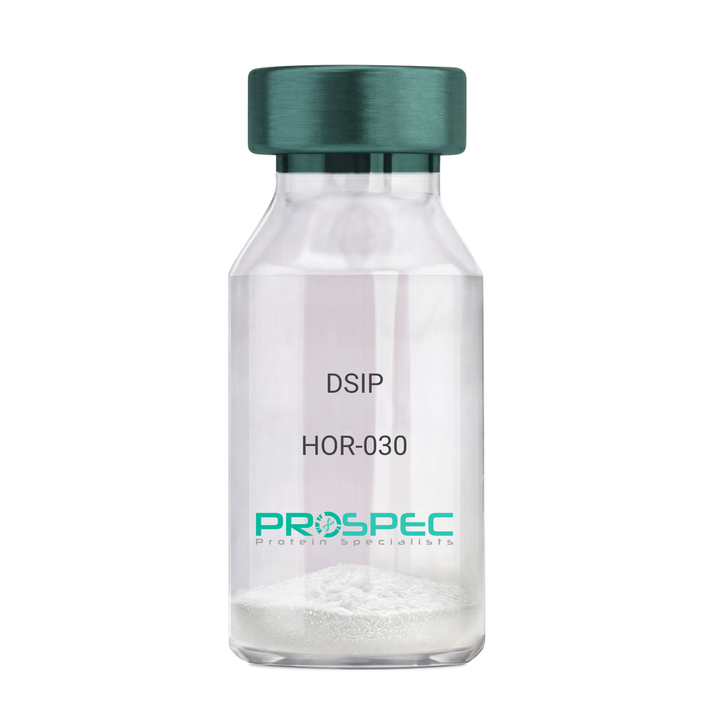
DSIPDescription:
Delta Sleep Inducing Peptide
Product # :
HOR-030Price :
Quantity :
Shipping Method :
Shipped at Room temp
More Info
- description
- formulation
- purity
- More Info
Description
DSIP Synthetic is a single, non-glycosylated polypeptide chain containing 15 amino acids, having a molecular mass of 1419.55 Dalton and a Molecular formula of C62H98N16O22.
Formulation
The protein was lyophilized with no additives.
Purity
Greater than 97.0% as determined by analysis by RP-HPLC.
More Info
-
Physical Appearance
Sterile Filtered White lyophilized (freeze-dried) powder.
-
Stability
Lyophilized DSIP although stable at room temperature for 3 weeks, should be stored desiccated below -18°C. Upon reconstitution DSIP should be stored at 4°C between 2-7 days and for future use below -18°C. For long term storage it is recommended to add a carrier protein (0.1% HSA or BSA). Please prevent freeze-thaw cycles.
-
Solubility
It is recommended to reconstitute the lyophilized DSIP in sterile 18MΩ-cm H2O not less than 100 µg/ml, which can then be further diluted to other aqueous solutions.
-
Amino Acid Sequence
H-Gly-Glu-Pro-Pro-Pro-Gly-Lys-Pro-Ala-Asp-Asp-Ala-Gly-Leu-Val-OH
-
Background
Delta Sleep-Inducing Peptide (DSIP), also known as Sleep-Promoting Peptide, is a neuropeptide that has been the subject of extensive research due to its potential role in sleep regulation, stress response, and neuroprotection. This nonapeptide, first isolated from the cerebral venous blood of rabbits during sleep, has been shown to induce slow-wave sleep, modulate pain perception, and exhibit potential antioxidant and immunomodulatory properties.
DSIP's primary function is its interaction with the sleep regulatory system. By modulating the release of certain neurotransmitters, DSIP can influence sleep patterns, particularly promoting slow-wave sleep, the most restorative stage of sleep. Studies by Kovalzon et al. (2011) have demonstrated that DSIP can enhance sleep quality in rats, suggesting potential applications in sleep disorders and the promotion of healthy sleep patterns.
In addition to its sleep-inducing effects, DSIP has been shown to possess neuroprotective properties. Research by Zolotarev et al. (2014) found that DSIP could protect neurons from oxidative stress, suggesting potential applications in the treatment of neurodegenerative diseases such as Alzheimer's and Parkinson's disease.
Given its sleep-inducing and neuroprotective effects, DSIP has been proposed as a potential therapeutic agent for a variety of conditions, including sleep disorders, chronic pain, and neurodegenerative diseases. For instance, a study by Spong et al. (2016) found that DSIP could improve sleep quality in patients with chronic insomnia, indicating its potential as a therapeutic agent in the treatment of sleep disorders.
While research on DSIP is promising, it is important to note that most studies have been conducted in animals or in vitro. More research is needed to fully understand the potential effects and applications of DSIP in humans. However, the existing body of research suggests that DSIP could be a promising tool in the treatment of sleep disorders, chronic pain, and neurodegenerative diseases.
ProSpec's products are furnished for LABORATORY RESEARCH USE ONLY. The product may not be used as drugs, agricultural or pesticidal products, food additives or household chemicals.
- View Data Sheet
Name :
COX5A AntibodyDescription:
Cytochrome C Oxidase Subunit Va, Mouse Anti Human
Cytochrome c oxidase subunit 5A, mitochondrial, Cytochrome c oxidase polypeptide Va, COX5A, VA, COX, COX-VA.
Product # :
ANT-605Price :
Quantity :
Shipping Method :
Shipped with Ice Packs
More Info
- formulation
- More Info
Formulation
1mg/ml containing PBS, pH-7.4, 10% Glycerol and 0.02% Sodium Azide.
More Info
-
Introduction
Cytochrome C Oxidase Subunit Va (COX5A) is a member of the cytochrome c oxidase subunit 5A family. COX (Cytochrome C Oxidase), which is the terminal component of the mitochondrial respiratory chain, catalyzes the electron transfer from reduced Cytochrome C to oxygen. The COX component is a heteromeric complex comprised of three catalytic subunits encoded by mitochondrial genes and multiple structural subunits encoded by nuclear genes. The mitochondrially-encoded subunits serve in electron transfer, and the nuclear-encoded subunits act in the regulation and assembly of the complex. COX5A is the heme A-containing chain of Cytochrome C Oxidase,which is the terminal oxidase in mitochondrial electron transport.
-
Synonyms
Cytochrome c oxidase subunit 5A, mitochondrial, Cytochrome c oxidase polypeptide Va, COX5A, VA, COX, COX-VA.
-
Physical Appearance
Sterile filtered colorless solution.
-
Immunogen
Anti-human COX5A mAb, is derived from hybridization of mouse F0 myeloma cells with spleen cells from BALB/c mice immunized with a recombinant human COX5A protein 42-150 amino acids purified from E. coli.
-
Ig Subclass
Mouse IgG1 heavy chain and k light chain.
-
Clone
PAT7A6AT.
-
Applications
COX5A antibody has been tested by ELISA, Western blot analysis, ICC/IF and Flow cytometry to assure specificity and reactivity. Since application varies, however, each investigation should be titrated by the reagent to obtain optimal results.
-
Type
Mouse Anti Human Monoclonal.
-
Storage Procedures
For periods up to 1 month store at 4°C, for longer periods of time, store at -20°C. Prevent freeze thaw cycles.
-
Purification Method
COX5A antibody was purified from mouse ascitic fluids by protein-A affinity chromatography.
ProSpec's products are furnished for LABORATORY RESEARCH USE ONLY. The product may not be used as drugs, agricultural or pesticidal products, food additives or household chemicals.