Search results
700 results found for “Transforming Growth Factor Beta Induced”
Name
Description
Product #
Price
Quantity
Shipping Method
- View Data Sheet
Name :
IGF1 RatDescription:
IGF-1 Rat Recombinant
Somatomedin C, IGF-I, IGFIA, IGF1.
Product # :
CYT-289Price :
Quantity :
Shipping Method :
Shipped at Room temp
More Info
- description
- source
- formulation
- purity
- biological activity
- More Info
Description
IGF-1 Rat Recombinant produced in E.Coli is a single, non-glycosylated, polypeptide chain containing 70 amino acids and having a molecular mass of 7.7kDa. IGF-I is purified by proprietary chromatographic techniques.
Source
Escherichia Coli.
Formulation
The protein was lyophilized with a 0.2µm filtered concentrated solution in 20mM PBS, pH 7.0.
Purity
Greater than 98.0% as determined by SDS-PAGE.
Biological Activity
The ED50 as determined by a cell proliferation assay using FDC-P1 cells is less than 2.0ng/ml, corresponding to a specific activity of >500,000units/mg.More Info
-
Introduction
The somatomedins, or IGFs, comprise a family of peptides that play important roles in mammalian growth and development. IGF1 mediates many of the growth-promoting effects of GH. Early studies showed that GH did not directly stimulate the incorporation of sulfate into cartilage, but rather acted through a serum factor, termed 'sulfation factor,' which later became known as 'somatomedin' (Daughaday et al., 1972). Three main somatomedins have been characterized: somatomedin C (IGF1), somatomedin A (IGF2; MIM 147470), and somatomedin B (MIM 193190) (Rotwein, 1986; Rosenfeld, 2003).
-
Synonyms
Somatomedin C, IGF-I, IGFIA, IGF1.
-
Physical Appearance
Sterile Filtered White lyophilized (freeze-dried) powder.
-
Stability
Lyophilized IGF-1 although stable at room temperature for 3 weeks, should be stored desiccated below -18°C. Upon reconstitution IGFI should be stored at 4°C between 2-7 days and for future use below -18°C.For long term storage it is recommended to add a carrier protein (0.1% HSA or BSA).Please prevent freeze-thaw cycles.
-
Solubility
It is recommended to reconstitute the lyophilized IGF1 in sterile 18MΩ-cm H2O not less than 100µg/ml, which can then be further diluted to other aqueous solutions.
-
Amino Acid Sequence
GPETLCGAEL VDALQFVCGP RGFYFNKPTG YGSSIRRAPQ TGIVDECCFR SCDLRRLEMY CAPLKPTKSA.
-
Background
What is the molecular weight/Mw of IGF1 RAT Protein?
IGF1 RAT Protein has a total Mw of 7.7kDa.
What is the source or expression system of IGF1 RAT Protein?
Escherichia Coli.
What is the Purity of IGF1 RAT Protein?
IGF1 RAT Protein is >98% pure as determined by SDS-PAGE.
What is the Biological Activity of IGF1 RAT Protein?
The ED50 as determined by a cell proliferation assay using FDC-P1 cells is less than 2.0ng/ml, corresponding to a specific activity of >500,000units/mg.
What is the amino acid sequence of IGF1 RAT Protein?
GPETLCGAEL VDALQFVCGP RGFYFNKPTG YGSSIRRAPQ TGIVDECCFR SCDLRRLEMY CAPLKPTKSA.
What applications can IGF1 RAT Protein be used in?
IGF1 RAT Protein can probably be used in western blot, ELISA and Lateral Flow.
What is the endotoxin level for IGF1 RAT Protein?
The endotoxin level is minimal, IGF1 RAT Protein was purified using conventional chromatography techniques.
ProSpec's products are furnished for LABORATORY RESEARCH USE ONLY. The product may not be used as drugs, agricultural or pesticidal products, food additives or household chemicals.
- View Data Sheet
Name :
PLGF1 Human, Sf9Description:
Placental Growth Factor-1 Human Recombinant, Sf9
PIGF, PGF, PLGF-1.
Product # :
CYT-419Price :
Quantity :
Shipping Method :
Shipped at Room temp
More Info
- description
- source
- formulation
- purity
- biological activity
- More Info
Description
Placenta Growth Factor-1 Human Recombinant produced in insect cells is a homodimer, glycosylated polypeptide chain containing 2 x 131 amino acids and having a total molecular mass of approximately 34 kDa. The PLGF-1 is purified by proprietary chromatographic techniques.
Source
Insect Cells.
Formulation
Lyophilized from a concentrated (1mg/ml) solution containing 50mM acetic acid.
Purity
Greater than 95.0% as determined by SDS-PAGE.
Biological Activity
The ED50 as determined by its ability to bind to immobilized rh-sFlt-1 in a functional ELISA. PlGF-1 human Recombinant can bind to immobilized rh-sFlt-1 (100ng/well) with a linear range at 0.5-10ng/ml, corresponding to a Specific Activity of 1x10⁵-2x10⁶units/mg.More Info
-
Introduction
PLGF is a growth factor active in angiogenesis, and endothelial cell growth, stimulating their proliferation and migration. It binds to receptor vegfr-1/flt1.
-
Synonyms
PIGF, PGF, PLGF-1.
-
Physical Appearance
Sterile Filtered White lyophilized (freeze-dried) powder.
-
Stability
Lyophilized Placenta Growth Factor 1 although stable at room temperature for 3 weeks, should be stored desiccated below -18°C. Upon reconstitution PLGF1 should be stored at 4°C between 2-7 days and for future use below -18°C.Please prevent freeze-thaw cycles.
-
Solubility
It is recommended to reconstitute the lyophilized Placenta Growth Factor 1 in sterile 0.1M acetic acid not less than 100µg/ml, which can then be further diluted to other aqueous solutions.
-
Amino Acid Sequence
The sequence of the first five N-terminal amino acids was determined and was found to be Met-Leu-Pro-Ala-Val.
ProSpec's products are furnished for LABORATORY RESEARCH USE ONLY. The product may not be used as drugs, agricultural or pesticidal products, food additives or household chemicals.
- View Data Sheet
Name :
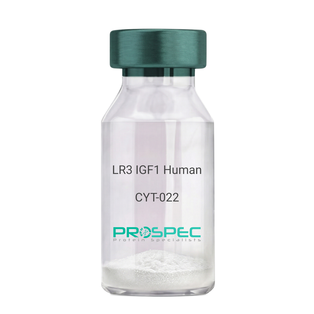
IGF1 LR3 HumanDescription:
LR3 Insulin Like Growth Factor-1 Human Recombinant
R3 IGF1, R3 IGF-1, R3IGF1, R3IGF-1, LONG IGF1, LONG IGF-1, LONG R3 IGF1, LONG R3IGF1, LONG R3 IGF-1, LONG R3IGF-1.
Product # :
CYT-022Price :
Quantity :
Shipping Method :
Shipped at Room temp
More Info
- description
- source
- formulation
- purity
- biological activity
- More Info
- sds-page
Description
The LR3 is a long-term analog of human IGF-1, specifically designed and manufactured for mammalian cell culture to support large-scale manufacturing of recombinant biopharmaceuticals. Recombinant Human LR3 Insulin Like Growth Factor-1 produced in E.Coli is a single, non-glycosylated, polypeptide chain containing 83 amino acids and having a molecular mass of 9.1kDa.
Source
Escherichia Coli.
Formulation
Lyophilized from a 0.2µm filtered concentrated solution in 20mM PB, pH 7.2.
Purity
Greater than 97.0% as determined by SDS-PAGE.
Biological Activity
The ED50 as determined by the stimulation of protein synthesis in L6 myoblasts is less than 10ng/ml, corresponding to a specific activity of 100,000units/mg.sds-page
More Info
-
Introduction
IGF-1 (Insulin-like growth factor-1) is a major hormonal mediator of statural growth. Under regular circumstances, GH (growth hormone) binds to its receptor in the liver, and other tissues, and stimulates the synthesis/secretion of IGF-1. In target tissues, the Type 1 IGF receptor, that is homologous to the insulin receptor, is activated by IGF-1, leading to intracellular signaling which stimulates multiple processes leading to statural growth. IGF-1 metabolic actions are partly directed at stimulating the uptake of glucose, fatty acids, and amino acids so that metabolism supports growing tissues.
-
Synonyms
R3 IGF1, R3 IGF-1, R3IGF1, R3IGF-1, LONG IGF1, LONG IGF-1, LONG R3 IGF1, LONG R3IGF1, LONG R3 IGF-1, LONG R3IGF-1.
-
Physical Appearance
Sterile Filtered White lyophilized (freeze-dried) powder.
-
Stability
Lyophilized LR3 IGF1 although stable at room temperature for 3 weeks, should be stored desiccated below -18°C. Upon reconstitution the LR3 IGF1 should be stored at 4°C between 2-7 days and for future use below -18°C.For long term storage it is recommended to add a carrier protein (0.1% HSA or BSA).Please prevent freeze-thaw cycles.
-
Solubility
It is recommended to reconstitute the lyophilized LR3 IGF1 in sterile 18M-cm H2O at a concentration of 100µg/ml, which can then be further diluted to other aqueous solutions.
-
Amino Acid Sequence
MFPAMPLSSLFVNGPRTLCGAELVDALQFVCGDRGFYFNKPTGYGSSSRRAPQTGIV DECCFRSCDLRRLEMYCAPLKPAKSA.
-
Background
What is the molecular weight/Mw of IGF1 LR3 HUMAN Protein?
IGF1 LR3 HUMAN Protein has a total Mw of 9.1kDa.
What is the source or expression system of IGF1 LR3 HUMAN Protein?
Escherichia Coli.
What is the Purity of IGF1 LR3 HUMAN Protein?
IGF1 LR3 HUMAN Protein is >98% pure as determined by SDS-PAGE.
What is the Biological Activity of IGF1 LR3 HUMAN Protein?
The ED50 as determined by the stimulation of protein synthesis in L6 myoblasts is less than 10ng/ml, corresponding to a specific activity of 100,000units/mg.
What is the amino acid sequence of IGF1 LR3 HUMAN Protein?
MFPAMPLSSLFVNGPRTLCGAELVDALQFVCGDRGFYFNKPTGYGSSSRRAPQTGIV DECCFRSCDLRRLEMYCAPLKPAKSA.
What applications can IGF1 LR3 HUMAN Protein be used in?
IGF1 LR3 HUMAN Protein can probably be used in western blot, ELISA and Lateral Flow.
What is the endotoxin level for IGF1 LR3 HUMAN Protein?
The endotoxin level is minimal, IGF1 LR3 HUMAN Protein was purified using conventional chromatography techniques.
ProSpec's products are furnished for LABORATORY RESEARCH USE ONLY. The product may not be used as drugs, agricultural or pesticidal products, food additives or household chemicals.
- View Data Sheet
Name :
FGF12 Human, HisDescription:
Recombinant Human Fibroblast Growth Factor 12, His Tag
FGF-12, FGF12, FGF12B, FHF1, Fibroblast growth factor 12, Fibroblast growth factor homologous factor 1, FHF-1, Myocyte-activating factor.
Product # :
CYT-620Price :
Quantity :
Shipping Method :
Shipped with Ice Packs
More Info
- description
- source
- formulation
- purity
- More Info
Description
The FGF-12 Human recombinant protein is a single, non-glycosylated polypeptide chain produced in E. coli, having a molecular weight of 22.6kDa and containing 201 amino acids (1-181). The FGF12 is fused to a 20 amino acid His tag at the N-terminus.
Source
Escherichia Coli.
Formulation
The FGF-12 solution (1mg/ml) contains 20mM Tris pH-7.5, 1mM DTT, 2mM EDTA and 10% glycerol.
Purity
Greater than 90% as determined by SDS-PAGE.
More Info
-
Introduction
FGF12 is part of the Fibroblast Growth Factor (FGF) family which has a vast mitogenic and cell survival functions, and play a role in a range of biological activities, among them are embryonic development, cell growth, morphogenesis, tissue repair, tumor growth, and invasion. FGF-12 doesn’t obtain the N-terminal signal sequence present in the majority of the FGF family members, but it contains clusters of basic residues that act as a nuclear localization signal. When transfected into mammalian cells, FGF12 accumulated in the nucleus, but was not secreted. FGF12 is involved in nervous system development and function. FGF12 binds to IB2 (islet brain-2), a cellular kinase scaffold, and voltage gated sodium channels and is also involved in intracellular signaling and ion exchange.
-
Synonyms
FGF-12, FGF12, FGF12B, FHF1, Fibroblast growth factor 12, Fibroblast growth factor homologous factor 1, FHF-1, Myocyte-activating factor.
-
Physical Appearance
Sterile liquid colorless solution.
-
Stability
Store FGF12 at -20°C. Can be stored at 4°C for a limited period of time of 7 days.
-
Amino Acid Sequence
MSSHHHHHH SSGLVPRGSH MESKEPQLKG IVTRLFSQQG YFLQMHPDGT IDGTKDENSD YTLFNLIPVG LRVVAIQGVK ASLYVAMNGE YLYSSDVFT PECKFKESVF ENYYVIYSST LYRQQESGRA WFLGLNKEGQ IMKGNRVKKT KPSSHFVPKP IEVCMYREQS LHEIGEKQGR RKSSGTPTM NGGKVVNQDS T.
-
Background
What is the molecular weight/Mw of FGF12 Protein?
FGF12 Protein has a total Mw of 22.6kDa.
What is the source or expression system of FGF12 Protein?
Escherichia Coli.
What is the Purity of FGF12 Protein?
FGF12 Protein is >90% pure as determined by SDS-PAGE.
What is the Biological Activity of FGF12 Protein?
The biological functionality of FGF12 Protein will be determined in the future.
What is the amino acid sequence of FGF12 Protein?
MSSHHHHHH SSGLVPRGSH MESKEPQLKG IVTRLFSQQG YFLQMHPDGT IDGTKDENSD YTLFNLIPVG LRVVAIQGVK ASLYVAMNGE YLYSSDVFT PECKFKESVF ENYYVIYSST LYRQQESGRA WFLGLNKEGQ IMKGNRVKKT KPSSHFVPKP IEVCMYREQS LHEIGEKQGR RKSSGTPTM NGGKVVNQDS T.
What applications can FGF12 Protein be used in?
FGF12 Protein can probably be used in western blot, ELISA and Lateral Flow.
What is the endotoxin level for FGF12 Protein?
The endotoxin level is minimal, FGF12 Protein was purified using conventional chromatography techniques.
ProSpec's products are furnished for LABORATORY RESEARCH USE ONLY. The product may not be used as drugs, agricultural or pesticidal products, food additives or household chemicals.
- View Data Sheet
Name :
TRAIL Human (114-281 a.a.)Description:
TRAIL/APO 2 Ligand (114-281 a.a.) Human Recombinant
Tumor necrosis factor ligand superfamily member 10, TNF-related apoptosis-inducing ligand, Protein TRAIL, Apo-2 ligand, Apo-2L, CD253 antigen, TL2, APO2L, TNFSF10.
Product # :
CYT-546Price :
Quantity :
Shipping Method :
Shipped with Ice Packs
More Info
- description
- source
- formulation
- purity
- More Info
Description
Soluble TNF-related apoptosis-inducing ligand Human Recombinant produced in E.Coli is a single, non-glycosylated polypeptide chain containing 169 amino acids (114-281) and having a molecular mass of 19.6 kDa. The sTRAIL is purified by proprietary chromatographic techniques.
Source
Escherichia Coli.
Formulation
1mg/ml in 20mM Tris-HCl pH-7.5, 300mM NaCl, 0.1mM DTT & 10% glycerol.
Purity
Greater than 95.0% as determined by SDS-PAGE.
More Info
-
Introduction
TNF-related apoptosis-inducing ligand (TRAIL) is a ligand molecule which induces apoptosis. It is a type II transmembrane protein with homology to other members of the tumor necrosis factor family.
In humans, the gene that encodes for TRAIL is located at chromosome 3q26.
TRAIL binds to the death receptors, DR4 and DR5. The process of apoptosis is caspase-8-dependent. This protein preferentially induces apoptosis in transformed and tumor cells, but does not appear to kill normal cells although it is expressed at a significant level in most normal tissues. -
Synonyms
Tumor necrosis factor ligand superfamily member 10, TNF-related apoptosis-inducing ligand, Protein TRAIL, Apo-2 ligand, Apo-2L, CD253 antigen, TL2, APO2L, TNFSF10.
-
Physical Appearance
Sterile Filtered colorless liquid.
-
Stability
Store at 4°C if entire vial will be used within 2-4 weeks. Store, frozen at -20°C for longer periods of time. For long term storage it is recommended to add a carrier protein (0.1% HSA or BSA).Avoid multiple freeze-thaw cycles.
-
Amino Acid Sequence
MVRERGPQRV AAHITGTRGR SNTLSSPNSK NEKALGRKIN SWESSRSGHS FLSNLHLRNGELVIHEKGFY YIYSQTYFRF QEEIKENTKN DKQMVQYIYK YTSYPDPILL MKSARNSCWSKDAEYGLYSI YQGGIFELKE NDRIFVSVTN EHLIDMDHEA SFFGAFLVG.
ProSpec's products are furnished for LABORATORY RESEARCH USE ONLY. The product may not be used as drugs, agricultural or pesticidal products, food additives or household chemicals.
- View Data Sheet
Name :
BDNF HumanDescription:
Brain-Derived Neurotrophic Factor Human Recombinant
Brain-Derived Neurotrophic Factor, BDNF, MGC34632.
Product # :
CYT-207Price :
Quantity :
Shipping Method :
Shipped at Room temp
More Info
- description
- source
- formulation
- purity
- biological activity
- More Info
- Activity
Description
BDNF Human Recombinant produced in E.Coli is a homodimer, non-glycosylated, polypeptide chain containing 2 x 119 amino acids (and an N-terminal Met) and having a total molecular mass of 28kDa. BDNF Human Recombinant is purified by proprietary chromatographic techniques.
Source
Escherichia Coli.
Formulation
The protein was lyophilized with 20mM PB and 400mM NaCl, pH 7.2.
Purity
BDNF is greater than 950% as determined SDS-PAGE.
Biological Activity
The activity was determined using Immobilized Human TrkB-His tag protein 2ug/ml (100 μl/well) for its binding to NHS-Biotin BDNF. The ED50 of was found to be ≤20ng/mLActivity
More Info
-
Introduction
BDNF promotes the survival of neuronal populations that are all located either in the central nervous system or directly connected to it. BDNF is a major regulator of synaptic transmission and plasticity at adult synapses in many regions of the cns. The versatility of BDNF is emphasized by its contribution to a range of adaptive neuronal responses including long-term potentiation (ltp), long-term depression (ltd), certain forms of short-term synaptic plasticity, as well as homeostatic regulation of intrinsic neuronal excitability.
-
Synonyms
Brain-Derived Neurotrophic Factor, BDNF, MGC34632.
-
Physical Appearance
Sterile Filtered White lyophilized (freeze-dried) powder.
-
Stability
Lyophilized BDNF although stable at room temperature for 3 weeks, should be stored desiccated below -18°C. Upon reconstitution BDNF should be stored at 4°C between 2-7 days and for future use below -18°C.For long term storage it is recommended to add a carrier protein (0.1% HSA or BSA).Please prevent freeze-thaw cycles.
-
Solubility
It is recommended to reconstitute the lyophilized BDNF in sterile 18M-cm H2O not less than 100µg/ml, which can then be further diluted to other aqueous solutions.
-
Amino Acid Sequence
HSDPARRGE LSVCDSISEW VTAADKKTAV DMSGGTVTVL EKVPVSKGQL KQYFYETKCN PMGYTKEGCR GIDKRHWNSQ CRTTQSYVRA LTMDSKKRIG WRFIRIDTSC VCTLTIKRGR.
-
Background
Final Thoughts
Although more research is needed on the safety and effectiveness of BDNF human recombinant, trials suggest that this laboratory-produced protein may be effective in managing and treating several neurological and psychiatric disorders. It's important for experts to stay up to date on the latest developments and research to learn more about potential risks and benefits.
What is the molecular weight/Mw of BDNF Protein?
BDNF Protein has a total Mw of 27kDa.
What is the source or expression system of BDNF Protein?
Escherichia Coli.
What is the Purity of BDNF Protein?
BDNF Protein is >97% pure as determined by SDS-PAGE.
What is the Biological Activity of BDNF Protein?
The ED50, as determined by the dose-dependent induction of C6 cells proliferation, is 1.3-2µg/ml.
What is the amino acid sequence of BDNF Protein?
MHSDPARRGE LSVCDSISEW VTAADKKTAV DMSGGTVTVL EKVPVSKGQL KQYFYETKCN PMGYTKEGCR GIDKRHWNSQ CRTTQSYVRA LTMDSKKRIG WRFIRIDTSC VCTLTIKRGR.
What applications can BDNF Protein be used in?
BDNF Protein can probably be used in western blot, ELISA and Lateral Flow.
What is the endotoxin level for BDNF Protein?
The endotoxin level is minimal, BDNF Protein was purified using conventional chromatography techniques.
-
Protein content
BDNF quantitation was carried out by two independent methods:1. UV spectroscopy at 280 nm using the absorbency value of 1.6 as the extinction coefficient for a 0.1% (1mg/ml) solution. This value is calculated by the PC GENE computer analysis program of protein sequences (IntelliGenetics). 2. Analysis by RP-HPLC, using a standard solution of Brain-derived Neurotrophic Factor as a Reference Standard.
ProSpec's products are furnished for LABORATORY RESEARCH USE ONLY. The product may not be used as drugs, agricultural or pesticidal products, food additives or household chemicals.
- View Data Sheet
Name :
BMP8B HumanDescription:
Bone Morphogenetic protein-8b Human Recombinant
Bone morphogenetic protein 8B, BMP-8, BMP-8B, Osteogenic protein 2, OP-2, BMP8B, BMP8, Bone Morphogenetic protein-8b, OP2.
Product # :
CYT-830Price :
Quantity :
Shipping Method :
Shipped with Ice Packs
More Info
- description
- source
- formulation
- purity
- More Info
- sds-page
Description
BMP8B Human Recombinant produced in E.Coli is a single, non-glycosylated polypeptide chain containing 162 amino acids (264-402a.a.) and having a molecular mass of 18.1kDa.BMP8B is fused to a 23 amino acid His-tag at N-terminus & purified by proprietary chromatographic techniques.
Source
E.coli.
Formulation
BMP8B protein solution (1mg/ml) containing 20mM Tris-HCl buffer (pH8.0), 0.4M Urea and 10% glycerol.
Purity
Greater than 90% as determined by SDS-PAGE.
sds-page
More Info
-
Introduction
Bone Morphogenetic protein-8b (BMP8B) belongs to a family of secreted signaling molecules which can induce ectopic bone growth. BMP8B is known for having a possible bone inductive activity as it is related to BMP5 and BMP7. BMP8B is the osteoinductive factor accountable for epithelial osteogenesis.
-
Synonyms
Bone morphogenetic protein 8B, BMP-8, BMP-8B, Osteogenic protein 2, OP-2, BMP8B, BMP8, Bone Morphogenetic protein-8b, OP2.
-
Physical Appearance
Sterile Filtered colorless solution.
-
Stability
Store at 4°C if entire vial will be used within 2-4 weeks. Store, frozen at -20°C for longer periods of time. For long term storage it is recommended to add a carrier protein (0.1% HSA or BSA).Avoid multiple freeze-thaw cycles.
-
Amino Acid Sequence
MGSSHHHHHH SSGLVPRGSH MGSAVRPLRR RQPKKSNELP QANRLPGIFD DVHGSHGRQV CRRHELYVSF QDLGWLDWVI APQGYSAYYC EGECSFPLDS CMNATNHAIL QSLVHLMMPD AVPKACCAPT KLSATSVLYY DSSNNVILRK HRNMVVKACG CH.
-
Background
Bone Morphogenetic Protein-8B Human Recombinant: Unveiling the Potential for Regenerative Medicine and Tissue Engineering
Abstract:
Bone Morphogenetic Protein-8B (BMP-8B) human recombinant is a key member of the bone morphogenetic protein family, renowned for its crucial role in tissue development, regeneration, and repair. This research paper aims to provide a comprehensive analysis of BMP-8B, including its characteristics, signaling pathways, and potential therapeutic applications. Furthermore, innovative methodologies for the production and optimization of BMP-8B human recombinant are proposed, shedding light on its future implications in the field of regenerative medicine and tissue engineering.
Introduction:
Regenerative medicine and tissue engineering offer promising solutions to address the challenges of tissue repair and regeneration. BMP-8B, a prominent member of the BMP family, plays a vital role in orchestrating cellular responses during tissue development and healing. This paper delves into the distinctive features of BMP-8B and presents novel approaches for the production and optimization of BMP-8B human recombinant, aiming to unleash its therapeutic potential in various regenerative contexts.
Characteristics and Signaling Pathways:
BMP-8B is a secreted growth factor belonging to the transforming growth factor-beta (TGF-β) superfamily. It exerts its biological effects by binding to specific cell surface receptors, thereby initiating intricate intracellular signaling cascades. BMP-8B signaling pathways, including Smad-dependent and Smad-independent pathways, regulate crucial processes such as cell differentiation, proliferation, and extracellular matrix synthesis, influencing tissue development and repair.
Production of BMP-8B Human Recombinant:
Efficient production methodologies are essential for harnessing the therapeutic potential of BMP-8B human recombinant. Various recombinant protein expression systems, such as mammalian cells or baculovirus-insect cell systems, have been utilized for the production of functional BMP-8B. Optimization strategies, including codon optimization, signal peptide engineering, and protein folding enhancement, have been employed to improve the yield and bioactivity of BMP-8B recombinant protein.
Potential Therapeutic Applications:
BMP-8B human recombinant holds immense promise in the field of regenerative medicine and tissue engineering. Its involvement in bone and cartilage formation, muscle regeneration, and wound healing makes it a potential candidate for the treatment of skeletal disorders, muscle injuries, and chronic wounds. Furthermore, the ability of BMP-8B to modulate cell behavior and tissue remodeling highlights its broader therapeutic applications in diverse regenerative processes.
Conclusion:
BMP-8B human recombinant emerges as a crucial regulator in regenerative medicine and tissue engineering, offering significant potential for tissue repair and regeneration. Optimizing production methodologies and further unraveling its signaling mechanisms will enhance its therapeutic applications. Given its involvement in bone, cartilage, and muscle formation, as well as wound healing, BMP-8B human recombinant represents a valuable tool for promoting tissue regeneration and addressing the unmet clinical needs in regenerative medicine.
What is the molecular weight/Mw of BMP8B Protein?
BMP8B Protein has a total Mw of 18.1kDa.
What is the source or expression system of BMP8B Protein?
Escherichia Coli.
What is the Purity of BMP8B Protein?
BMP8B Protein is >90% pure as determined by SDS-PAGE.
What is the Biological Activity of BMP8B Protein?
The biological functionality of BMP8B Protein will be determined in the future.
What is the amino acid sequence of BMP8B Protein?
MGSSHHHHHH SSGLVPRGSH MGSAVRPLRR RQPKKSNELP QANRLPGIFD DVHGSHGRQV CRRHELYVSF QDLGWLDWVI APQGYSAYYC EGECSFPLDS CMNATNHAIL QSLVHLMMPD AVPKACCAPT KLSATSVLYY DSSNNVILRK HRNMVVKACG CH.
What applications can BMP8B Protein be used in?
BMP8B Protein can probably be used in western blot, ELISA and Lateral Flow.
What is the endotoxin level for BMP8B Protein?
The endotoxin level is minimal, BMP8B Protein was purified using conventional chromatography techniques.
ProSpec's products are furnished for LABORATORY RESEARCH USE ONLY. The product may not be used as drugs, agricultural or pesticidal products, food additives or household chemicals.
- View Data Sheet
Name :
Activin-A MouseDescription:
Activin-A Mouse Recombinant
Inhba, Inhibin beta A, FSH releasing protein.
Product # :
CYT-146Price :
Quantity :
Shipping Method :
Shipped at Room temp
More Info
- description
- source
- formulation
- purity
- biological activity
- More Info
Description
Active form Activin-A Murine Recombinant produced in e.coli is a homodimeric, non-glycosylated, polypeptide chain containing 2 x 117 amino acids and having a molecular weight of 26.2kDa.The Active form Activin-A is purified by standard chromatographic techniques.
Source
E.Coli.
Formulation
Mouse Activin-A lyophilized from a concentrated 1mg/ml protein solution containing 0.1% TFA.
Purity
Greater than 95% as obsereved by SDS-PAGE.
Biological Activity
Biological activity is assessed by the ability to induce cytoxicity of MPC-11 cells and was found to be 8.8ng/ml corresponding to a specific activity of 1.1 x 10⁵ units/mg.
More Info
-
Introduction
Activins are homodimers or heterodimers of the different β subunit isoforms, part of the TGFβ family. Mature Activin A has two 116 amino acids residues βA subunits (βA-βA). Activin displays an extensive variety of biological activities, including mesoderm induction, neural cell differentiation, bone remodelling, haematopoiesis, and reproductive physiology. Activins takes part in the production and regulation of hormones such as FSH, LH, GnRH and ACTH. Cells that are identified to express Activin A include fibroblasts, endothelial cells, hepatocytes, vascular smooth muscle cells, macrophages, keratinocytes, osteoclasts, bone marrow monocytes, prostatic epithelium, neurons, chondrocytes, osteoblasts, Leydig cells, Sertoli cells, and ovarian granulosa cells.
-
Synonyms
Inhba, Inhibin beta A, FSH releasing protein.
-
Physical Appearance
Lyophilized freeze dried powder.
-
Stability
Lyophilized Activin-A although stable at room temperature for 3 weeks, should be stored desiccated below -18°C. Upon reconstitution Activin-A should be stored at 4°C between 2-7 days and for future use below -18°C.For long term storage it is recommended to add a carrier protein (0.1% HSA or BSA).Please prevent freeze-thaw cycles.
-
Solubility
Murine INHBA protein should be reconstituted in distilled pyrogen free water to a concentration of 100ug /ml which can then be further diluted to other aqueous solutions.
-
Amino Acid Sequence
MGLECDGKVN ICCKKQFFVS FKDIGWNDWI IAPSGYHANY CEGECPSHIA GTSGSSLSFH STVINHYRMR GHSPFANLKS CCVPTKLRPM SMLYYDDGQN IIKKDIQNMI VEECGCS.
-
Background
What is the molecular weight / Mw of Activin A Protein?
Activin A Protein has a total Mw of 26.2 kDa.What is the source or expression system of Activin A Protein?
Ecoli
What is the Purity of Activin A Protein?
Activin A Protein is >95% pure as determined by SDS-PAGE.
What is the Biological Activity of Activin A Protein?
Biological activity is assessed by the ability to induce cytoxicity of MPC-11 cells and was found to be 8.8ng/ml corresponding to a specific activity of 110,000 units/mg.
What is the endotoxin level for Activin A Protein?
The endotoxin level is minimal, ACTIVIN A Protein was purified using conventional chromatography techniques.
What is the amino acid sequence of ACTIVIN A Protein?
MGLECDGKVN ICCKKQFFVS FKDIGWNDWI IAPSGYHANY CEGECPSHIA GTSGSSLSFH STVINHYRMR GHSPFANLKS CCVPTKLRPM SMLYYDDGQN IIKKDIQNMI VEECGCS.
What applications can ACTIVIN A Protein be used in?
ACTIVIN A Protein can probably be used in western blot, ELISA and Lateral Flow.
ProSpec's products are furnished for LABORATORY RESEARCH USE ONLY. The product may not be used as drugs, agricultural or pesticidal products, food additives or household chemicals.
- View Data Sheet
Name :
GDF15 MouseDescription:
Growth and Differentiation factor 15 Mouse Recombinant
Growth/differentiation factor 15, GDF-15.
Product # :
CYT-857Price :
Quantity :
Shipping Method :
Shipped with Ice Packs
More Info
- description
- source
- formulation
- purity
- More Info
- sds-page
Description
GDF15 Mouse Recombinant produced in E.Coli is a single, non-glycosylated polypeptide chain containing 138 amino acids (189-303 a.a) and having a molecular mass of 14.9kDa. GDF15 is fused to a 23 amino acid His-tag at N-terminus & purified by proprietary chromatographic techniques.
Source
Escherichia Coli.
Formulation
GDF15 protein solution (1.0mg/ml) containing 20mM Phosphate buffer (pH 8.0) and 10% glycerol.
Purity
Greater than 90.0% as determined by SDS-PAGE.
sds-page
More Info
-
Introduction
GDF15 is part of the TGF-Beta superfamily which is involved in regulating inflammatory and apoptotic pathways in injured tissues and throughout disease processes. GDF15 is most abundant in the liver. Its expression in liver can be considerably up-regulated in during injury of organs such as liver, kidney, heart and lung. GDF-15 promotes proliferation or growth arrest and differentiation due to differences in cellular differentiation. GDF15 prevents apoptosis in cerebellar granule neurons by activating Akt and inhibiting endogenously active ERK. GDF15 is a novel autocrine/endocrine factor that antagonizes the hypertrophic response and loss of ventricular performance.
-
Synonyms
Growth/differentiation factor 15, GDF-15.
-
Physical Appearance
Sterile Filtered colorless solution.
-
Stability
Store at 4°C if entire vial will be used within 2-4 weeks. Store, frozen at -20°C for longer periods of time. For long term storage it is recommended to add a carrier protein (0.1% HSA or BSA).Avoid multiple freeze-thaw cycles.
-
Amino Acid Sequence
MGSSHHHHHH SSGLVPRGSH MGSSAHAHPR DSCPLGPGRC CHLETVQATL EDLGWSDWVL SPRQLQLSMC VGECPHLYRS ANTHAQIKAR LHGLQPDKVP APCCVPSSYT PVVLMHRTDS GVSLQTYDDL VARGCHCA.
-
Background
What is the molecular weight/Mw of GDF15 MOUSE Protein?
GDF15 MOUSE Protein has a total Mw of 14.9kDa.
What is the source or expression system of GDF15 MOUSE Protein?
Escherichia Coli.
What is the Purity of GDF15 MOUSE Protein?
GDF15 MOUSE Protein is >90% pure as determined by SDS-PAGE.
What is the Biological Activity of GDF15 MOUSE Protein?
The biological functionality of GDF15 MOUSE Protein will be determined in the future.
What is the amino acid sequence of GDF15 MOUSE Protein?
MGSSHHHHHH SSGLVPRGSH MGSSAHAHPR DSCPLGPGRC CHLETVQATL EDLGWSDWVL SPRQLQLSMC VGECPHLYRS ANTHAQIKAR LHGLQPDKVP APCCVPSSYT PVVLMHRTDS GVSLQTYDDL VARGCHCA.
What applications can GDF15 MOUSE Protein be used in?
GDF15 MOUSE Protein can probably be used in western blot, ELISA and Lateral Flow.
What is the endotoxin level for GDF15 MOUSE Protein?
The endotoxin level is minimal, GDF15 MOUSE Protein was purified using conventional chromatography techniques.
ProSpec's products are furnished for LABORATORY RESEARCH USE ONLY. The product may not be used as drugs, agricultural or pesticidal products, food additives or household chemicals.
- View Data Sheet
Name :
Activin-A Human ActiveDescription:
Activin-A Human Recombinant, Active
Inhba, Inhibin beta A, FSH releasing protein.
Product # :
CYT-145Price :
Quantity :
Shipping Method :
Shipped at Room temp
More Info
- description
- source
- formulation
- purity
- biological activity
- More Info
Description
Active form Activin-A Human Recombinant produced in e.coli is a homodimeric, non-glycosylated, polypeptide chain containing 2 x 117 amino acids and having a molecular weight of 26.2kDa.The Active form Activin-A is purified by standard chromatographic techniques.
Source
E.Coli.
Formulation
Human Activin-A was lyophilized from a concentrated 1mg/ml protein solution containing 0.1% TFA.
Purity
Greater than 95% as obsereved by SDS-PAGE.
Biological Activity
Biological activity is assessed by the ability to induce cytotoxicity of MPC-11 cells and was found to be 8.95ng/ml corresponding to a specific activity of 1.1 x 10⁵ units/mg.
More Info
-
Introduction
Activins are homodimers or heterodimers of the different β subunit isoforms, part of the TGFβ family. Mature Activin A has two 116 amino acids residues βA subunits (βA-βA). Activin displays an extensive variety of biological activities, including mesoderm induction, neural cell differentiation, bone remodelling, haematopoiesis, and reproductive physiology. Activins takes part in the production and regulation of hormones such as FSH, LH, GnRH and ACTH. Cells that are identified to express Activin A include fibroblasts, endothelial cells, hepatocytes, vascular smooth muscle cells, macrophages, keratinocytes, osteoclasts, bone marrow monocytes, prostatic epithelium, neurons, chondrocytes, osteoblasts, Leydig cells, Sertoli cells, and ovarian granulosa cells.
-
Synonyms
Inhba, Inhibin beta A, FSH releasing protein.
-
Physical Appearance
Lyophilized freeze dried powder.
-
Stability
Lyophilized Activin-A although stable at room temperature for 3 weeks, should be stored desiccated below -18°C. Upon reconstitution Activin-A should be stored at 4°C between 2-7 days and for future use below -18°C.For long term storage it is recommended to add a carrier protein (0.1% HSA or BSA).Please prevent freeze-thaw cycles.
-
Solubility
Human INHBA protein should be reconstituted in distilled pyrogen free water to a concentration of 100ug /ml which can then be further diluted to other aqueous solutions.
-
Amino Acid Sequence
MGLECDGKVN ICCKKQFFVS FKDIGWNDWI IAPSGYHANY CEGECPSHIA GTSGSSLSFH STVINHYRMR GHSPFANLKS CCVPTKLRPM SMLYYDDGQN IIKKDIQNMI VEECGCS.
-
Background
Title: Research on Activin A Human Recombinant: Molecular Characteristics, Signaling Pathways, Physiological Functions, and Therapeutic Potential
Introduction:
Activin A, a member of the transforming growth factor-beta (TGF-β) superfamily, is a multifunctional cytokine that plays a significant role in various biological processes in the human body. Its involvement in diverse physiological and pathological functions has garnered considerable attention in scientific research. This paper aims to provide an overview of Activin A, encompassing its molecular characteristics, signaling pathways, physiological functions, and therapeutic potential.
Activin A is encoded by the INHBA gene and is produced as a precursor protein that undergoes post-translational modifications to generate the mature form. The mature Activin A protein consists of two β-subunits held together by disulfide bonds. These structural features contribute to its functional properties and interactions with specific receptors.
Upon binding to its cell surface receptors, Activin A triggers intracellular signaling cascades, leading to various cellular responses. Canonical SMAD-dependent pathway as well as non-SMAD pathways, such as MAPK/ERK, PI3K/Akt, and JNK signaling, are activated by Activin A. The intricate network of signaling pathways enables Activin A to regulate diverse biological processes, including cell proliferation, differentiation, apoptosis, and tissue homeostasis.
Activin A exerts its physiological functions in a tissue-specific manner. It plays a critical role in embryonic development, particularly in organogenesis and patterning. Additionally, Activin A is involved in reproductive biology, where it participates in folliculogenesis, spermatogenesis, and hormonal regulation. It also contributes to neural development, immune system modulation, and skeletal homeostasis.
The multifunctional properties of Activin A have positioned it as a potential therapeutic target for various diseases. Its involvement in cancer, neurodegenerative disorders, fibrosis, and reproductive disorders has prompted extensive research to explore its therapeutic potential. Understanding the molecular mechanisms underlying Activin A's actions provides valuable insights for developing innovative therapeutic strategies.
In conclusion, Activin A is a versatile cytokine with diverse roles in human biology. This research aims to deepen our understanding of its molecular characteristics, signaling pathways, physiological functions, and therapeutic potential. By elucidating the complexities of Activin A, we strive to pave the way for novel therapeutic interventions in various human diseases.
ProSpec's products are furnished for LABORATORY RESEARCH USE ONLY. The product may not be used as drugs, agricultural or pesticidal products, food additives or household chemicals.
- View Data Sheet
Name :
GM-CSF K9Description:
Granulocyte Macrophage-Colony Stimulating Factor Canine Recombinant
CSF-2, MGI-1GM, GM-CSF, Pluripoietin-alpha, MGC131935, MGC138897.
Product # :
CYT-724Price :
Quantity :
Shipping Method :
Shipped at Room temp
More Info
- description
- source
- formulation
- purity
- biological activity
- More Info
Description
GMCSF k9 Recombinant produced in E.Coli is a single, non-glycosylated, polypeptide chain containing 128 amino acids and having a molecular mass of 14.2 kDa. GM-CSF is purified by proprietary chromatographic techniques.
Source
Escherichia Coli.
Formulation
GMCSF was lyophilized after extensive dialysis against 1xPBS pH 7.4.
Purity
Greater than 96.0% as determined by
1. Analysis by RP-HPLC.
2. Analysis by SDS-PAGE.Biological Activity
The ED50 as calculated by the dose-dependent stimulation of the proliferation of human TF1 erythroleukemic cells is typically 1-4 ng/ml.More Info
-
Introduction
GMCSF is a cytokine that controls the production, differentiation, and function of granulocytes and macrophages. The active form of the protein is found extracellularly as a homodimer. This gene has been localized to a cluster of related genes at chromosome region 5q31, which is known to be associated with interstitial deletions in the 5q- syndrome and acute myelogenous leukemia. Other genes in the cluster include those encoding interleukins 4, 5, and 13.
GM-CSF stimulates the growth and differentiation of hematopoietic precursor cells from various lineages, including granulocytes, macrophages, eosinophils and erythrocytes. -
Synonyms
CSF-2, MGI-1GM, GM-CSF, Pluripoietin-alpha, MGC131935, MGC138897.
-
Physical Appearance
Sterile Filtered White lyophilized (freeze-dried) powder.
-
Stability
Lyophilized GMCSF although stable at room temperature for 3 weeks, should be stored desiccated below -18°C. Upon reconstitution GMCSF should be stored at 4°C between 2-7 days and for future use below -18°C. For long term storage it is recommended to add a carrier protein (0.1% HSA or BSA).Please prevent freeze-thaw cycles.
-
Solubility
It is recommended to reconstitute the lyophilized GMCSF in sterile 18MΩ-cm H2O not less than 100µg/ml, which can then be further diluted to other aqueous solutions.
-
Amino Acid Sequence
APTRSPTLVT RPSQHVDAIQ EALSLLNNSN DVTAVMNKAV KVVSEVFDPE
GPTCLETRLQ LYKEGLQGSL TSLKNPLTMM ANHYKQHCPP TPESPCATQN
INFKSFKENL KDFLFNIPFD CWKPVKK. -
Background
What is the molecular weight/Mw of GM-CSF K9 Protein?
GM-CSF K9 Protein has a total Mw of 14.2kDa.
What is the source or expression system of GM-CSF K9 Protein?
Escherichia Coli.
What is the Purity of GM-CSF K9 Protein?
GM-CSF K9 Protein is >96% pure as determined by SDS-PAGE.
What is the Biological Activity of GM-CSF K9 Protein?
The ED50 as calculated by the dose-dependent stimulation of the proliferation of human TF1 erythroleukemic cells is typically 1-4 ng/ml.
What is the amino acid sequence of GM-CSF K9 Protein?
APTRSPTLVT RPSQHVDAIQ EALSLLNNSN DVTAVMNKAV KVVSEVFDPE
GPTCLETRLQ LYKEGLQGSL TSLKNPLTMM ANHYKQHCPP TPESPCATQN
INFKSFKENL KDFLFNIPFD CWKPVKK.
What applications can GM-CSF K9 Protein be used in?
GM-CSF K9 Protein can probably be used in western blot, ELISA and Lateral Flow.
What is the endotoxin level for GM-CSF K9 Protein?
The endotoxin level is minimal, GM-CSF K9 Protein was purified using conventional chromatography techniques.
ProSpec's products are furnished for LABORATORY RESEARCH USE ONLY. The product may not be used as drugs, agricultural or pesticidal products, food additives or household chemicals.
- View Data Sheet
Name :
TIFA HumanDescription:
TRAF-Interacting Protein with Forkhead-Associated Domain Human Recombinant
TRAF-interacting protein with FHA domain-containing protein A, Putative MAPK-activating protein PM14, Putative NF-kappa-B-activating protein 20, TRAF2-binding protein, TIFA, T2BP, T6BP, TIFAA.
Product # :
PRO-1041Price :
Quantity :
Shipping Method :
Shipped with Ice Packs
More Info
- description
- source
- formulation
- purity
- More Info
Description
TIFA Human Recombinant produced in E.Coli is a single, non-glycosylated polypeptide chain containing 208 amino acids (1-184 a.a.) and having a molecular mass of 24kDa.TIFA is fused to a 24 amino acid His-tag at N-terminus & purified by proprietary chromatographic techniques.
Source
Escherichia Coli.
Formulation
TIFA protein solution (0.5mg/ml) containing 20mM Tris-HCl buffer (pH8.0), 20% glycerol and 1mM DTT.
Purity
Greater than 90.0% as determined by SDS-PAGE.
More Info
-
Introduction
TRAF-interacting protein with FHA domain-containing protein A (TIFA) is an adapter protein that mediates the IRAK1 and TRAF6 interaction following IL-1 stimulation, triggering the downstream activation of NF-kappa-B and AP-1 pathways. The TIFA protein stimulates the oligomerization and polyubiquitination of TRAF6, leading to the activation of TAK1 and IKK through a proteasome-independent mechanism.
-
Synonyms
TRAF-interacting protein with FHA domain-containing protein A, Putative MAPK-activating protein PM14, Putative NF-kappa-B-activating protein 20, TRAF2-binding protein, TIFA, T2BP, T6BP, TIFAA.
-
Physical Appearance
Sterile Filtered clear solution.
-
Stability
Store at 4°C if entire vial will be used within 2-4 weeks. Store, frozen at -20°C for longer periods of time. For long term storage it is recommended to add a carrier protein (0.1% HSA or BSA).Avoid multiple freeze-thaw cycles.
-
Amino Acid Sequence
MGSSHHHHHH SSGLVPRGSH MGSHMTSFED ADTEETVTCL QMTVYHPGQL QCGIFQSISF NREKLPSSEV VKFGRNSNIC HYTFQDKQVS RVQFSLQLFK KFNSSVLSFE IKNMSKKTNL IVDSRELGYL NKMDLPYRCM VRFGEYQFLM EKEDGESLEF FETQFILSPR SLLQENNWPP HRPIPEYGTY SLCSSQSSSP TEMDENES.
ProSpec's products are furnished for LABORATORY RESEARCH USE ONLY. The product may not be used as drugs, agricultural or pesticidal products, food additives or household chemicals.
- View Data Sheet
Name :
TNFRSF17 Human, Sf9Description:
B-Cell Maturation Antigen, Sf9 Human Recombinant
BCMA, CD269, Tumor Necrosis Factor Receptor Superfamily Member 17, BCM, TNFRSF17, B-cell maturation protein, CD269 antigen
Product # :
CYT-1148Price :
Quantity :
Shipping Method :
Shipped with Ice Packs
More Info
- description
- source
- formulation
- purity
- More Info
Description
TNFRSF17 Human Recombinant produced in Baculovirus is a single glycosylated polypeptide chain containing 296 amino acids (1-54 aa) and having a molecular mass of 33.1Da.TNFRSF17 is fused to a 242 amino acid hIgG-His-Tag at C-terminus and purified by proprietary chromatographic techniques.
Source
Sf9, Baculovirus cells.
Formulation
TNFRSF17 protein (0.5mg/ml) contains 10% glycerol and Phosphate-Buffered Saline (pH 7.4).
Purity
Greater than 95.0% as determined by SDS-PAGE.
More Info
-
Introduction
B-Cell Maturation Antigen or TNFRSF17 or tumor necrosis factor receptor superfamily member 17, is part of the TNF receptor protein family. This protein is a TNFSF13B/BLyS/BAFF, TNFSF13/APRIL & promotes B-cell survival receptor. TNFRSF17 has a crucial part in the humoral immunity regulation. This protein is responsible for the activation of NF-kappa-B & JNK.
-
Synonyms
BCMA, CD269, Tumor Necrosis Factor Receptor Superfamily Member 17, BCM, TNFRSF17, B-cell maturation protein, CD269 antigen
-
Physical Appearance
Sterile Filtered colorless solution.
-
Stability
Store at 4°C if entire vial will be used within 2-4 weeks. Store, frozen at -20°C for longer periods of time. For long term storage it is recommended to add a carrier protein (0.1% HSA or BSA).Avoid multiple freeze-thaw cycles.
-
Amino Acid Sequence
ADPMLQMAGQ CSQNEYFDSL LHACIPCQLR CSSNTPPLTC QRYCNASVTN SVKGTNALEP KSCDKTHTCP PCPAPELLGG PSVFLFPPKP KDTLMISRTP EVTCVVVDVS HEDPEVKFNW YVDGVEVHNA KTKPREEQYN STYRVVSVLT VLHQDWLNGK EYKCKVSNKA LPAPIEKTIS KAKGQPREPQ VYTLPPSRDE LTKNQVSLTC LVKGFYPSDI AVEWESNGQP ENNYKTTPPV LDSDGSFFLY SKLTVDKSRW QQGNVFSCSV MHEALHNHYT QKSLSLSPGK HHHHHH
ProSpec's products are furnished for LABORATORY RESEARCH USE ONLY. The product may not be used as drugs, agricultural or pesticidal products, food additives or household chemicals.
- View Data Sheet
Name :
TNFRSF12A Human, sf9Description:
TNF Ligand Receptor Superfamily Member 12A Human Recombinant, Sf9
Tumor necrosis factor receptor superfamily member 12A, FN14, CD266 antigen, TweakR, tweak-receptor, Fibroblast growth factor-inducible immediate-early response protein 14, FGF-inducible 14, type I transmembrane protein Fn14.
Product # :
CYT-1021Price :
Quantity :
Shipping Method :
Shipped with Ice Packs
More Info
- description
- source
- formulation
- purity
- More Info
Description
TNFRSF12A Human Recombinant produced in Sf9 Baculovirus cells is a single, glycosylated polypeptide chain containing 292 amino acids (28-80a.a.) and having a molecular mass of 32.6kDa (Molecular size on SDS-PAGE will appear at approximately 28-40kDa). TNFRSF12A is expressed with a 239 amino acids hIgG- His tag at C-Terminus and purified by proprietary chromatographic techniques.
Source
Sf9, Baculovirus cells.
Formulation
TNFRSF12A protein solution (1mg/ml) contains Phosphate Buffered Saline (pH 7.4), 1mM DTT and 20% glycerol.
Purity
Greater than 90% as determined by SDS-PAGE.
More Info
-
Introduction
The gene for TNFRSF12A was initially recognized as a fibroblast growth factor inducible immediate early response gene Fn14 in mouse NIH 3T3 fibroblasts. Human TNFRSF12A cDNA encodes a 129 amino acid residue type I transmembrane protein with a 27 aa signal peptide, a 53 aa extracellular domain, a 21 aa transmembrane domain and a 28 aa cytoplasmic domain. Human and mouse TNFRSF12A hold 82% aa sequence identity. TNFRSF12 is the tiniest member of the TNF receptor superfamily and has only one cysteine rich region in its extracellular domain. The TNFRSF12A cytoplasmic domain holds one TRAF binding motif which binds TRAFs 1, 2, and 3. TNFRSF12A binds its ligand TWEAK/TNFSF12A with high affinity to initiate a signal transduction cascade which subject to the cell type, causes different cellular responses such as cell death, cell proliferation, and angiogenesis.
-
Synonyms
Tumor necrosis factor receptor superfamily member 12A, FN14, CD266 antigen, TweakR, tweak-receptor, Fibroblast growth factor-inducible immediate-early response protein 14, FGF-inducible 14, type I transmembrane protein Fn14.
-
Physical Appearance
Sterile Filtered colorless solution.
-
Stability
Store at 4°C if entire vial will be used within 2-4 weeks. Store, frozen at -20°C for longer periods of time. For long term storage it is recommended to add a carrier protein (0.1% HSA or BSA).Avoid multiple freeze-thaw cycles.
-
Amino Acid Sequence
EQAPGTAPCS RGSSWSADLD KCMDCASCRA RPHSDFCLGC AAAPPAPFRL LWPLEPKSCD KTHTCPPCPA PELLGGPSVF LFPPKPKDTL MISRTPEVTC VVVDVSHEDP EVKFNWYVDG VEVHNAKTKP REEQYNSTYR VVSVLTVLHQ DWLNGKEYKC KVSNKALPAP IEKTISKAKG QPREPQVYTL PPSRDELTKN QVSLTCLVKG FYPSDIAVEW ESNGQPENNY KTTPPVLDSD GSFFLYSKLT VDKSRWQQGN VFSCSVMHEA LHNHYTQKSL SLSPGKHHHH HH.
ProSpec's products are furnished for LABORATORY RESEARCH USE ONLY. The product may not be used as drugs, agricultural or pesticidal products, food additives or household chemicals.
- View Data Sheet
Name :
EGF HumanDescription:
Epidermal Growth Factor Human Recombinant
Urogastrone, URG, EGF.
Product # :
CYT-217Price :
Quantity :
Shipping Method :
Shipped at Room temp
More Info
- description
- source
- formulation
- purity
- biological activity
- More Info
Description
Epidermal Growth Factor Human Recombinant produced in E.Coli is a single, non-glycosylated, polypeptide chain containing 53 amino acids and having a molecular mass of 6.2kDa. The EGF is purified by proprietary chromatographic techniques.
Source
Escherichia Coli.
Formulation
EGF was lyophilized from a concentrated (1mg/ml) solution containing PBS pH-7.4.
Purity
Greater than 98.0% as determined by SDS-PAGE.
Biological Activity
The ED50 as determined by a cell proliferation assay using murine Balb/c 3T3 cells is less than 0.1 ng/ml, corresponding to a specific activity of >1.0x10⁷ IU/mg.
More Info
-
Introduction
Epidermal growth factor has a profound effect on the differentiation of specific cells in vivo and is a potent mitogenic factor for a variety of cultured cells of both ectodermal and mesodermal origin. The EGF precursor is believed to exist as a membrane-bound molecule which is proteolytically cleaved to generate the 53-amino acid peptide hormone that stimulates cells to divide. EGF stimulates the growth of various epidermal and epithelial tissues in vivo and in vitro and of some fibroblasts in cell culture.
-
Synonyms
Urogastrone, URG, EGF.
-
Physical Appearance
Sterile Filtered White lyophilized (freeze-dried) powder.
-
Stability
Lyophilized Epidermal Growth Factor Recombinant although stable at room temperature for 3 weeks, should be stored desiccated below -18°C. Upon reconstitution EGF should be stored at 4°C between 2-7 days and for future use below -18°C.For long term storage it is recommended to add a carrier protein (0.1% HSA or BSA).Please prevent freeze-thaw cycles.
-
Solubility
It is recommended to reconstitute the lyophilized Epidermal Growth Factor in sterile 18MΩ-cm H2O not less than 100µg/ml, which can then be further diluted to other aqueous solutions.
-
Amino Acid Sequence
NSDSECPLSH DGYCLHDGVC MYIEALDKYA CNCVVGYIGE RCQYRDLKWW ELR.
-
Background
About EGF:
In the sphere of biomedical studies, epidermal boom factor (EGF) is a cornerstone that gives precious insights into the mechanisms underlying tissue healing, differentiation, and mobile proliferation. In this article we will explore the characteristics and uses of epidermal growth factor (EGF).
Description:
Epidermal growth factor (EGF) is a 6-kDa protein consisting of 53 amino acid residues and 3 intramolecular disulfide linkages. Human tissues, such as platelets, the parotid gland, and the submandibular gland, are rich in EGF. EGF, which was first discovered in human urine and the submaxillary glands of mice, functions as a major modulator of cell proliferation by attaching to its receptor, EGFR, which is found on the cell membrane. EGF triggers autophosphorylation of transmembrane protein tyrosine kinase EGFR upon binding, hence initiating downstream signaling cascades through pathways such as phosphatidylinositol and ras. Beyond the cell membrane, EGF has a variety of roles as it also initiates cytoplasmic processes such actin depolymerization and membrane ruffle formation. Studies indicate that EGF and its receptor might possibly be important components of the nucleus, highlighting the complexity of EGF-mediated cellular responses.
Function:
By attaching to the epidermal growth factor receptor (EGFR), EGF promotes the survival, differentiation, and multiplication of cells. This connection is essential for boosting many physiological processes and stimulating cell proliferation. The preservation of oro-esophageal and stomach tissue integrity is greatly supported by salivary EGF, which is regulated by dietary inorganic iodine. Its actions include the healing of gastric and oral ulcers, the inhibition of gastric acid secretion, the stimulation of DNA synthesis, and the protection of mucosal surfaces against harmful substances such as bile acids, gastric acid, and bacteria. Salivary EGF's role extends to repairing gastric tissue and addressing oro-esophagal issues, showcasing its healing ability in resolving oral and gastrointestinal ailments, including ulcers.
Mechanism:
EGF functions by forming a strong bond with the cell surface's epidermal growth factor receptor (EGFR), which triggers ligand- induced dimerization. This incident sets off the intrinsic protein-tyrosine kinase activity of EGFR, which in turn initiates a signal transduction cascade inside the cell. Numerous biochemical changes are brought about by this cascade, such as increased intracellular calcium levels, increased glycolysis and protein synthesis, and increased expression of particular genes, most notably the EGFR gene. These carefully planned alterations eventually promote DNA synthesis and cell division, illuminating the complex process by which EGF directs basic biological functions and modulates cellular responses.
What is the molecular weight/Mw of EGF Protein?
EGF Protein has a total Mw of 6.2kDa.
What is the source or expression system of EGF Protein?
Escherichia Coli.
What is the Purity of EGF Protein?
EGF Protein is >98% pure as determined by SDS-PAGE.
What is the Biological Activity of EGF Protein?
The ED50 as determined by a cell proliferation assay using murine Balb/c 3T3 cells is less than 0.1 ng/ml, corresponding to a specific activity of >1.0x10⁷ IU/mg.
What is the amino acid sequence of EGF Protein?
NSDSECPLSH DGYCLHDGVC MYIEALDKYA CNCVVGYIGE RCQYRDLKWW ELR.
What applications can EGF Protein be used in?
EGF Protein can probably be used in western blot, ELISA and Lateral Flow.
What is the endotoxin level for EGF Protein?
The endotoxin level is minimal, EGF Protein was purified using conventional chromatography techniques.
ProSpec's products are furnished for LABORATORY RESEARCH USE ONLY. The product may not be used as drugs, agricultural or pesticidal products, food additives or household chemicals.
- View Data Sheet
Name :
GM-CSF Poricne, HisDescription:
Granulocyte Macrophage-Colony Stimulating Factor Porcine Recombinant, His Tag
CSF-2, MGI-1GM, GM-CSF, Pluripoietin-alpha, Molgramostin, Sargramostim
Product # :
CYT-1160Price :
Quantity :
Shipping Method :
Shipped with Ice Packs
More Info
- description
- source
- formulation
- purity
- biological activity
- More Info
Description
GMCSF Poricne Recombinant produced in E.Coli is a single, non-glycosylated polypeptide chain containing 148 amino acids (18-144a.a.) and having a molecular mass of 16.6kDa.GMCSF is fused to a 21 amino acid His tag at N-Terminus and purified by proprietary chromatographic techniques.
Source
Escherichia Coli.
Formulation
GMCSF protein solution (0.5mg/ml) containing Phosphate-Buffered Saline (pH7.4).
Purity
Greater than 95.0% as determined by SDS-PAGE.
Biological Activity
The ED50 range is ≤ 40 ng/ml, and is measured in a cell proliferation assay using TF-1 human erythroleukemic cells.
More Info
-
Introduction
The hematopoietic growth factor GM-CSF or granulocyte macrophage colony-stimulating factor, stimulates the development of neutrophils & macrophages, enhance proliferation and development of early erythroid megakaryocytic & eosinophilic progenitor cells. GM-CSF is secreted from the fibroblasts, monocytes, T-lymphocytes & endothelial cells. This protein blocks the migration of neutrophils & induces the biological activity of mature end-cells.
-
Synonyms
CSF-2, MGI-1GM, GM-CSF, Pluripoietin-alpha, Molgramostin, Sargramostim
-
Physical Appearance
Sterile Filtered colorless solution.
-
Stability
Store at 4°C if entire vial will be used within 2-4 weeks. Store, frozen at -20°C for longer periods of time. For long term storage it is recommended to add a carrier protein (0.1% HSA or BSA).Avoid multiple freeze-thaw cycles.
-
Amino Acid Sequence
MGSSHHHHHH SSGLVPRGSH MAPTRPPSPV TRPWQHVDAI KEALSLLNNS NDTAAVMNET VDVVCEMFDP QEPTCVQTRL NLYKQGLRGS LTRLKSPLTL LAKHYEQHCP LTEETSCETQ SITFKSFKDS LNKFLFTIPF DCWGPVKK
-
Background
What is the molecular weight/Mw of GM-CSF PORCINE, HIS Protein?
GM-CSF PORCINE, HIS Protein has a total Mw of 16.6kDa.
What is the source or expression system of GM-CSF PORCINE, HIS Protein?
Escherichia Coli.
What is the Purity of GM-CSF PORCINE, HIS Protein?
GM-CSF PORCINE, HIS Protein is >95% pure as determined by SDS-PAGE.
What is the Biological Activity of GM-CSF PORCINE, HIS Protein?
The ED50 range is ≤ 40 ng/ml, and is measured in a cell proliferation assay using TF-1 human erythroleukemic cells.
What is the amino acid sequence of GM-CSF PORCINE, HIS Protein?
MGSSHHHHHH SSGLVPRGSH MAPTRPPSPV TRPWQHVDAI KEALSLLNNS NDTAAVMNET VDVVCEMFDP QEPTCVQTRL NLYKQGLRGS LTRLKSPLTL LAKHYEQHCP LTEETSCETQ SITFKSFKDS LNKFLFTIPF DCWGPVKK
What applications can GM-CSF PORCINE, HIS Protein be used in?
GM-CSF PORCINE, HIS Protein can probably be used in western blot, ELISA and Lateral Flow.
What is the endotoxin level for GM-CSF PORCINE, HIS Protein?
The endotoxin level is minimal, GM-CSF PORCINE, HIS Protein was purified using conventional chromatography techniques.
ProSpec's products are furnished for LABORATORY RESEARCH USE ONLY. The product may not be used as drugs, agricultural or pesticidal products, food additives or household chemicals.
- View Data Sheet
Name :
HIF1A HumanDescription:
Hypoxia-Inducible Factor-1 Alpha Human Recombinant
Hypoxia-inducible factor 1 alpha, HIF-1 alpha, HIF1 alpha, ARNT-interacting protein, Member of PAS protein 1, Basic-helix-loop-helix-PAS protein MOP1, HIF1A, MOP1, HIF1, PASD8, HIF-1A.
Product # :
PRO-477Price :
Quantity :
Shipping Method :
Shipped with Ice Packs
More Info
- description
- source
- formulation
- purity
- More Info
Description
HIF1A Human Recombinant produced in E.Coli is a single, non-glycosylated polypeptide chain containing 298 amino acids (530-826) and having a molecular mass of 32.8kDa. The protein migrates as a 32.8kDa band on SDS-PAGE. The HIF1-A is purified by proprietary chromatographic techniques.
Source
Escherichia Coli.
Formulation
The HIF1A recombinant Human solution (1mg/ml) is formulated in PBS pH-7.4 and 10% glycerol.
Purity
Greater than 90.0% as determined by SDS-PAGE.
More Info
-
Introduction
Hypoxia-inducible factor-1 (HIF-1), identified as one of the transcription factors, has been found to play an essential role in cellular and systemic oxygen homeostasis. HIF-1 is a heterodimer composed of HIF-1b subunit and one of three subunits (Hif-1a, Hif-2 (or Hif-3)). The activation of Hif-1 (is closely associated with a variety of tumors and oncogenic pathways. Hif-1 (consists of DNA binding domain (DBD domain), Dimerization domain and C-terminal regulatory domains, including two transactivation domains (TAD), an oxygen-dependent degradation (ODD) domain, and inhibitory domains. Under hypoxic conditions HIF1A activates the transcription of more than 40 genes, including, erythropoietin, glucose transporters, glycolytic enzymes, VEGF, and other genes whose protein products increase oxygen delivery or facilitate metabolic adaptation to hypoxia. HIF-1A also plays a crucial role in embryonic vascularization, tumor angiogenesis and pathophysiology of ischemic disease. It binds to core DNA sequence 5'-[AG]CGTG-3' within the hypoxia response element (HRE) of target gene promoters. Activation involves recruitment of transcriptional coactivators such as CREBPB and EP300. Its activity is improved by interaction with both, NCOA1 or NCOA2. Interaction with redox regulatory protein APEX appears to activate CTAD and potentiates activation by NCOA1 and CREBBP. The induction is under reduced oxygen tension. HIF1A is also induced by a variety of receptor-mediated factors such as growth factors, cytokines, and circulatory factors for example PDGF, EGF, FGF-2, IGF-2, TGF-1 beta, HGF, TNF alpha, IL-1 beta, angiotensin-2 and thrombin. Nevertheless, this induction is less intense than that stimulated by hypoxia.
-
Synonyms
Hypoxia-inducible factor 1 alpha, HIF-1 alpha, HIF1 alpha, ARNT-interacting protein, Member of PAS protein 1, Basic-helix-loop-helix-PAS protein MOP1, HIF1A, MOP1, HIF1, PASD8, HIF-1A.
-
Physical Appearance
Sterile filtered colorless solution.
-
Stability
Store at 4°C if entire vial will be used within 2-4 weeks. Store, frozen at -20°C for longer periods of time. For long term storage it is recommended to add a carrier protein (0.1% HSA or BSA).Avoid multiple freeze-thaw cycles.
-
Amino Acid Sequence
MEFKLELVEK LFAEDTEAKN PFSTQDTDLD LEMLAPYIPM DDDFQLRSFD QLSPLESSSA SPESASPQST VTVFQQTQIQ EPTANATTTT ATTDELKTVT KDRMEDIKIL IASPSPTHIH KETTSATSSP YRDTQSRTAS PNRAGKGVIE QTEKSHPRSP NVLSVALSQR TTVPEEELNP KILALQNAQR KRKMEHDGSL FQAVGIGTLL QQPDDHAATT SLSWKRVKGC KSSEQNGMEQ KTIILIPSDL ACRLLGQSMD ESGLPQLTSY DCEVNAPIQG SRNLLQGEEL LRALDQVN.
ProSpec's products are furnished for LABORATORY RESEARCH USE ONLY. The product may not be used as drugs, agricultural or pesticidal products, food additives or household chemicals.
- View Data Sheet
Name :
LIF Human, Sf9Description:
Leukemia Inhibitory Factor Human Recombinant, Sf9
Leukemia Inhibitory Factor, Differentiation Inhibitory Activity, Cholinergic Differentiation Factor, Differentiation-Stimulating Factor, Hepatocyte-Stimulating Factor III, Differentiation-Inducing Factor, Melanoma-Derived LPL Inhibitor, Human Interleukin In DA Cells, D Factor, HILDA, MLPLI, Emfilermin, DIA, CDF, Leukemia inhibitory factor, LIF, Differentiation-stimulating factor, D factor, Melanoma-derived LPL inhibitor, MLPLI, Emfilermin.
Product # :
CYT-1003Price :
Quantity :
Shipping Method :
Shipped with Ice Packs
More Info
- description
- source
- formulation
- purity
- biological activity
- More Info
Description
LIF Human Recombinant produced in Sf9 Baculovirus cells is a single, glycosylated polypeptide chain containing 189 amino acids (23-202a.a.) and having a molecular mass of 20.8kDa (Molecular size on SDS-PAGE will appear at approximately 18-40kDa). LIF is expressed with a 6 amino acid His tag at C-Terminus and purified by proprietary chromatographic techniques.
Source
Sf9, Baculovirus cells.
Formulation
LIF protein solution (0.25mg/ml) contains Phosphate Buffered Saline (pH 7.4) and 10% glycerol.
Purity
Greater than 90.0% as determined by SDS-PAGE.
Biological Activity
Measured in a cell proliferation assay using TF-1 human erythroleukemic cell. The ED50 for this effects is less or equal to 0.5 ng/ml.
More Info
-
Introduction
Leukemia Inhibitory Factor also called LIF is a lymphoid factor that promotes long-term maintenance of embryonic stem cells by suppressing spontaneous differentiation. Leukemia Inhibitory Factor has several functions such as cholinergic neuron differentiation, control of stem cell pluripotency, bone & fat metabolism, mitogenesis of factor dependent cell lines & promotion of megakaryocyte production in vivo. Human and mouse LIF exhibit a 78% identity in its amino acid sequence.
-
Synonyms
Leukemia Inhibitory Factor, Differentiation Inhibitory Activity, Cholinergic Differentiation Factor, Differentiation-Stimulating Factor, Hepatocyte-Stimulating Factor III, Differentiation-Inducing Factor, Melanoma-Derived LPL Inhibitor, Human Interleukin In DA Cells, D Factor, HILDA, MLPLI, Emfilermin, DIA, CDF, Leukemia inhibitory factor, LIF, Differentiation-stimulating factor, D factor, Melanoma-derived LPL inhibitor, MLPLI, Emfilermin.
-
Physical Appearance
Sterile filtered colorless solution.
-
Stability
Store at 4°C if entire vial will be used within 2-4 weeks. Store, frozen at -20°C for longer periods of time. For long term storage it is recommended to add a carrier protein (0.1% HSA or BSA). Avoid multiple freeze-thaw cycles.
-
Amino Acid Sequence
ADPSPLPITP VNATCAIRHP CHNNLMNQIR SQLAQLNGSA NALFILYYTA QGEPFPNNLD KLCGPNVTDF PPFHANGTEK AKLVELYRIV VYLGTSLGNI TRDQKILNPS ALSLHSKLNA TADILRGLLS NVLCRLCSKY HVGHVDVTYG PDTSGKDVFQ KKKLGCQLLG KYKQIIAVLA
QAFHHHHHH.
ProSpec's products are furnished for LABORATORY RESEARCH USE ONLY. The product may not be used as drugs, agricultural or pesticidal products, food additives or household chemicals.
- View Data Sheet
Name :
LITAF AntibodyDescription:
Lipopolysaccharide-induced TNF factor, Mouse Anti Human
Lipopolysaccharide-induced tumor necrosis factor-alpha factor, LPS-induced TNF-alpha factor, p53-induced gene 7 protein, Small integral membrane protein of lysosome/late endosome, LITAF, PIG7, SIMPLE, TP53I7, FLJ38636, MGC116698, MGC116700, MGC116701, MGC125274, MGC125275, MGC125276.
Product # :
ANT-443Price :
Quantity :
Shipping Method :
Shipped with Ice Packs
More Info
- formulation
- More Info
Formulation
1mg/ml containing PBS, pH-7.4, & 0.02% Sodium Azide and 10% Glycerol.
More Info
-
Introduction
Lipopolysaccharide is a potent stimulator of monocytes and macrophages, causing secretion of tumor necrosis factor-alpha (TNF-alpha) and other inflammatory mediators. LITAF is a lipopolysaccharide-induced TNF-alpha factor, which is a DNA-binding protein and can mediate the TNF-alpha expression by direct binding to the promoter region of the TNF-alpha gene. The transcription of the LITAF gene is induced by tumor suppressor p53 and has been implicated in the p53-induced apoptotic pathway. Mutations in the LITAF gene cause Charcot-Marie-Tooth disease type 1C (CMT1C) and may be involved in the carcinogenesis of extramammary Paget''s disease (EMPD).
-
Synonyms
Lipopolysaccharide-induced tumor necrosis factor-alpha factor, LPS-induced TNF-alpha factor, p53-induced gene 7 protein, Small integral membrane protein of lysosome/late endosome, LITAF, PIG7, SIMPLE, TP53I7, FLJ38636, MGC116698, MGC116700, MGC116701, MGC125274, MGC125275, MGC125276.
-
Physical Appearance
Sterile Filtered clear solution.
-
Immunogen
Anti-human LITAF mAb, is derived from hybridization of mouse F0 myeloma cells with spleen cells from BALB/c mice immunized with recombinant human LITAF amino acids 1-161 purified from E. coli.
-
Ig Subclass
Mouse IgG2b heavy chain and k light chain.
-
Clone
PAT5C10AT.
-
Applications
LITAF antibody has been tested by ELISA and Western blot analysis to assure specificity and reactivity. Since application varies, however, each investigation should be titrated by the reagent to obtain optimal results. Recommended dilution range for Western blot analysis is 1:1000. Recommended starting dilution is 1:1000.
-
Type
Mouse Anti Human Monoclonal.
-
Storage Procedures
For periods up to 1 month store at 4°C, for longer periods of time, store at -20°C. Prevent freeze thaw cycles.
-
Purification Method
LITAF antibody was purified from mouse ascitic fluids by protein-G affinity chromatography.
ProSpec's products are furnished for LABORATORY RESEARCH USE ONLY. The product may not be used as drugs, agricultural or pesticidal products, food additives or household chemicals.
- View Data Sheet
Name :
EIF3I Human, Sf9Description:
Eukaryotic Translation Initiation Factor 3I Human Recombinant, Sf9
eIF3-beta, eIF3-p36, EIF3S2, PRO2242, TRIP-1, TRIP1, Eukaryotic translation initiationfactor 3 subunit I, eIF3i, TGF-beta receptor-interacting protein 1, eIF-3-beta.
Product # :
PRO-2611Price :
Quantity :
Shipping Method :
Shipped with Ice Packs
More Info
- description
- source
- formulation
- purity
- More Info
Description
EIF3I Human Recombinant produced in Sf9 Baculovirus cells is a single, glycosylated polypeptide chain containing 331 amino acids (1-325 a.a) and having a molecular mass of 37.3kDa (Migrates at 40-57kDa on SDS-PAGE under reducing conditions).EIF3I is fused to an 6 amino acid His-tag at C-terminus & purified by proprietary chromatographic techniques.
Source
Sf9, Baculovirus cells.
Formulation
EIF3I protein solution (0.25mg/ml) 20mM Tris-HCl buffer (pH 8.0), 0.1M NaCl, 40% Glycerol and 1mM DTT.
Purity
Greater than 90.0% as determined by SDS-PAGE.
More Info
-
Introduction
Eukaryotic translation initiation factor 3, subunit I (EIF3I) is part of the eukaryotic translation initiation factor 3 (eIF-3) complex, which is essential for numerous steps in the initiation of protein synthesis. The eIF-3 complex links with the 40S ribosome and facilitates the recruitment of eIF-1, eIF-1A, eIF-2: GTP: methionyl-tRNAi and eIF-5 to form the 43S pre-initiation complex (43S PIC). The eIF-3 complex stimulates mRNA recruitment to the 43S PIC and scanning of the mRNA for AUG recognition. The eIF-3 complex is also essential for disassembly and recycling of post-termination ribosomal complexes and subsequently prevents premature joining of the 40S and 60S ribosomal subunits prior to initiation. Among the diseases associated with EIF3I are clonorchiasis, and tonsillitis.
-
Synonyms
eIF3-beta, eIF3-p36, EIF3S2, PRO2242, TRIP-1, TRIP1, Eukaryotic translation initiation
factor 3 subunit I, eIF3i, TGF-beta receptor-interacting protein 1, eIF-3-beta. -
Physical Appearance
Sterile Filtered colorless solution.
-
Stability
Store at 4°C if entire vial will be used within 2-4 weeks. Store, frozen at -20°C for longer periods of time. For long term storage it is recommended to add a carrier protein (0.1% HSA or BSA).Avoid multiple freeze-thaw cycles.
-
Amino Acid Sequence
MKPILLQGHE RSITQIKYNR EGDLLFTVAK DPIVNVWYSV NGERLGTYMG HTGAVWCVDA
DWDTKHVLTG SADNSCRLWD CETGKQLALL KTNSAVRTCG FDFGGNIIMF STDKQMGYQC
FVSFFDLRDP SQIDNNEPYM KIPCNDSKIT SAVWGPLGEC IIAGHESGEL NQYSAKSGEV
LVNVKEHSRQ INDIQLSRDM TMFVTASKDN TAKLFDSTTL EHQKTFRTER PVNSAALSPN
YDHVVLGGGQ EAMDVTTTST RIGKFEARFF HLAFEEEFGR VKGHFGPINS VAFHPDGKSY SSGGEDGYVR IHYFDPQYFE FEFEAHHHHH H.
ProSpec's products are furnished for LABORATORY RESEARCH USE ONLY. The product may not be used as drugs, agricultural or pesticidal products, food additives or household chemicals.
- View Data Sheet
Name :
TNF a Human, Sf9Description:
Tumor Necrosis Factor-alpha Human Recombinant, Sf9
Tumor Necrosis Factor, TNFA, Tumor Necrosis Factor Ligand Superfamily Member 2, Cachectin, TNF-Alpha, TNFSF2, TNF-A, Tumor Necrosis Factor (TNF Superfamily, Member 2), Tumor Necrosis Factor-Alpha, TNF, Macrophage-Derived, TNF, Monocyte-Derived, TNF Superfamily, APC1 Protein, Member 2, DIF, Tumor necrosis factor.
Product # :
CYT-903Price :
Quantity :
Shipping Method :
Shipped with Ice Packs
More Info
- description
- source
- formulation
- purity
- biological activity
- More Info
Description
TNF a produced in Sf9 Baculovirus cells is a single, glycosylated polypeptide chain containing 163 amino acids (77-233a.a.) and having a molecular mass of 18.1kDa. (Molecular size on SDS-PAGE will appear at approximately 18-28kDa). TNF a is expressed with a 6 amino acid His tag at C-Terminus and purified by proprietary chromatographic techniques.
Source
Sf9, Baculovirus cells.
Formulation
TNF a protein solution (1mg/ml) contains Phosphate Buffered Saline (pH 7.4) and 10% glycerol.
Purity
Greater than 90.0% as determined bySDS-PAGE.
Biological Activity
Measured in a cytotoxicity assay using L929 mouse fibrosarcoma cells in the presence of the metabolic inhibitor actinomycin D.The ED50 for this effect is ≤ 0.2 ng/ml.More Info
-
Introduction
Tumor necrosis factor is a cytokine involved in systemic inflammation and is a member of a group of cytokines that all stimulate the acute phase reaction. TNF is mainly secreted by macrophages.
TNF causes apoptotic cell death, cellular proliferation, differentiation, inflammation, tumorigenesis and viral replication, TNF is also involved in lipid metabolism, and coagulation. TNF's primary role is in the regulation of immune cells.
Dysregulation and, in particular, overproduction of TNF have been implicated in a variety of human diseases- autoimmune diseases, insulin resistance, and cancer. -
Synonyms
Tumor Necrosis Factor, TNFA, Tumor Necrosis Factor Ligand Superfamily Member 2, Cachectin, TNF-Alpha, TNFSF2, TNF-A, Tumor Necrosis Factor (TNF Superfamily, Member 2), Tumor Necrosis Factor-Alpha, TNF, Macrophage-Derived, TNF, Monocyte-Derived, TNF Superfamily, APC1 Protein, Member 2, DIF, Tumor necrosis factor.
-
Physical Appearance
Sterile Filtered clear solution.
-
Stability
Store at 4°C if entire vial will be used within 2-4 weeks. Store, frozen at -20°C for longer periods of time. For long term storage it is recommended to add a carrier protein (0.1% HSA or BSA).Please avoid freeze thaw cycles.
-
Amino Acid Sequence
VRSSSRTPSD KPVAHVVANP QAEGQLQWLN RRANALLANG VELRDNQLVV PSEGLYLIYS QVLFKGQGCP STHVLLTHTI SRIAVSYQTK VNLLSAIKSP CQRETPEGAE AKPWYEPIYL GGVFQLEKGD RLSAEINRPD YLDFAESGQV YFGIIALHHH HHH.
ProSpec's products are furnished for LABORATORY RESEARCH USE ONLY. The product may not be used as drugs, agricultural or pesticidal products, food additives or household chemicals.
- View Data Sheet
Name :
VEGF D HumanDescription:
Vascular Endothelial Growth Factor D Human Recombinant
c-fos induced growth factor (vascular endothelial growth factor D), FIGF, VEGFD.
Product # :
CYT-045Price :
Quantity :
Shipping Method :
Shipped at Room temp
More Info
- description
- source
- formulation
- purity
- biological activity
- More Info
Description
VEGFD Human Recombinant produced in HEK-293 cells is a secreted protein (amino acids Phe93-Ser201) fused to a polyhistidine tag at the C-terminus.
Source
HEK293.
Formulation
The recombinant VEGF-D was lyophilized after extensive dialysis against PBS.
Purity
Greater than 95.0% as determined by SDS-PAGE.
Biological Activity
The ED50 of 3-4ng/ml is measured by its ability to stimulate the proliferation of human microvascular endothelial cells (HMVECs).More Info
-
Introduction
VEGF-D belongs to the VEGF/PDGF family of proteins. VEGF-D promotes lymphangiogesis, endothelial cell growth, and regulates vascular permeability. In addition, VEGF-D has an important part in the creation of the venous and lymphatic vascular systems and in the growth and maintenance of differentiated lymphatic endothelium Mature VEGF-D forms a noncovalently linked homodimer, and binds to and activate both VEGFR-2 (flk1) and VEGFR-3 (flt4).
-
Synonyms
c-fos induced growth factor (vascular endothelial growth factor D), FIGF, VEGFD.
-
Physical Appearance
Sterile Filtered White lyophilized (freeze-dried) powder.
-
Stability
Lyophilized VEGF-D although stable at room temperature for 3 weeks, should be stored desiccated below -18°C. Upon reconstitution VEGF-D should be stored at 4°C between 2-7 days and for future use below -18°C.For long term storage it is recommended to add a carrier protein (0.1% HSA or BSA).Please prevent freeze-thaw cycles.
-
Solubility
It is recommended to reconstitute the Vascular Endothelial Growth Factor D in sterile 18M-cm H2O not less than 100 µg/ml, which can then be further diluted to other aqueous solutions.
ProSpec's products are furnished for LABORATORY RESEARCH USE ONLY. The product may not be used as drugs, agricultural or pesticidal products, food additives or household chemicals.
- View Data Sheet
Name :
FGF 2 BovineDescription:
Fibroblast Growth Factor-Basic Bovine Recombinant
HBGH-2, HBGF-2, Prostatropin, FGF-2, FGB-b.
Product # :
CYT-288Price :
Quantity :
Shipping Method :
Shipped at Room temp
More Info
- description
- source
- formulation
- purity
- biological activity
- More Info
Description
FGF-2 Bovine Recombinant produced in E.Coli is a single, non-glycosylated, polypeptide chain containing 155 amino acids and having a molecular mass of 17250 Dalton. The Fibroblast Growth Factor 2 is purified by proprietary chromatographic techniques.
Source
Escherichia Coli.
Formulation
The FGF-b Bovine was lyophilized from a concentrated (1mg/ml) sterile solution containing 1% HSA.
Purity
Greater than 97.0% as determined by:
(a) Analysis by RP-HPLC.
(b) Analysis by SDS-PAGE.Biological Activity
The ED50, measured in a mitogenic assay using quiescent NR6R-3T3 fibroblasts was found to be <0.1ng/ml, corresponding to a specific activity of 3 x 10⁶ Units/mg.More Info
-
Introduction
FGF-basic is a member of the fibroblast growth factor (FGF) family. FGF family members bind heparin and possess broad mitogenic and angiogenic activities. This protein has been implicated in diverse biological processes, such as limb and nervous system development, wound healing, and tumor growth. The mRNA for this gene contains multiple polyadenylation sites, and is alternatively translated from AUG and non-AUG (CUG) initiation codons resulting in five different isoforms with distinct properties. The CUG-initiated isoforms are localized in the nucleus and are responsible for the intracrine effect, whereas, the AUG-initiated form is mostly cytosolic and is responsible for the paracrine and autocrine effects of this FGF.
The heparin-binding growth factors are angiogenic agents in vivo and are potent mitogens for a variety of cell types in vitro. there are differences in the tissue distribution and concentration of these 2 growth factors. -
Synonyms
HBGH-2, HBGF-2, Prostatropin, FGF-2, FGB-b.
-
Physical Appearance
Sterile Filtered White lyophilized (freeze-dried) powder.
-
Stability
Lyophilized Fibroblast Growth Factor 2 Bovine although stable at room temperature for 3 weeks, should be stored desiccated below -18°C. Upon reconstitution FGF-b Bovine Recombinant should be stored at 4°C between 2-7 days and for future use below -18°C.Please prevent freeze-thaw cycles.
-
Solubility
It is recommended to reconstitute the lyophilized Fibroblast Growth Factor-2 Bovine Recombinant in sterile 18MΩ-cm H2O not less than 100µg/ml, which can then be further diluted to other aqueous solutions.
-
Amino Acid Sequence
The sequence of the first five N-terminal amino acids was determined and was found to be Met-Ala-Ala-Gly-Ser.
-
Background
What is the molecular weight/Mw of FGF-2 BOVINE Protein?
FGF-2 BOVINE Protein has a total Mw of 17.5kDa.
What is the source or expression system of FGF-2 BOVINE Protein?
Escherichia Coli.
What is the Purity of FGF-2 BOVINE Protein?
FGF-2 BOVINE Protein is >97% pure as determined by SDS-PAGE.
What is the Biological Activity of FGF-2 BOVINE Protein?
The ED50, measured in a mitogenic assay using quiescent NR6R-3T3 fibroblasts was found to be <0.1ng/ml, corresponding to a specific activity of 3 x 10⁶ Units/mg.
What is the amino acid sequence of FGF-2 BOVINE Protein?
FGF-2 BOVINE Protein is composed from 155 amino acids.
What applications can FGF-2 BOVINE Protein be used in?
FGF-2 BOVINE Protein can probably be used in western blot, ELISA and Lateral Flow.
What is the endotoxin level for FGF-2 BOVINE Protein?
The endotoxin level is minimal, FGF-2 BOVINE Protein was purified using conventional chromatography techniques.
-
Protein content
Protein quantitation was carried out by two independent methods:1. UV spectroscopy at 280 nm using the absorbency value of 0.85 as the extinction coefficient for a 0.1% (1mg/ml) solution. This value is calculated by the PC GENE computer analysis program of protein sequences (IntelliGenetics).2. Analysis by RP-HPLC, using a calibrated solution of FGF-2 Bovine as a Reference Standard.
ProSpec's products are furnished for LABORATORY RESEARCH USE ONLY. The product may not be used as drugs, agricultural or pesticidal products, food additives or household chemicals.
- View Data Sheet
Name :
G CSF Human, HisDescription:
Granulocyte-Colony Stimulating Factor Human Recombinant, His Tag
CSF-3, MGI-1G, GM-CSF beta, Pluripoietin, Filgrastim, Lenograstim, G-CSF, MGC45931, GCSF.
Product # :
CYT-476Price :
Quantity :
Shipping Method :
Shipped with Ice Packs
More Info
- description
- source
- formulation
- purity
- More Info
Description
Granulocyte Colony Stimulating Factor-His Tag Human Recombinant produced in E.coli is a single, non-glycosylated, polypeptide chain containing 174 amino acids, fragment (31-204) and having a molecular mass of 23.19 kDa with an amino-terminal hexahistidine tag.G-CSF-His is purified by proprietary chromatographic techniques.
Source
Escherichia Coli.
Formulation
Granulocyte Colony Stimulating Factor His is supplied in 1x PBS and 50% glycerol.
Purity
Greater than 95.0% as determined by:
(a) Analysis by RP-HPLC.
(b) Analysis by SDS-PAGE.More Info
-
Introduction
Granulocyte Colony Stimulating Factor is a growth factor and/or cytokine produced by the endothelium, macrophages and a number of other immune cells. GCSF stimulates the bone marrow to produce granulocytes and also to stimulate the survival, proliferation, differentiation and function of neutrophil granulocyte progenator cells and mature neutrophils.
-
Synonyms
CSF-3, MGI-1G, GM-CSF beta, Pluripoietin, Filgrastim, Lenograstim, G-CSF, MGC45931, GCSF.
-
Physical Appearance
Sterile Filtered clear solution.
-
Stability
Store at 4°C if entire vial will be used within 2-4 weeks. Store, frozen at -20°C for longer periods of time. Please avoid freeze thaw cycles.
-
Background
What is the molecular weight/Mw of G CSF Protein?
G CSF Protein has a total Mw of 23.19kDa.
What is the source or expression system of G CSF Protein?
Escherichia Coli.
What is the Purity of G CSF Protein?
G CSF Protein is >95% pure as determined by SDS-PAGE.
What is the Biological Activity of G CSF Protein?
The biological functionality of G CSF Protein will be determined in the future.
What is the amino acid sequence of G CSF Protein?
G CSF Protein is composed from 174 amino acids.
What applications can G CSF Protein be used in?
G CSF Protein can probably be used in western blot, ELISA and Lateral Flow.
What is the endotoxin level for G CSF Protein?
The endotoxin level is minimal, G CSF Protein was purified using conventional chromatography techniques.
ProSpec's products are furnished for LABORATORY RESEARCH USE ONLY. The product may not be used as drugs, agricultural or pesticidal products, food additives or household chemicals.