Search results
1000 results found for “Calmodulin”
Name
Description
Product #
Price
Quantity
Shipping Method
- View Data Sheet
Name :
BMP 7 Human, HisDescription:
Bone Morphogenetic Protein-7 Human Recombinant, His Tag
Osteogenic Protein 1, OP-1, BMP-7, Bone morphogenetic protein 7, BMP7, OP1.
Product # :
CYT-629Price :
Quantity :
Shipping Method :
Shipped with Ice Packs
More Info
- description
- source
- formulation
- purity
- More Info
- sds-page
Description
BMP7 Human Recombinant produced in E.Coli is a monomeric, non-glycosylated, polypeptide chain containing 148 amino acids (293-431) and having a molecular mass of 16.8 kDa. The BMP-7 is fused to 8 amino acid His Tag at C-terminus & purified by proprietary chromatographic techniques.
Source
Escherichia Coli.
Formulation
BMP-7 protein (0.5mg/ml) solution contains 10mM sodium citrate pH3.5 and 10% glycerol.
Purity
Greater than 95.0% as determined by SDS-PAGE.
sds-page
More Info
-
Introduction
The bone morphogenetic proteins (BMPs) are a family of secreted signaling molecules that can induce ectopic bone growth. Many BMPs are part of the transforming growth factor-beta (TGFB) superfamily. BMPs were originally identified by an ability of demineralized bone extract to induce endochondral osteogenesis in vivo in an extraskeletal site. Based on its expression early in embryogenesis, the BMP encoded by this gene has a proposed role in early development. In addition, the fact that this BMP is closely related to BMP5 and BMP7 has lead to speculation of possible bone inductive activity.
-
Synonyms
Osteogenic Protein 1, OP-1, BMP-7, Bone morphogenetic protein 7, BMP7, OP1.
-
Physical Appearance
Sterile Filtered colorless solution.
-
Stability
Store at 4°C if entire vial will be used within 2-4 weeks. Store, frozen at -20°C for longer periods of time.For long term storage it is recommended to add a carrier protein (0.1% HSA or BSA).Please avoid freeze thaw cycles.
-
Amino Acid Sequence
MSTGSKQRSQ NRSKTPKNQE ALRMANVAEN SSSDQRQACK KHELYVSFRD LGWQDWIIAP EGYAAYYCEG ECAFPLNSYM NATNHAIVQTLVHFINPETV PKPCCAPTQL NAISVLYFDD SSNVILKKYR NMVVRACGCH LEHHHHHH.
-
Background
Research Paper on Bone Morphogenetic Protein-7 Human Recombinant, His Tag, Monomer, HEK
Abstract:
Step into the fascinating world of Bone Morphogenetic Protein-7 Human Recombinant, His Tag, Monomer (BMP-7 HR) in Human Embryonic Kidney Cells (HEK). In this research paper, we embark on an exciting journey to uncover the wonders of BMP-7 HR and its significance in cellular differentiation. As a pivotal member of the transforming growth factor-beta (TGF-β) superfamily, BMP-7 HR holds immense potential in tissue regeneration and development. Join us as we delve into the intricate molecular mechanisms of BMP-7 HR signaling in HEK cells while also exploring its friendly interactions with key cytokines, including Tumor Necrosis Factor-alpha (TNF-α) and Tumor Necrosis Factor-alpha Superfamily Member 2 (TNFα SF2 or TNFSF2).
Introduction:
Welcome to the world of BMP-7 HR! In this section, we introduce the remarkable BMP-7 HR and its essential role in guiding cellular differentiation. Let's get to know our loyal companion, Human Embryonic Kidney Cells (HEK), as they help us unveil the secrets of BMP-7 HR signaling.
BMP-7 HR Signaling in HEK Cells:
Be amazed by the graceful dance of BMP-7 HR signaling within HEK cells! Uncover the captivating process of ligands binding to specific receptors, setting the stage for both the canonical SMAD-dependent and non-canonical SMAD-independent pathways. This harmonious interplay orchestrates various cellular processes, including gene transcription, cell proliferation, and differentiation.
Influential Role in Cellular Differentiation:
Watch in awe as BMP-7 HR takes center stage as a master conductor of cellular differentiation within HEK cells. Marvel at its ability to promote osteogenic differentiation, leading to the expression of vital osteogenic markers like RUNX2 and Osteocalcin. But that's not all! Join us in exploring BMP-7 HR's versatile nature, influencing other forms of differentiation, such as chondrogenic and adipogenic pathways.
Interplay with Key Cytokines:
Uncover the intriguing interactions between BMP-7 HR and key cytokines like TNF-α and TNFSF2. Witness how BMP-7 HR modulates the expression and activity of these cytokines, hinting at potential cross-talk between BMP-7 HR and inflammatory pathways, fostering a harmonious cellular environment.
Therapeutic Implications and Tissue Regeneration:
The therapeutic potential of BMP-7 HR in tissue regeneration comes to the forefront. Together, we explore the exciting possibilities of utilizing BMP-7 HR in regenerative medicine, offering hope for healing and tissue repair. As we navigate this path, we also address challenges, such as optimal dosage, innovative delivery methods, and safety considerations, ensuring the best outcomes.
Conclusion:
As we conclude our exploration of BMP-7 HR in HEK cells, we stand in awe of its role in guiding cellular differentiation and tissue regeneration. Equipped with this knowledge, we look forward to a future where BMP-7 HR opens doors to innovative applications in regenerative medicine, making a positive impact on human health and well-being.
What is the molecular weight/Mw of BMP7 Protein?
BMP7 Protein has a total Mw of 16.8kDa.
What is the source or expression system of BMP7 Protein?
Escherichia Coli.
What is the Purity of BMP7 Protein?
BMP7 Protein is >95% pure as determined by SDS-PAGE.
What is the Biological Activity of BMP7 Protein?
The biological functionality of BMP7 Protein will be determined in the future.
What is the amino acid sequence of BMP7 Protein?
MSTGSKQRSQ NRSKTPKNQE ALRMANVAEN SSSDQRQACK KHELYVSFRD LGWQDWIIAP EGYAAYYCEG ECAFPLNSYM NATNHAIVQTLVHFINPETV PKPCCAPTQL NAISVLYFDD SSNVILKKYR NMVVRACGCH LEHHHHHH.
What applications can BMP7 Protein be used in?
BMP7 Protein can probably be used in western blot, ELISA and Lateral Flow.
What is the endotoxin level for BMP7 Protein?
The endotoxin level is minimal, BMP7 Protein was purified using conventional chromatography techniques.
ProSpec's products are furnished for LABORATORY RESEARCH USE ONLY. The product may not be used as drugs, agricultural or pesticidal products, food additives or household chemicals.
- View Data Sheet
Name :
CCL28 HumanDescription:
Mucosae-Associated Epithelial Chemokine Human Recombinant (CCL28)
MEC, CCK1, SCYA28, MGC71902, CCL28, C-C motif chemokine 28, Small-inducible cytokine A28, Mucosae-associated epithelial chemokine, Protein CCK1.
Product # :
CHM-353Price :
Quantity :
Shipping Method :
Shipped at Room temp
More Info
- description
- source
- formulation
- purity
- biological activity
- More Info
Description
CCL28 Human Recombinant produced in E.Coli is a single, non-glycosylated, polypeptide chain containing 108 amino acids and having a molecular mass of 12.3 kDa. The CCL28 is purified by proprietary chromatographic techniques.
Source
Escherichia Coli.
Formulation
Lyophilized from a concentrated (1mg/ml) solution in water containing 20mM PBS and 150mM NaCl.
Purity
Greater than 97.0% as determined by:
(a) Analysis by RP-HPLC.
(b) Analysis by SDS-PAGE.Biological Activity
Determined by its ability to chemoattract human lymphocytes using a concentration range of 1-10ng/ml corresponding to a Specific Activity of 100,000-1,000,000IU/mg.More Info
-
Introduction
CCL28 is part of the subfamily of small cytokine CC genes. CCL28 shows chemotactic activity for resting CD4 or CD8 T cells and eosinophils. CCL28 binds to chemokine receptors CCR3 and CCR10. CCL28 is involved in the physiology of extracutaneous epithelial tissues, including diverse mucosal organs. CCL28 mediates mucosal immunity in HIV exposure and infection. CCL28 is involved in the pathogenesis of inflammatory skin diseases.
Human CCL28 cDNA encodes a 127 amino acid residue precursor protein with a putative 22 amino acid residue signal peptide that is cleaved to produce the 105 amino acid residue mature protein. Human and mouse CCL28 are highly conserved, sharing 83% amino acid identity in their mature regions. CCL28 shares the most homology with CCL27/CTACK. Human and mouse CCL28 RNA expression was found to be highest in normal and pathologic colon with the protein being expressed by epithelial cells. Human CCL28 RNA was also present in normal and asthmatic lung tissues. -
Synonyms
MEC, CCK1, SCYA28, MGC71902, CCL28, C-C motif chemokine 28, Small-inducible cytokine A28, Mucosae-associated epithelial chemokine, Protein CCK1.
-
Physical Appearance
Sterile Filtered White lyophilized (freeze-dried) powder.
-
Stability
Lyophilized CCL28 although stable at room temperature for 3 weeks, should be stored desiccated below -18°C. Upon reconstitution CCL28 should be stored at 4°C between 2-7 days and for future use below -18°C.For long term storage it is recommended to add a carrier protein (0.1% HSA or BSA).Please prevent freeze-thaw cycles.
-
Solubility
It is recommended to reconstitute the lyophilized CCL28 in sterile 18MΩ-cm H2O not less than 100µg/ml, which can then be further diluted to other aqueous solutions.
-
Amino Acid Sequence
SEAILPIASS CCTEVSHHIS RRLLERVNMC RIQRADGDCD LAAVILHVKR RRICVSPHNH TVKQWMKVQA AKKNGKGNVC HRKKHHGKRN SNRAHQGKHE TYGHKTPY.
-
Background
What is the molecular weight/Mw of CCL28 HUMAN Protein?
CCL28 HUMAN Protein has a total Mw of 12.3kDa.
What is the source or expression system of CCL28 HUMAN Protein?
Escherichia Coli.
What is the Purity of CCL28 HUMAN Protein?
CCL28 HUMAN Protein is > 97% pure as determined by SDS-PAGE.
What is the Biological Activity of CCL28 HUMAN Protein?
Determined by its ability to chemoattract human lymphocytes using a concentration range of 1-10ng/ml corresponding to a Specific Activity of 100,000-1,000,000IU/mg.
What is the amino acid sequence of CCL28 HUMAN Protein?
SEAILPIASS CCTEVSHHIS RRLLERVNMC RIQRADGDCD LAAVILHVKR RRICVSPHNH TVKQWMKVQA AKKNGKGNVC HRKKHHGKRN SNRAHQGKHE TYGHKTPY.
What applications can CCL28 HUMAN Protein be used in?
CCL28 HUMAN Protein can probably be used in western blot, ELISA and Lateral Flow.
What is the endotoxin level for CCL28 HUMAN Protein?
The endotoxin level is minimal, CCL28 HUMAN Protein was purified using conventional chromatography techniques.
ProSpec's products are furnished for LABORATORY RESEARCH USE ONLY. The product may not be used as drugs, agricultural or pesticidal products, food additives or household chemicals.
- View Data Sheet
Name :
CX3CL1 MouseDescription:
Fractalkine Mouse Recombinant (CX3CL1)
Fractalkine, C-X3-C motif chemokine 1, CX3C membrane-anchored chemokine, Neurotactin, Small-inducible cytokine D1, Cx3cl1, Cx3c, Fkn, Scyd1, CX3C, ABCD-3, AB030188, AI848747, D8Bwg0439e.
Product # :
CHM-017Price :
Quantity :
Shipping Method :
Shipped at Room temp
More Info
- description
- source
- formulation
- purity
- biological activity
- More Info
Description
Fractalkine Mouse Recombinant produced in E.Coli is a single, non-glycosylated, polypeptide chain containing 76 amino acids and having a molecular mass of 8.7kDa. The CX3CL1 is purified by proprietary chromatographic techniques.
Source
Escherichia Coli.
Formulation
Lyophilized from a 0.2µm filtered concentrated solution in PBS, pH 7.4.
Purity
Greater than 97.0% as determined by:
(a) Analysis by RP-HPLC.
(b) Analysis by SDS-PAGE.Biological Activity
The ED50as determined by a cell proliferation assay using human peripheral blood lymphocytes (PBL) is less than 0.5 µg/ml, corresponding to a specific activity of > 2000IU/mg.More Info
-
Introduction
Fractalkine soluble form is chemotactic for t-cells and monocytes, but not for neutrophils. Fractalkine membrane-bound form promotes adhesion of those leukocytes to endothelial cells. Fractalkine regulates leukocyte adhesion and migration processes at the endothelium and binds to CX3CR1. Natural Human Fractalkine is produced as a long protein (373-amino acid) with an extended mucin-like stalk and a chemokine domain on top. The mucin-like stalk permits it to bind to the cell surface. Fractalkine gene is located on human chromosome 16 along with some CC chemokines known as CCL17 and CCL22.
-
Synonyms
Fractalkine, C-X3-C motif chemokine 1, CX3C membrane-anchored chemokine, Neurotactin, Small-inducible cytokine D1, Cx3cl1, Cx3c, Fkn, Scyd1, CX3C, ABCD-3, AB030188, AI848747, D8Bwg0439e.
-
Physical Appearance
Sterile Filtered White lyophilized (freeze-dried) powder.
-
Stability
Lyophilized CX3CL1 although stable at room temperature for 3 weeks, should be stored desiccated below -18°C. Upon reconstitution CX3CL1 should be stored at 4°C between 2-7 days and for future use below -18°C.For long term storage it is recommended to add a carrier protein (0.1% HSA or BSA).Please prevent freeze-thaw cycles.
-
Solubility
It is recommended to reconstitute the lyophilized CX3CL1 in sterile 18MΩ-cm H2O not less than 100µg/ml, which can then be further diluted to other aqueous solutions.
-
Amino Acid Sequence
QHLGMTKCEI MCGKMTSRIP VALLIRYQLN QESCGKRAIV LETTQHRRFC ADPKEKWVQD AMKHLDHQAA ALTKNG.
-
Background
What is the molecular weight/Mw of CX3CL1 MOUSE Protein?
CX3CL1 MOUSE Protein has a total Mw of 8.7kDa.
What is the source or expression system of CX3CL1 MOUSE Protein?
Escherichia Coli.
What is the Purity of CX3CL1 MOUSE Protein?
CX3CL1 MOUSE Protein is >97% pure as determined by SDS-PAGE.
What is the Biological Activity of CX3CL1 MOUSE Protein?
The ED50as determined by a cell proliferation assay using human peripheral blood lymphocytes (PBL) is less than 0.5 µg/ml, corresponding to a specific activity of > 2000IU/mg.
What is the amino acid sequence of CX3CL1 MOUSE Protein?
QHLGMTKCEI MCGKMTSRIP VALLIRYQLN QESCGKRAIV LETTQHRRFC ADPKEKWVQD AMKHLDHQAA ALTKNG.
What applications can CX3CL1 MOUSE Protein be used in?
CX3CL1 MOUSE Protein can probably be used in western blot, ELISA and Lateral Flow.
What is the endotoxin level for CX3CL1 MOUSE Protein?
The endotoxin level is minimal, CX3CL1 MOUSE Protein was purified using conventional chromatography techniques.
ProSpec's products are furnished for LABORATORY RESEARCH USE ONLY. The product may not be used as drugs, agricultural or pesticidal products, food additives or household chemicals.
- View Data Sheet
Name :
LGALS2 MouseDescription:
Galectin-2 Mouse Recombinant
Galectin-2, Gal-2, Lgals2, AI324147, 2200008F12Rik.
Product # :
CYT-019Price :
Quantity :
Shipping Method :
Shipped with Ice Packs
More Info
- description
- source
- formulation
- purity
- More Info
- SDS-PAGE
Description
LGALS2 mouse Recombinant produced E. coli is a single polypeptide chain containing 153 amino acids (1-130) and having a molecular mass of 17.3kDa.LGALS2 is fused to a 23 amino acid His-tag at N-terminus & purified by proprietary chromatographic techniques.
Source
Escherichia Coli.
Formulation
The LGALS2 solution (1mg/ml) contains 20mM Tris-HCl buffer (pH 8.0), 0.1M NaCl, 10% glycerol and 1mM DTT.
Purity
Greater than 95.0% as determined by SDS-PAGE.
SDS-PAGE
More Info
-
Introduction
LGALS2 is a soluble beta-galactoside binding lectin that controls cell-to-cell adhesion and cell-to-extracellular matrix interactions and takes part in tumor progression, pre-mRNA splicing and apoptosis. LGALS2 induces apoptosis in activated T cells and binds to the cytokine lymphotoxin-alpha (LTA) with threat of myocardial infarction.
-
Synonyms
Galectin-2, Gal-2, Lgals2, AI324147, 2200008F12Rik.
-
Physical Appearance
Sterile Filtered colorless solution.
-
Stability
Store at 4°C if entire vial will be used within 2-4 weeks. Store, frozen at -20°C for longer periods of time. For long term storage it is recommended to add a carrier protein (0.1% HSA or BSA).Avoid multiple freeze-thaw cycles.
-
Amino Acid Sequence
MGSSHHHHHH SSGLVPRGSH MGSMSEKFEV KDLNMKPGMS LKIKGKIHND VDRFLINLGQ GKETLNLHFN PRFDESTIVC NTSEGGRWGQ EQRENHMCFS PGSEVKITIT FQDKDFKVTL PDGHQLTFPN RLGHNQLHYL SMGGLQISSF KLE.
-
Background
What is the molecular weight/Mw of LGALS2 MOUSE Protein?
LGALS2 MOUSE Protein has a total Mw of 17.3kDa.
What is the source or expression system of LGALS2 MOUSE Protein?
Escherichia Coli.
What is the Purity of LGALS2 MOUSE Protein?
LGALS2 MOUSE Protein is >95% pure as determined by SDS-PAGE.
What is the Biological Activity of LGALS2 MOUSE Protein?
The biological functionality of LGALS2 MOUSE Protein will be determined in the future.
What is the amino acid sequence of LGALS2 MOUSE Protein?
MGSSHHHHHH SSGLVPRGSH MGSMSEKFEV KDLNMKPGMS LKIKGKIHND VDRFLINLGQ GKETLNLHFN PRFDESTIVC NTSEGGRWGQ EQRENHMCFS PGSEVKITIT FQDKDFKVTL PDGHQLTFPN RLGHNQLHYL SMGGLQISSF KLE.
What applications can LGALS2 MOUSE Protein be used in?
LGALS2 MOUSE Protein can probably be used in western blot, ELISA and Lateral Flow.
What is the endotoxin level for LGALS2 MOUSE Protein?
The endotoxin level is minimal, LGALS2 MOUSE Protein was purified using conventional chromatography techniques.
ProSpec's products are furnished for LABORATORY RESEARCH USE ONLY. The product may not be used as drugs, agricultural or pesticidal products, food additives or household chemicals.
- View Data Sheet
Name :
CCL7 MouseDescription:
Monocyte Chemotactic Protein-3 Mouse Recombinant (CCL7)
Small inducible cytokine A7, CCL7, Monocyte chemotactic protein 3, MCP-3, Monocyte chemoattractant protein 3, NC28, chemokine (C-C motif) ligand 7, FIC, MARC, MCP3, SCYA6, SCYA7, MGC138463, MGC138465.
Product # :
CHM-318Price :
Quantity :
Shipping Method :
Shipped at Room temp
More Info
- description
- source
- formulation
- purity
- biological activity
- More Info
Description
Monocyte Chemotactic Protein-3 Mouse Recombinant produced in E.Coli is a non-glycosylated, Polypeptide chain containing 74 amino acids and having a molecular mass of 8510 Dalton. The MCP-3 is purified by proprietary chromatographic techniques.
Source
Escherichia Coli.
Formulation
The protein was lyophilized from a concentrated (1mg/ml) sterile solution containing no additives.
Purity
Greater than 98.0% as determined by:
(a) Analysis by RP-HPLC.
(b) Analysis by SDS-PAGE.Biological Activity
The specific activity as determined by the ability of Mouse MCP-3 to chemoattract Balb/C mouse spleen MNCs at 10-100ng/ml corresponding to a Specific Activity of 10,000-100,000IU/mg.More Info
-
Introduction
Chemokine (C-C motif) ligand 7 (CCL7) is a small cytokine known as a chemokine that was previously called monocyte-specific chemokine 3 (MCP3). Due to CCL7 possessing two adjacent N-terminal cysteine residues in its mature protein, it is classified among the subfamily of chemokines known as CC chemokines. CCL7 specifically attracts monocytes, and regulates macrophage function. It is produced by certain tumor cell lines and by macrophages. This chemokine is located on chromosome 17 in humans, in a large cluster containing many other CC chemokines and is most closely related to CCL2(previously called MCP1).
-
Synonyms
Small inducible cytokine A7, CCL7, Monocyte chemotactic protein 3, MCP-3, Monocyte chemoattractant protein 3, NC28, chemokine (C-C motif) ligand 7, FIC, MARC, MCP3, SCYA6, SCYA7, MGC138463, MGC138465.
-
Physical Appearance
Sterile Filtered White lyophilized (freeze-dried) powder.
-
Stability
Lyophilized MCP-3 although stable at room temperature for 3 weeks, should be stored desiccated below -18°C. Upon reconstitution CCL7 should be stored at 4°C between 2-7 days and for future use below -18°C. For long term storage it is recommended to add a carrier protein (0.1% HSA or BSA).Please prevent freeze-thaw cycles.
-
Solubility
It is recommended to reconstitute the lyophilized Monocyte Chemotactic Protein-3 in sterile 18MΩ-cm H2O not less than 100µg/ml, which can then be further diluted to other aqueous solutions.
-
Amino Acid Sequence
QPDGPNASTC CYVKKQKIPK RNLKSYRRIT SSRCPWEAVI FKTKKGMEVC AEAHQKWVEE AIAYLDMKTP TPKP.
-
Background
What is the molecular weight/Mw of CCL7 MOUSE Protein?
CCL7 MOUSE Protein has a total Mw of 8.51kDa.
What is the source or expression system of CCL7 MOUSE Protein?
Escherichia Coli.
What is the Purity of CCL7 MOUSE Protein?
CCL7 MOUSE Protein is >98% pure as determined by SDS-PAGE.
What is the Biological Activity of CCL7 MOUSE Protein?
The specific activity as determined by the ability of Mouse MCP-3 to chemoattract Balb/C mouse spleen MNCs at 10-100ng/ml corresponding to a Specific Activity of 10,000-100,000IU/mg.
What is the amino acid sequence of CCL7 MOUSE Protein?
QPDGPNASTC CYVKKQKIPK RNLKSYRRIT SSRCPWEAVI FKTKKGMEVC AEAHQKWVEE AIAYLDMKTP TPKP.
What applications can CCL7 MOUSE Protein be used in?
CCL7 MOUSE Protein can probably be used in western blot, ELISA and Lateral Flow.
What is the endotoxin level for CCL7 MOUSE Protein?
The endotoxin level is minimal, CCL7 MOUSE Protein was purified using conventional chromatography techniques.
ProSpec's products are furnished for LABORATORY RESEARCH USE ONLY. The product may not be used as drugs, agricultural or pesticidal products, food additives or household chemicals.
- View Data Sheet
Name :
CCL22 Human, HisDescription:
Macrophage-Derived Chemokine Human Recombinant (CCL22), His Tag
C-C motif chemokine 22, Small-inducible cytokine A22, Macrophage-derived chemokine, MDC(1-69), Stimulated T-cell chemotactic protein 1, CC chemokine STCP-1, CCL22, MDC, SCYA22, ABCD-1, DC/B-CK, MGC34554, A-152E5.1.
Product # :
CHM-367Price :
Quantity :
Shipping Method :
Shipped with Ice Packs
More Info
- description
- source
- formulation
- purity
- More Info
- SDS-PAGE
Description
MDC Human Recombinant produced in E.Coli is a non-glycosylated, Polypeptide chain containing 90 amino acids (25-93 a.a.) and having a molecular mass of 10.3 kDa. The MDC is fused to 21 amino acid His-Tag at N-terminus purified by proprietary chromatographic techniques.
Source
Escherichia Coli.
Formulation
The MDC protein contains phosphate-buffered Saline (PBS) pH7.4 and 10% glycerol.
Purity
Greater than 95% as determined by Analysis by SDS-PAGE.
SDS-PAGE
More Info
-
Introduction
MDC (CCL22) is a small cytokine that belongs to the CC chemokine family. CCL22 is one of several Cys-Cys (CC) cytokine genes clustered on the q arm of chromosome 16. MDC shows chemotactic activity for natural killer cells, chronically activated T lymphocytes, monocytes and dendritic cells. On the other hand, MDC shows a mild activity for primary activated T lymphocytes and has no chemoattractant activity for neutrophils, eosinophils and resting T lymphocytes. MDC may also have a role in the trafficking of activated T lymphocytes to inflammatory sites and other aspects of activated T lymphocyte physiology. MDC interacts with cell surface chemokine receptors CCR4.
CCL22 is vastly expressed in macrophage and in monocyte-derived dendritic cells, and thymus. CCL22 is also found in the lymph node, appendix, activated monocytes, resting and activated macrophages. Lower expression of CCL22 can be seen in the lung and the spleen and very weak expression in the small intestine. In the lymph node CCL22 is expressed in a mature subset of Langerhans' cells (CD1a+ and CD83+).
Furthermore, CCL22 is expressed in atopic dermatitis, allergic contact dermatitis skin, and psoriasis, in both the epidermis and dermis. In addition, MDC has a role in hindering progression of lung cancer. Moreover, significantly higher CCL22 expression is linked to gastric cancer. -
Synonyms
C-C motif chemokine 22, Small-inducible cytokine A22, Macrophage-derived chemokine, MDC(1-69), Stimulated T-cell chemotactic protein 1, CC chemokine STCP-1, CCL22, MDC, SCYA22, ABCD-1, DC/B-CK, MGC34554, A-152E5.1.
-
Physical Appearance
Sterile filtered colorless solution.
-
Stability
Store at 4°C if entire vial will be used within 2-4 weeks. Store, frozen at -20°C for longer periods of time. For long term storage it is recommended to add a carrier protein (0.1% HSA or BSA). Avoid multiple freeze-thaw cycles.
-
Amino Acid Sequence
MGSSHHHHHH SSGLVPRGSH MGPYGANMED SVCCRDYVRY RLPLRVVKHF YWTSDSCPRP GVVLLTFRDK EICADPRVPW VKMILNKLSQ.
-
Background
What is the molecular weight/Mw of CCL22 HUMAN, HIS Protein?
CCL22 HUMAN, HIS Protein has a total Mw of 10.3kDa.
What is the source or expression system of CCL22 HUMAN, HIS Protein?
Escherichia Coli.
What is the Purity of CCL22 HUMAN, HIS Protein?
CCL22 HUMAN, HIS Protein is > 95% pure as determined by SDS-PAGE.
What is the Biological Activity of CCL22 HUMAN, HIS Protein?
The biological functionality of CCL22 HUMAN, HIS Protein will be determined in the future.
What is the amino acid sequence of CCL22 HUMAN, HIS Protein?
MGSSHHHHHH SSGLVPRGSH MGPYGANMED SVCCRDYVRY RLPLRVVKHF YWTSDSCPRP GVVLLTFRDK EICADPRVPW VKMILNKLSQ
What applications can CCL22 HUMAN, HIS Protein be used in?
CCL22 HUMAN, HIS Protein can probably be used in western blot, ELISA and Lateral Flow.
What is the endotoxin level for CCL22 HUMAN, HIS Protein?
The endotoxin level is minimal, CCL22 HUMAN, HIS Protein was purified using conventional chromatography techniques.
ProSpec's products are furnished for LABORATORY RESEARCH USE ONLY. The product may not be used as drugs, agricultural or pesticidal products, food additives or household chemicals.
- View Data Sheet
Name :
Activin-A Human ActiveDescription:
Activin-A Human Recombinant, Active
Inhba, Inhibin beta A, FSH releasing protein.
Product # :
CYT-145Price :
Quantity :
Shipping Method :
Shipped at Room temp
More Info
- description
- source
- formulation
- purity
- biological activity
- More Info
Description
Active form Activin-A Human Recombinant produced in e.coli is a homodimeric, non-glycosylated, polypeptide chain containing 2 x 117 amino acids and having a molecular weight of 26.2kDa.The Active form Activin-A is purified by standard chromatographic techniques.
Source
E.Coli.
Formulation
Human Activin-A was lyophilized from a concentrated 1mg/ml protein solution containing 0.1% TFA.
Purity
Greater than 95% as obsereved by SDS-PAGE.
Biological Activity
Biological activity is assessed by the ability to induce cytotoxicity of MPC-11 cells and was found to be 8.95ng/ml corresponding to a specific activity of 1.1 x 10⁵ units/mg.
More Info
-
Introduction
Activins are homodimers or heterodimers of the different β subunit isoforms, part of the TGFβ family. Mature Activin A has two 116 amino acids residues βA subunits (βA-βA). Activin displays an extensive variety of biological activities, including mesoderm induction, neural cell differentiation, bone remodelling, haematopoiesis, and reproductive physiology. Activins takes part in the production and regulation of hormones such as FSH, LH, GnRH and ACTH. Cells that are identified to express Activin A include fibroblasts, endothelial cells, hepatocytes, vascular smooth muscle cells, macrophages, keratinocytes, osteoclasts, bone marrow monocytes, prostatic epithelium, neurons, chondrocytes, osteoblasts, Leydig cells, Sertoli cells, and ovarian granulosa cells.
-
Synonyms
Inhba, Inhibin beta A, FSH releasing protein.
-
Physical Appearance
Lyophilized freeze dried powder.
-
Stability
Lyophilized Activin-A although stable at room temperature for 3 weeks, should be stored desiccated below -18°C. Upon reconstitution Activin-A should be stored at 4°C between 2-7 days and for future use below -18°C.For long term storage it is recommended to add a carrier protein (0.1% HSA or BSA).Please prevent freeze-thaw cycles.
-
Solubility
Human INHBA protein should be reconstituted in distilled pyrogen free water to a concentration of 100ug /ml which can then be further diluted to other aqueous solutions.
-
Amino Acid Sequence
MGLECDGKVN ICCKKQFFVS FKDIGWNDWI IAPSGYHANY CEGECPSHIA GTSGSSLSFH STVINHYRMR GHSPFANLKS CCVPTKLRPM SMLYYDDGQN IIKKDIQNMI VEECGCS.
-
Background
Title: Research on Activin A Human Recombinant: Molecular Characteristics, Signaling Pathways, Physiological Functions, and Therapeutic Potential
Introduction:
Activin A, a member of the transforming growth factor-beta (TGF-β) superfamily, is a multifunctional cytokine that plays a significant role in various biological processes in the human body. Its involvement in diverse physiological and pathological functions has garnered considerable attention in scientific research. This paper aims to provide an overview of Activin A, encompassing its molecular characteristics, signaling pathways, physiological functions, and therapeutic potential.
Activin A is encoded by the INHBA gene and is produced as a precursor protein that undergoes post-translational modifications to generate the mature form. The mature Activin A protein consists of two β-subunits held together by disulfide bonds. These structural features contribute to its functional properties and interactions with specific receptors.
Upon binding to its cell surface receptors, Activin A triggers intracellular signaling cascades, leading to various cellular responses. Canonical SMAD-dependent pathway as well as non-SMAD pathways, such as MAPK/ERK, PI3K/Akt, and JNK signaling, are activated by Activin A. The intricate network of signaling pathways enables Activin A to regulate diverse biological processes, including cell proliferation, differentiation, apoptosis, and tissue homeostasis.
Activin A exerts its physiological functions in a tissue-specific manner. It plays a critical role in embryonic development, particularly in organogenesis and patterning. Additionally, Activin A is involved in reproductive biology, where it participates in folliculogenesis, spermatogenesis, and hormonal regulation. It also contributes to neural development, immune system modulation, and skeletal homeostasis.
The multifunctional properties of Activin A have positioned it as a potential therapeutic target for various diseases. Its involvement in cancer, neurodegenerative disorders, fibrosis, and reproductive disorders has prompted extensive research to explore its therapeutic potential. Understanding the molecular mechanisms underlying Activin A's actions provides valuable insights for developing innovative therapeutic strategies.
In conclusion, Activin A is a versatile cytokine with diverse roles in human biology. This research aims to deepen our understanding of its molecular characteristics, signaling pathways, physiological functions, and therapeutic potential. By elucidating the complexities of Activin A, we strive to pave the way for novel therapeutic interventions in various human diseases.
ProSpec's products are furnished for LABORATORY RESEARCH USE ONLY. The product may not be used as drugs, agricultural or pesticidal products, food additives or household chemicals.
- View Data Sheet
Name :
PSMD10 AntibodyDescription:
Gankyrin, Mouse Anti Human
26S proteasome non-ATPase regulatory subunit 10, 26S proteasome regulatory subunit p28, Gankyrin, PSMD10, p28, dJ889N15.2.
Product # :
ANT-609Price :
Quantity :
Shipping Method :
Shipped with Ice Packs
More Info
- formulation
- More Info
Formulation
1mg/ml containing PBS, pH-7.4, 10% Glycerol and 0.02% Sodium Azide.
More Info
-
Introduction
Gankyrin (proteasome 26S subunit) is a multicatalytic proteinase oncoprotein commonly overexpressed in most hepatocellular carcinomas. Proteasomes are found throughout eukaryotic cells at a high concentrations and cleave peptides in an ATP/ubiquitin-dependent process in a non-lysosomal pathway. Gankyrin interacts with S6 ATPase of the 19S regulatory particle of the 26S proteasome. Gankyrin is involved in theregulation of the phosphorylation of the retinoblastoma protein by CDK4, and to enhance the ubiquitinylation of p53 by MDM2. Gankyrin consists of 7 ankyrin repeats and is structurally similar to I kappa Bs. Gankyrin acts as a regulatory subunit of the 26s proteasome which is involved in the atp-dependent degradation of ubiquitinated proteins. Gankyrin is involved in progression of esophageal squamous cell carcinoma. gankyrin plays an oncogenic role especially in early stages of human epatocarcinogenesis. Gankyrin binds to NF-kappaB and suppresses its activity at the transcription level by modulating acetylation through SIRT1. Structural comparison between Gankyrin & p16(INK4A) identified numerous residues of gankyrin that are potentially important for CDK4 binding.
-
Synonyms
26S proteasome non-ATPase regulatory subunit 10, 26S proteasome regulatory subunit p28, Gankyrin, PSMD10, p28, dJ889N15.2.
-
Physical Appearance
Sterile filtered colorless solution.
-
Immunogen
Anti-human PSMD10 mAb, is derived from hybridization of mouse F0 myeloma cells with spleen cells from BALB/c mice immunized with a recombinant human PSMD10 protein 1-226 amino acids purified from E.coli.
-
Ig Subclass
Mouse IgG1 heavy chain and k light chain.
-
Clone
PAT1F4AT.
-
Applications
PSMD10 antibody has been tested by ELISA, Western blot analysis, Flow cytometry and ICC/IF to assure specificity and reactivity. Since application varies, however, each investigation should be titrated by the reagent to obtain optimal results.
-
Type
Mouse Anti Human Monoclonal.
-
Storage Procedures
For periods up to 1 month store at 4°C, for longer periods of time, store at -20°C. Prevent freeze thaw cycles.
-
Purification Method
PSMD10 antibody was purified from mouse ascitic fluids by protein-A affinity chromatography.
ProSpec's products are furnished for LABORATORY RESEARCH USE ONLY. The product may not be used as drugs, agricultural or pesticidal products, food additives or household chemicals.
- View Data Sheet
Name :
Vimentin BovineDescription:
Bovine Vimentin
Vimentin, Vim, FLJ36605.
Product # :
PRO-525Price :
Quantity :
Shipping Method :
Shipped at Room temp
More Info
- description
- source
- formulation
- purity
- More Info
Description
Bovine Vimentin protein produced in Bovine Lens, having a molecular weight of 57 kDa.
Source
Bovine Lens.
Formulation
The protein (1mg/ml) was lyophilized from a 1mg/ml solution containing 10mM sodium phosphate pH 7.5, 6M urea, 2mM DTT, 1mM EDTA and 10mM methylammonium chloride.
Purity
Greater than 98.0% as determined by SDS-PAGE.
More Info
-
Introduction
Vimentin expression in human malignant glioma cells depends on cellular density, algorithms of drug delivery and chemo/radio treatment. Vimentin and detyrosinated microtubules provide structural support for the extensive microtentacles observed in detached tumor cells and a mechanism to promote successful metastatic spread. Primary colorectal carcinomas display aberrant expression of vimentin, and have activated Notch and TGFbeta signaling pathways. Vimentin is a strong arterial substrate for transglutaminases. Transglutaminase-mediated vimentin dimerization results in a novel unifying pathway by which vasodilatory and remodeling responses may be regulated. Ablation of vimentin expression inhibits migration and invasion of colon and breast cancer cell lines. Vimentin is the main intermediate filament protein in mesenchymal cells and is therefore of value in the differential diagnosis of undifferentiated neoplasms.
-
Synonyms
Vimentin, Vim, FLJ36605.
-
Physical Appearance
Sterile Filtered White lyophilized (freeze-dried) powder.
-
Stability
Lyophilized Bovine Vimentin although stable at room temperature for 3 weeks, should be stored desiccated below -18°C. Upon reconstitution BovineVimentin should be stored at 4°C between 2-7 days and for future use below -18°C.For long term storage it is recommended to add a carrier protein (0.1% HSA or BSA).Please prevent freeze-thaw cycles.
-
Solubility
It is recommended to reconstitute the Bovine Vimentin in sterile 18MΩ-cm H2O not less than 100µg/ml, which can then be further diluted to other aqueous solutions.
-
Reconstitution to filaments
After vimentin is dissolved in 6M urea buffer (see above), protofilaments and filament complexes are obtained by dialyzing the resulting polypeptide solution stepwise to a concentration of 4M urea and then to low salt condition (50mM NaCl, 2mM dithiothreitol, 10mM Sodium Phosphate, pH 7.4). For immunization purposes, the solution can be further dialyzed against PBS (phosphate buffered saline, e.g. Dulbecco''s PBS).- Hatzfeld M. and Franke W.W. (1985). J. Cell Biol. 101, 1826-1841.- Hatzfeld M. et al. (1987). J. Mol. Biol. 197, 237-255.
ProSpec's products are furnished for LABORATORY RESEARCH USE ONLY. The product may not be used as drugs, agricultural or pesticidal products, food additives or household chemicals.
- View Data Sheet
Name :
LMO1 HumanDescription:
LIM Domain Only 1 Human Recombinant
Rhombotin-1, Cysteine-rich protein TTG-1, LIM domain only protein 1, T-cell translocation protein 1, RBTN1, RHOM1, TTG1, LMO-1, LMO1.
Product # :
PRO-1457Price :
Quantity :
Shipping Method :
Shipped with Ice Packs
More Info
- description
- source
- formulation
- purity
- More Info
Description
LMO1 Human Recombinant produced in E.Coli is a single, non-glycosylated polypeptide chain containing 179 amino acids (1-156 a.a) and having a molecular mass of 20.2kDa.LMO1 is fused to a 23 amino acid His-tag at N-terminus & purified by proprietary chromatographic techniques.
Source
Escherichia Coli.
Formulation
LMO1protein solution (0.25mg/ml) containing 20mM Tris-HCl buffer (pH 8.0), 0.4M UREA and 10% glycerol.
Purity
Greater than 85.0% as determined by SDS-PAGE.
More Info
-
Introduction
LIM domain only 1 (LMO1) is a protein-coding gene. LMO1 encodes a transcriptional regulator which contains two cysteine-rich LIM domains but lacks a DNA-binding domain. LIM domains might play a part in protein interactions; hence the protein may regulate transcription by competitively binding to specific DNA-binding transcription factors.
-
Synonyms
Rhombotin-1, Cysteine-rich protein TTG-1, LIM domain only protein 1, T-cell translocation protein 1, RBTN1, RHOM1, TTG1, LMO-1, LMO1.
-
Physical Appearance
Sterile Filtered colorless solution.
-
Stability
Store at 4°C if entire vial will be used within 2-4 weeks. Store, frozen at -20°C for longer periods of time. For long term storage it is recommended to add a carrier protein (0.1% HSA or BSA).Avoid multiple freeze-thaw cycles.
-
Amino Acid Sequence
MGSSHHHHHH SSGLVPRGSH MGSMMVLDKE DGVPMLSVQP KGKQKGCAGC NRKIKDRYLL KALDKYWHED CLKCACCDCR LGEVGSTLYT KANLILCRRD YLRLFGTTGN CAACSKLIPA FEMVMRARDN VYHLDCFACQ LCNQRFCVGD KFFLKNNMIL CQMDYEEGQL NGTFESQVQ
ProSpec's products are furnished for LABORATORY RESEARCH USE ONLY. The product may not be used as drugs, agricultural or pesticidal products, food additives or household chemicals.
- View Data Sheet
Name :
YWHAG Human, HisDescription:
Tyr-3/Trp-5 Monooxygenase Activation Protein Gamma Human Recombinant, His Tag
14-3-3 protein gamma, Protein kinase C inhibitor protein 1, KCIP-1, YWHAG, Tyrosine 3-monooxygenase/tryptophan 5-monooxygenase activation protein, gamma polypeptide.
Product # :
PKA-262Price :
Quantity :
Shipping Method :
Shipped with Ice Packs
More Info
- description
- source
- formulation
- purity
- More Info
Description
YWHAG Human Recombinant produced in E.Coli is a single, non-glycosylated, polypeptide chain containing 246 amino acids (2-247) and having a molecular mass of 36 kDa. YWHAG is fused to His Tag and purified by proprietary chromatographic techniques.
Source
Escherichia Coli.
Formulation
YWHAG solution containing 1x PBS and 50% glycerol.
Purity
Greater than 95.0% as determined by SDS-PAGE.
More Info
-
Introduction
The 14-3-3 family of proteins plays a key regulatory role in signal transduction, checkpoint control, apoptotic and nutrient-sensing pathways. 14-3-3 proteins are highly conserved and ubiquitously expressed. There are at least seven isoforms that have been identified in mammals. The 14-3-3gamma, a subtype of the 14-3-3 family of proteins, was thought to be brain and neuron-specific. It has been shown to interact with RAF1 and protein kinase C, proteins involved in various signal transduction pathways.
-
Synonyms
14-3-3 protein gamma, Protein kinase C inhibitor protein 1, KCIP-1, YWHAG, Tyrosine 3-monooxygenase/tryptophan 5-monooxygenase activation protein, gamma polypeptide.
-
Physical Appearance
Sterile filtered colorless solution.
-
Stability
Store at 4°C if entire vial will be used within 2-4 weeks. Store, frozen at -20°C for longer periods of time. Avoid multiple freeze-thaw cycles.
ProSpec's products are furnished for LABORATORY RESEARCH USE ONLY. The product may not be used as drugs, agricultural or pesticidal products, food additives or household chemicals.
- View Data Sheet
Name :
Activin-A Human Plant-ActiveDescription:
Activin-A Human Recombinant, Plant-Active
Inhba, Inhibin beta A, FSH releasing protein.
Product # :
CYT-414Price :
Quantity :
Shipping Method :
Shipped at Room temp
More Info
- description
- source
- formulation
- purity
- biological activity
- More Info
Description
Active form Activin-A Human Recombinant produced in Plant is a homodimeric, glycosylated, polypeptide chain containing 2 x 116 amino acids and having a molecular weight of 27.4kDa.The Active form Activin-A is fused to a 6-His tag at N-terminus and purified by standard chromatographic techniques.
Source
Nicotiana benthamiana.
Formulation
Active form Activin-A was lyophilized from a concentrated 1mg/ml protein solution containing 50mM Tris-HCl pH-7.4
Purity
Greater than 98% as obsereved by SDS-PAGE.
Biological Activity
The biological activity of INHBA is measured by its ability to inhibit mouse plasmacytoma cell line (MPC-11) cells proliferation ([3H]thymidine incorporation). ED50<5ng/ml.More Info
-
Introduction
Activins are homodimers or heterodimers of the different β subunit isoforms, part of the TGFβ family. Mature Activin A has two 116 amino acids residues βA subunits (βA-βA). Activin displays an extensive variety of biological activities, including mesoderm induction, neural cell differentiation, bone remodelling, haematopoiesis, and reproductive physiology. Activins takes part in the production and regulation of hormones such as FSH, LH, GnRH and ACTH. Cells that are identified to express Activin A include fibroblasts, endothelial cells, hepatocytes, vascular smooth muscle cells, macrophages, keratinocytes, osteoclasts, bone marrow monocytes, prostatic epithelium, neurons, chondrocytes, osteoblasts, Leydig cells, Sertoli cells, and ovarian granulosa cells.
-
Synonyms
Inhba, Inhibin beta A, FSH releasing protein.
-
Physical Appearance
Lyophilized freeze dried powder.
-
Stability
For long term storage it is recommended to add a carrier protein (0.1% HSA or BSA). Repeated freezing and thawing is not recommended.
-
Solubility
INHBA protein should be reconstituted in distilled water to a concentration of 50 ug /ml. Due to the protein nature, dimmers and multimers may be observed.
-
Amino Acid Sequence
HHHHHHGLECDGKVNICCKKQFFVSFKDIGWNDWIIAPSG
YHANYCEGECPSHIAGTSGSSLSFHSTVINHYRMRGHSPFA
NLKSCCVPTKLRPMSMLYYDDGQNIIKKDIQNMIVEECGCS. -
Background
What is the molecular weight / Mw of Activin A Protein?
Activin A Protein has a total Mw of 27.4 kDa.What is the source or expression system of Activin A Protein?
Nicotiana benthamiana.What is the Purity of Activin A Protein?
Activin A Protein is >98% pure as determined by SDS-PAGE.
What is the Biological Activity of Activin A Protein?
The biological activity of INHBA is measured by its ability to inhibit mouse plasmacytoma cell line (MPC-11) cells proliferation ([3H]thymidine incorporation). ED50<5ng/ml.What is the endotoxin level for Activin A Protein?
The endotoxin level is minimal, ACTIVIN A Protein was purified using conventional chromatography techniques.What is the amino acid sequence of ACTIVIN A Protein?HHHHHHGLECDGKVNICCKKQFFVSFKDIGWNDWIIAPSGYHANYCEGECPSHIAGTSGSSLSFHSTVINHYRMRGHSPFANLKSCCVPTKLRPMSMLYYDDGQNIIKKDIQNMIVEECGCS
What applications can ACTIVIN-A Protein be used in?
ACTIVIN A Protein can probably be used in western blot, ELISA and Lateral Flow.
ProSpec's products are furnished for LABORATORY RESEARCH USE ONLY. The product may not be used as drugs, agricultural or pesticidal products, food additives or household chemicals.
- View Data Sheet
Name :
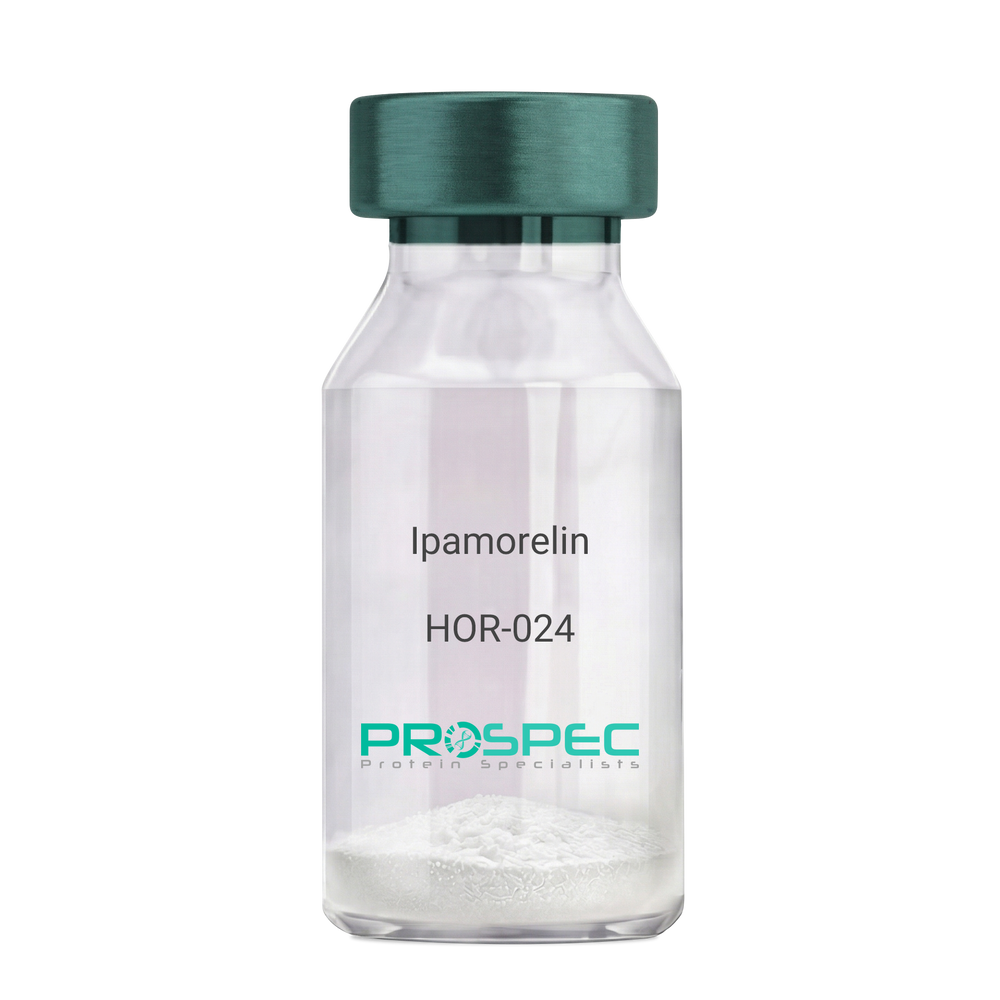
IpamorelinDescription:
Ipamorelin
Ipamorelin
Product # :
HOR-024Price :
Quantity :
Shipping Method :
Shipped at Room temp
More Info
- description
- formulation
- purity
- More Info
Description
Ipamorelin Synthetic is a single, non-glycosylated polypeptide chain containing 4 amino acids, having a molecular mass of 711.85 Dalton and a Molecular formula of C38H49N9O5.
Formulation
The protein was lyophilized with no additives.
Purity
Greater than 97.0% as determined by analysis by RP-HPLC.
More Info
-
Introduction
Ipamorelin is a peptide selective agonist of the ghrelin/growth hormone secretagogue receptor and a growth hormone secretagogue. Ipamorelin is a pentapeptide that was derived from GHRP1. Ipamorelin significantly increases plasma growth hormone levels in both animals and humans. Like pralmorelin and GHRP-6, ipamorelin does not affect prolactin, FSH, LH or TSH levels. However, unlike GHRP2 and GHRP6, but as growth hormone-releasing hormone (GHRH), ipamorelin does not stimulate the secretion of adrenocorticotropic hormone (ACTH) or cortisol, and is highly selective for inducing the secretion only of GH.
-
Synonyms
Ipamorelin
-
Physical Appearance
Sterile Filtered White lyophilized (freeze-dried) powder.
-
Stability
Lyophilized Ipamorelin although stable at room temperature for 3 weeks, should be stored desiccated below -18°C. Upon reconstitution Ipamorelin should be stored at 4°C between 2-7 days and for future use below -18°C. For long term storage it is recommended to add a carrier protein (0.1% HSA or BSA). Please prevent freeze-thaw cycles.
-
Solubility
It is recommended to reconstitute the lyophilized Ipamorelin in sterile 18MΩ-cm H2O not less than 100 µg/ml, which can then be further diluted to other aqueous solutions.
-
Amino Acid Sequence
Aib-His-D-2-Nal-D-Phe-Lys-NH2.
ProSpec's products are furnished for LABORATORY RESEARCH USE ONLY. The product may not be used as drugs, agricultural or pesticidal products, food additives or household chemicals.
- View Data Sheet
Name :
MALD3Description:
Non-Specific Lipid-Transfer Protein Mal d 3 Recombinant
Non-specific lipid-transfer protein, LTP, MALD3.
Product # :
ALR-023Price :
Quantity :
Shipping Method :
Shipped with Ice Packs
More Info
- description
- source
- formulation
- purity
- More Info
Description
Recombinant Non-Specific Lipid-Transfer Protein Mal d 3 produced in SF9 is a glycosylated, polypeptide chain having a calculated molecular mass of 12kDa.MALD3 is expressed with a 6xHis tag and purified by proprietary chromatographic techniques.
Source
Sf9 insect cells.
Formulation
MALD3 is supplied in 20mM HEPES buffer pH-8.0, 200mM NaCl and 20% glycerol.
Purity
Greater than 80.0% as determined by SDS-PAGE.
More Info
-
Introduction
MALD3 (a non-specific lipid transfer protein) is a major apple allergen. LTPs are found mainly in aerial plant organs such as leaves, seeds, flowers, and fruit. MALD3 is a heat-stable protein which is characterized by having resistance to pepsin hydrolysis.
-
Synonyms
Non-specific lipid-transfer protein, LTP, MALD3.
-
Physical Appearance
Sterile Filtered colorless solution.
-
Stability
Store at 4°C if entire vial will be used within 2-4 weeks.Store, frozen at -20°C for longer periods of time.Avoid multiple freeze-thaw cycles.
-
Immunological Functions
1. Binds IgE-type human antibodies.2. Immunodot test with positive/negative samples
-
Applications
Tested by LAL (Limulus Amoebocyte Lysate) chromogenic endotoxin assay.
ProSpec's products are furnished for LABORATORY RESEARCH USE ONLY. The product may not be used as drugs, agricultural or pesticidal products, food additives or household chemicals.
- View Data Sheet
Name :
FABP1 Mouse, HisDescription:
Fatty Acid Binding Protein-1, His Tag Mouse Recombinant
Fatty acid-binding protein 1 liver, L-FABP, FABPL, FABP-1, FABP1, Z-protein, Fatty acid-binding protein, liver.
Product # :
PRO-2512Price :
Quantity :
Shipping Method :
Shipped with Ice Packs
More Info
- description
- source
- formulation
- purity
- More Info
Description
FABP1 Mouse Recombinant produced in E.Coli is a single, non-glycosylated, polypeptide chain containing 150 amino acids (1-127 a.a.) and having a molecular mass of 16.6 kDa. The FABP1 is fused to a 23 amino acid His Tag at N-terminus and purified by proprietary chromatographic techniques.
Source
Escherichia Coli.
Formulation
The FABP1 solution (0.25mg/ml) contains PBS (pH7.4), 20% glycerol and 1mM DTT.
Purity
Greater than 95.0% as determined by SDS-PAGE.
More Info
-
Introduction
FABP1 (Fatty acid binding protein1) encodes the fatty acid binding protein found in liver. FABP1 is composed of ten antiparallel beta strands that form a barrel with a bigger binding pocket than the other FABPs allowing it to accommodate two fatty acid. This protein binds free fatty acids and their coenzyme A derivatives, bilirubin, and some other small molecules in the cytoplasm; it may be involved in intracellular lipid transport and metabolism.
-
Synonyms
Fatty acid-binding protein 1 liver, L-FABP, FABPL, FABP-1, FABP1, Z-protein, Fatty acid-binding protein, liver.
-
Physical Appearance
Sterile Filtered colorless solution.
-
Stability
Store at 4°C if entire vial will be used within 2-4 weeks. Store, frozen at -20°C for longer periods of time. For long term storage it is recommended to add a carrier protein (0.1% HSA or BSA).Avoid multiple freeze-thaw cycles.
-
Amino Acid Sequence
MGSSHHHHHH SSGLVPRGSH MGSMNFSGKY QLQSQENFEP FMKAIGLPED LIQKGKDIKG VSEIVHEGKK IKLTITYGPK VVRNEFTLGE ECELETMTGE KVKAVVKLEG DNKMVTTFKG IKSVTELNGD TITNTMTLGD IVYKRVSKRI.
ProSpec's products are furnished for LABORATORY RESEARCH USE ONLY. The product may not be used as drugs, agricultural or pesticidal products, food additives or household chemicals.
- View Data Sheet
Name :
FABP7 Human, HisDescription:
Fatty Acid Binding Protein-7 Human Recombinant, His Tag
MRG, BLBP, FABPB, B-FABP, DKFZp547J2313, Fatty acid-binding protein brain, Fatty acid-binding protein 7, Brain lipid-binding protein, Mammary-derived growth inhibitor related, FABP7.
Product # :
PRO-661Price :
Quantity :
Shipping Method :
Shipped with Ice Packs
More Info
- description
- source
- formulation
- purity
- More Info
Description
FABP7 Human Recombinant produced in E.Coli is a single, non-glycosylated polypeptide chain containing 132 amino acids and having a molecular mass of 19.39kDa. FABP7 is fused to His-Tag at N-terminus and purified by standard chromatography techniques.
Source
Escherichia Coli.
Formulation
The FABP7 protein solution contains 20mM Tris-HCl pH-8 and 50% Glycerol.
Purity
Greater than 95.0% as determined by SDS-PAGE.
More Info
-
Introduction
FABP7 is a brain fatty acid binding protein. Fatty acid binding proteins (FABPs) are a family of small, highly conserved, cytoplasmic proteins that bind long-chain fatty acids and other hydrophobic ligands. FABPs are are inovlved in fatty acid uptake, transport, and metabolism. FABP7 is expressed in radial glia by the activation of Notch receptors and binds DHA with the highest affinity among all of FABPs. FABP7 plays an important role in transport of hydrophobic ligand with potential morphogenic activity during cns development. FABP7 is required for the establishment of the radial glial fiber system in developing brain, a system that is necessary for the migration of immature neurons to establish cortical layers (by similarity).
-
Synonyms
MRG, BLBP, FABPB, B-FABP, DKFZp547J2313, Fatty acid-binding protein brain, Fatty acid-binding protein 7, Brain lipid-binding protein, Mammary-derived growth inhibitor related, FABP7.
-
Physical Appearance
Sterile Filtered colorless solution.
-
Stability
Store at 4°C if entire vial will be used within 2-4 weeks.Store, frozen at -20°C for longer periods of time. Avoid multiple freeze-thaw cycles.
ProSpec's products are furnished for LABORATORY RESEARCH USE ONLY. The product may not be used as drugs, agricultural or pesticidal products, food additives or household chemicals.
- View Data Sheet
Name :
TACSTD2 Human, sf9Description:
Tumor-Associated Calcium Signal Transducer 2 Human Recombinant, Sf9
Tumor-Associated Calcium Signal Transducer 2,Membrane Component Chromosome 1 Surface Marker 1, Pancreatic Carcinoma Marker Protein GA733-1,Cell Surface Glycoprotein Trop-2, GA733-1,TROP2, M1S1, 40kD Glycoprotein, Identified By Monoclonal Antibody GA733,Membrane Component, Chromosome 1, Surface Marker 1, Gastrointestinal Tumor-Associated Antigen GA7331,Pancreatic Carcinoma Marker Protein GA7331,Cell Surface Glycoprotein TROP2,Epithelial Glycoprotein-1Truncated TACSTD2,GA7331,EGP-1,EGP1,GP50, TACSTD2.
Product # :
PRO-2364Price :
Quantity :
Shipping Method :
Shipped with Ice Packs
More Info
- description
- source
- formulation
- purity
- More Info
Description
TACSTD2 Human Recombinant produced in Sf9 Baculovirus cells is a single, non-glycosylated polypeptide chain containing 253 amino acids (31-274a.a) and having a molecular mass of 28.6kDa (Molecular size on SDS-PAGE will appear at approximately 28-40kDa).TACSTD2 is fused to a 6 amino acid His-tag at C-terminus & purified by proprietary chromatographic techniques.
Source
Sf9, Baculovirus cells.
Formulation
TACSTD2 protein solution (1mg/ml) contains Phosphate buffered saline (pH7.4) and 10% glycerol.
Purity
Greater than 95.0% as determined by SDS-PAGE.
More Info
-
Introduction
Tumor-associated calcium signal transducer 2 (TACSTD2), is a member of to the EPCAM family. TACSTD2 is an intronless gene which encodes a carcinoma-associated antigen. TACSTD2 may act as a growth factor receptor and as a cell surface receptor that transduces calcium signals. Mutations of TACSTD2 have been associated with gelatinous drop-like corneal dystrophy.
-
Synonyms
Tumor-Associated Calcium Signal Transducer 2,Membrane Component Chromosome 1 Surface Marker 1, Pancreatic Carcinoma Marker Protein GA733-1,Cell Surface Glycoprotein Trop-2, GA733-1,TROP2, M1S1, 40kD Glycoprotein, Identified By Monoclonal Antibody GA733,Membrane Component, Chromosome 1, Surface Marker 1, Gastrointestinal Tumor-Associated Antigen GA7331,Pancreatic Carcinoma Marker Protein GA7331,Cell Surface Glycoprotein TROP2,Epithelial Glycoprotein-1Truncated TACSTD2,GA7331,EGP-1,EGP1,GP50, TACSTD2.
-
Physical Appearance
Sterile filtered colorless solution.
-
Stability
Store at 4°C if entire vial will be used within 2-4 weeks. Store, frozen at -20°C for longer periods of time. For long term storage it is recommended to add a carrier protein (0.1% HSA or BSA).Avoid multiple freeze-thaw cycles.
-
Amino Acid Sequence
ADPQDNCTCP TNKMTVCSPD GPGGRCQCRA LGSGMAVDCS TLTSKCLLLK ARMSAPKNAR TLVRPSEHAL VDNDGLYDPD CDPEGRFKAR QCNQTSVCWC VNSVGVRRTD KGDLSLRCDE LVRTHHILID LRHRPTAGAF NHSDLDAELR RLFRERYRLH PKFVAAVHYE QPTIQIELRQNTSQKAAGDV DIGDAAYYFE RDIKGESLFQ GRGGLDLRVR GEPLQVERTL IYYLDEIPPK FSMKRLTHHH HHH.
ProSpec's products are furnished for LABORATORY RESEARCH USE ONLY. The product may not be used as drugs, agricultural or pesticidal products, food additives or household chemicals.
- View Data Sheet
Name :
CX3CL1 Human, HisDescription:
Fractalkine Human Recombinant (CX3CL1), His Tag
Fractalkine, CX3CL1, Neurotactin, CX3C membrane-anchored chemokine, Small inducible cytokine D1, NTN, NTT, CXC3, CXC3C, SCYD1, ABCD-3, C3Xkine.
Product # :
CHM-360Price :
Quantity :
Shipping Method :
Shipped with Ice Packs
More Info
- description
- source
- formulation
- purity
- More Info
- SDS-PAGE
Description
Fractalkine Human Recombinant fused with a 20 amino acid His tag at N-terminus produced in E.Coli is a single, non-glycosylated, polypeptide chain containing 97 amino acids (25-100 a.a.) and having a molecular mass of 10.9kDa. The Fractalkine is purified by proprietary chromatographic techniques.
Source
Escherichia Coli.
Formulation
The Fractalkine solution (0.5 mg/ml) contains Phosphate Buffered Saline pH7.4 and 10% glycerol.
Purity
Greater than 90.0% as determined by SDS-PAGE.
SDS-PAGE
More Info
-
Introduction
Fractalkine soluble form is chemotactic for t-cells and monocytes, but not for neutrophils. Fractalkine membrane-bound form promotes adhesion of those leukocytes to endothelial cells. Fractalkine regulates leukocyte adhesion and migration processes at the endothelium and binds to CX3CR1. Natural Human Fractalkine is produced as a long protein (373-amino acid) with an extended mucin-like stalk and a chemokine domain on top. The mucin-like stalk permits it to bind to the cell surface. Fractalkine gene is located on human chromosome 16 along with some CC chemokines known as CCL17 and CCL22.
-
Synonyms
Fractalkine, CX3CL1, Neurotactin, CX3C membrane-anchored chemokine, Small inducible cytokine D1, NTN, NTT, CXC3, CXC3C, SCYD1, ABCD-3, C3Xkine.
-
Physical Appearance
Sterile Filtered clear solution.
-
Stability
Store at 4°C if entire vial will be used within 2-4 weeks.Store, frozen at -20°C for longer periods of time.For long term storage it is recommended to add a carrier protein (0.1% HSA or BSA).Avoid multiple freeze-thaw cycles.
-
Amino Acid Sequence
MGSSHHHHHH SSGLVPRGSH MQHHGVTKCN ITCSKMTSKI PVALLIHYQQ NQASCGKRAI ILETRQHRLF CADPKEQWVK DAMQHLDRQA AALTRNG.
-
Background
What is the molecular weight/Mw of CX3CL1 HUMAN, HIS Protein?
CX3CL1 HUMAN, HIS Protein has a total Mw of 10.9kDa.
What is the source or expression system of CX3CL1 HUMAN, HIS Protein?
Escherichia Coli.
What is the Purity of CX3CL1 HUMAN, HIS Protein?
CX3CL1 HUMAN, HIS Protein is >90% pure as determined by SDS-PAGE.
What is the Biological Activity of CX3CL1 HUMAN, HIS Protein?
The biological functionality of CX3CL1 HUMAN, HIS Protein will be determined in the future.
What is the amino acid sequence of CX3CL1 HUMAN, HIS Protein?
MGSSHHHHHH SSGLVPRGSH MQHHGVTKCN ITCSKMTSKI PVALLIHYQQ NQASCGKRAI ILETRQHRLF CADPKEQWVK DAMQHLDRQA AALTRNG.
What applications can CX3CL1 HUMAN, HIS Protein be used in?
CX3CL1 HUMAN, HIS Protein can probably be used in western blot, ELISA and Lateral Flow.
What is the endotoxin level for CX3CL1 HUMAN, HIS Protein?
The endotoxin level is minimal, CX3CL1 HUMAN, HIS Protein was purified using conventional chromatography techniques.
ProSpec's products are furnished for LABORATORY RESEARCH USE ONLY. The product may not be used as drugs, agricultural or pesticidal products, food additives or household chemicals.
- View Data Sheet
Name :
MALD1Description:
Major Allergen Mal d 1 Recombinant (Mal d 1.0108)
Major allergen Mal d 1, Ypr10 protein, MALD1, ypr10, Mal d 1.0108
Product # :
ALR-013Price :
Quantity :
Shipping Method :
Shipped with Ice Packs
More Info
- description
- source
- formulation
- purity
- More Info
Description
Recombinant MALD1 produced in SF9 is a glycosylated, polypeptide chain having a calculated molecular mass of 17,492 Dalton. MALD1 purified by proprietary chromatographic techniques.
Source
Sf9 insect cells.
Formulation
MALD1 is supplied in 20mM HEPES buffer pH-7.6, 250mM NaCl and 20% glycerol.
Purity
Greater than 80.0% as determined by SDS-PAGE.
More Info
-
Introduction
MALD1 is a ribonuclease and a heat-sensitive allergen which is a member of the pathogenesis-related protein class. MALD1 shares homologous IgE epitopes with major birch pollen allergen Bet v 1 and Cor a 1 from hazelnut pollen.
-
Synonyms
Major allergen Mal d 1, Ypr10 protein, MALD1, ypr10, Mal d 1.0108
-
Physical Appearance
Sterile Filtered clear solution.
-
Stability
Store at 4°C if entire vial will be used within 2-4 weeks. Store, frozen at -20°C for longer periods of time. Avoid multiple freeze-thaw cycles.
-
Immunological Functions
1. Binds IgE type human antibodies. 2. Immunodot test with positive/negative sera panels.
ProSpec's products are furnished for LABORATORY RESEARCH USE ONLY. The product may not be used as drugs, agricultural or pesticidal products, food additives or household chemicals.
- View Data Sheet
Name :
S100P HumanDescription:
S100 Calcium Binding Protein P Human Recombinant
Protein S100-P, S100 calcium-binding protein P, S100P, S100E, MIG9.
Product # :
PRO-425Price :
Quantity :
Shipping Method :
Shipped at Room temp
More Info
- description
- source
- formulation
- purity
- More Info
Description
The Recombinant Human S100P has a molecular mass of 10.4 kDa containing 95 amino acid residues of the human S100P.
Source
Escherichia Coli.
Formulation
Filtered (0.4µm) and lyophilized from 0.5 mg/ml in 0.05M Phosphate buffer pH7.2, 0.1M NaCl.
Purity
Greater than 95% as determined by SDS-PAGE.
More Info
-
Introduction
S100P was originally described as a placental protein of 95 amino acid residues shares about 50% sequence identity with the brain S100 proteins alpha and beta.
S100 proteins are small dimeric members of the EF-hand superfamily of Ca(2+) binding proteins thought to participate in mediating intracellular Ca(2+) signals by binding to and thereby regulating target proteins in a Ca(2+)-dependent manner. S100P in addition to binding Ca2+, also binds Zn2+ and Mg2+. S100P gene is located on chromosome 4p16.
S100P is dysregulated in the androgen-independent prostate cancer cell lines LNCaP-R, DU145, and PC3 and may play a role in the etiology of prostate cancer.
In ductal hyperplasias, in situ and invasive ductal carcinoma, but not in the normal tissues, S100P overexpression is an early event that might play an important role in the immortalization of human breast epithelial cells in vitro and tumor progression in vivo.
In NIH3T3 cells, the expression of S100P led to the presence of S100P in the culture medium, increased cellular proliferation, and enhanced survival following detachment from the culture substrate or after exposure to the chemotherapeutic agent 5-flurouracil. The proliferation and survival effects of S100P expression were duplicated in a time- and concentration-dependent manner by extracellular addition of purified S100P to wild-type NIH3T3 cells and correlated with the activation of Erks and NFB.
To determine the mechanisms involved in these effects, we tested the hypothesis that S100P activated RAGE (Receptor for Activated Glycation End-Products). It was found that S100P coimmunoprecipitated with RAGE. Furthermore, the effects of S100P on cell signaling, proliferation and survival were blocked by agents that interfere with RAGE including administration of an amphoterin derived peptide known to antagonize RAGE activation, anti-RAGE antibodies and by expression of a dominant negative RAGE. These data suggest that S100P can act in an autocrine manner via RAGE to stimulate cell proliferation and survival. -
Synonyms
Protein S100-P, S100 calcium-binding protein P, S100P, S100E, MIG9.
-
Physical Appearance
Filtered White lyophilized (freeze-dried) powder.
-
Stability
Store lyophilized protein at -20°C. Aliquot the product after reconstitution to avoid repeated freezing/thawing cycles. Reconstituted protein can be stored at 4°C for a limited period of time; it does not show any change after two weeks at 4°C.
-
Solubility
Add deionized H2O and let the lyophilized pellet dissolve completely.
-
Amino Acid Sequence
MTELEAAMGM IIDVFSRYSG SEGSTQTLTK GELKVLMEKE LPGFLQSGKD KDAVDKLLKD LDANGDAQVD FSEFIVFVAA ITSACHKYFE KAGLK.
ProSpec's products are furnished for LABORATORY RESEARCH USE ONLY. The product may not be used as drugs, agricultural or pesticidal products, food additives or household chemicals.
- View Data Sheet
Name :
CXCL17 RatDescription:
VEGF Co-regulated Chemokine 1 Rat Recombinant
Protein Cxcl17, Cxcl17, RGD1304717, C-X-C motif chemokine 17, VEGF co-regulated chemokine 1, Vcc1, VCC-1.
Product # :
CHM-281Price :
Quantity :
Shipping Method :
Shipped at Room temp
More Info
- description
- source
- formulation
- purity
- biological activity
- More Info
Description
CXCL17 Rat Recombinant produced in E.coli is a single, non-glycosylated polypeptide chain containing 97 amino acids and having a molecular mass of 11.5kDa.The CXCL17 is purified by proprietary chromatographic techniques.
Source
Escherichia Coli.
Formulation
CXCL17 protein was lyophilized from a 0.2µm filtered concentrated solution in 20mM PB, 300mM NaCl, pH 7.4.
Purity
Greater than 95.0% as determined by:
(a) Analysis by RP-HPLC.
(b) Analysis by SDS-PAGE.Biological Activity
Measured by its ability to induce VEGF expression in mouse endothelial cells. The ED50 for this effect is typically 1-5 µg/ml.More Info
-
Introduction
Dendritic cell and monocyte chemokinelike protein (DMC/CXCL17/VEGF-correlated chemokine 1/VCC1), is a secreted molecule with a size and predicted 3-dimensional folding pattern similar to that of chemokines CXCL8/IL8 and CXCL14/BRAK. CXCL17 is constitutively generated by airway and intestinal epithelium. CXCL17 induces the chemotaxis of quiescent, but not LPS-activated peripheral blood monocytes and dendritic cells, and it also binds these cells specifically. The expression of CXCL17 is increased in endothelial cells when they are induced to form tubes in vitro. CXCL17, CXCL1/GRO and CXCL8/IL8 which have roles in angiogenesis, show significantly correlated expression with that of VEGF in primary lung, breast and esophageal tumors. Therefore, CXCL17 is suggested to have a role in tumor angiogenesis. The mature Rat CXCL17 shares 82%, 71% amino acid sequence identity with mouse, human CXCL17, respectively.
-
Synonyms
Protein Cxcl17, Cxcl17, RGD1304717, C-X-C motif chemokine 17, VEGF co-regulated chemokine 1, Vcc1, VCC-1.
-
Physical Appearance
Sterile Filtered White lyophilized (freeze-dried) powder.
-
Stability
Lyophilized CXCL17 although stable at room temperature for 3 weeks, should be stored desiccated below -18°C. Upon reconstitution CXCL17 should be stored at 4°C between 2-7 days and for future use below -18°C. For long term storage it is recommended to add a carrier protein (0.1% HSA or BSA). Please prevent freeze-thaw cycles.
-
Solubility
It is recommended to reconstitute the lyophilized CXCL17 in sterile 18M-cm H2O not less than 100µg/ml, which can then be further diluted to other aqueous solutions.
-
Amino Acid Sequence
SPNQEVARHH GDQHQAPRRW LWEGGQECDC KDWSLRVSKR KTTAVLEPPR KQCPCDHVKG SEKKNRRQKH HRKSQRPSRT CQQFLKRCQL ASFTLPL.
-
Background
What is the molecular weight/Mw of CXCL17 RAT Protein?
CXCL17 RAT Protein has a total Mw of 11.5kDa.
What is the source or expression system of CXCL17 RAT Protein?
Escherichia Coli.
What is the Purity of CXCL17 RAT Protein?
CXCL17 RAT Protein is >95% pure as determined by SDS-PAGE.
What is the Biological Activity of CXCL17 RAT Protein?
Measured by its ability to induce VEGF expression in mouse endothelial cells. The ED50 for this effect is typically 1-5 µg/ml.
What is the amino acid sequence of CXCL17 RAT Protein?
SPNQEVARHH GDQHQAPRRW LWEGGQECDC KDWSLRVSKR KTTAVLEPPR KQCPCDHVKG SEKKNRRQKH HRKSQRPSRT CQQFLKRCQL ASFTLPL.
What applications can CXCL17 RAT Protein be used in?
CXCL17 RAT Protein can probably be used in western blot, ELISA and Lateral Flow.
What is the endotoxin level for CXCL17 RAT Protein?
The endotoxin level is minimal, CXCL17 RAT Protein was purified using conventional chromatography techniques.
ProSpec's products are furnished for LABORATORY RESEARCH USE ONLY. The product may not be used as drugs, agricultural or pesticidal products, food additives or household chemicals.
- View Data Sheet
Name :
Agrin RatDescription:
Agrin Rat Recombinant
Agrin, Agrn, C90, C22, Agrin N-terminal 110 kDa subunit, Agrin C-terminal 110 kDa subunit, Agrin C-terminal 90 kDa fragment, Agrin C-terminal 22 kDa fragment, AGR
Product # :
PRO-2627Price :
Quantity :
Shipping Method :
Shipped with Ice Packs
More Info
- description
- source
- formulation
- purity
- More Info
Description
Agrin Rat produced in Sf9 Baculovirus cells is a single, glycosylated polypeptide chain containing 766 amino acids (997-1753 a.a.) and having a molecular mass of 82.5kDa.Agrin is fused to a 9 amino acid His tag at C-Terminus and purified by proprietary chromatographic techniques.
Source
Sf9, Baculovirus cells.
Formulation
The Agrin solution (0.5mg/ml) contains 10% Glycerol in Phosphate-Buffered Saline (pH 7.4).
Purity
Greater than 90.0% as determined by SDS-PAGE.
More Info
-
Introduction
Agrin or AGRN is a large protein (proteoglycan) that has a crucial part in the development of neuromuscular junction amid embryogenesis. The protein has an involvement in the collection and aggregation of acetylcholine receptors through synaptogenesis. The agrin gene can be found and expressed in rat embryonic nervous system andmuscle tissue. This Agrin protein is aggregated in the synapses, there it can take part in regeneration & development. The protein binds to receptors on the surface of skeletal muscle.
-
Synonyms
Agrin, Agrn, C90, C22, Agrin N-terminal 110 kDa subunit, Agrin C-terminal 110 kDa subunit, Agrin C-terminal 90 kDa fragment, Agrin C-terminal 22 kDa fragment, AGR
-
Physical Appearance
Sterile Filtered colorless solution.
-
Stability
Store at 4°C if entire vial will be used within 2-4 weeks. Store, frozen at -20°C for longer periods of time. For long term storage it is recommended to add a carrier protein (0.1% HSA or BSA).Avoid multiple freeze-thaw cycles.
-
Amino Acid Sequence
ADPSCYNSPL GCCSDGKTPS LDSEGSNCPA TKAFQGVLEL EGVEGQELFY TPEMADPKSE LFGETARSIE STLDDLFRNS DVKKDFWSVR LRELGPGKLV RAIVDVHFDP TTAFQASDVG QALLRQIQVS RPWALAVRRP LQEHVRFLDF DWFPTFFTGA ATGTTAAMAT ARATTVSRLP ASSVTPRVYP SHTSRPVGRT TAPPTTRRPP TTATNMDRPR TPGHQQPSKS CDSQPCLHGG
TCQDQDSGKG FTCSCTAGRG GSVCEKVQPP SMPAFKGHSF LAFPTLRAYH TLRLALEFRA LETEGLLLYN GNARGKDFLA LALLDGRVQF RFDTGSGPAV LTSLVPVEPG RWHRLELSRH WRQGTLSVDG ETPVVGESPS GTDGLNLDTN LYVGGIPEEQ VAMVLDRTSV GVGLKGCIRM LDINNQQLEL SDWQRAAVQS SGVGECGDHP CLPNPCHGGA LCQALEAGMF LCQCPPGRFG PTCADEKSPC QPNPCHGAAP CRVLSSGGAK CECPLGRSGT FCQTVLETAG SRPFLADFNG FSYLELKGLH TFERDLGEKM ALEMVFLARG PSGLLLYNGQ KTDGKGDFVS LALHNRHLEF CYDLGKGAAV IRSKEPIALG TWVRVFLERN GRKGALQVGD GPRVLGESPK SRKVPHTMLN LKEPLYIGGA PDFSKLARGA AVSSGFSGVI QLVSLRGHQL LTQEHVLRAV DVSPFADHPC TQALGNPCLN GGSCVPREAT YECLCPGGFS GLHCEKGLVE HHHHHH
ProSpec's products are furnished for LABORATORY RESEARCH USE ONLY. The product may not be used as drugs, agricultural or pesticidal products, food additives or household chemicals.
- View Data Sheet
Name :
DCAF7 HumanDescription:
DDB1 and CUL4 Associated Factor 7 Human Recombinant
DDB1- and CUL4-associated factor 7, WD repeat-containing protein 68, WD repeat-containing protein An11 homolog, DCAF7, HAN11, WDR68, AN11, SWAN-1.
Product # :
PRO-909Price :
Quantity :
Shipping Method :
Shipped with Ice Packs
More Info
- description
- source
- formulation
- purity
- More Info
Description
DCAF7 Human Recombinant produced in E.Coli is a single, non-glycosylated polypeptide chain containing 298 amino acids (19-295 a.a) and having a molecular mass of 33.6kDa.DCAF7 is fused to a 21 amino acid His-tag at N-terminus & purified by proprietary chromatographic techniques.
Source
Escherichia Coli.
Formulation
DCAF7 protein solution (1mg/ml) containing 20mM Tris-HCl buffer (pH8.0), 0.4M urea and 1mM DTT.
Purity
Greater than 90.0% as determined by SDS-PAGE.
More Info
-
Introduction
DDB1-and CUL4-associated factor (DCAF7) is involved in craniofacial development. The DCAF7 protein acts upstream of the EDN1 pathway and is essential for formation of the upper jaw equivalent, the palatoquadrate. The activity necessary for the EDN1 pathway function varies between the first and second arches. DCAF7 associates with DIAPH1 and controls GLI1 transcriptional activity. DCAF7 may be involved in normal and diseased skin development.
-
Synonyms
DDB1- and CUL4-associated factor 7, WD repeat-containing protein 68, WD repeat-containing protein An11 homolog, DCAF7, HAN11, WDR68, AN11, SWAN-1.
-
Physical Appearance
Sterile filtered colorless solution.
-
Stability
Store at 4°C if entire vial will be used within 2-4 weeks. Store, frozen at -20°C for longer periods of time. For long term storage it is recommended to add a carrier protein (0.1% HSA or BSA).Avoid multiple freeze-thaw cycles.
-
Amino Acid Sequence
MGSSHHHHHH SSGLVPRGSH MVYAMNWSVR PDKRFRLALG SFVEEYNNKV QLVGLDEESS EFICRNTFDH PYPTTKLMWI PDTKGVYPDL LATSGDYLRV WRVGETETRL ECLLNNNKNS DFCAPLTSFD WNEVDPYLLG TSSIDTTCTI WGLETGQVLG RVNLVSGHVK TQLIAHDKEV YDIAFSRAGG GRDMFASVGA DGSVRMFDLR HLEHSTIIYE DPQHHPLLRL CWNKQDPNYL ATMAMDGMEV VILDVRVPCT PVARLNNHRA CVNGIAWAPH SSCHICTAAD DHQALIWD.
ProSpec's products are furnished for LABORATORY RESEARCH USE ONLY. The product may not be used as drugs, agricultural or pesticidal products, food additives or household chemicals.
- View Data Sheet
Name :
SDF 1b RatDescription:
Stromal Cell-Derived Factor-1 beta Rat Recombinant (CXCL12)
SDF-1, CXCL12, Pre-B cell growth-stimulating factor, PBSF, hIRH, chemokine (C-X-C motif) ligand 12, SDF1, SDF1B, TPAR1, SCYB12, SDF-1b, TLSF-b, 12-O-tetradecanoylphorbol 13-acetate repressed protein 1, Thymic lymphoma cell-stimulating factor, TLSF.
Product # :
CHM-248Price :
Quantity :
Shipping Method :
Shipped at Room temp
More Info
- description
- source
- formulation
- purity
- biological activity
- More Info
Description
SDF-1 beta Rat Recombinant produced in E.Coli is a non-glycosylated, Polypeptide chain containing 72 amino acids and having a molecular mass of 8.4 kDa. The Rat SDF-1b is purified by proprietary chromatographic techniques.
Source
Escherichia Coli.
Formulation
The Rat SDF1B protein was lyophilized from a concentrated (1mg/ml) sterile solution containing 20mM Phsophate buffer pH-7.4 and 0.15M NaCl.
Purity
Greater than 97.0% as determined by:
(a) Analysis by RP-HPLC.
(b) Analysis by SDS-PAGE.Biological Activity
The specific activity as determined by its ability to chemoattract human peripheral blood monocytes at 100-150ng/ml corresponding to a Specific Activity of 6,667-10,000IU/mg.More Info
-
Introduction
SDF-1 (stromal cell-derived factor-1) is small cytokine belonging to the chemokine family that is officially designated Chemokine (C-X-C motif) ligand 12 (CXCL12). It is produced in two forms, SDF-1?/CXCL12a and SDF-1?/CXCL12b, by alternate splicing of the same gene. Chemokines are characterized by the presence of four conserved cysteines, which form two disulfide bonds. The CXCL12 proteins belong to the group of CXC chemokines, whose initial pair of cysteines are separated by one intervening amino acid. CXCL12 is strongly chemotactic for lymphocytes and has been implicated as an important cell co-ordinator during development. During embryogenesis it directs the migration of hematopoietic cells from foetal liver to bone marrow. Mice which were knocked-out for CXCL12 gene were lethal before the birth or within just 1 hour of life. As another role, CXCL12a alters also the electrophysiology of neurons. CXCL12 was shown to be expressend in many tissues in mice (including brain, thymus, heart, lung, liver, kidney, spleen and bone marrow).
The receptor for this chemokine is CXCR4, which was previously called fusin. This CXCL12-CXCR4 interaction used to be considered exclusive (unlike for other chemokines and their receptors), but recently it was suggested that CXCL12 is also bound by CXCR7 receptor.
The gene for CXCL12 is located on human chromosome 10. In human and mouse both CXCL12 and CXCR4 show high identity of sequence: 99% and 90%, respectively. -
Synonyms
SDF-1, CXCL12, Pre-B cell growth-stimulating factor, PBSF, hIRH, chemokine (C-X-C motif) ligand 12, SDF1, SDF1B, TPAR1, SCYB12, SDF-1b, TLSF-b, 12-O-tetradecanoylphorbol 13-acetate repressed protein 1, Thymic lymphoma cell-stimulating factor, TLSF.
-
Physical Appearance
Sterile Filtered White lyophilized (freeze-dried) powder.
-
Stability
Lyophilized SDF-1b although stable at room temperature for 3 weeks, should be stored desiccated below -18°C. Upon reconstitution CXCL12 should be stored at 4°C between 2-7 days and for future use below -18°C.For long term storage it is recommended to add a carrier protein (0.1% HSA or BSA).Please prevent freeze-thaw cycles.
-
Solubility
It is recommended to reconstitute the lyophilized SDF-1b in sterile 18MΩ-cm H2O not less than 100µg/ml, which can then be further diluted to other aqueous solutions.
-
Amino Acid Sequence
KPVSLSYRCP CRFFESHVAR ANVKHLKILN TPNCALQIVA RLKSNNRQVC IDPKLKWIQE YLDKALNKRL KM.
ProSpec's products are furnished for LABORATORY RESEARCH USE ONLY. The product may not be used as drugs, agricultural or pesticidal products, food additives or household chemicals.